CRANIOVERTEBRAL JUNCTION
REALIGNMENT FOR BASILAR
INVAGINATION
Dissertation submitted to the Dr.M.G.R. Medical University, Chennai,
DEPARTMENT OF NEUROLOGICAL SCIENCES CHRISTIAN MEDICAL COLLEGE
VELLORE CERTIFICATE
This is to certify that the dissertation titled “Craniovertebral junction
realignment for basilar invagination” is the bonafide original work of Dr. Manikandan, S. N submitted in partial fulfillment of the rules and
regulations, for Branch-II M.Ch. Neurosurgery, final examination of the Tamil Nadu Dr. M.G.R. Medical University to be held in August 2010.
Signature of guide:
Dr. Ari G. Chacko
Professor & Head, Neurosurgery Unit I Head, Department of Neurological Sciences Christian Medical College Vellore
ACKNOWLEDGEMENT
I would like to express my gratitude to all those who gave me the possibility to complete this thesis.
I am deeply indebted to my guide, Prof. Dr. Ari. G. Chacko for his stimulating suggestions and encouragement, which helped me in this study and writing of this thesis and for his role as an Observer for calculating craniometry.
I would thank Dr. Roy T Daniel, Professor of Neurosurgery, for his valuable suggestions and support and who gave this study as my thesis and for his role as an Observer for calculating craniometry.
I would also thank Dr. Sunithi E Mani, Assistant professor, Dept of Radiology for her role as an Observer for calculating craniometry.
Table of contents Page. No.
1. List of figures………..….. 05
2. List of tables……….…. 06
3. Aim……… 07
4. Introduction……….…. 07
5. Literature review………..… 08
6. Materials and Methods……….….... 37
7. Results……….… 41
8. Discussion……… 64
10. Conclusions………...………. 75
11. References………. 76
List of figures Figure Legends
1. Coronal view of a CT scan showing Occipitoatlantoaxial complex.
2. The bony landmarks of the skull base and the craniovertebral junction. 3. Illustration showing Chamberlain’s line.
4. Illustration showing Wackenheim clival baseline and clival canal angle. 5. Imaging showing Group B type of basilar invagination.
6. Illustration showing Modified omega angle.
7. Final construct of C1-2 distraction and fusion with bone graft.
8. Illustrated examples of various craniometry findings measured before and after surgery.
9. Graph showing mean value (of 3 Observers) of the relation of the odontoid to Wackenheim’s line in 17 patients.
10. Graph showing mean value (of 3 Observers) of the relation of the odontoid to Chamberlain’s line in 17 patients.
11. Graph showing mean value (of 3 Observers) of the relation of the odontoid to Mac Rae’s line in 17 patients.
12. Graph showing mean value (of 3 Observers) of the atlantoaxial distance in 17 patients.
13. Graph showing mean value (of 3 Observers) of the sagittal canal diameter in 17 patients.
14. Graph showing mean value (of 3 Observers) of the Clival canal angle in 17 patients. 15. Graph showing mean value (of 3 Observers) of the Modified Omega canal angle in
17 patients.
16. Implants and instruments used in craniovertebral junction realignment surgery. 17. Midsagittal CT of a patient who underwent reoperation.
18.A Immediate postoperative scan after the 2 nd surgery showing realignment of craniovertebral junction and widening of the space available for the cord of a patient who underwent reoperation
B Immediate postoperative scan after Ist surgery showing slippage of the spacer anteriorly of a patient who underwent reoperation.
List of Tables Table Legends
1. Major Transoral series and their results. 2. Preoperative symptoms.
3. Preoperative and follow-up Nuricks grade. 4. Preoperative and follow-up Modified JOA score.
5. Level of odontoid in relation to Wackenheim’s line preoperatively, immediate postop and at follow-up in all patients (mean value of 3 Observers).
6. Level of odontoid in relation to Chamberlain’s line preoperatively, immediate postop and at follow-up in all patients (mean value of 3 Observers).
7. Level of odontoid in relation to Mac Rae’s line in all patients (mean value of 3 Observers).
8. Atlantoaxial distance all patients (preop, immediate postop and follow-up) (mean value of 3 Observers).
9. Sagittal canal diameter in all patients (preop, immediate postop and follow-up) (mean value of 3 Observers).
10. Clival canal angle in all patients (preop, immediate postop and follow-up) (mean value of 3 Observers).
11. Modified omega angle in all patients (preop, immediate postop and follow-up) (mean value of 3 Observers).
AIM:
y To assess the clinical, functional and radiological outcome after craniovertebral realignment surgery for basilar invagination.
INTRODUCTION:
Basilar invagination is a congenital condition where the tip of the odontoid process invaginates into the foramen magnum. It may result in lower brain stem or upper spinal cord compression producing progressive neurological deficits.
Primary or true congenital basilar invagination may be associated with other vertebral anomalies including occipito-atlantal fusion, hypoplasia of atlas, and hemirings of C1 with spreading of lateral masses, odontoid abnormalities and Klippel-Feil deformity (1).
REVIEW OF LITERATURE:
Development of Craniovertebral junction:
The craniovertebral junction develops from four occipital sclerotomes and from the first and second cervical sclerotomes (1). The occipital bone, clivus and occipital condyles are derived from four occipital sclerotomes. The fourth occipital sclerotome forms the occipital condyles, paracondylar process and the bones surrounding the foramen magnum. The anterior arch of the atlas is derived from a band of tissue, the hypochondral bow (1), which is also derived from the fourth occipital sclerotome. The posterior arch of atlas is derived from both the fourth occipital sclerotome and from the first cervical sclerotome. The atlas ossifies from a single ventral and paired dorsal ossification centres.
The axis is derived from the fourth occipital and the first two cervical sclerotomes. The tip of the odontoid is derived from the fourth occipital sclerotome; the odontoid process from the first cervical sclerotome and body of the axis and dorsal vertebral arch are derived from the second cervical sclerotome. Fusion of the odontoid and axis body begins at 4 years and is completed at the age of 8. Fusion of the apex of the odontoid to the odontoid proper occurs at 12 years.
Surgical anatomy of craniovertebral junction
Foramen Magnum:
The foramen magnum has three parts: (a) the squamosal portion which is located in the dorsal aspect of foramen magnum, (b) the basal or clival portion located anterior to the foramen magnum, and (c) the condylar part that connects the squamosal and the clival parts that articulates the atlas lateral mass (2). The hypoglossal canal perforates the skull base at the lateral part of the condyles and transmits the hypoglossal nerve along with the branch of the posterior meningeal artery. The most posterior margin of the foramen magnum is called the opisthion. The anterior most midline of the foramen magnum is termed the basion.
Atlas:
Axis:
The axis is the second cervical vertebra. The odontoid process projects cephalad from its articulation with the axis body. On the ventral odontoid surface is an oval facet, which articulates with the dorsal surface of the anterior atlas arch. In the dorsal aspect of the dens is a transverse groove over which passes the transverse ligament of the atlas. The axis spinous process is large, deeply concave on its caudal border and is the first bifid vertebra in the cervical spine.
Ligamentous anatomy:
divide the atlas ring into a dorsal and ventral component. The ventral component contains the odontoid process and the dorsal component encompasses the spinal cord and the spinal accessory nerves. The ligament presents a fibrocartilaginous surface allowing for free gliding motion to occur over the posterior facet of the dens. The tectorial membrane is dorsal to the cruciate ligament and a strong band of longitudinally oriented fibers that are attached to the dorsal surface of C3 vertebrae, the axis body and to the body of the dens. The ligament ascends upward and widens to attach to the base of the occipital bone. This membrane is the rostral extension of the posterior longitudinal ligament of the vertebral column. Ruptures of the cruciate ligament are easily identified and can aid in the decision making of craniocervical stability.
Biomechanics of craniovertebral junction:
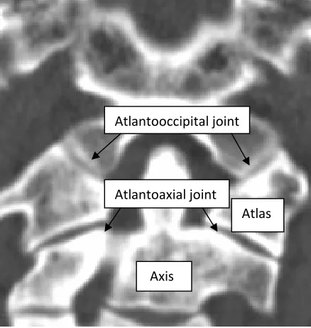
Figure 1 Coronal view of a CT scan showing Occipitoatlantoaxial complex
The largest degree of rotation occurs at the atlantoaxial joint (Figure 1). Usually, a rotation past 25–30° brings the middle and lower cervical segments into play. This prevents increased rotation at the atlantoaxial joint. Anatomic studies have shown that stretching and kinking of the contralateral vertebral artery occurs between 30 and 35° of atlantoaxial rotation (2). When rotation exceeds 40°, an interlocking of the facets occurs between the atlas and the axis vertebra. When an acute rotation of the atlantoaxial joint is made exceeding 45°, the ipsilateral vertebral artery may demonstrate angulation and occlusion. This has particular significance in children with atlas assimilation, with individuals participating in football and wrestling, and those who undergo excessive rotation of the head during general anesthesia or forceful head manipulations (2). The unique anatomic configuration of the craniovertebral junction creates a distinct biomechanical behavior that differs from that of other spinal joints. There is no intervertebral disc between the occiput and C1 and C2. The ball-and-socket shaped occiput-C1 joint allows slightly more flexion-extension than the other levels of the spine. The biconvex articular surfaces of the C1-2 joints allow gliding and wide rotation of
Atlas
Axis
Atlantooccipital joint
the C1 around the dens. The atlanto-axial joint is more flexible and allows more than 90 degrees of rotation bilaterally. When the transverse component of the cruciate ligament has been disrupted, the alar ligaments are still intact. Hence the amount of displacements remains between 5 and 6 mm until the alar ligaments become incompetent (2). The transverse atlantal ligament is the strongest and thickest ligaments of the entire spine.
Pathogenesis of basilar invagination:
Several theories have been suggested to clarify the probable cause and origin of basilar invagination. They include mechanical, embryological dysgenesis, genetic abnormalities or viral infections (4). Goel et al speculated that basilar invagination is secondary to an abnormally inclined alignment of the facets of the atlas and axis (4). The progressive slippage of the atlas over the axis secondary to this malalignment, results in invagination of the odontoid process into the foramen magnum (5).
Classification of basilar invagination:
Radiological criteria:
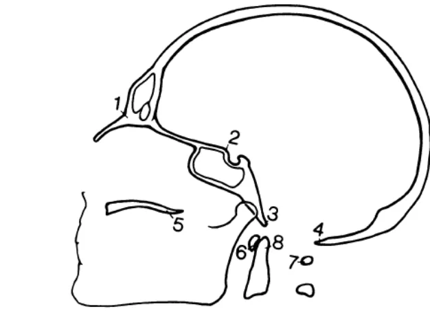
[image:15.612.151.464.144.377.2]The bony landmarks of the skull base and the craniovertebral junction are shown in Figure 2
Figure 2
1. Nasion
2. Tuberculum sella 3. Basion
4. Opisthion
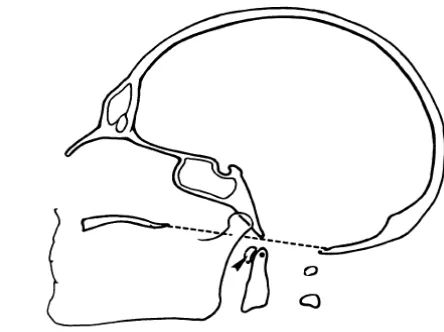
Chamberlain's line:
Chamberlain’s line (Fig 3) extends from the posterior pole of the hard palate to the opisthion (6). Mc gregor’s line extends from the posterior pole of the hard palate to the lowest point of the occipital squamousal surface. McRae’s line is between basion and opisthion (7).
[image:16.612.258.480.209.375.2]
Figure 3
Chamberlain’s line
Basilar invagination is diagnosed when the tip of the odontoid process was at least 2 mm above Chamberlain's line (4).
Mc gregor’s line:
McRae line:
McRae line is between basion and opisthion. The odontoid tip lies below this line in normal subjects and if the odontoid tip is above this line then it is diagnostic of basilar invagination.
Atlanto-dental distance:
The distance between anterior arch of atlas and the odontoid is the atlanto-dental distance. The atlanto-dental or atlanto-axial distance more than 5 mm is considered as abnormal in children and more than 3 mm is considered as abnormal in adults.
Wackenheim's clival line:
Wackenheim clivus baseline (Fig 4) is a line along the clivus extrapolated inferiorly into the upper cervical spinal canal (9). The angle formed at the intersection of the Wackenheim clivus baseline with a line constructed along the posterior surface of the axis body and odontoid process is the clivus canal angle. It normally ranges from 150 in flexion to 180 degrees in extension.
Figure 4: Wackenheim clival baseline and clival canal angle
The tip of the odontoid process is significantly superior to Wackenheim’s clival line in Group A patients. In Group B patients, the relationship of the tip of the odontoid process and the lower end of the clivus and the atlanto-dental interval is normal. In Group B patients (4) the basilar invagination is due to the rostral positioning of the plane of the foramen magnum in relation to the brainstem as shown in the Fig 5.
Figure 5: Imaging showing Group B type of basilar invagination
Omega angle:
Goel et al described a modified Omega angle as a measurement of the angle of the odontoid in the sagittal plane in relation to the plane of the hard palate (10). A vertical line is drawn traversing through the centre of the base of the axis parallel to the line of the hard palate. The line of the hard palate is unaffected by the relative movement of the head and the cervical spine during the movement of the neck in these 'fixed' craniovertebral anomalies (10). The Omega angle (Fig 6) is the angle between the line B and line C. The Omega angle is severely reduced in Group A patients while it is much larger in Group B patients.
Figure 6 : Modified omega angle
METHODS OF TREATMENT OF BASILAR INVAGINATION Transoral odontoidectomy:
The transoral approach for craniospinal malformations was first used by Kanavell and Le Fort in 1918, Fang and Ong in 1962, Greenberg et al in 1968, Grison in 1967, Fokes and Jomin and Bouasakao in 1977, Menezes et al in 1980, and more recently by Crockard in 1985 (11).
Procedure
A Spetzler-Sonntag retractor is placed by retracting the tongue and endotracheal tube to allow maximum exposure of the posterior pharyngeal area. The uvula and the soft palate are retracted superiorly with a red rubber catheter inserted through the nose. This retraction improves exposure of the upper portion of the posterior pharyngeal wall overlying the tip of the odontoid and prevents secretions from running into the incision. The posterior pharynx is then infiltrated with 1% lidocaine with epinephrine. The incision is typically 1.5 to 2 cm in length and is carried through the posterosuperior pharyngeal constrictor muscle in the midline raphe with “no touch” oral cavity technique.
mandatory to prevent the acute or delayed effects of transoral odontoidectomy (14). Cervical fusion alone may not be possible when the ring of C1 is fractured, C1 incorporated into the occiput or in patients with rheumatoid arthritis; in this case occipitocervical fusion is the preferred method of stabilization.
Results of transoral odontoidectomy:
Menezes et al (16) studied 72 patients between the ages of 6 and 82 years who underwent ventral transoral transpharyngeal decompression of the craniocervical junction. Pluridirectional lateral tomography of the CVJ was obtained 7 days after surgery to determine craniovertebral stability. This was done in the flexed and extended positions, as well as with and without traction. Of the 72 individuals who underwent a ventral decompression, 52 patients showed instability and required a dorsal fixation procedure. All patients showed neurological improvement. Six individuals who were ventilator-dependent following either trauma or a previous primary posterior decompression had resolution of their neurological symptoms and signs in the postoperative period. Downbeat nystagmus, sleep apnea and brain-stem signs were prominent features in 15 individuals with basilar invagination and the Chiari malformation. These signs regressed following the ventral decompressive procedure. Two patients died within the 1 st month of operation, one due to a myocardial infarction and other due to Escherichia coli septicemia from a urinary tract infection. One patient had a postoperative pharyngeal wound infection and a retropharyngeal abscess that required drainage.
fixation was performed in the same surgical session following a transoral surgical procedure in 18 Group I patients. In these cases the indication for immediate fixation was relatively high mobility of the cervical vertebral bodies during drilling. In 39 other Group I patients, fixation was performed as a second stage surgery. Excessive pain and spasm of the neck muscles and suboccipital radicular pain formed the primary indication for fixation in these patients. No patient worsened in motor function prior to second-stage fixation. In this group fixation was performed after the initial surgery within 15 days in 16 patients, within 2 months in 11 patients, and between 2 and 6 months in 12 patients. In Group II, a posterior fixation procedure was conducted following transoral decompression in the same surgical sitting in one patient. In four patients fixation was performed within 2 weeks after transoral surgery. No patient needed a fixation procedure as a delayed measure. In six Group II patients, no fixation was necessary, even after both anterior and posterior decompressive operations. They concluded that the transoral surgery is indicated in Group I patients whereas Group II patients warrants foramen magnum decompression only.
Disadvantages of transoral odontoidectomy:
The worsening of basilar invagination as a cause of failure of transoral odontoidectomy has been reported earlier (12, 23). Transoral odontoidectomy causes further cranial settling of the upper cervical spine (C2 body) causing brain stem compression. This is caused by the horizontal separation of the lateral mass of C1 due to removal of anterior arch and ligaments. A partial resection of anterior arch of C1 will minimize horizontal separation of lateral mass and thus cranial settling of C2. Such a worsening has been seen even when a posterior fixation was performed with wires as the pullout strength of wire is less than the screw (23).
Neurological deterioration after transoral odontoidectomy:
Cranial Settling:
The unique anatomy and biomechanics of the CVJ differentiate this region from other spinal segments. Naderi et al (23) reported further cranial settling in two patients whom underwent transoral decompression and occipitocervical fusion which necessitated a second decompressive procedure in one of the cases. The other patient was asymptomatic, and an osseous fusion was demonstrated between the C-2 vertebral body and lower aspect of the clivus. Transoral odontoidectomy results in a severe ligamentous and osseous destruction and it alters the CVJ anatomy and affects the biomechanics of the region. Both these patients underwent C1 anterior arch excision which probably caused further cranial settling in these patients. The author in another study (25) demonstrated the effects of odontoidectomy in a cadaveric model by compressing the occiput–C3 complex before and after resection of the anterior arch of C-1. In the specimen in which the C-1 anterior arch had been sectioned, horizontal separation of the lateral masses of C-1 occurred and resulted in further cranial settling of the C-2 body. The author determined that the preservation of C-1 anterior arch and lamina minimizes the horizontal separation of the C-1 lateral masses.
Author patients No of Preoperative Traction
Improved/ Stabilized
neurological status Complications & Incidence Hospital stay Follow up details Menezes (16)
et al 1998
72 No All 2 mortality due to MI and Septicemia N.A N.A Mark (22)
et al 1989
53 No All Morbidity-3 (6)% -wound dehiscence, brainstem stroke, CSF leak
& mortality-3 (6%)-pneumonia and pulmonary embolus.
N.A 24 months (median)
Laborde (11)
et al 1992 15 No Not available Morbidity-12 (80%), atlantoaxial dislocation, CSF leak, hemorrhage, infection & hydrocephalus Mortality-2 (13.3%), infection & hemorrhage
N.A N.A
Goel (10) et al 1998
99 Yes All Morbidity- 1(1%) N.A 2 months to 9
years (average 43 months).
Jain (15) et al 1999
74 Yes 55.3% improved 29.8% stabilized
22.9% - deterioration in neurological status Dehiscence (20.3%) and hemorrhage (4%),
Velopharyngeal insufficiency (8.1%), CSF leak (6.7%) and inadequate decompression (6.7%). Mortality-6.1%
N.A 47 (63.5%) patients were followed up for 3 months to 2 years Landeiro (27)
et al 2007
38 No 36.8% improved 60.2% stabilized
Morbidity-18.4% due to dehiscence, pulmonary infection & CSF leak. Mortality-1(2.6%)
[image:27.612.25.775.100.408.2]N.A N.A
Transcondylar approach for resection of the dens:
Al-Mefty et al (28) introduced the transcondylar approach for resection of the dens as an alternative to the transoral approaches. Ture et al (28) described modifications of transcondylar approach for resection of dens via transatlas route advantage being preserving the jugular bulb, hypoglossal nerve, and facial nerve and able to perform the occipital condyle–C2 fusion is one stage. This extreme lateral–transatlas approach was used for the resection of the dens of the axis in five patients. Unilateral occipitocervical fusion was performed during the same procedure. There were no cases of intra- or postoperative complications. There were no cases of postoperative infections, wound infection, or CSF leakage. The follow-up period ranged from 13 to 24 months (median 17.2 months) within which no craniocervical instability was demonstrated.
Halo traction and fusion:
resolved under cervical halter traction therapy for 4 weeks without any operative intervention. Kyoshima et al (29) was the first to report a simple cervical traction method with the halo vest apparatus for the unstable CVJ injuries. Moreover, bed rest is not necessary during the procedure; it may be an advantageous point for preventing deep venous thrombosis and pulmonary embolism, particularly in elder patients. However Goel et al (10) performed a posterior fixation procedure in a reduced position of the basilar invagination and the atlantoaxial dislocation following cervical traction in four patients. All these patients needed a transoral surgery at a later stage because the reduced position could not be maintained by the implant.
Craniovertebral realignment for basilar invagination:
Surgical procedure described by Goel et al (31)
The patients are placed in cervical traction prior to induction of anesthesia, and the weights are progressively increased to approximately one fifth of the total body weight. The patient is positioned prone with the head end elevation to 35°. The atlantoaxial facet joints are approached via the pars of C2 and exposed after sectioning of the large C-2 ganglion. The joint capsule is excised and the articular cartilages are removed with a micro drill. The joint is distracted bilaterally. The pieces of corticocancellous bone with metal plate spacer are used as strut graft and are packed into the joints. The size of the spacers depends on the space available within the distracted joint space. Posteriorly bone graft was placed between the posterior elements of C1–suboccipital bone complex and C-2 after decorticating the host bone. The neck is immobilized in a Philadelphia collar for 3 months. The patients were followed up with MRI imaging 7 days postoperatively and after a follow up of every 6 months.
of implant fixation. Immediate postoperative and follow-up imaging confirmed fixation and fusion as well as reduction of the basilar invagination in all cases. Fusion was considered successful when the implant was shown to maintain the distraction and reduction of the basilar invagination on dynamic radiography 6 months after surgery. Successful and sustained distraction and reduction of basilar invagination was observed in all patients. Torticollis improved significantly following surgery in all patients and in four patients there was a complete symptomatic recovery. On examination there was at least some degree of C-2 sensory loss in all cases.
Distraction and fusion for basilar invagination with syringomyelia:
Distraction and fusion for rheumatoid arthritis:
Goel et al (33) also reported a case series of 9 patients of rheumatoid arthritis with basilar impression treated with the same surgical strategy of distraction and fusion. Follow up range was 4 to 48 months (mean 28 months). None suffered a delayed neurological worsening and none required a reexploration for failure of implant fixation. Immediate postoperative and follow-up radiography confirmed fixation and fusion as well as reduction of the basilar invagination. The authors speculated that the main pathogenesis of basilar invagination is an abnormally inclined position of C1-2 joint as a result of congenital bone abnormality, and progressive worsening of the dislocation is likely secondary to increasing subluxation of C-1 onto C-2.
Posterior fusion Of C1and C2 and their biomechanics: Gallie fusion and modified Gallie’s fusion:
The Gallie technique was modified by Sonntag in the early 1990s. Sonntag's modified technique (34) improves the rotational stability of the Gallie fusion technique while avoiding the bilateral sublaminar C1-C2 cable passage of the Brooks-Jenkins technique. In the Sonntag technique, (34) a sublaminar cable is passed under the posterior C1 arch from inferior to superior. Next a notched iliac crest is placed in between the spinous process of C2 and wedged underneath the posterior arch of C1 unlike the Gallie technique where the bone graft is notched over the spinous process of C2 and simply leaned onto the posterior arch of C1. Both the superior aspect of the C2 spinous process and the inferior arch of C1 are decorticated before graft placement. The cable is then looped over the iliac crest autograft and placed into a notch created on the inferior aspect of the C2 spinous process. The cable is then tightened and crimped. In patients treated with a wiring procedure only, Sonntag recommends the use of a halo to immobilize patients for three months after surgery and the use of a rigid cervical collar for an additional one to two months after that. With this kind of immobilization he has demonstrated a 97% fusion rate with the technique (34).
Brooks-Jenkins fusion:
technique has been reported to be as high as 93% and is improved by the use of halo immobilization. The disadvantages of the Brooks-Jenkins fusion technique include the need for passage of bilateral sublaminar cables beneath both C1 and C2. This carries a higher potential rate of neurological or dural injury than does the single cable passage under the posterior C1 arch for the Gallie technique.
Atlantoaxial fixation biomechanics:
The major posterior fixation methods include various bone graft and wiring techniques, atlantoaxial screw fixation, and interlaminar clamps. Posterior wiring of the atlas and axis with the incorporation of a bone graft has been described with various modifications by Gallie, Brooks and Jenkins, and Papadopoulos et al. Monofilament wire has been replaced by a variety of more flexible and stronger cable systems. Biomechanical studies examining the stabilizing potential of internal fixation of C1 and C2 are generally compared to posterior wiring and graft techniques.
Hanley and Harvell (36) evaluated the immediate stability of midline, Gallie, and Brooks wiring techniques in a spinal injury model consisting of a type II odontoid fracture and transected transverse ligament. All methods restored the stability of the injured segment to at least the level of the intact specimen when tested in flexion, extension, and rotation. The Brooks fixation, however, resulted in the stiffest stabilization, being at least twice as stiff as the midline wiring procedure or the Gallie technique.
technique), and bilateral laminar clamps with a midline graft (Halifax technique). After creation of a soft tissue type injury consisting of transaction of the alar, transverse, and capsular ligaments, ten cadaveric spines were stabilized with these four techniques applied in random fashion. After fixation, motion stability was assessed in flexion/extension, lateral bending, and axial rotation. In the intact specimens, the mean range flexion across this segment was 12.7°. After injury, sagittal plane rotation increased to 30.2°. The Gallie procedure, however, provided significantly less stability in flexion, extension and axial rotation and lateral bending. Magerl C1–C2 transarticular screws provided the greatest stability in axial rotation.
Occipitocervical fixation biomechanics:
Disadvantages of occipitocervical fusion:
METHODS AND MATERIALS: Patients:
This prospective study included all consecutive patients with basilar invagination and impression in one neurosurgery unit of CMC Vellore admitted from April 2007 to February 2009 who had at least 6 months follow-up except 2 patients. Patients with irreducible atlanto-axial dislocation were excluded. Thus 20 patients, 14 males and 6 females were included in this study. 17 patients with basilar invagination were in Group I and three with basilar impressions were in Group II.
Preoperative evaluation:
• Clinical:
The various symptoms with the durations were noted. Detailed neurological status of the patients was assessed including ability or inability to walk with or without support, involvement of sensory tracts, autonomic functions and the cranial nerve dysfunction. The Nuricks score and the modified Japanese orthopedic association scores were assessed before surgery. The proforma used is shown as Appendix 1.
• Plain X-rays:
Flexion, extension and neutral X-rays of craniovertebral junction were done.
• 3D CT angiogram:
• MRI:
MRI of craniovertebral junctions was done in all patients to assess the degree of compression at the upper cervical cord. The narrowest sagittal canal diameter at the level of the upper cervical cord and the presence or absence of Chiari malformation was noted.
Craniometry:
The various radiological craniometry measures were assessed on preoperative, immediate postoperative and in the follow-up CT scans in the mid sagittal view by 2 neurosurgeons and 1 neuroradiologist. The three observers had a discussion regarding the method of doing these measurements to achieve uniformity. Subsequently the assessments were done independently. The level of the odontoid in relation to the Wackenheim’s line, Chamberlain’s line, and Mc Rae line were measured. The atlanto-axial distance, narrowest sagittal canal diameter, clival canal angle and the modified omega angle were also measured. The reduction of basilar invagination and the craniovertebral realignment after surgery was determined by comparing the preoperative and postoperative craniometry values. The craniometry measurements of the entire series are tabulated in the appendices 2 and 3.
Surgical technique:
cable (Medtronic), C1 or occiput to C3 lateral mass screws and rods were inserted. The final construct is shown in the Fig 7.
Figure 7 Final construct of C1-2 distraction and fusion with bone graft
Postoperative management:
Patients are kept in the neurointensive care for 1 day immediately after surgery. They are mobilized the next day with a Philadelphia collar. The postoperative CT scan was done within 1-3 days of surgery. Patients were discharged on the seventh postoperative day after 5 days of intravenous antibiotics. They were advised to wear a cervical collar continuously for a period of 6 months and to review in our outpatient department with a CT and plain X-ray (dynamic view) of craniovertebral junction between 6 months to 1 year of surgery.
Bone graft Titanium cable
Statistical methods:
Both the preoperative and follow-up craniometry findings of three observers were compared and subjected to statistical methods to calculate mean and the significance of reduction of basilar invagination was assessed based on Wilcoxon test using SPSS software version 17 . The mean values of preoperative, follow-up and their difference of the all the three observers were calculated and the inter class correlation was obtained using SPSS software version 17.
Sample size and rationale (41):
The required sample size to show 3 mm reduction as a significant improvement was found to be 20 subjects after surgery for basilar invagination with 80% power and 5% level of significance. The 3 mm was the minimum reduction of odontoid by Chamberlain’s line after realignment surgery for basilar invagination by Goel et al (32).
Formula:
where,
RESULTS:
There were 14 males and 6 females aged 14-62 years mean being 32.3 + 14.8 years. Two patients had undergone previous surgery for basilar invagination; one underwent C1-2 distraction with bone cement inserted into the C1-2 joint as a spacer done by us while the other patient had a foramen magnum decompression and posterior fusion done elsewhere.
Clinical features:
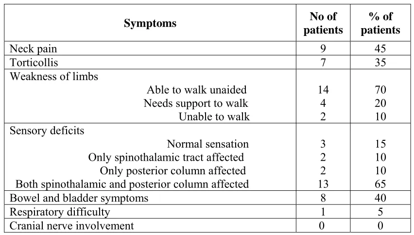
19 out of 20 patients (95%) presented with features of high cervical myelopathy. 45% of them had neck pain and 35% had torticollis. Fourteen patients were able to walk unaided (70%), four needed support to walk (20%) and two patients were unable to walk even with support (10%). Both spinothalamic and posterior column sensations were affected in 13 patients (65%), posterior column sensations were affected in two patients (10%) and two patients (10%) had only spinothalamic tract involvement. Three patients (15%) had normal sensation. Eight patients (40%) had bowel and bladder symptoms. None had lower cranial nerve symptoms and one patient (5%) had features of respiratory embarrassment which improved postoperatively. He did not require ventilator support postoperatively.
Follow-up:
follow up none of these patients had respiratory embarrassment. Overall, 88.8% patients had improvement in their clinical symptoms and signs. The various clinical features are summarized in Table 2.
Table 2 Preoperative symptoms:
Symptoms No of
patients
% of patients
Neck pain 9 45
Torticollis 7 35
Weakness of limbs
Able to walk unaided Needs support to walk Unable to walk
14 4 2 70 20 10 Sensory deficits Normal sensation Only spinothalamic tract affected Only posterior column affected Both spinothalamic and posterior column affected
3 2 2 13 15 10 10 65
Bowel and bladder symptoms 8 40
Respiratory difficulty 1 5
Cranial nerve involvement 0 0
Functional scores:
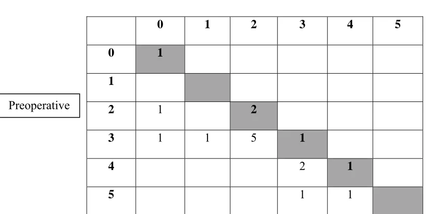
Table.3 Nurick grade
[image:43.612.82.504.143.355.2]
Table 3 shows that 12 patients had at least a grade 1 improvement in their Nuricks score at follow-up while 5 patients (those along the diagonal) had the same Nuricks grade before surgery and at follow-up.
0 1 2 3 4 5
0 1
1
2 1 2
3 1 1 5 1
4 2 1
5 1 1
Follow-up
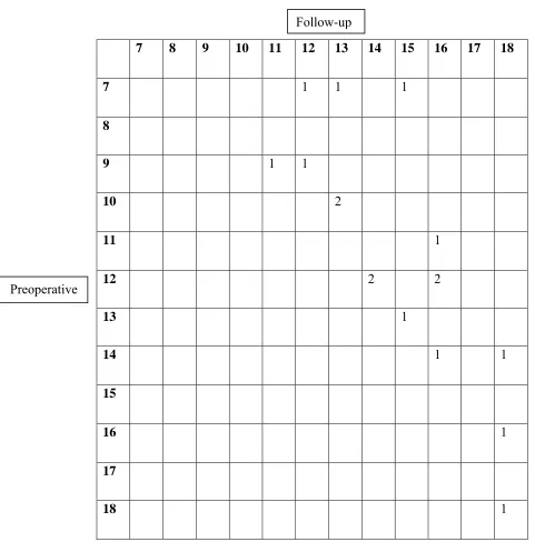
Table. 4 JOA score.
Table 4 shows that all the patients had a significant improvement of JOA score at follow-up. 7 8 9 10 11 12 13 14 15 16 17 18
7 1 1 1
8
9 1 1
10 2
11 1
12 2 2
13 1
14 1 1
15
16 1
17
18 1
Follow-up
Realignment in immediate postoperative and at follow-up scans:
The following (Fig 8) are the illustrated examples of various craniometry findings measured before and after surgery.
Figures 8: Preoperative and postoperative images
1. Chamberlain’s line
2. Mac Rae’s line
This figures shows excellent
3. Wackenheim’s Clival line
4. Clivus canal angle
5. Atlantoaxial distance
6. Sagittal canal diameter or (Space Available for Cord)
7. Modified Omega angle
This figure shows the reduction of
atlantoaxial distance after surgery
This figure shows the increased space available for cord after surgery.
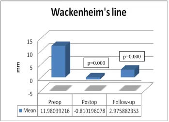
Figure 9: Mean value (of 3 Observers) of the relation of the odontoid to Wackenheim’s line in 17 patients.
The mean levels of the odontoid tip above Wackenheim’s Clival line on preoperative, postoperative and at follow-up scans are 11.9+ 5.2, -0.81+ 5.0and 2.97+ 5.6 respectively. The minus sign indicates the odontoid tip is below the particular line. The difference of 12.5+ 4.4mm is achieved due to surgery; however the odontoid has gone back by 3.7+ 4.8 mm at the follow-up scans. This is depicted in Figure 9.
p=0.000
Craniometry measurements:
Table 5 Level of odontoid in relation to Wackenheim’s line preoperatively, immediate postop and at follow-up in all patients (mean value of 3 Observers).
Change I indicates the difference between the level of odontoid before and immediately after surgery. Change II indicates the difference at the follow-up scan compared to preoperative scan. The patient no 9 showed worsening at follow-up who required a resurgery.
Wilcoxon signed-rank test p=0.000 p=0.000
S. No Preop Immediate
Postop Change 1 Follow-up Change 2
1 6.7 -8.3 15 -3 9.4 2 14 7.1 7.1 9.2 5
3 11 1.83 9.1 4 6.9
4 16 2.97 13 0.3 16 5 15 0 15 5.1 9.5 6 12 -14 23 2 10 7 7.2 -1.2 8.4 -1 7.8 8 6.6 -2.3 8.9 0.7 5.9
9 14 1.65 13 16 -1.2
10 14 -2.1 16 2.3 11 11 10 1.33 8.6 4.5 5.4 12 5.1 -0.5 5.6 -0 5.5 13 10 -3.2 14 -3 13 14 27 6.57 20 14 13 15 6.8 -5.4 12 -2 8.4 16 12 0 12 0.6 11 17 16 1.53 14 0 16
18 5.1 -4.4 9.5 N.A N.A
19 16 4.37 12 N.A N.A
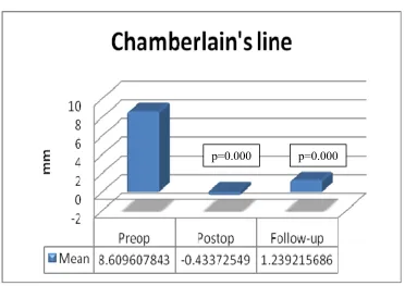
Figure 10: Mean value (of 3 Observers) of the relation of the odontoid to Chamberlain’s line in 17 patients.
The mean level of odontoid tip in relation to Chamberlain’s line at on preoperative, postoperative and at follow-up scans are 8.60+ 4.3, -0.43+ 4.0and 1.23+ 4.28 mm respectively and a mean change of 9 + 2.7 mm was achieved due to surgery. There was ascending of odontoid tip by 1.7+ 3.3mm at follow-up scans. This is depicted in Figure 10.
Table 6 Level of odontoid in relation to Chamberlain’s line preoperatively, immediate postop and at follow-up in all patients (mean value of 3 Observers).
All the patients showed a good reduction at the follow-up scan except patient no 2 who did not have worsening of neurological status at follow-up.
Wilcoxon signed-rank test p=0.000 p=0.000 S.No Preop Immediate Postop Change 1 Follow-up Change 2
1 5.7 -4.4 10 -3 8.2
2 8.6 1.53 7.1 12 -2.9
3 9.5 2.56 7 0 9.5
4 6.9 1.27 5.6 0.4 6.5
5 14 5.57 8.7 7.4 6.8
6 7.1 -1.7 8.7 3.2 3.9 7 10 -0.3 11 1.8 8.4 8 3.4 -0.8 4.3 -1 4.4 9 -0.1 -8.4 8.4 -4 4.4 10 5.8 -7.1 13 -4 9.6 11 7.8 -3.8 12 0 7.8 12 12 1.1 11 0 14 13 12 3.36 8.8 -2 14 14 17 1.43 15 2.8 14 15 6.2 -1.8 7.9 0 6.2 16 5.2 -2.5 7.7 0 5.3 17 14 6.64 7.8 7.9 6.5
18 14 -10 24 N.A N.A
19 17 5.69 11 N.A N.A
Figure 11: Mean value (of 3 Observers) of the relation of the odontoid to Mac Rae’s line in 17 patients.
It was seen that the odontoid was 3.9 + 5.7 mm above Mac Rae’s line preoperatively, while in the immediate postoperative period it was 4.7 mm below this line. The odontoid tip is brought down by 8.6+ 6.3 mm postoperatively and it is 6.1+ 3.7 mm below at the follow-up scans when compared to its position in the preoperative scan in relation to Mac Rae’s line. This is depicted in Figure 11.
Table 7 shows the level of odontoid in relation to Mac Rae’s line in all patients (mean value of 3 Observers)
All the patients showed good reduction of odontoid in relation to Mac Rae’s line except patient no 2 who did not require a intervention because of stable neurological status.
Wilcoxon signed-rank test p=0.000 p=0.000
S.No Preop Immediate
Postop Change 1 Follow-up Change 2
1 -0.2 -11 11 -3.5 3.3
2 3.06 -3.3 6.4 3.1 -0
3 10.3 -0.8 11 3.33 6.9
4 2.2 -2.6 4.8 -2.4 4.6
5 7.13 -1.9 9.1 0.67 6.5 6 0.76 -10 11 -5.9 6.7 7 3.03 -4.5 7.6 -1.5 4.5
8 0.2 -7.2 7.4 -9.6 9.8
Figure 12: Mean value (of 3 Observers) of the atlantoaxial distance in 17 patients.
It was shown that the mean value of atlantoaxial distance before surgery was 7.1 + 2.4 and it was 3.2 + 2.7 mm postoperatively and it is increased to 4.3 + 3.2 mm at follow-up. The difference of 2.83+ 2.6 mm is achieved due to realignment surgery at the follow-up scans. This is depicted in Figure 12.
p=0.000
Table 8 shows the atlantoaxial distance in all patients (mean value of 3 Observers)
Patient no. 2 and 9 showed increase in the atlantoaxial distance at the follow-up scan. Rest of the patients showed a decrease in the atlantoaxial distance after surgery and at follow-up. As already mentioned patient no 9 underwent a resurgery whereas the patient no 2 was stable neurologically at the follow-up and he did not require any intervention.
Wilcoxon signed-rank test p=0.000 p=0.002
S.No Preop Immediate
Postop Change 1 Follow-up Change 2
1 6.13 5.33 0.8 5.1 1
2 11.5 10.3 1.2 13.3 -2
3 6.21 1.1 5.1 3.03 3.2
4 12 6.2 5.8 4.77 7.2
5 8.63 1.13 7.5 3.7 4.9
6 9.13 2.77 6.4 6.87 2.3
7 5.93 0.83 5.1 1.87 4.1
8 5.33 0.9 4.4 0 5.3
9 4.95 3.3 1.6 8.08 -3
10 11.4 5.71 5.7 7.35 4.1 11 4.53 1.95 2.6 2.4 2.1 12 6.24 4.95 1.3 4.67 1.6 13 6.48 2.73 3.8 2.37 4.1 14 3.87 5.67 -2 3.87 0 15 7.33 0.67 6.7 2.19 5.1 16 6.37 1.07 5.3 3.2 3.2 17 5.65 0.33 5.3 0.73 4.9 18 6.47 2.3 4.2 N.A N.A
19 3.5 3.2 0.3 N.A N.A
Figure 13: Mean value (of 3 Observers) of the sagittal canal diameter in 17 patients.
It was shown that the mean value of sagittal canal diameter before surgery was 10.5 + 2.9 and it was increased to 18.5 + 4 mm postoperatively and it is decreased to 16 + 4 mm at follow-up. The difference in the increase in the size of the canal was 5.55+ 3.6 mm at the follow-follow-up. This is depicted in Figure 13.
p=0.000
Table 9 shows the sagittal canal diameter in all patients (mean value of 3 Observers)
All the patients had a significant increase in the size of sagittal canal diameter which is the space available for the cord except the patient no 6 who had worsening of the space at the follow-up scan. She had a very good neurological recovery at the last follow-up.
Wilcoxon signed-rank test p=0.000 p=0.000 S.No Preop Immediate Postop Change 1 Follow-up Change 2
1 8.5 18.8 10 18 9.3
2 12 16.4 4.6 13 1.7
3 8.2 16.9 8.7 18 9.5
4 7.6 14.7 7.1 16 8.6
5 9.5 19.5 10 16 7
6 10 21.1 11 9.3 -0.9
7 11 15.7 4.9 13 2.6
8 12 16.8 4.7 18 6
9 6.4 13 6.6 7.8 1.4 10 12 22 10 17 5.2 11 17 23.3 6.3 22 5.1 12 14 20.7 6.3 17 2.6 13 7.8 13.7 5.9 13 5.7 14 9.7 12.9 3.2 11 1.8 15 7.5 18.6 11 17 9.5 16 15 26.2 11 22 6.9 17 9.1 24.1 15 22 13
18 12 18.6 6.2 N.A N.A
19 12 16.2 4.1 N.A N.A
Figure 14: Mean value (of 3 Observers) of the Clival canal angle in 17 patients.
It was shown that the mean value of clival canal angle before surgery was 119 + 17 degrees and it was increased to 143.1 + 12.8 degrees postoperatively and it is decreased to 134 + 18 degrees at the follow-up. The change in the angle at the follow-up scan, 15.4 + 10.2 degrees is achieved due to surgery. This is depicted in the Figure 14
Table 10 shows the clival canal angle in all patients (mean value of 3 Observers)
All the patients showed increase in the clival canal angle which is a consistent finding among all the craniometry measured. This is due to downward as well as anterior realignment of odontoid due to surgery.
Wilcoxon signed-rank test p=0.000 p=0.000 S.No Preop Immediate Postop Change 1 Follow-up Change 2
Figure 15: Mean value (of 3 Observers) of the Modified Omega canal angle in 17 patients.
It was shown that the mean value of Modified Omega angle before surgery was 76.3 + 16 degrees and it was increased to 86 + 11.8 degrees postoperatively and it is decreased to 79 + 12.1 degrees at follow-up. This is depicted in Figure 15.
p=0.001
Table 11 shows the modified omega angle in all patients (mean value of 3 Observers)
Patient no. 2, 5, 8, and 16 showed minimal decrease in the angle at the follow-up scans. The patients who had an increase in the angle at the follow-up scans are shown to have no significance. This change in the angle is very inconsistent among the craniometry measured.
Wilcoxon signed-rank test p=0.001 p=0.124
S.No Preop Immediate
Postop Change 1 Follow-up Change 2
1 60 81.7 21 85 25
2 60 60.3 0 55 -4.9
3 79 86.9 8.1 80 1.5
4 70 91.2 21 85 15
5 100 92.1 -8 88 -12
6 64 89.1 25 64 0.3 7 77 87.6 11 78 1.6
8 96 104 8.6 74 -22
9 37 59.1 22 53 15
10 83 94.3 12 89 5.9 11 74 85.9 12 77 3.6 12 84 91.6 7.2 89 4.2
13 77 74.7 -2.1 82 5.5
14 82 88.5 6.9 82 0.3 15 66 88 22 73 7
16 95 89.2 -6.1 92 -3.5
17 94 97.9 4.1 96 2.7
18 80 88 7.9 N.A N.A
19 77 81.3 4.1 N.A N.A
Inter class correlation (ICC):
The inter class correlation between all the three observers was calculated based on the preoperative and follow-up craniometry data are shown in Table 12.
Table 12
ICC between observers based on preoperative and the follow-up craniometry findings:
Variables ICC ICC
Wackenheim’s Clival line .849 .881
Chamberlain’s line .885 .889
Mac Rae’s line .718 .787
Atlantoaxialdistance .627 .922
Space Available for Cord .825 .826
Clival canal angle .853 .941
Modified omega angle .859 .308
DISCUSSION:
Craniovertebral anomalies are frequently found in the Indian subcontinent particularly in Uttar Pradesh, Bihar, Rajasthan and parts of Gujarat (4). The surgical management of congenital craniovertebral anomalies is complex due to the relative difficulty in accessing the region, critical relationships of neurovascular structures and the biomechanical issues involved.
Morbidity of transoral surgery:
unrelated deaths, one due to a myocardial infarction and the other was due to septicemia among 72 patients of a transoral series. Goel had a morbidity of 1% in his transoral series of 99 (10).
Pathogenesis of basilar invagination:
Goel et al speculated that basilar invagination is probably secondary to “slippage” of the atlas over the axis (31). This slippage can be severe enough to cause spondyloptosis of the atlas over the axis. These authors reported a novel surgical technique in which the cranially migrated odontoid is brought down to its normal relationship with the atlas and fixed in position. In their technique a spacer is connected to a stainless steel plate and is used for distracting the C1-2 joint. The mean reduction of the odontoid achieved was 7.5, 8.7, and 6.2 mm corresponding to Wackenheim’s, Chamberlain’s and Mac Rae’s line respectively (31). All their patients improved postoperatively and there were no complications reported in their series. They considered distraction and fusion surgery superior to other treatment options for basilar invagination as it is deals directly with the pathogenesis of basilar invagination and has less morbidity.
Basis for our study:
Figure 16 Implants and instruments used in craniovertebral junction realignment surgery Types of spacers
Parallel
Convex
Lordotic
Trial spacer Spacer holder
Rod
Locking caps
Adjustable drill guide
Occipital clamp
Lateral mass screws Rod reducer
Depth gauge
C 1
Description of surgical technique:
Clinical outcome:
On follow-up of 7-24 (13.1+5.23) months, there was significant improvement in neurological deficits. There was improvement of neck pain; torticollis was corrected in all these patients. All patients were able to walk unaided but there were five (25%) who had some sensory deficits. In general 88.8% of our patients had improvement in their clinical symptoms and signs. There are 12 patients who had improvement by at least 1 grade at follow-up. Three patients (two patients who needed support to walk and one was bed bound) are able to walk without support at follow-up. These functional scores are neither assessed nor compared before surgery and at follow up in other studies (4). These results are comparable to the study done by Goel et al (4). In their series, neck pain was observed in 77% and torticollis in 41% of their patients and all of them improved postoperatively. The preoperative and follow-up Nuricks grade and JOA score are plotted in table 3.
Radiological outcome: Immediate:
Follow up radiology:
During the up period (13.1+5.23 months) the craniometry findings in the follow-up scans showed a slight change as compared to the immediate postop CT scan indicating settling. When early CT scan are done 1-3 days after surgery most patients may not be fully ambulated and this may not really reflect the true position of the odontoid in relation to all the lines and it seems to reduce on follow-up. However the importance of the immediate postoperative CT is to show good alignment before sending the patient home. Patients tend to have some degree of cranial settling in the follow-up scans when compared to the immediate post operative scans but bony fusion occurred in most of them (94%). One patient had significant worsening of the craniometry values when compared to the immediate postoperative scan along with worsening symptoms. He underwent realignment of C1 and C2 and posterior decompression following which his neurological status was stable.
The mean preoperative and follow-up craniometry subjected to statistical methods was shown statistically significant by all the observers except the modified omega angle which was not statistically significant. The modified omega angle corresponds to the anteroposterior tilt of the odontoid and postoperatively this angle did not change enough to cause statistical significance.
Wackenheim’s line:
Chamberlain’s line:
The mean reduction of odontoid tip in relation to Chamberlain’s line is 7.46 + 4.2 mm which is comparable to the results by Goel et al which is 8.7 mm.
Mac Rae’s line:
The mean reduction of the odontoid in relation to Mac Rae’s line is 6.1 + 3.7 which is comparable to achieved by Goel et al which is 6.2+ mm corresponding to Mac Rae’s line (31). Atlantoaxial distance:
The mean reduction of atlantoaxial distance achieved due to realignment surgery is 2.83+ 2.6 mm which is 4.7 mm mean by Goel et al (31).
Space available for cord or Sagittal canal diameter:
The space available for the cord is increased to 16 + 4.1 mm due to surgery at the follow-up scans. The difference in the increase in the size of the canal was 5.55+ 3.6 mm. This extensive increase in the size of the canal is directly proportional to the excellent clinical improvement noticed in these patients.
Clival canal angle:
The mean Clival canal angle at follow up is 134.44 + 18 degrees which is the normal range (130-150 degrees). The change in the angle due to surgery is 15.5+ 10.2 degrees. This indicates the downward as well as anterior realignment of odontoid due to surgery.
Modified omega angle:
Interoberver correlation coefficient:
ICC analysis showed an excellent agreement between observers in measuring various craniometries. The preoperative atlantoaxial distance had a agreement of only 0.627 and at the follow-up scan is .922. The reason for this excellent agreement is the atlantoaxial distance value is close to zero in the follow-up scans. The follow-up modified omega had a value of .308 which is a poor agreement. This poor agreement is probably because in some of the postoperative scans the anterior end of the hard palate is not adequately covered to draw the tangential line which passes through the base of C2 body.
Complications:
Reoperation:
In our series, one out of 20 patients underwent re-alignment after first surgery. He presented with Nuricks grade of 4 and JOA score of 7/15. Immediate postoperative period his Nuricks grade improved to 3. On follow up after 1 year his Nuricks grade improved to 3 and JOA to 13/18. Although there was improvement of his tightness and weakness of limbs and sensory deficits he continued to have worsening of bowel and bladder symptoms. There was worsening of craniometry findings when compared to the scan done in the immediate postoperative period as shown in Fig 17.
[image:72.612.74.524.405.612.2]Preoperative Immediate postoperative Follow-up A B C
His neurological status stabilized after the second surgery. This is probably due to the implant related complications which is shown in figure 18 A. The spacer was placed more anteriorly in the left C1-2 joint which probably caused the reinvagination.
Figure 18 A
B
Immediate postoperative scan after the 2 nd surgery showing
realignment of craniovertebral junction and widening of the space available for the cord.
Realignment surgery in comparison with transoral odontoidectomy:
CONCLUSION:
Bibliography
1. Klimo P Jr, Rao G, Brockmeyer D. Congenital anomalies of the cervical spine. Neurosurg Clin N Am. 2007 Jul;18(3):463-78. Review.
2. Menezes AH, Traynelis VC. Anatomy and biomechanics of normal craniovertebral junction (a) and biomechanics of stabilization (b). Childs Nerv Syst. 2008 Oct;24(10):1091-100.
3. MenezesAH Developmental abnormalities of the craniocervical
junction. In: Winn RH (5ed) Youmans neurological surgery, pp 3331-3345.
4. Goel A. Basilar invagination, Chiari malformation, syringomyelia: a review.Neurol India. 2009 May-Jun;57(3):235-46. Review.
5. Goel A. Progressive basilar invagination after transoral odontoidectomy: treatment by atlantoaxial facet distraction and craniovertebral realignment. Spine. 2005 Sep 15;30(18):E551-5.
6. Chamberlain WE: Basilar impression (Platybasia). A bizarre developmental anomaly of the occipital bone and upper cervical spine with striking and misleading neurologic manifestations. Yale J Biol Med 1939 11:487–496.
7. McRae DL: Bony abnormalities in the region of foramen magnum: correlation of the anatomic and neurologic findings. Acta Radiol 1953 40:335–354.
8. Smoker WR. Craniovertebral junction: normal anatomy, craniometry, and congenital anomalies. Radiographics. 1994 Mar;14(2):255-77.
9. Thiebaut F, Wackenheim A, Vrousos C: [New median sagittal pneumostratigraphical finding concerning the posterior fossa.] J Radiol Electrol Med Nucl 1961 42:1–7.
10. Goel A, Bhatjiwale M, Desai K. Basilar invagination: a study based on 190 surgically treated patients. J Neurosurg. 1998 Jun;88(6):962-8.
11. Laborde G, Gilsbach J, Bertalanffy H, Harders A, Hardenack M. Limits of the transoral approach in craniospinal malformations. Skull Base Surg. 1992;2(1):6-10.
12. Curtis A, Dickman, Jacqueline Locantro, Richarrd G Fessler: The influence of transoral odontoid resection on stability of craniovertebral junction. J Neurosurg 1992
77:525-530.
13.Mummaneni PV, Haid RW. Transoral odontoidectomy. Neurosurgery. 2005 May;56(5):1045-50.
15. Jain VK , Behari S, Banerji D, Bhargava V, Chhabra D. Transoral decompression for craniovertebral osseous anomalies : perioperative management dilemmas. Neurology India 1999 Volume 47 Issue 3 p.188.
16. Menezes AH, VanGilder JC. Transoral-transpharyngeal approach to the anterior craniocervical junction. Ten-year experience with 72 patients. J Neurosurg. 1988 Dec;69(6):895-903.
17.Apuzzo ML, Weiss MH, Heiden JS: Transoral exposure of the atlantoaxial region. Neurosurgery 1978 3:201–207.
18. Di Lorenzo N: Craniocervical junction malformation treated by transoral approach. A survey of 25 cases with emphasis on postoperative instability and outcome. Acta Neurochir (Wien)1992 118:112–116.
19.Cantarella G, Mazzola RF, Benincasa A: A possible sequela of transoral approach to the upper cervical spine. Velopharyngeal incompetence. J Neurosurg Sci 1998 42:51–55. 20. Kingdom TT, Nockels RP, Kaplan MJ: Transoral-transpharyngeal approach to the
craniocervical junction. Otolaryngol Head Neck Surg 1995 113:393–400.
21. Shaha AR, Johnson R, Miller J, Milhorat T: Transoral-transpharyngeal approach to the upper cervical vertebrae. Am J Surg 1993 166:336–340.
22.Hadley MN, Spetzler RF, Sonntag VK.: The transoral approach to the superior cervical spine. J Neurosurg. 1989 Jul;71(1):16-23.
23.Naderi S, Pamir MN. Further cranial settling of the upper cervical spine following odontoidectomy. Report of two cases. J Neurosurg. 2001 Oct;95(2 Suppl):246-9.
24. Tuite GF, Veres R, Crockard A et al : Pediatric transoral surgery : Indications, complications and long term outcome. J Neurosurg 1996; 84 : 573-583.
25. Naderi S, Crawford NR, Melton S, et al: Biomechanical analysis of cranial settling after transoral odontoidectomy. Neurosurg Focus 1999 6(6):Article 7.
26. Frempong-Boadu AK, Faunce WA, Fessler RG. Endoscopically assisted transoral-transpharyngeal approach to the craniovertebral junction. Neurosurgery. 2002 Nov;51(5 Suppl):S60-6.
27. Landeiro JA, Boechat S, Christoph Dde H, Gonçalves MB, Castro I, Lapenta MA, Ribeiro CH. Transoral approach to the craniovertebral junction. Arq Neuropsiquiatr. 2007 Dec;65(4B):1166-71.
29.Simsek S, Yigitkanli K, Belen D, Bavbek M. Halo traction in basilar invagination: technical case report. Surg Neurol. 2006 Sep;66(3):311-4.
30. Joseph V, Rajshekhar V. Resolution of syringomyelia and basilar invagination after traction. J Neurosurg (Spine 3) 2003;98:298.
31. Goel.A Treatment of basilar invagination by atlantoaxial joint distraction and direct lateral mass fixation. J Neurosurg Spine. 2004 Oct;1(3):281-6.
32.Goel A, Sharma P. Craniovertebral junction realignment for the treatment of basilar invagination with syringomyelia: preliminary report of 12 cases. Neurol Med Chir (Tokyo). 2005 Oct;45(10):512-7.
33. Goel A, Sharma P. Craniovertebral realignment for basilar invagination and atlantoaxial dislocation secondary to rheumatoid arthritis. Neurol India. 2004 Sep;52(3):338-41.
34. Mummaneni PV, Haid RW Atlantoaxial fixation: overview of all techniques. Neurol India. 2005 Dec;53(4):408-15.
35. Coyne TJ, Fehlings MG, Wallace MC, Bernstein M, Tator CH. C1-C2 Posterior Cervical Fusion: Long Term Evaluation of Results and Efficacy. Neurosurg 1995; 37:688-693.
36. Hanley EN, Harvell JC (1992) Immediate postoperative stability of atlantoaxial
articulation: a biomechanical study comparing simple midline wiring and the Gallie and Brooks procedures. J Spinal Disord 5:306.
37. Grob D, Crisco JJ, Panjabi MM (1992) Biomechanical evaluation of four different
atlantoaxial fixation techniques. Spine 17:480.
38. Oda I, Abumi K, Sell LC, Haggerty CJ, Cunningham BW, McAfee PC (1999)
Biomechanical evaluation of five different occipito-atlanto-axial fixation techniques. Spine 24:2377–2382.
39.Moorthy RK, Rajshekhar V . Changes in cervical spine curvature in pediatric patients following occipitocervical fusion. Childs Nerv Syst. 2009 Aug;25(8):961-7.
40. Goel A, Muzumdar D, Dange N. One stage reduction and fixation for atlantoaxial spondyloptosis: report of four cases British journal of Neurosurgery 2006 Aug 20(4) 209-213.
Appendix -1
C1-2 DISTRACTION AND FUSION PROFORMA
Name- Hospital no- Age- Sex Occupation
Address-
Nuricks grade-
JOA score-
CLINICAL FEATURES: Neck pain: Present / absent
Torticollis/ restricted neck movements: Present / Absent. Weakness of limbs:
Able to walk unaided/ Needs support to walk/ Unable to walk with support Sensation:
Normal sensation/Spinothalamic function affected/ Posterior column affected/ Both spinothalamic and posterior column affected.
Bowel/bladder symptoms- Present / Absent. Cranial nerve involvement – Present/ Absent Respiratory embarrassment- Present/ Absent. History of trauma- Present/Absent
History suggestive of infection (TB) -Present/ Absent
Preoperative traction applied- Yes/ No Improvement of neck pain- Yes/ no
Improvement of weakness of limbs Yes/ no Improvement of sensation- Yes/ no
Improvement of respiratory embarrassment - Yes/ no
OPERATION DONE
C1-2 DISTRACTION WITH SPACER AND POSTERIOR FUSION WITH 1--3 /1-2 LATERAL MASS SCREW AND ROD FIXATION
Size of spacer:
Right Left
Postoperative clinical findings; Improvement of neck pain- Yes/ no
Improvement of weakness of limbs Yes/ no Improvement of sensation- Yes/ no
Improvement of respiratory embarrassment - Yes/ no Radiographic findings:
MRI findings
CT FINDINGS
CT findings Preoperative Postoperative
Wackenheim’s line Above /below
mm
Above/below mm
Chamberlain’s line mm mm
Mc Rae’s line Above/below
mm
Above/below mm Atlantoaxial distance mm mm
Clival canal angle Omega angle. Sagittal canal diameter
C1-2 Distraction follow up proforma Follow up in months ---
Symptoms
Neck pain – Improved/static / worsened
Weakness of limbs – Improved/static / worsened Sensory symptoms – Improved/static / worsened
Bowel or bladder symptoms- – Improved/static / worsened Respiratory embarrassment – Improved/static / worsened New symptoms present/ absent
Nuricks grade- JOA score- CT FINDINGS
CT findings preop -- months follow up Wackenheim’s line Above /below
mm
Above/below mm
Chamberlain’s line mm mm Mc Rae’s line Above/below mm Above/below mm
Atlantoaxial distance mm mm Clival canal angle
Omega angle