Copyright © 1999, American Society for Microbiology. All Rights Reserved.
Microglia Express CCR5, CXCR4, and CCR3, but of These, CCR5 Is
the Principal Coreceptor for Human Immunodeficiency
Virus Type 1 Dementia Isolates
ANDREW V. ALBRIGHT,
1JOSEPH T. C. SHIEH,
1TAKAYUKI ITOH,
2BENHUR LEE,
3DAVID PLEASURE,
2MICHAEL J. O’CONNOR,
4ROBERT W. DOMS,
3AND
FRANCISCO GONZA
´ LEZ-SCARANO
1*
Departments of Neurology and Microbiology
1and Department of Pathology and Laboratory Medicine,
3University of
Pennsylvania School of Medicine, Division of Neurology, The Children’s Hospital of Philadelphia,
2and Department of Neurosurgery, Thomas Jefferson University School
of Medicine,
4Philadelphia, Pennsylvania
Received 22 July 1998/Accepted 9 October 1998
Microglia are the main human immunodeficiency virus (HIV) reservoir in the central nervous system and
most likely play a major role in the development of HIV dementia (HIVD). To characterize human adult
mi-croglial chemokine receptors, we analyzed the expression and calcium signaling of CCR5, CCR3, and CXCR4
and their roles in HIV entry. Microglia expressed higher levels of CCR5 than of either CCR3 or CXCR4. Of
these three chemokine receptors, only CCR5 and CXCR4 were able to transduce a signal in microglia in
response to their respective ligands, MIP-1
b
and SDF-1
a
, as recorded by single-cell calcium flux experiments.
We also found that CCR5 is the predominant coreceptor used for infection of human adult microglia by the
HIV type 1 dementia isolates HIV-1
DS-br, HIV-1
RC-br, and HIV-1
YU-2, since the anti-CCR5 antibody 2D7 was
able to dramatically inhibit microglial infection by both wild-type and single-round luciferase pseudotype
reporter viruses. Anti-CCR3 (7B11) and anti-CXCR4 (12G5) antibodies had little or no effect on infection.
Last, we found that virus pseudotyped with the DS-br and RC-br envelopes can infect cells transfected with
CD4 in conjunction with the G-protein-coupled receptors APJ, CCR8, and GPR15, which have been previously
implicated in HIV entry.
Human immunodeficiency virus (HIV) dementia (HIVD) is
a central nervous system (CNS) complication that affects 20 to
30% of individuals infected with HIV and is a defining
condi-tion for AIDS (24). The underlying cause of HIVD is
un-known, but since productive HIV infection in the CNS
oc-curs mostly in microglia, or brain macrophages, it is generally
thought that these cells play a key role in the development of
neurological abnormalities. HIVD might then be caused by
neuronal damage or dysfunction resulting from the release of
putative neurotoxic products by infected microglia or,
alterna-tively, by neuronal interaction with viral proteins released or
expressed by the infected cells.
The propensity for certain viral isolates to infect the CNS
and mediate neuronal damage is one of the major unanswered
questions of HIVD. A proportion of HIV isolates replicate in
cultured microglia (44), resulting in prominent syncytial
for-mation, which is an important signature of HIV replication in
the CNS (39). This cytopathology is presumably the result of
membrane fusion between microglia mediated by HIVD
en-velope proteins.
Cellular entry by HIV is now known to require at least two
cell membrane proteins, CD4, and one of several
seven-trans-membrane domain G-protein-coupled receptors (GPCRs),
principally CXCR4, an
a
-chemokine receptor, and CCR5,
whose natural ligands are
b
-chemokines (7). CXCR4 mediates
infection of T-tropic HIV strains, i.e., those, that replicate in
T-cell lines, whereas CCR5 is the most important coreceptor
for M-tropic strains, which replicate both in monocyte-derived
macrophages (MDM) and in microglia. Studies with cultured
fetal and adult microglia have shown that CCR5 is sufficient for
HIV entry (19, 43). The role of CCR3, another
b
-chemokine
receptor, is more controversial. Several HIVD isolates isolated
from the CNS can use CCR3 to enter cells dually transfected
with CCR3 and CD4 and to enter fetal microglia, which
ex-press CCR3 on their cell surface. However, studies that
exam-ined the inhibition of microglial infection by CCR3
anti-bodies or the CCR3 ligand eotaxin have yielded conflicting
results (16, 19). Microglia also express CXCR4 in vivo and
in vitro (27), but in general T-tropic strains do not replicate
very well in microglia or MDM (42, 48). Whether microglial
GPCRs can respond to their natural chemokine ligands, and
what role signal transduction may play in HIV infection of
microglia or CNS pathogenesis, is thus far unknown.
Recent studies have demonstrated that HIV and simian
immunodeficiency virus (SIV) envelopes can also use other
GPCRs, besides CCR5, CCR3, and CXCR4, for viral entry
and fusion. Among these are CCR8 (21, 40), the receptor for
I309, and the orphan receptors GPR1 (8, 12), GPR15 (6, 8,
12), STRL33 (6, 8, 29), and APJ (3, 10). The mRNAs for
GPR1 (31) and APJ (3, 32, 36) are expressed in the brain, but
their cellular localization is unknown. Choe and colleagues
have recently demonstrated that APJ is not utilized by the
HIVD isolates JrFL and YU-2 (3), although JrFL has been
reported to use STRL33 (29) and YU-2 utilizes GPR15 (6, 12).
Little else is known regarding the ability of HIVD envelopes to
use CCR8 or orphan receptors as HIV coreceptors. However,
it is quite conceivable that preferential replication in the brain
is a consequence of the utilization of one or more of these
alternate coreceptors by HIV isolates.
* Corresponding author. Mailing address: University of
Pennsylva-nia, Department of Neurology, Clinical Research Building, 415 Curie
Blvd., Room 264, Philadelphia, PA 19104-6146. Phone: (215)
662-3389. Fax: (215) 573-2029. E-mail: Scarano@mail.med.upenn.edu.
205
on November 9, 2019 by guest
http://jvi.asm.org/
To begin to develop a more detailed understanding of the
role of each of the established coreceptors (CCR5, CCR3, and
CXCR4) in HIV entry into adult microglia, we have assayed
their surface expression by flow cytometry. We have also
ad-dressed the functionality of these GPCRs by determining the
microglial response to
a
- and
b
-chemokines. To determine
whether viruses obtained from the brain can use CCR5, CCR3,
or CXCR4 as a coreceptor, we have looked at infection with
pseudotyped viruses expressing the luciferase reporter gene, as
well as with wild-type viruses.
MATERIALS AND METHODS
Cells.Microglial cultures were prepared as previously described from fresh adult human brain tissue obtained during temporal lobectomy for medication-resistant epilepsy (1, 43, 44, 49). Microglia were cultured in 10% Dulbecco modified Eagle medium (DMEM; GIBCO-BRL) with 5% heat-inactivated fetal calf serum (FCS; Atlanta Biologicals, Norcross, Ga.), 5% Giant Cell Tumor Supernatant (Fisher), 50mg of gentamicin (GIBCO-BRL)/ml, and 1 mM sodium pyruvate. 293T and U87 cells were cultured in DMEM with 10% FCS (43).
Detection of cell surface CCR5 and CCR3.Microglial cells were cultured for 5 to 7 days prior to staining for CCR5 or CCR3. All staining steps were per-formed on ice. For each staining condition, 83105cells were washed with 13
phosphate-buffered saline (PBS), without Ca21or Mg21(GIBCO-BRL), and
detached by treatment with 0.5 mM EDTA and mechanical dissociation. Cells were immediately diluted 15-fold in DMEM with 10% FCS and then centrifuged for 5 min at 1,000 rpm in a Beckman tabletop centrifuge. The supernatant was removed; the cells were resuspended in staining buffer with blocking solution (PBS with 0.1% bovine serum albumin [BSA], 0.02% sodium azide, and 8% rabbit serum), divided into staining tubes (Robbins Scientific, Sunnyvale, Calif.), and washed; and 50ml of primary antibody was added per tube. Cells were incubated on ice for 45 min with the following primary antibodies diluted in staining buffer (PBS with 0.1% BSA and 0.02% sodium azide): 807.09 isotype control (17), 7B11 (anti-CCR3) (20), and 2D7 (anti-CCR5) (47) (all at 5mg/ml) and a 1:10 dilution of an anti-major histocompatibility complex MHC class I monoclonal antibody (MAb) supernatant (W6/32) (11, 37). Cells were washed with staining buffer without rabbit serum and were resuspended in 1 drop of heat-inactivated FCS, 25ml of secondary antibody (10mg of rabbit anti-mouse biotin/ml) was added per tube (Dako, Carpinteria, Calif.), and the mixture was incubated on ice for 30 min. Cells were washed and resuspended in 25ml of 1.3
mg of streptavidin/ml coupled to fluorescein isothiocyanate (Dako), incubated on ice for 30 min, washed, and fixed with 300ml of freshly prepared 2% parafor-maldehyde diluted in PBS. U87 and 293T cells transiently transfected with CCR5 and CCR3 plasmids (as described below) were used as controls. All flow cytom-etry analysis was performed on the FACScan (Becton Dickinson) by using CellQuest flow cytometry (Cancer Center, University of Pennsylvania).
Quantification of CCR5, CCR3, and CXCR4 antibody binding sites on micro-glia.Quantitative flow cytometry was performed by converting the mean channel fluorescence into the number of antibody binding sites, or the number of target molecules per cell (if antibody-binding valency is known), by using a standardized microbead kit (Quantum Simply Cellular Microbeads Kit; Sigma, St. Louis, Mo.). This is a mixture of five microbead populations of uniform size, coated with goat anti-mouse antibodies, that have differing abilities to bind mouse antibodies (one bead population has no specific ability to bind mouse immunoglobulin G and is included as a baseline control). Each MAb is then added at saturating amounts to approximately 100,000 beads. After a 1-h incubation, the beads were washed and stained with secondary antibodies in a manner identical to that used for microglia. The beads were then analyzed by using the same instrument settings as those for the microglia. The binding capacities of the stained mi-crobeads were then regressed against the corresponding geometric mean of each bead population. Subsequently, the mean fluorescence intensity of the antigen analyzed on microglia can be converted to the number of antibody binding sites per cell by comparison with the regression curve generated. The parameters of the regression curve permit a determination of the linear deviation and hence provide an estimate of the degree of confidence one should have in the values generated. Regression curves are acceptable only if r.0.995 and the deviation from linearity (average residual percent) is less than 5%.
Microglial response to chemokines.All intracellular calcium level ([Ca21] i)
measurement procedures were performed at room temperature (23 to 25°C). Microglia (23104to 43104) were plated on 22- by 22-mm coverslips and
cultured for 2 days (as described above); coverslips were then inverted and attached to the upper side of a perfusion chamber (RC-21B; Warner Instrument Corp., Hamden, Conn.) that was mounted on the stage of an upright microscope (Optiphot; Nikon, Tokyo, Japan) equipped for epifluorescence. The microglia were then loaded with 5mM fura-2/AM in 0.02% Pluronic F-127 in standard recording medium for 30 min (22). Excess fura-2/AM was washed out of the chamber, and the cells were maintained in standard recording medium for an additional 15 min in order to hydrolyze loaded fura-2/AM completely. Microglial cells were exposed to 30 to 150 nM eotaxin, 60 nM macrophage inflammatory protein 1b(MIP-1b), or 60 nM stromal cell-derived factor 1a(SDF-1a)
(Pep-rotech, Rocky Hill, N.J.) while being alternatively illuminated by a 75-W Xe arc lamp through 340- and 380-nm excitation filters controlled by a computer-assisted filter changing device (LAMBDA-10; Sutter Instrument Co., Novato, Calif.). Emission fluorescence images through a 203objective lens (CF Fluor DL; numerical aperture, 0.75; Nikon) and a 510-nm barrier filter were collected with a SIT camera (C2400-08; Hamamatsu Photonics K. K., Hamamatsu City, Japan) and converted to digital data by an image-processing system (ARGUS-50; Hamamatsu Photonics K. K.). Each frame of a digital image consisted of 512 by 382 pixels. Every frame was stored in a PC-based computer and converted to single-cell temporal 510-nm emission plots.
Preparation of luciferase reporter virus pseudotyped with high-expression HIVD envelope clones.The HIVD env genes DS-br (clone C17), RC-br (clone 56), KJ-br (clone A1), and YU-2 (clone A10) were obtained from viruses from individuals with HIVD or encephalopathy (15, 28). Prior to cloning, these viruses were obtained from brains by cocultivation with MDM (14, 15). They were expanded by a single additional culture in MDM prior to cloning of their enve-lopes (43). The previously cloned enveenve-lopes in pCR3.1-Uni (Invitrogen) (43) were subcloned into pEXV3, a eukaryotic expression plasmid with a simian virus 40 (SV40) promoter (a gift from N. Harel) (34). pEXV3 was linearized by digestion with SmaI (New England Biolabs), and the HIVD envelope genes were removed from pCR3.1-Uni by digestion with PmeI. The HIVD envelope genes were blunt ligated into pEXV3 and cotransfected with pNL-4-3-LucR1E2into
293T cells to generate one-round infectious pseudotype virus as previously de-scribed (5, 43). Other envelopes (BaL and NL43) were obtained from J. Moore (Aaron Diamond AIDS Research Center), and SIVmac251 was obtained from A. Edinger (8).
Infection of U87 cells expressing chemokine and orphan coreceptors.U87 cells (33105/well) were transiently transfected in a six-well plate with 2mg of
pT4 (30), which expresses the CD4 molecule, in each well and 3mg of a plasmid expressing CCR3 (40), CCR5 (5), CXCR4 (13), APJ (10), CCR8 (40, 41), GPR1 or GPR15 (9, 31), or STRL33 (9) in each well by using a calcium phosphate transfection kit (5 Prime33 Prime, Boulder, Colo.) as previously described (43). The following day, 7.53103transfected cells per well were plated in 96-well
plates, incubated overnight, and infected with pseudotype virus in the presence of 8mg of Polybrene (Sigma)/ml. Cultures were exposed to the inocula overnight at 37°C and refed with 200ml of medium. Three days after infection, the cells were lysed with 60ml of luciferase assay buffer (Promega). Luciferase activity was measured by adding 50ml of luciferase assay substrate (Promega) to 50ml of lysate and reading light activity in a Wallac 1450 Microbeta luminometer detec-tor. The light activity is reported as relative light units (RLU) per second.
Infection of microglia with HIVD envelope-pseudotyped viruses in the pres-ence of anti-chemokine receptor antibodies.Microglial cells were cultured in 96-well plates for 2 to 7 days and were then pretreated with anti-CCR5 (2D7), anti-CCR3 (7B11), or anti-CXCR4 (12G5; a gift from J. Hoxie) at 20mg/ml for 45 min at 4°C. The cells were then infected with 200ml of pseudotyped virus for 16 h at 37°C, the medium was replaced, the cells were lysed 4 to 5 days postin-fection with 100ml of lysis buffer, and then 40ml of the lysate was combined with 100ml of luciferase substrate, and the chemiluminescence was read as indicated above.
Inhibition of virus infection of microglia with chemokine receptor anti-bodies.Microglia were preincubated with anti-chemokine receptor antibodies, as described above, and then infected with HIV-1DS-br, HIV-1RC-br(14, 15),
HIV-1YU-2(RF-1)(originally cloned by Li et al. [28] and obtained from R. Fouchier
[University of Pennsylvania]), or the microglia-passaged virus HIV-1BORI-20(44)
at 4 ng of p24gagper well. The next day the inocula were removed, the cells were
washed, and medium was replaced with 5mg of the corresponding antibody/ml. A 2D7 antibody with no azide and low endotoxin levels (Pharmingen) was used in these experiments. The cell cultures were observed for cytopathicity, and the supernatants were assayed for viral p24gagantigen at intervals of several days
with maintenance of antibody throughout (43).
Expression and infection of U87 cells transfected with chemokine receptors (CCR5, CCR3, and CXCR4).U87 cells were transiently transfected with 2mg of CD4 and a total of 1.5 to 2.0mg of pCDNA3.1 and the chemokine receptor CCR5, CCR3, or CXCR4 or a combination of CCR5 plus CXCR4, CCR5 plus CCR3, or CCR3 plus CXCR4. pCDNA3.1 was used to equilibrate the quantity of total DNA used in each transfection. The cells were plated, cultured over-night, infected with HIV env/pNL-4-3-LucR1E2 pseudotyped viruses,
con-structed with the HIVD envs from HIV-1YU-2and HIV-1DS-br, lysed, and
ana-lyzed by a luciferase assay.
RESULTS
Chemokine receptor expression on adult microglia.
To
de-termine whether there were detectable levels of CCR5 or CCR3
on the surfaces of microglia, we used flow cytometry analysis.
Microglia were cultured for 5 to 7 days under the conditions
described in Materials and Methods and were stained with
MAbs directed against each of the chemokine receptors. As
shown in Fig. 1A, there was a prominent shift in the
fluores-cence profile with a MAb against CCR5 (2D7) in comparison
on November 9, 2019 by guest
http://jvi.asm.org/
with an isotype-matched control. CCR3 expression was
con-siderably lower or undetectable in four experiments performed
with different microglial preparations. The histogram in Fig.
1A is a representative experiment where there was a slight shift
with the anti-CCR3 MAb (7B11). CXCR4 expression has been
shown previously in similar microglial preparations (27).
Fig-ures 1B and C are antibody controls using cells transfected
with CCR5 and CCR3 plasmids, respectively.
To quantify the expression of CCR3 on microglial cells, and
to eliminate any potential effects of the culture conditions on
chemokine receptor expression, we used a flow cytometry
as-say, as described in Materials and Methods. Microglia were
stained less than 24 h after isolation, and the numbers of
antibody binding sites for CCR3, CCR5, and CXCR4 were
calculated. Figure 2 demonstrates that the findings with this
assay were consistent with the data shown in Fig. 1 and
fur-thermore that microglia express high levels of CCR5 before
prolonged culture. The levels of CCR5 found in these
micro-glia are comparable to those seen in MDM cultured in
mac-rophage colony-stimulating factor (data not shown).
Microglial calcium signaling in response to MIP-1
b
,
eotax-in, SDF-1
a
, and RANTES.
To determine whether the
chemo-kine receptors present on adult microglia could transduce a
calcium flux in response to their chemokine ligands, changes in
intracellular calcium levels were measured in single-cell
exper-iments using a calcium-sensitive dye (Fig. 3). Following
stim-ulation with 60 nM MIP-1
b
, which binds CCR5, the majority of
the microglial cells in the culture responded with a marked
change in the ratio of emission (recorded at 510 nm) at
[image:3.612.135.469.78.380.2]340-and 380-nm excitation wavelengths (Fig. 3A). In contrast to the
results with MIP-1
b
, we were unable to detect a response after
exposure to eotaxin (30 to 150 nM), a chemokine that
trans-duces signals through CCR3 (Fig. 3A), in five different
exper-iments with microglia from four different donors. To control
FIG. 1. Surface expression of chemokine receptors CCR3 and CCR5 on microglia. (A) Adult human microglia were cultured for 6 days, detached, stained with a MAb for CCR5 (2D7) or CCR3 (7B11), an isotype-matched monoclonal control (807.09), or an anti-class I hybridoma supernatant, and analyzed by flow cytometry as described in Materials and Methods. Microglia were positive for CCR5, whereas levels of CCR3 staining were low or undetectable. Similar staining patterns were seen in microglia from three other donors. (B and C) As positive controls, 293T cells transiently transfected with CCR5 (B) and CCR3 (C) were stained with the same chemokine receptor MAbs.FIG. 2. Anti-chemokine receptor antibody binding sites on microglia cul-tured less than 24 h. To determine the number of antibody binding sites per cell, microglia were stained and analyzed with the MAbs 2D7 (anti-CCR5), 7B11 (anti-CCR3), and 12G5 (anti-CXCR4), as described in Materials and Methods. Error bars indicate the deviations from linearity obtained from the linear regres-sion curve. The CCR3 quantification was repeated on microglia that had been cultured for 11 days with similar results.
on November 9, 2019 by guest
http://jvi.asm.org/
[image:3.612.316.546.515.669.2]for potential generalized unresponsiveness of some of the cells,
ATP was used in experiments where there was no response to
ligands, since microglia have metabotropic ATP receptors (35).
There was a marked response to ATP (data not shown).
Since CXCR4 was also detected in these microglial
prepa-rations (Fig. 2) (27), we performed experiments with SDF-1
a
,
its natural ligand. As shown in the representative curve in Fig.
3A (bottom panel), a proportion of the cells responded with a
change in the emission ratio after excitation of the fura-2. In
four separate experiments, 10 to 50% of the cells demonstrated
this phenomenon. These signaling data reinforce the receptor
expression analysis, as the microglia responded to the ligands
whose receptors are most readily detected on the cell surface.
CCR3, on the other hand, demonstrated no response to its
ligand.
RANTES, a potent
b
-chemokine that binds and signals
through several receptors, was also used to stimulate signal
transduction in microglia. As shown in Fig. 3B (top panel),
there was a strong response to this chemokine when it was used
at concentrations ranging from 60 to 80 nM. Since microglia
had low levels of CCR3 and high levels of CCR5 on their
surfaces, it is likely that the changes in free calcium
concen-tration were due to an interaction between RANTES and
CCR5, although signaling through other receptors cannot be
ruled out. Microglia pretreated with 0.5 to 1.0
m
g of pertussis
toxin/ml did not respond to RANTES, indicating that the
mi-croglia respond to these chemokines by the pertussis
toxin-sensitive GPCR pathway (Fig. 3B, bottom panel).
Inhibition of infection with antibodies:
envelope-pseudo-typed viruses.
We have previously shown that envelope
pro-teins derived from microglia-tropic virus strains used CCR5,
and, to a lesser extent, CCR3 and CXCR4 to infect transfected
cells (43). To determine which of the receptors can mediate
entry of these HIVD isolates into microglia, we exposed
mi-croglia to pseudotyped viruses in the presence of chemokine
receptor antibodies. Pretreatment of microglia with the
anti-CCR5 antibody 2D7 consistently inhibited, by 2 log units,
in-fection by virus pseudotyped with envelopes obtained from
HIV-1
DS-brand HIV-1
RC-br(Fig. 4). In contrast, antibodies
against either CCR3 or CXCR4 showed either very slight or no
inhibition of infection by the same pseudotypes (Fig. 4).
Pseudotypes prepared with the envelope from HIV-1
YU-2did
not result in as high a signal as pseudotypes made with other
envelopes, particularly that from HIV-1
DS-br. Nevertheless, the
YU-2 pseudotypes followed the general trend of marked
re-duction in the signal with anti-CCR5 and much less
pro-nounced effects with anti-CCR3 or anti-CXCR4 antibody. As a
control, the anti-CCR3 antibody inhibited infection of DS-br
and RC-br pseudotype viruses in CCR3-transfected U87 cells
(data not shown).
Inhibition of infection with antibodies: wild-type viruses.
In
the next series of infections we used wild-type viruses. In
agree-FIG. 3. Exposure to chemokines triggers changes in microglial intracellular free [Ca21]. Microglia cultured for 5 to 10 days (33104/coverslip) were loaded with2.5mM fura-2/AM and prepared for single-cell calcium flux experiments as described in Materials and Methods. (A) Cells were exposed to 60 nM MIP-1b, 60 nM eotaxin, or 60 nM SDF-1afor 5 min, and changes in free Ca21were expressed as the emission ratio, at 510 nm, following excitation at 340 and 380 nm. These results are representative of experiments repeated multiple times with different microglial preparations. (B) Microglia that were or were not pretreated with 0.5 to 1.0mg of pertussis toxin (PTX)/ml were exposed to 60 nM RANTES.
on November 9, 2019 by guest
http://jvi.asm.org/
[image:4.612.74.534.70.392.2]ment with the single-round pseudotype infection data (Fig. 4),
when microglia were pretreated with anti-CCR5 MAb, there
was a marked reduction in the production of p24
gagantigen
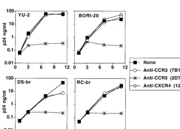
after infection with several HIV isolates. Figure 5 shows
rep-resentative growth curves from isolates HIV-1
YU-2,
HIV-1
BORI-20, HIV-1
DS-br, and HIV-1
RC-br. All viral inocula were
normalized by p24
gagantigen concentration, and peak viral
outputs in five similar experiments were consistent. Infections
performed after preincubation with anti-CCR3 or anti-CXCR4
antibody demonstrated low or no inhibition of viral replication
in four of five experiments. In one experiment there was
sig-nificant (100- to 1,000-fold) diminution in p24
gagoutput by two
other isolates, HIV-1
BORIand HIV-1
89.6, after pretreatment
with anti-CCR3 and less inhibition of HIV-1
BORI-20(data not
shown).
Analysis of HIVD envelope utilization of cells transfected
with two chemokine receptors.
We used a double chemokine
receptor system to determine whether two chemokine
recep-tors present together on the surface of a cell, which we assume
occurs in microglia, could have either a synergistic or an
in-hibitory role in HIV entry, since such a scenario has been
proposed for this cell type (19). 293T cells transiently
trans-fected with CD4 and various combinations of the CCR5,
CCR3, and CXCR4 plasmids (Fig. 6) were infected with
pseu-dotypes expressing the HIV-1
DS-brand HIV-1
YU-2envelopes,
and single-cycle infection was measured by
chemilumines-cence. As shown in the summary of several experiments in Fig.
6, the presence of two receptors was not synergistic, and
max-imum entry was achieved with CCR5 alone. The addition of
either CCR3 or CXCR4 had no effect on pseudotype entry.
Since the degree of CD4 expression may affect the importance
of chemokine receptor concentration (38), similar experiments
were performed with different levels of CD4 expression. The
results were comparable to those depicted in Fig. 6 (data not
shown).
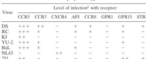
[image:5.612.122.484.422.681.2]HIVD envelope utilization of alternate coreceptors.
We
re-cently studied the ability of HIVD envelope-pseudotyped virus
to infect transiently transfected cells through CCR5-, CCR3-,
and CXCR4 (CD4-dependent)-mediated entry pathways, and
we concluded that all HIVD envelopes tested used CCR5, with
some evidence for low-level CCR3 and CXCR4 use by some
of the envelope clones (43). To improve expression, HIVD
FIG. 4. Inhibition of env-pseudotyped virus infection of microglia withanti-bodies to chemokine receptors. Microglia were preincubated with MAb to CCR5 (2D7), CCR3 (7B11), or CXCR4 (12G5) at a concentration of 20mg/ml for 45 min at 4°C and were then infected with pseudotyped viruses. Cells were lysed 4 to 5 days postinfection, and infection was measured as luciferase activity (RLU per second). Data are expressed as percentages of luciferase activity obtained in the absence of antibody. DS, envelope from HIV-1DS-br; RC, envelope from
HIV-1RC-br.
FIG. 5. Inhibition of HIVD isolate infection of microglia in the presence of antibodies to chemokine receptors. Microglia were preincubated with MAb to CCR3 (7B11), CCR5 (2D7), or CXCR4 (12G5) at a concentration of 20mg/ml for 45 min at 4°C and were then infected with 4 ng of p24gagof HIV-1
YU-2, HIV-1BORI-20,
HIV-1DS-br, or HIV-1RC-br. The following day, the inocula were removed, the cells were washed, and medium was replaced with the appropriate antibody (5mg/ml).
Infected cultures were maintained with the appropriate MAb at a constant concentration. Supernatants were collected over the course of infection and assayed for viral p24gagantigen, expressed in nanograms per milliliter.
on November 9, 2019 by guest
http://jvi.asm.org/
envelopes were cloned into EXV3, an SV40 expression vector
(34). Luciferase reporter virus was then pseudotyped with the
EXV3-based HIVD envelope clones DS, RC, KJ, and YU-2.
Virus pseudotypes were also generated with the BaL, NL43,
and SIVmac251 envelopes to ensure that adequate expression
of the various coreceptors was obtained. These pseudotyped
viruses were tested in triplicate on U87 cells transiently
trans-fected with plasmids encoding CD4 and either CCR5, CCR3,
CXCR4, APJ, CCR8, GPR1, GPR15, or STRL33 (Fig. 7). A
signal 10 times over background (CD4 only) was considered to
be positive. In agreement with our previous report (43), HIVD
envelope-pseudotyped viruses predominantly used CCR5, with
some other coreceptors being used less efficiently. These data
are depicted graphically in Fig. 7 and summarized in Table 1.
Because reagents are not available, except for CCR5, CXCR4,
and CCR3 (see Fig. 1), we could not quantify the level of
expression of the alternate receptors. Several envelopes used
CCR3 and GPR15, albeit at lower levels than CCR5. There
was also some use of APJ, CCR8, and STRL33, particularly by
the DS envelope. The KJ envelope, cloned from a virus
iso-lated from a pediatric encephalitic case, used CCR5 only.
DS-br, the pseudotyped virus with the highest luciferase activity,
was unable to use any of the coreceptors tested when CD4 was
not coexpressed (data not shown).
DISCUSSION
Microglia are the cells primarily responsible for viral load in
the CNS, where they probably play a role in the development
of HIVD, and because of poor penetration of antiretrovirals
into the CNS, they may serve as a potential “sanctuary” site for
the virus (4). Since virus replication can differ between
micro-glia and MDM (44), understanding the virus-cell biology in
microglia is critical for the development of strategies to treat
HIVD. The discovery that chemokine receptors act as HIV
coreceptors has been a major advance in delineating tropism,
and the role of these receptors in microglial entry and
repli-cation has been the subject of recent publirepli-cations (16, 19, 43).
Here we have shown that three major chemokine receptors
implicated in HIV entry, CCR5, CCR3, and CXCR4, are
ex-pressed on adult microglial cells, albeit at different levels (Fig.
2). Two of these, CCR5 and CXCR4, are functional
chemo-kine receptors, as measured by their abilities to mediate signal
transduction after stimulation with their respective ligands. In
these experiments we noted robust Ca
21responses with
che-mokine concentrations within the same range as (or lower
than) those used for stimulation of MDM (20a, 42). Although
we did not detect eotaxin-mediated calcium signaling, it is
formally possible that CCR3 could mediate a signal in the
absence of changes in intracellular calcium levels, as such
sig-naling has been reported with other GPCRs (45). While our
primary interest in chemokine receptors on human microglia
relates to their role in HIV infection, these and future
exper-iments will help us understand the roles of chemokine
recep-tors in a number of inflammatory conditions involving
micro-glia (2, 33).
Our results demonstrating that SDF-1
a
, MIP-1
b
, and
RANTES induced internal calcium fluxes in microglia are not
entirely unexpected, insomuch as MDM and microglia share
many of the same phenotypes, with some notable differences
(44). Two groups have demonstrated MDM signaling in
re-sponse to treatment with SDF-1
a
(20a, 42), and a recent report
by Herbein and colleagues demonstrated that MDM signal
in response to the CCR5 ligands MIP-1
b
and RANTES
(20a). However, there are other differences between
micro-glia and MDM, including the time course of expression of
chemokine receptors and differences in the replication
po-tential among different isolates. Furthermore, whereas the
re-sponses of MDM and microglia were qualitatively similar, we
cannot make any quantitative statements based on our data.
Depending on the assay, several viruses isolated from
indi-viduals with HIVD can use CCR5, CCR3, and, to some extent,
CXCR4 as coreceptors for entry into cells transfected with
plasmids expressing these receptors (43). Theoretically, HIV
could utilize any of these chemokine receptors for entry into
microglial cells, and He et al. have proposed that CCR3 and
CCR5 function as coreceptors for fetal microglia (19). Using
antibodies, we demonstrated that CCR5 is the predominant
coreceptor involved in HIVD virus infection of adult microglia,
with antibodies against either CCR3 or CXCR4 having only a
modest or no effect on viral replication. Results with
pseudo-typed or wild-type viruses were quite congruent with each
other and previous data (43). It is possible that the isolates
utilize the CCR3 or CXCR4 on microglia inefficiently simply
because of the low number of chemokine receptor
mole-cules on the cell surface or, alternatively, because CCR3 and
CXCR4 are not presented in the right context. For example,
there may be an optimal CD4/coreceptor ratio that can be
achieved only with a coreceptor that is expressed at high levels,
such as CCR5 (25, 38). Another possibility is that a virion
could simultaneously use two coreceptors complexed together,
e.g., CCR5 and CCR3, and that under some circumstances
antibodies against the coreceptor present in the lower
concen-tration (CCR3) could partially block entry. This would explain
why we see some inhibition with anti-CCR3 antibodies but
have not been able to infect microglia with a pseudotyped virus
that uses CCR3 but not CCR5 (20b). Therefore, CCR3 and
CXCR4 may play roles in microglial entry, but for an infection
of microglia to occur at relevant levels, HIV must use CCR5,
which is both necessary and sufficient for infection.
[image:6.612.53.292.69.245.2]Additionally, we found that envelopes from several HIVD
isolates can mediate infection of cells transfected with other
GPCRs previously described as HIV coreceptors. But they
do so with apparently reduced efficiency in comparison with
CCR5, at least within the constraints of this assay system,
which did not quantify the level of expression of each of the
coreceptors on the transfected cell surface. This area will need
FIG. 6. HIVD env-pseudotyped virus infection of 293T cells transfected withtwo chemokine receptors. 293T cells were transiently transfected with CD4 alone or with CD4 and CCR5, CCR3, CXCR4, CCR5 plus CXCR4, CCR5 plus CCR3, or CCR3 plus CXCR4. pCDNA3.1 was used to equalize the quantity of DNA in each well. Data are expressed as percentages of infection of 293T cells with CD4 and CCR5. Error bars, standard deviations. DS, envelope from HIV-1DS-br;
YU-2, envelope from HIV-1YU-2.
on November 9, 2019 by guest
http://jvi.asm.org/
FIG. 7. Alternative coreceptor use by HIVD viral envelopes. Pseudotyped viruses prepared with HIV envelopes DS-br, RC-br, and KJ-br and control envelopes YU-2, BaL, NL-43, and SIVmac251 were assayed for infectivity as measured by luciferase activity (in RLU per second) on U87 cells transiently transfected with CD4 and CCR5, CCR3, CXCR4, APJ, CCR8, GPR1, GPR15, or STRL33. Infections were performed in triplicate. Error bars, standard deviations.
on November 9, 2019 by guest
http://jvi.asm.org/
further clarification when antibodies against these alternative
coreceptors become available.
We have previously suggested that direct amplification of
envelope genes from HIV-infected brains may clarify the
po-tential role of these other coreceptors, since it does not
intro-duce the selection bias associated with viral isolation (43). For
infection of microglia, it is also quite possible that CCR3,
CXCR4, and other coreceptors are expressed at higher levels
in the CNS of individuals with HIVD and that under those
circumstances they play a more significant role in HIV entry.
In contrast to microglia, which can be infected soon after
isolation (43a), undifferentiated monocytes are relatively
resis-tant to infection on day 1 after isolation (46). Monocytes have
low levels of CCR5 until cultured, whereas microglia have high
levels of CCR5 soon after purification from brain tissue (Fig.
2). These high levels of CCR5 may explain why the CNS is
infected early during the course of HIV infection, at a time
when most viruses use CCR5 as a coreceptor, and why virus is
present in the brains of many patients with or without HIVD
(23). Chemokine receptor levels may also contribute to the
differences we have previously noted between replication in
MDM and in microglia (44). Given the detectable levels of
CXCR4 present in microglia, it is somewhat surprising that
more CXCR4-using viruses have not been isolated from
brains, particularly since these isolates are particularly
promi-nent in the late stages of HIV disease, when HIVD is more
prevalent. In MDM, blocks to replication beyond the entry
step have been identified for some HIV strains (42, 48), and
further experimentation may clarify this issue in microglia.
Alternatively, parenchymal microglia may become infected
only after the CNS perivascular population of macrophages/
microglia has been infected (26). This perivascular population,
which may be phenotypically different with regard to HIV
in-fection, may act as a filter, allowing only viruses using a certain
coreceptor repertoire to infect the parenchymal microglia,
which are used in our experimental system (18).
ACKNOWLEDGMENTS
J.T.C.S. and A.V.A. contributed equally to this work.
This work was supported in part by NS-27405, NS-35743, and
MH-58958.
We thank Wei Cao for excellent technical assistance and Benjamin
Doranz, Trevor Hoffman, Aimee Edinger, and Joseph Rucker from
the R.W.D. laboratory for sharing reagents and for helpful discussions.
Sarina Berger (F.G.-S. laboratory) helped clone the KJ-br envelope,
and Julie Turner (J. Hoxie laboratory) provided advice on flow
cytom-etry. The anti-chemokine receptor antibodies were obtained from the
AIDS Reagent Program. Suzanne Gartner (Johns Hopkins University)
kindly provided the HIVD viral isolates.
REFERENCES
1. Albright, A. V., J. Strizki, J. M. Harouse, E. Lavi, M. O’Connor, and F. Gonza´lez-Scarano.1996. HIV-1 infection of cultured human adult oligoden-drocytes. Virology 217:211–219.
2. Benveniste, E. N. 1997. Role of macrophages/microglia in multiple sclerosis and experimental allergic encephalomyelitis. J. Mol. Med. 75:165–173. 3. Choe, H., M. Farzan, M. Konkel, K. Martin, Y. Sun, L. Marcon, M.
Cayab-yab, M. Berman, M. E. Dorf, N. Gerard, C. Gerard, and J. Sodroski.1998. The orphan seven-transmembrane receptor Apj supports the entry of pri-mary T-cell-line-tropic and dualtropic human immunodeficiency virus type 1. J. Virol. 72:6113–6118.
4. Cohen, J. 1998. AIDS therapies: exploring how to get at—and eradicate— hidden HIV. Science 279:1854.
5. Deng, H., R. Liu, W. Ellmeier, S. Choe, D. Unutmaz, M. Burkhart, P. Di Marzio, S. Marmon, R. E. Sutton, C. M. Hill, C. B. Davis, S. C. Peiper, T. J. Schall, D. R. Littman, and N. R. Landau.1996. Identification of a major co-receptor for primary isolates of HIV-1. Nature 381:661–666.
6. Deng, H. K., D. Unutmaz, V. N. KewalRamani, and D. R. Littman. 1997. Expression cloning of new receptors used by simian and human immunode-ficiency viruses. Nature 388:296–300.
7. Doms, R. W., and S. C. Peiper. 1997. Unwelcomed guests with master keys: how HIV uses chemokine receptors for cellular entry. Virology 235:179–190. 8. Edinger, A. L., J. L. Mankowski, B. J. Doranz, B. J. Margulies, B. Lee, J. Rucker, M. Sharron, T. L. Hoffman, J. F. Berson, M. C. Zink, V. M. Hirsch, J. E. Clements, and R. W. Doms.1997. CD4-independent, CCR5-dependent infection of brain capillary endothelial cells by a neurovirulent simian im-munodeficiency virus strain. Proc. Natl. Acad. Sci. USA 94:14742–14747. 9. Edinger, A. L., T. L. Hoffman, M. Sharron, B. Lee, B. O’Dowd, and R. W.
Doms.1998. Use of GPR1, GPR15, and STRL33 as coreceptors by diverse HIV-1 and SIV envelope proteins. Virology 249:367–378.
10. Edinger, A. L., T. L. Hoffman, M. Sharron, B. Lee, Y. Yi, W. Choe, D. L. Kolson, B. Mitrovic, Y. Zhou, D. Faulds, R. G. Collman, J. Hesselgesser, R. Horuk, and R. W. Doms.1998. An orphan seven-transmembrane domain receptor expressed widely in the brain functions as a coreceptor for human immunodeficiency virus type 1 and simian immunodeficiency virus. J. Virol. 72:7934–7940.
11. Endres, M. J., P. R. Clapham, M. Marsh, M. Ahuja, J. D. Turner, A. McKnight, J. F. Thomas, B. Stoebenau-Haggerty, S. Chose, P. J. Vance, T. N. C. Wells, C. A. Power, N. R. Landau, and J. A. Hoxie.1996. CD4-independent infection of HIV-2 is mediated by fusin/CXCR4. Cell 87:745– 756.
12. Farzan, M., H. Choe, K. Martin, L. Marcon, W. Hofmann, G. Karlsson, Y. Sun, P. Barrett, N. Marchand, N. Sullivan, N. Gerard, C. Gerard, and J. Sodroski.1997. Two orphan seven-transmembrane segment receptors which are expressed in CD4-positive cells support simian immunodeficiency virus infection. J. Exp. Med. 186:405–411.
13. Feng, Y., C. C. Broder, P. E. Kennedy, and E. A. Berger. 1996. HIV-1 entry cofactor: functional cDNA cloning of a seven-transmembrane, G protein-coupled receptor. Science 272:872–877.
14. Gartner, S., and M. Popovic. 1990. Macrophage tropism of HIV-1. AIDS Res. Hum. Retroviruses 6:1017–1021.
15. Gartner, S., R. A. McDonald, E. A. Hunter, F. Bouwman, Y. Liu, and M. Popovic.1997. gp120 sequence variation in brain and in T-lymphocyte hu-man immunodeficiency virus type 1 primary isolates. J. Hum. Virol. 1:3–18. 16. Ghorpade, A., M. Q. Xia, B. T. Hyman, Y. Persidsky, A. Nukuna, P. Bock, M. Che, J. Limoges, H. E. Gendelman, and C. R. Mackay.1998. Role of the
b-chemokine receptors CCR3 and CCR5 in human immunodeficiency virus type 1 infection of monocytes and microglia. J. Virol. 72:3351–3361. 17. Gonza´lez-Scarano, F., R. E. Shope, C. E. Calisher, and N. Nathanson. 1982.
Characterization of monoclonal antibodies against the G1 and N proteins of LaCrosse and Tahyna, two California serogroup bunyaviruses. Virology 120: 42–53.
18. Gonza´lez-Scarano, F., J. M. Strizki, A. Albright, and J. Shieh. 1997. Use of primary CNS cultures to investigate HIV neurotropism. J. Neurovirol. 3: S11–S13.
19. He, J., Y. Chen, M. Farzan, H. Choe, A. Ohagen, S. Gartner, J. Busciglio, X. Yang, W. Hoffman, W. Newman, C. R. Mackay, J. Sodroski, and D. Gabuzda. 1997. CCR3 and CCR5 are coreceptors for HIV-1 infection of microglia. Nature 385:645–649.
20. Heath, H., S. Qin, P. Rao, L. Wu, G. LaRosa, N. Kassam, P. D. Ponath, and C. R. Mackay.1997. Chemokine receptor usage by human eosinophils. The importance of CCR3 demonstrated using an antagonistic monoclonal anti-body. J. Clin. Investig. 99:178–184.
20a.Herbein, G., U. Mahlknecht, F. Batliwalla, P. Gregersen, T. Pappas, J. Butler, W. A. O’Brien, and E. Verdin.1998. Apoptosis of CD81T cells is
mediated by macrophages through interaction of HIV gp120 with chemokine receptor CXCR4. Nature 395:189–194.
20b.Hoffman, T., and J. Shieh. Unpublished data.
[image:8.612.53.295.91.189.2]21. Horuk, R., J. Hesselgesser, Y. Zhou, D. Faulds, M. Halks-Miller, S. Harvey, D. Taub, M. Samson, M. Parmentier, J. Rucker, B. J. Doranz, and R. W. Doms.1998. The CC chemokine I-309 inhibits CCR8-dependent infection by diverse HIV-1 strains. J. Biol. Chem. 273:386–391.
TABLE 1. CCR5, CCR3, and orphan receptor use by
HIVD envelope-pseudotyped viruses
Virus Level of infection
awith receptor:
CCR5 CCR3 CXCR4 APJ CCR8 GPR1 GPR15 STRL33
DS 111 11 2 1 1 2 1 1
RC 111 1 2 1 1 2 1 2
KJ 11 2 2 2 2 2 2 2
YU-2 111 1 2 2 2 2 1 2
BaL 111 1 2 1 2 2 2 2
NL43 2 2 11 2 2 2 2 2
251 11 2 2 2 2 2 11 11
aInfection was measured in RLU per second.2, less than 1 log unit over
background;1, 1 log unit over background;11, 2 log units over background;
111, 3 log units over background. Background, infection with CD4 alone and no chemokine receptor (see text for details).
on November 9, 2019 by guest
http://jvi.asm.org/
22. Itoh, T., A. Itoh, K. Horiuchi, and D. Pleasure. AMPA receptor-mediated excitotoxicity in human NT2-N neurons results from loss of intracellular Ca21homeostasis following marked elevation of intracellular Na21. J.
Neu-rochem., in press.
23. Johnson, R. T., J. D. Glass, J. C. McArthur, and B. W. Chesebro. 1996. Quantitation of human immunodeficiency virus in brains of demented and nondemented patients with acquired immunodeficiency syndrome. Ann. Neurol. 39:392–395.
24. Kolson, D. L., E. Lavi, and F. Gonza´lez-Scarano. 1998. The effects of human immunodeficiency virus in the central nervous system. Adv. Virus Res. 50:1– 47.
25. Kozak, S. L., E. J. Platt, N. Madani, F. E. Ferro, K. Peden, and D. Kabat. 1997. CD4, CXCR-4, and CCR-5 dependencies for infections by primary patient and laboratory-adapted isolates of human immunodeficiency virus type 1. J. Virol. 71:873–882.
26. Lane, J. H., V. G. Sasseville, M. O. Smith, P. Vogel, D. R. Pauley, M. P. Heyes, and A. A. Lackner.1996. Neuroinvasion by simian immunodeficiency virus coincides with increased numbers of perivascular macrophages/micro-glia and intrathecal immune activation. J. Neurovirol. 2:423–432. 27. Lavi, E., J. M. Strizki, A. M. Ulrich, W. Zhang, L. Fu, Q. Wang, M.
O’Connor, J. A. Hoxie, and F. Gonza´lez-Scarano.1997. CXCR-4 (Fusin), a co-receptor for the type 1 human immunodeficiency virus (HIV-1), is ex-pressed in the human brain in a variety of cell types, including microglia and neurons. Am. J. Pathol. 151:1035–1042.
28. Li, Y., J. C. Kappes, J. A. Conway, R. W. Price, G. M. Shaw, and B. H. Hahn. 1991. Molecular characterization of human immunodeficiency virus type 1 cloned directly from uncultured human brain tissue: identification of repli-cation-competent and -defective viral genomes. J. Virol. 65:3973–3985. 29. Liao, F., G. Alkhatib, K. W. Peden, G. Sharma, E. A. Berger, and J. M.
Farber.1997. STRL33, a novel chemokine receptor-like protein, functions as a fusion cofactor for both macrophage-tropic and T cell line-tropic HIV-1. J. Exp. Med. 185:2015–2023.
30. Maddon, P. J., A. G. Dalgleish, J. S. McDougal, P. R. Clapham, R. A. Weiss, and R. Axel.1986. The T4 gene encodes the AIDS virus receptor and is expressed in the immune system and the brain. Cell 47:333–348. 31. Marchese, A., J. M. Docherty, T. Nguyen, M. Heiber, R. Cheng, H. H. Heng,
L. C. Tsui, X. Shi, S. R. George, and B. F. O’Dowd.1994. Cloning of human genes encoding novel G protein-coupled receptors. Genomics 23:609–618. 32. Matsumoto, M., K. Hidaka, H. Akiho, S. Tada, M. Okada, and T.
Yamagu-chi.1996. Low stringency hybridization study of the dopamine D4 receptor revealed D4-like mRNA distribution of the orphan seven-transmembrane receptor, APJ, in human brain. Neurosci. Lett. 219:119–122.
33. McRae, A., A. Dahlstrom, and E. A. Ling. 1997. Microglia in neurodegen-erative disorders: emphasis on Alzheimer’s disease. Gerontology 43:95–108. 34. Miller, J., and R. N. Germain. 1986. Efficient cell surface expression of class II MHC molecules in the absence of associated invariant chain. J. Exp. Med. 164:1478–1489.
35. Moeller, T., C. Nolte, R. Burger, A. Verkhratsky, and H. Kettenmann. 1997. Mechanisms of C5a and C3a complement fragment-induced [Ca21]
i
signal-ing in mouse microglia. J. Neurosci. 17:615–624.
36. O’Dowd, B. F., M. Heiber, A. Chan, H. H. Heng, L. C. Tsui, J. L. Kennedy, X. Shi, A. Petronis, S. R. George, and T. Nguyen.1993. A human gene that shows identity with the gene encoding the angiotensin receptor is located on
chromosome 11. Gene 136:355–360.
37. Parham, P., C. J. Barnstable, and W. F. Bodmer. 1979. Use of a monoclonal antibody (W6/32) in structural studies of HLA-A,B,C antigens. J. Immunol. 123:342–349.
38. Platt, E. J., K. Wehrly, S. E. Kuhmann, B. Chesebro, and D. Kabat. 1998. Effects of CCR5 and CD4 cell surface concentrations on infections by mac-rophagetropic isolates of human immunodeficiency virus type 1. J. Virol. 72: 2855–2864.
39. Price, R. W., B. Brew, J. Sidtis, M. Rosenblum, A. C. Scheck, and P. Cleary. 1988. The brain in AIDS: central nervous system HIV-1 infection and AIDS dementia complex. Science 239:586–592.
40. Rucker, J., A. L. Edinger, M. Sharron, M. Samson, B. Lee, J. F. Berson, Y. Ti, B. Margulies, R. G. Collman, B. J. Doranz, M. Parmentier, and R. W. Doms.1997. Utilization of chemokine receptors, orphan receptors, and her-pesvirus-encoded receptors by diverse human and simian immunodeficiency viruses. J. Virol. 71:8999–9007.
41. Samson, M., P. Stordeur, O. Labbe, P. Soularue, G. Vassart, and M. Par-mentier.1996. Molecular cloning and chromosomal mapping of a novel human gene, ChemR1, expressed in T lymphocytes and polymorphonuclear cells and encoding a putative chemokine receptor. Eur. J. Immunol. 26: 3021–3028.
42. Schmidtmayerova, H., M. Alfano, G. Nuovo, and M. Bukrinsky. 1998. Hu-man immunodeficiency virus type 1 T-lymphotropic strains enter macro-phages via a CD4- and CXCR4-mediated pathway: replication is restricted at a postentry level. J. Virol. 72:4633–4642.
43. Shieh, J. T. C., A. V. Albright, M. Sharron, S. Gartner, J. Strizki, R. W. Doms, and F. Gonza´lez-Scarano.1998. Chemokine receptor utilization by human immunodeficiency virus type 1 isolates that replicate in microglia. J. Virol. 72:4243–4249.
43a.Shieh, J. T. C. Unpublished data.
44. Strizki, J. M., A. V. Albright, H. Sheng, M. O’Connor, L. Perrin, and F. Gonza´lez-Scarano.1996. Infection of primary human microglia and mono-cyte-derived macrophages with human immunodeficiency virus type 1 iso-lates: evidence of differential tropism. J. Virol. 70:7654–7662.
45. Turner, L., S. G. Ward, and J. Westwick. 1995. RANTES-activated human T lymphocytes. A role for phosphoinositide 3-kinase. J. Immunol. 155:2437– 2444.
46. Tuttle, D. L., J. K. Harrison, C. Anders, J. W. Sleasman, and M. M. Good-enow.1998. Expression of CCR5 increases during monocyte differentiation and directly mediates macrophage susceptibility to infection by human im-munodeficiency virus type 1. J. Virol. 72:4962–4969.
47. Wu, L., W. A. Paxton, N. Kassam, N. Ruffing, J. B. Rottman, N. Sullivan, H. Choe, J. Sodroski, W. Newman, R. A. Koup, and C. R. Mackay.1997. CCR5 levels and expression pattern correlate with infectability by macrophage-tropic HIV-1, in vitro. J. Exp. Med. 185:1681–1691.
48. Yi, Y., S. Rana, J. D. Turner, N. Gaddis, and R. G. Collman. 1998. CXCR-4 is expressed by primary macrophages and supports CCR5-independent in-fection by dual-tropic but not T-tropic isolates of human immunodeficiency virus type 1. J. Virol. 72:772–777.
49. Yong, V. W., and J. P. Antel. 1992. Culture of glial cells from human brain biopsies, p. 81–96. In S. Fedoroff and A. Richardson (ed.), Protocols for neural cell culture. Humana Press, Totowa, N.J.
on November 9, 2019 by guest
http://jvi.asm.org/