STUDY ON EXTENDED SPECTRUM
BETALACTAMASE (ESBL) PRODUCING GRAM
NEGATIVE BACILLI ISOLATED FROM
INTENSIVE CARE UNITS
Dissertation Submitted to
THE TAMIL NADU DR. M.G.R. MEDICAL UNIVERSITY
in partial fulfillment of the regulations for the award of the degree of
M.D. (MICROBIOLOGY)
BRANCH – IV
GOVT. STANLEY MEDICAL COLLEGE & HOSPITAL
THE TAMIL NADU DR. M.G.R. MEDICAL UNIVERSITY
CHENNAI, INDIA.
DECLARATION
I solemnly declare that this dissertation “STUDY ON
EXTENDED SPECTRUM BETALACTAMASE (ESBL)
PRODUCING GRAM NEGATIVE BACILLI ISOLATED FROM
INTENSIVE CARE UNITS”
is the bonafide work done by me at
the Department of Microbiology, Govt. Stanley Medical College and
Hospital, Chennai, under the guidance and supervision of
Prof. Dr. P. R. THENMOZHI VALLI, M.D., Professor of
Microbiology, Govt. Stanley Medical College, Chennai-600 001.
This dissertation is submitted to The Tamil Nadu Dr. M.G.R.
Medical University, Chennai in partial fulfillment of the University
regulations for the award of degree of M.D. Branch IV Microbiology
examinations to be held in September 2006.
Place : Chennai.
Date :
CERTIFICATE
This is to certify that this dissertation entitled “STUDY ON
EXTENDED SPECTRUM BETALACTAMASE (ESBL)
PRODUCING GRAM NEGATIVE BACILLI ISOLATED FROM
INTENSIVE CARE UNITS”
is the bonafide original work done by
Dr. A. VASUMATHI, Post graduate in Microbiology, under myoverall supervision and guidance in the department of Microbiology,
Stanley Medical College, Chennai, in partial fulfillment of the
regulations of The Tamil Nadu Dr. M.G.R. Medical University for the
award of M.D Degree in Microbiology (Branch IV).
Dr. M. VASANTHA, M.D., Dr. P. R. Thenmozhi Valli M.D.,
Dean Prof. & H.O.D.
Stanley Medical College Department of Microbiology
Chennai-600 001 Stanley Medical College
ACKNOWLEDGEMENT
My sincere thanks to Dr. M. VASANTHA, M.D., Dean, Government
Stanley Medical College and Hospital for giving me permission to commence
this dissertation and use the resources of this Institution.
I owe my gratitude to Dr. P.R. THENMOZHI VALLI, M.D.,
Professor and H.O.D., Department of Microbiology for her unflinching
interest, relentless efforts, valuable advice, excellent guidance and
encouragement given to me through out this study.
I am immensely grateful to Dr. S. SHANTHA. M.D., Ph.D.,
Professor of Immunology for her kind helps to carry out this work
My profound thanks to Dr. S. GEETHALAKSHMI, M.D., Former
Professor, for her encouragement in this study.
My sincere and special thanks to Dr. G. JAYALAKSHMI, M.D.,
D.T.C.D., Former Professor, Department of Microbiology for her constant
support and encouragement.
My sincere thanks to Dr. THIAGARAJAN RAVINDER, M.D.,
Professor of Microbiology for his valuable advice and timely help in carrying
My sincere thanks to Dr. RAJASEKAR, M.D., Professor and Head,
Department of Intensive Medical Care Unit and Dr. MEENAKSHI, M.D.,
D.A., Professor, Department of Anaesthesia, Govt. Stanley Hospital, for
allowing me to collect samples for this study.
I extend my sincere thanks to all Assistant Professors, Department of
Microbiology for their help, support, interest and valuable hints.
My sincere thanks to Mrs. Florida Tilton, M..Sc., M.phil HOD,
Department of Biotechnology, Hindustan College of Arts and Sciences and
the M.Sc. students of that college for their help in carrying out the molecular
part of my work
I also thank all my department colleagues for their timely help, co-
operation and moral support.
I express many thanks to all the technical staff and other staff members
of the Department of Microbiology and Immunology for their kind
co-operation to carryout this work successfully.
I also extend my thanks to all the patients who participated in my
CONTENTS
SL.NO. TITLE PAGE NO.
1. INTRODUCTION 1
2. REVIEW OF LITERATURE 4
3. AIM AND OBJECTIVES 33
4. MATERIALS AND METHODS 34
5. RESULTS 46
6. DISCUSSION 64
7. SUMMARY 72
8. CONCLUSION 74
9. ANNEXURES
I) APPENDIX
INTRODUCTION
Incidence of bacterial infection from Hospitals accounts for
5-10% in most developed nations while in India one in four patients
admitted into hospitals acquire bacterial infection59. Around 30% of
patients in Intensive Care Units were contracting infections during their
stay in hospital3, which leads to many complications in them.
Since 1940, Penicillin- the first beta lactam antibiotic
remained the antibiotic of choice for many years. Bacteria were so
inventive that they developed many mechanisms to escape the action of
antibiotics and became resistant. Main mechanism was by the
production of β-lactamases. The first plasmid mediated β-lactamase
was TEM – 1 which was described in 1960s. Antibiotic resistance was
high among organisms isolated from Intensive Care Units3.
Over the last 25 years, many new β-lactam antibiotics have
been developed, that were specifically designed to be resistant to the
hydrolytic action of β-lactamases. However with each new class of
antibiotic used to treat patients, because of inappropriate and extensive
use new β-lactamases emerged that caused resistance to that particular
group of drug. This was due to the continuous mutation of the
enzymes.22 Major effort to overcome this lead to introduction of the new
Cefotaxime, which became widely used for the treatment of serious
infection due to gram negative bacilli, in 1980s.49 Because of their
expanded spectrum of action against bacteria, they were also called as
Expanded Spectrum Antibiotics. Due to extensive use of third
generation antibiotics, bacteria soon developed resistance, by means of a
new β- lactamase. Because of their increased spectrum of activity
against most of the β-lactam antibiotics including third generation
cephalosporins, these enzymes were called Extended Spectrum Beta
Lactamases (ESBL).
First ESBL was identified in Klebsiella pneumoniae in Germany
in 1983, 49 it was found to be a derivative of SHV type Beta Lactamase.
They were encoded by transferable plasmids and hence dissemination of
resistance to other members of gram negative bacilli occurred.
Major factor contributing to the production of ESBL is prolonged
stay in Intensive Care Units.38
The incidence and type of ESBL vary with geographical
locations.22
Though Extended spectrum beta lactamases are responsible for
nosocomial outbreaks of infections like urinary tract infections,
pneumonia, septicemia etc, the real incidence and prevalence of ESBL
control outbreaks has resulted in the appearance of new mutant type
Extended spectrum beta lactamase in the same institution and the same
patient source.
The appearance of eight different Extended Spectrum Beta
Lactamases (TEM-3, TEM-5, CAZ-2, CAI-3, CAI-6, CAI-7, SHV-4 and
SHV-5) at Clermont-Ferrand hospital, since the first one in 1984 is a
classic example.37
Hence the present study was undertaken to study the Extended
Spectrum Beta Lactamase producing gram negative bacilli isolated from
REVIEW OF LITERATURE
HISTORY:
Antibiotic era started with discovery of penicillin by Alexander
Flemming in 1928.23 Use of Penicillin started in 1941. Emergence of
resistance to β-lactam antibiotic began even before the introduction of
penicillin.18 First β-Lactamase was found in E-coli prior to introduction
of penicillin. Next is the emergence of penicillin resistance in
staphylococcus aureus due to plasmid encoded β-lactamase.
First plasmid mediated β-lactamase in gram negative organisms-
TEM-1 was described in early 1960’s. It was first isolated in Escherichia
coli from a patient Temoniera in Greece. It spread to other genera soon.
Another common plasmid mediated β-lactamase found in
Klebsiella pneumoniae and Escherichia coli are SHV-1 (for Sulph
Hydryl Variable).
Over the last 20 years many new β-lactam antibiotics have been
developed which were resistant to hydrolytic action of β-lactamases but
because of over usage, these antibiotics also became resistant.
To overcome it, around 1980 3rd generation cephalosporins, also
called broad spectrum cephalosporins were introduced. They were very
use, they also became resistant. In 1983 in Germany, isolates of
Klebsiella pneumoniae and other enterobacteriaceae were found to
produce a plasmid-determined β-lactamase that hydrolyzed cefotaxime,
as well as other newer cephalosporins. This new β-lactamase, called
SHV-2 was derived from a mutation in the well-known SHV-1
β-lactamase commonly found in Klebsiella,37 since they lead to
resistance of extended spectrum cephalosporins they are called extended
spectrum β-lactamases.
β- Lactam antibiotics: 23
They are antibiotics having a β-lactam ring. It comprises of
1) Penicillins
2) Cephalosporins
3) Monobactams
4) Carbapenems
All the β-lactam antibiotics have β-lactam ring in common which
is made of 3 carbon atoms and one nitrogen atom. The other rings vary
Penicillins:
The structure of Penicillin is
1. β-lactam ring
2. Thiazolidone ring
3. site where β-lactamase will act
Types of Penicillins
I. Natural Penicillins eg., Penicillin G
II. Semi synthetic Penicillins
1. Acid resistant Penicillins eg., Penicillin V
2. Penicillinase resistant Penicillins eg., Methicillin, Oxacillin,
Cloxacillin
3. Extended spectrum Penicillins.
a. Amino penicillins eg., Ampicillin, Amoxycillin
b. Carboxy penicillins e.g., Carbenicillin
Cephalosporins:
Have β- lactam ring attached to dihyro thiazine ring. By addition of different side chains to dihyro thiazine ring a large number of semi synthetic compounds have been produced. They are divided into four generations
I – Generation: Cephalothin, Cephalexin, Cefadroxil, Cefazolin.
II – Generation: Cefuroxime, Cefuroxime Axetil, Cefaclor, Cefoxitin.
III – Generation: Cefotaxim, Ceftazidime, Cefixim, Cefoperazone.
IV – Generation: Cefepime, Cefpirome.
Monobactams:
Monobactams have a monocyclic β-lactam ring and are
resistant to β-lactamase. They are active against gram negative
bacteria but not against gram positive bacteria. e.g., Aztreonam.
Carbapenems:
Mechanism of Action of β-lactam antibiotics:
β-lactam antibiotics act by inhibiting cell wall synthesis of
bacteria.
Bacteria synthesise UDP – N – Acetyl muramic acid pentapeptide
and UDP – N – Acetyl glucosamine. Peptidoglycan residues are linked
together and UDP is split off. Final step is cleavage of the terminal
D-alanine of the peptide chains by Tran’s peptidases and cross linking
between peptide chains of the neighbouring strands. β-lactam
antibiotics inhibits trans peptidases so that cross linking is not formed.23
Mechanism of resistance to β-lactam antibiotics
:-1) Enzymatic inhibition: plasmid mediated e.g., β-lactamase
2) Membrane impermeability - both chromosome and plasmid
mediated
3) Alteration of target protein e.g., Penicillin binding protein.
The β- Lactamases
:-This is a heterogeneous group of penicillin recognizing proteins. They are members of a super family of active site serine proteases. They act by cleaving an amide bond of beta lactam ring to form an acyl-enzyme complex. Any β-lactam antibiotic may be inactivated by these enzymes. There are about > 170 enzymes of this kind.38
1. Early classification scheme was developed by Richmond and Sykes
based on substrate profile and the location of genes encoding the
β-lactamases.
2. Modern scheme based on molecular structure was proposed by
Ambler.
Most important is class A which is serine proteases that have either preference for penicillin or) broad spectrum activities.
They are found either on chromosomes (or) plasmids and are
easily transferable from one bacteria to another.
They may be produced constitutively and may be induced. In
this group are the staphylococcus and some Gram negative bacilli.
Class C: Primarily cephalosporinases, either constitutive (or) inducible.
Found on chromosomes of Gram Negative Bacilli.
CLASSIFICATION OF β-LACTAMASES
Class Examples Location Bacteria
Gram positive β- lactamases
Enterococcal Staphylococca
l Streptococcal
Serological types
A-D PlasmidsPlasmid
Plasmid
Staphylococcus aureus, Staphylococcus epidermidis Enterococcus faecalis Streptococcus uberis
Gram Negative β- Lactamase
Class A TEM-1&2 Plasmid; broad
spectrum Enterobacteriaceae, N.gonorrhoea, H.influenzae, V. cholerae
SHV – 1 Plasmid; Broad
spectrum Enterobacteriaceae, also found in chromosomes of Klebsiella pneumoniae.
OXA-1 Plasmid;
oxacillinases Escherichia coli
PSE-1 Plasmid;
carbenicillinase in pseudomonas
Enterobacteriaceae, most common type in
Pseudomonas aeruginosa.
BRO – 1 Plasmid;
carbenicillinase base
Branhamella, moraxella
Class B Cephalosporinase Bacillus cereus
Class C Broad spectrum Chromosome Klebsiella pneumoniae,
Proteus vulgaris, Klebsiella oxytoca, K. aerogenes K-1 Cephalosporinase Chromosome Escherichia coli, Bacteroides
fragilis
Inducible Chromosome Enterobacter aerogenes,
E.cloacae, Providencia rettgeri, Pseudomonas. aeruginosa,
Serratia marcescens, Acinetobacter baumannii Carbapenamase
(Imipenem) Chromosome Xanthomonas maltophilia, B. fragilis, Serratia marcescens, Enterobacter cloacae.
-Recent Classification of β-lactamases is by Bush-Jacoby – Medeiros
scheme according to substrate profile and inhibition by clavulanic acid.38
The Bush Jacoby – Medeiros functional classification scheme for β-lactamames
Group Enzyme Type Inhibition by Clavulanat e Mole-cular Class No. of Enzymes Example
1 Cephalosporinase No C 53 E.cloacae P99,M
2a Penicillinase Yes A 20 Staphylococcus
aureus,
Staphylococcus albus.
2b Broad spectrum Yes A 16 TEM-1, SHV-1
2be Extended spectrum Yes A 38 TEM-3, SHV-2, K.
2br Inhibitor resistant Diminished A 9 TEM-30,
TRC-1
2c Carbenicillinase Yes A 15 PSE-1, CARB-3, BR
2d Cloxacillinase Yes D or A 18 OXA-1, PSE-2, Str.
Cacaoi
2e Cephalosporinase Yes A 19 Proteus vulgaris, B.
fragilis Cep. A
2f Carbapenemase Yes A 3 E.cloacae
IMI-1 N
3 Metalloenzyme No B 15 Xanthomonas
maltophilia LI
4 Penicillinase N 7 Pseudomonas
cepacia. Some β-lactamases like that produced by Staphylococcus aureus
are stable over several decades. These enzymes have a rather narrow
The broad spectrum, plasmid mediated β-lactamases of Gram
negative bacilli (GNB) such as TEM-1 and SHV-1 were stable for many
years.
From 1980, a series of enzymatic variants appeared that had a
broadened spectrum of activity against the newly developed β-lactam
antibiotics. These ESBLs were first found in Europe most commonly in
Klebsiella species, less commonly in Escherichia coli. The number of
enzymes continues to increase.
The new enzymes TEM-1 and SHV-1 are located on plasmids.
But they might have been derived originally from a chromosomal
enzyme. Many of the new β-lactamases differ from each other only in
single aminoacid substitution but the changes have profound
Key Dates Showing Emergence of Β-Lactamases
Year Enzyme Organism Place
1944 Penicillinase Staphylococcus
aureus
-1963 TEM-1 Escherichia coli Athens
1974 SHV-1 Escherichia coli Switzerland
1978 OXA-10 Pseudomonas.
aeruginosa
-1982 SME-1 Serratia
marcescens London
1984 IMI-1 E. cloacae California
1988 Metallo
β-lactamase Pseudomonas aeruginosa Japan
1989 Inhibitor –
resistant Penicillinase Escherichia coli, Klebsiella pneumoniae France, Spain
1990 NMcA E. cloacae Paris
1991 OXA-11
OXA-14
Pseudomonas
aeruginosa Turkey
1991 PER-1 Pseudomonas
aeruginosa, Salmonella typhimurium
Turkey
1992 MEN-1 Escherichia coli,
K. pneumoniae
France
1994 TOHO-1 Escherichia coli Japan
1996 PER-2 Escherichia coli,
K. pneumoniae, S. typhimurium, P. mirabilis
Germany
Detection of β-lactamases 36
Detection of β-lactamases was done by biochemical tests for the
enzymes. This was by measuring penicilloic acids which was produced
when β-lactamases hydrolyse benzyl penicillins. The acid production
was detected by
1. Measuring the change in pH of an indicator dye (acidometric
method)
2. By exploiting the ability of penicilloic acid to reduce iodine and
reverse the formation of the blue colour when iodine complexes with
starch. (Iodometric method)
3. Chromogenic cephalosporin method – usually nitrocephin was used.
Nitrocephin was normally yellow but when the β-lactam ring was
hydrolysed it turns red.
β -lactamase inhibitors : 16
These compounds resemble β-lactam antibiotics. They can bind
to β-lactam antibiotics either reversibly or irreversibly protecting the
antibiotics from destruction. They serve as suicide bombers utilizing all
available enzymes. These compounds have weak antibacterial activity
but are potent inhibitors of many plasmid-encoded and some
Three important β-lactamase inhibitors are:
1) Clavulanic acid
2) Sulbactam
3) Tazobactam
Clavulanic acid show only low level of antibacterial action but
when combined with β-lactam antibiotics inhibition of bacteria which
are otherwise resistant to β-lactam antibiotics was noted.
Sulbactam has broader spectrum of inhibition but less potent.
Tazobactam is as potent as clavulanic acid
EXTENDED SPECTRUM BETA LACTAMASES :
Definition:
Enzymes capable of hydrolyzing major β – lactam antibiotics
including third generation cephalosporins are called as extended
spectrum beta lactamases.
CHARACTERISTICS OF ESBLs: 16
1) They are mostly class- A cephalosporinases carried on plasmids
2) They are more common in Klebsiella species followed by
Escherichia coli
3) Described first in Germany and France.
5) Imipenem and Cefoxitin not hydrolysed.
6) Comparative activity against Cefotaxime and Ceftazidine varies
with enzymes.
7) Some enzymes active against Astreonam.
8) Resistance may not be detected by standard susceptibility tests.
9) Inhibition of activity by β-lactamase inhibitors can be
demonstrated.
Majority of ESBLs contain a serine at the active site and belong
to Ambler’s Molecule. Class A enzymes are characterized by an active
site serine, a molecular mass of 29,000 Dalton and hydrolysis of
penicillin.
ESBLs contain a number of mutations that allow them to
hydrolyse ESBL antibiotics. While TEM and SHV type ESBLs retain
their ability to hydrolyse penicillin, but they are not catalytically as
efficient as the parent enzymes.
ESBLs are not active against cephamycin and most strains
expressing ESBLs are susceptible to Cefoxitin and Cefotetan. But it has
been reported that ESBL producing strains can become resistant to
Major risk factors for ESBL production
1) Prolonged stay in ICU
2) Long term use of antibiotics
3) Nursing home residency
4) Severe illness
5) High rate use of ceftazidime and other Third Generation
Cephalosporins
6) Use of lifelines and catheters
Types of ESBLs:
-Most ESBLs are derivatives of TEM & SHV. Now > 90 TEM
type and > 25 SHV types are there. A few point mutations at selected
loci with in the gene give rise to the extended spectrum phenotype.
TEM & SHV type ESBLs are most often found in Escherichia
coli and Klebsiella. pneumoniae but also found in Proteus species,
Providentia species and other genera of Enterobacteriaceae.
TEM
:-TEM-2 was the 1st derivative of TEM which had a single
aminoacid substitution from the original β-lactamase. This did not
change the substrate profile. TEM-3 reported in 1989 was the first TEM
Aminoacid residues important for ESBL production are
Glutamate → Lysine at position 104
Arginine → Serine (or) histidine at position 164
Glycine → Serine at Position 238
Glutamate → Lysine at position 240
TEM type is often found in Escherichia coli and Klebsiella.
Pneumoniae, but also reported in other members of Enterobacteriaceae
and some other gram negative bacilli.
TEM- 42 β-lactamase was found in p. aeruginosa
TEM-17 β-lactamase reported in a blood culture isolate of
Capnocytophaga ochracia.
Characteristics of TEM-type β-lactamases
PI Enzyme type
Broad spectrum
ESBL Inhibitor Resistant
5.2 TEM-12, TEM-55,
TEM-57, TEM-58
TEM-30, TEM-31, TEM-35, TEM-36, TEM-37, TEM-38, TEM-41, TEM-45, TEM-51, TEM-73, TEM-74
5.3 TEM-25
5.4 TEM-1 TEM-7, TEM-19,
TEM-20, TEM-65 TEM-32, TEM-33, TEM-34, TEM-39, TEM-40, TEM-44
5.55 TEM-5, TEM-17
5.59 TEM-9
5.6 TEM-2 TEM-10, TEM-11,
TEM-13, TEM-26, TEM-63, TEM-50
TEM-50, TEM-59
5.7 TEM-68 TEM-68
5.8 TEM-42
5.9 TEM-4, TEM-6, TEM-8,
TEM-27, TEM-72
6.0 TEM-15, TEM-47,
TEM-48, TEM-49, TEM-52, TEM-66, TEM-92
6.1 TEM-28, TEM-43
6.3 TEM-3, TEM-16,
TEM-21, TEM-22
6.4 TEM-56, TEM-60
6.5 TEM-24, TEM-46,
TEM-61 Not
determined
TEM-14, TEM-53, TEM-54
SHV
:-SHV β- lactamases are most commonly found in K. pneumoniae
and responsible for plasmid mediated resistance.
There are only few derivatives of SHV-1 most of them have
substitution of serine for glycine at position 238. Lysine for glutamate at
position 240.
Serine residues responsible for efficient hydrolysis of ceftazidime
and lysine residue critical for the efficient hydrolysis of cefotaxime.
Characteristics of SHV-type -lactamases
pI Enzyme type
Broad spectrum ESBL Inhibitor resistant
7.0 OHIO-1, LEN-1 SHV-3, SHV-14
7.5 SHV-24
7.6 SHV-1, SHV-11 SHV-2, SHV-2a, SHV-6,
SHV-8, SHV-13, SHV-19, SHV-20,SHV-21, SHV-22
7.8 SHV-4, SHV-7, SHV-18
CTX-M
:-CTX M are plasmid mediated ESBLS which preferentially
hydrolyse cefotaxime.
Found mainly in salmonella. enterica serovar typhimurium and
Escherichia coli. But also found in other enterobacteriaceae.
It is suggested that it has originated from Amp-C β-lactamase
enzymes, which are chromosomal mediated.
Major types are → CTX M1, CTX-M3
CTx-M2 Type includes CTX-M2, CTX-M4
CTx-M5, CTX M-6, CTX M-7, and Toho-1, Toho-2, and
CTX M-8.
CTX-M types hydrolyse cefotaxime than ceftazidime.
Serine residue at position 237 plays an important role in their ESBL activity.
Another unique feature is that they are better inhibited by
β-lactamase inhibitor tazobactam than by sulbactam and clavulanate.
Characteristics of CTX-M-type ESBLs
β -Lactamase
Alternative
name pI Country of origin Bacterial species
CTX-M-1 MEN-1 8.9 Germany, Italy E. coli
CTX-M-2 7.9 Argentina S. enterica
CTX-M-3 8.4 Poland C. freundii, E. coli
CTX-M-4 8.4 Russia S. enterica
CTX-M-5 CTX-M-3 8.8 Latvia S. enterica
CTX-M-6 8.4 Greece S. enterica
CTX-M-7 CTX-M-5 8.4 Greece S. enterica
CTX-M-8 7.6 Brazil P. mirabilis,
E. cloacae, E. aerogenes, C. amalonaticus
CTX-M-9 8.0 Spain E. coli
CTX-M-10 8.1 Spain E. coli
Toho-1 7.8 Japan E. coli
Toho-2 7.7 Japan E. coli
OXA
Belongs to Class d and functional group 2d. Characteristics
feature is that they are able to hydrolyse ampicilin, cephalosporin,
cloxacillin, oxacillin and are poorly inhibited by clavulanic acid.
Characteristics of OXA-type ESBLs
β -lactamase
Derivation pI Amino acid substitutions vs. OXA-10
Country
of origin Bacterial species
OXA-11 OXA-10 6.4 Asn143Ser,
Gly157Asp Turkey Pseudomonas aeruginosa
OXA-13 OXA-10 8.0 Ile10Thr,
Gly20Ser, Asp55N, Asn73Ser, Thr107Ser, Tyr174Phe, Glu229Gly, Ser245Asn, Glu259Ala
France Pseudomonas
aeruginosa
OXA-14 OXA-10 6.2 Gly157Asp Turkey Pseudomonas
aeruginosa
OXA-15 OXA-2 8.7,
8.9 dou blet
NA Turkey Pseudomonas
aeruginosa
OXA-16 OXA-10 6.2 Ala124Thr,
Gly157Asp Turkey Pseudomonas aeruginosa
OXA-17 OXA-10 6.1 Asn73Ser Turkey Pseudomonas
aeruginosa
OXA-18 OXA-9,
OXA-12 5.5 NA France Pseudomonas aeruginosa
OXA-19 OXA-10 7.6 Ile10Thr,
Gly20Ser, Asp55Asn, Thr107Ser, Gly157Asp,Tyr17 4Phe, Glu229Gly,Ser24 5Asn,Glu259Ala
France Pseudomonas
aeruginosa
OXA-28 OXA-10 7.6 Ile10Thr, ly20Ser,
Thr107Ser, Trp154Gly, Gly157Asp,Tyr17 4Phe, Glu229Gly,Ser24 5Asn, Glu259Ala
France Pseudomonas
OTHER ESBLs
:-1) PER-1 β-lactamase 18f isolated from Pseudomonas aeruginosa in
Turkey.
2) PER-2 found in Salmonella. enterica from Argentina.
3) VEB-1 1st found in E-coli isolate from Vietnam.
4) CME-1
5) TLA-1 identified in Escherichia coli isolate from Mexico
6) SFO-1 cannot hydrolyse cephamycin and inhibited by clavulanic
acid.
Characteristics of novel, unrelated ESBLs
β -Lactamase
Closest
relative pI Preferred substrate Country of origin Bacterial species
BES-1 Penicllinase
from Yersinia enterocolitica
7.5 CTX,
CAZ, ATM Brazil marcescensSerratia
FEC-1 8.2 CTX Japan Escherichia coli
GES-1 Penicillinase
from
P. mirabilis
5.8 CAZ French
Guiana pneumoniaeKlebsiella
CME-1 VEB-1 >9.0 CAZ Isolated
from reference
strain
Chryseobacterium meningosepticum
PER-1 PER-2 5.4 CAZ France Pseudomonas
aeruginosa
PER-2 PER-1 5.4 CAZ Argentina S. enterica serovar
Typhimurium
SFO-1 AmpA from
S. fonticola 7.3 CTX Japan E. cloacae
TLA-1 CME-1 9.0 CAZ,
CTX, ATM
Mexico Escherichia coli
VEB-1 PER-1,
PER-2
5.35 CAZ, ATM Vietnam/
DETECTION METHODS
1) Double-disk approximation test of Tarlier et al:
Organism was swabbed onto a Muller – Hinton agar plate. An
antibiotic disk containing one of the oxyimino β-lactam antibiotics
placed 30mm (centre to centre) from the amoxicillin – clavulanic acid
disk. Enhancement of zone of inhibition of the oxyimino β-lactam
caused by synergy of clavulanate present in Amoxy-clav disk was a
positive result. This test is more reliable. Sensitivity is increased by
reducing the distance between the disks to 20mm. Addition of
clavulanate (4µg/ml) to the MHA increases the sensitivity.
Disc strength: - 3rd generation cephalosporins 30µg/ disc,
Coamoxyclav - 20µg amoxicillin + 10µg clavulanic acid.36
Ceftazidime is the best sentinel antibiotic for suspecting resistance to 3rd
generation cephaloporins.16
2) Three Dimensional Test:
Advantage is simultaneous determination of antibiotic
2 types of inoculam are prepared.
1) Inoculum-1: contains 109 – 1010 CFU/ml of active ESBL producers.
2)Inoculum-2: Contains 0.5 Mc Farland Std. (150 million organisms/ml)
Plate was inoculated as for disc diffusion procedure with
inoculam-2. A circular slit was cut on the agar 4mm inside the position
at which the antibiotic discs was placed and 109-1010 inoculum was
poured into it. Distortion or discontinuity in the circular inhibition zone
is interpreted as positive for ESBL production.
3) Inhibitor Potentiated Disc Diffusion Test :
Cephalosporin disc is placed on clavulanate containing and
without clavulanate MHA plates. More than 10mm increase in the zone
of inhibition of the clavulanate containing MHA plate indicates ESBL
production.
Disk Approximation test : Cefoxitin (inducer) disc in placed at a
distance of 2.5cm from cephalosporin disc, production of inducible
β-lactamase was indicated by flattening of the zone of inhibition of
cephalosporin disk towards inducer disk by > 1mm indicates ESBL
4) MIC Reduction test:
An eight fold reduction in the MIC of 3rd generation
cephalosporins in the presence of clavulanic acid indicates production of
ESBL.
5) Vitek ESBL test :
Four wells containing cards are inoculated. A predetermined reduction in growth of cephalosporin well containing clavulanic acid compared with level of growth in well with cephalosporin alone indicates the presence of ESBL.
6) E-test :
E test ESBL strips have 2 gradients, on one end ceftazidime and
on the opposite end ceftazidime plus clavulanic acid. MIC is the point
of intersection of the inhibition ellipse with the E-test strip edge.
Ratio of ceftazidime MIC and ceftazidime clavulanic acid MIC >
8 indicates presence of ESBLs.
7) Phenotypic Confirmation Test
Antibiotic susceptibility testing done on Miller Hinton Agar with
0.5 Mc Farland’s standard of the organism.38
Lawn culture of the organism was made and 3rd generation
cephalosporin, Ceftazidime (30µg) disc was tested alone and along with
increase in zone of inhibition for ceftazidime / clavulanic acid
(30µg/10µg) are confirmed as ESBLs.
(NCCLS recommends MIC > 2µg/ml for cefotaxime,
ceftazidime, astreonam, ceftriaxone (or) cefpodoxime as potential ESBL
producers).
2 indicators of ESBLs are
i) 8 fold reduction in MIC when 3 GC are used with clavulanic acid.
ii) > 5mm increase in diameter of inhibition zone when using disc
diffusion method with 3rd generation cephalosporin and clavulanic acid
combined disc.
8) Molecular detection methods:
Tests already described only presumptively identify the presence
of ESBL.
Earlier, determination of isoelectric point was sufficient for
studying ESBL, But nowadays since there are >90 TEM type and >25
SHV type of β-lactamase and many of them have same isoelectric point,
it has become impossible to detect the individual ESBLs.
So detection of β-lactamases using DNA probes that were
Easiest and most reliable molecular method used to detect ESBLs
is PCR with oligonucleotide primers that are specific for a β-lactamase
gene Oligonucleotide primers can be chosen from sequence available in
Gen Bank. Primers are usually chosen to anneal to regions where
various point mutation are known to occur.
MEDICAL SIGNIFICANCE OF DETECTION OF ESBL
Patients having infections caused by ESBL – producing
organisms are at increased risk of treatment failure with expanded
spectrum β-lactam antibiotics. So it is recommended that if an organism
was confirmed to produce ESBL it is considered as resistant to all 3rd
Generation cephalosporins.
Many ESBL isolates will not be phenotypically resistant; even
through their MIC is so high. ESBL producing strains have been
established in many hospitals producing epidemic diseases especially in
Intensive Care Units.16 Failure to control outbreaks has resulted in new
mutant types in some institution.
Problems in
2) Inoculum effect: - MIC for 3rd generation cephalosporins
increases if the inoculum is 107 CFU instead of 105 CFU. So we must
be very careful.
TREATMENT:-Carbapenems are most effective and reliable as they are highly
resistant to the hydrolytic activity of all ESBLs due to the
Trans 6 – hydroxy ethyl group.
Meropenem is the most active drug with MIC lower than that of
Imipenem.59
Few β - lactam, 7α- methoxy cephalosporins such as Cefoxitin,
cefotetan and latamoxcef are effective. Alternatively, fluoroquinolones
and amino glycosides may be used if they show in vitro activity,30
although clinical data for their use are absent, a Beta lactam-Beta
lactamase inhibitor combination such as amoxicillin-clavulanate or
Piperacillin Tazobactam may also be a further option to consider. All
these agents should be used with caution, as their susceptibility varies
among ESBL producers. Cephamycin, such as Cefoxitin and cefotetan,
although active in vitro are not recommended for treating such
infections, because of the relative ease with which these strains decrease
In urinary tract infection combination with clavulanic acid can be
used.
PREVENTION AND CONTROL
MEASURES:-Proper infection control practices and barrier methods are
essential to prevent spreading and outbreaks of ESBL producing
bacteria. The reservoir for these bacteria seems to be the gastrointestinal
tract of patients. Alternative reservoirs could be the oropharynx,
colonised wounds, and urine. The contaminated hands and stethoscopes
of healthcare providers are important factors in spreading infection
between patients. Essential infection control practices should include
hand washing by hospital personnel, increased barrier precautions, and
isolation of patients colonized or infected with ESBL producers. Other
practices that have minimized the spread of such organisms include
clinical and bacteriological surveillance of patients admitted to intensive
care units and antibiotic cycling, as well as policies of restriction,
especially on the empirical use of broad spectrum antimicrobial agents
AIM AND OBJECTIVES OF THE STUDY
To find the incidence of ESBL producing organisms in Intensive
care units of Government Stanley Hospital.
To prove association between prolonged stay in intensive care units
and production of ESBL.
To assess the outcome of treatment in patients with infections due to
ESBL producing bacteria.
To formulate treatment policy for those patients who are having
severe infection resistant to treatment with routine antibiotics for
MATERIALS AND METHODS
This study was a ‘descriptive study’ done among patients in
Intensive Care Units from December 2004 to February, 2006 at
Government Stanley Hospital, Chennai.
A total of 100 patients from both sexes in age group between 20
to 60 years admitted in Intensive Care Units and having infection for
more than a week were included in the study. Isolates obtained from
various clinical samples like blood, urine, pus, sputum, tracheobronchial
aspirates, drainage tube tips taken from Intensive medical and intensive
surgical care units were included in the study.
Inclusion criteria:
Presence of obvious prolonged infections not responding to treatment with routine drugs used in intensive care units, like ciprofloxacin, Gentamycin, Amikacin and Cefotaxim.
METHODOLOGY:-
Methodology includes
1. Collection of samples
2. Identification of the organisms
3. Selection of resistant strains
4. Detection of ESBL production
SPECIMEN COLLECTION :9,16
Urine – Clean-catch midstream urine was collected in non-catheterized
patients
In case of indwelling catheter, tubing was clamped off above the
port to allow collection of freshly voided urine. The catheter port or
wall of the tubing was then cleaned vigorously with 70 % ethanol, and
urine was aspirated via a needle and syringe; the integrity of the closed
drainage system was maintained to prevent the introduction of
micro-organisms into the bladder.
Blood - Venipuncture site was cleaned with betadine and 70% alcohol.
Blood was drawn at time of febrile episodes. 5 ml of blood was
collected in 50 ml of Brain Heart Infusion broth to give a dilution of one
in ten and incubated at 37o C.
Wound Swab : Wound area was wiped with sterile saline or 70%
alcohol. Swab was rolled along leading edge of the wound.
Endotracheal Aspirate collected in a sterile container under a septic
precaution.
IDENTIFICATION OF THE ORGANISM :-
Organisms were identified by colony morphology Gram’s
staining and biochemical reactions such as oxidase test, catalase test,
indole test, citrate utilisation test, triple sugar iron (TSI) test,
carbohydrate fermentation tests, oxidation- fermentation (OF) test and
motility after inoculating them on routine media used in our
microbiology laboratory namely nutrient agar, blood agar and
Macconkey’s agar. Main isolates included Escherichia coli, Klebsiella
pneumoniae, Klebsiella oxytoca, Proteus mirabilis, Proteus vulgaris,
Pseudomonas aeruginosa, and few other species. .
Differentiating Characters of some isolates commonly producing ESBL
Organism TSI C it ra te In d ol e O xi d as e C at al as e G lu co se L ac to se S u cr os e M al to se M an n os e M ot il it y
Escherichia coli A/A Not
utilised + - + + + + + + +
Klebsiella
Pneumonia A/A utilised - - + + + + + +
-Klebsiella
oxytoca A/A utilised + - + + + + + +
-Proteus. species
A/A with
H2S + + - + + - + - - +
Pseudomonas
.aeruginosa K/No change utilised + + + - - - + - +
Acinetobacter baumannii
K/No Change
Not
utilised - - + - - -
SELECTION OF RESISTANT STRAINS
Routine disc diffusion susceptibility testing of the strains was
performed by modified Kirby Bauer Method46 in Mueller-Hinton agar
medium.
Storage of Antimicrobial Disks
The container was refrigerated at 4 – 8o C or kept frozen at
minus 14o C. β- Lactam antibiotics were stored frozen. Some labile
agents like imipenem and clavulanic acid retained greater stability when
stored frozen until the day of use.
Disk container was removed from refrigerator one or two hours
before use and brought to room temperature.
Once a cartridge of disks has been removed from its sealed
package, it was placed in a tightly sealed, desiccated container.
Turbidity Standard for Inoculam Preparation
To standardize the inoculam density for a susceptibility test, a
BaSO4 turbidity standard equivalent to a 0.5 McFarland standard was
Preparation of Inoculum :
Tip of 3-5 representative colonies were picked up and put in
4-5ml of broth and incubated at 37oC for 2 – 6 hrs to attain 0.5 Mc
Farland’s standard which corresponds to 150 million organisms/ml.
If it was more turbid then some more broth was added and
adjusted to 0.5 Mc Farland’s standard by comparing against a card with
white background and contrasting black lines.
Inoculation of MHA Plates:
Within 15 minutes of adjusting the turbidity of the inoculum
suspension, a sterile cotton swab was dipped into it. The swab was
rotated several times and pressed firmly on the inside wall of the tube
above the fluid level to remove excess broth from the swab.
Dried surface of Muller Hinton agar plate was inoculated by
streaking the swab over the entire sterile agar surface. This procedure
was repeated by streaking two more times rotating the plate
approximately 60o each time to ensure an even distribution of inoculum.
As a final step, the rim of the agar was swabbed.
The lid was let ajar for 3 to 5 minutes but not more than 15
minutes to allow any excess surface moisture to be absorbed before
Application of discs to Inoculated agar plates.
The predetermined battery of antimicrobial discs which included
Co-trimoxazole, Amoxycillin, Gentamycin, Amikacin, Ciprofloxacin,
Cefotaxime, Ceftazidime, Ceftriaxone and meropenem were dispensed
on agar plates and pressed down to ensure completes contact with the
agar surface. Discs were distributed evenly so that they were not closer
than 24mm from centre to centre. Not more than 5 discs for a 100mm
plate.46 Because some of the drug diffuses almost instantaneously, ‘a
disc was not relocated once it has come into contact with the agar
surface. Instead, a new disc was placed in another location on the agar’.
Plates were inverted and incubated at 37oC for 16-18 hrs.
Reading plates and interpretation of results :
After 16-18 hrs of incubation each plate was examined. If the
plate was satisfactorily streaked, and the inoculum was correct, the
resulting zones of inhibition will be uniformly circular and there will be
a confluent lawn of growth. If individual colonies were apparent, then
the inoculum was too light and the test was repeated. The diameter of
the zones of complete inhibition was measured, including the diameter
of the disc. Zones were measured to the nearest whole millimeter using
The Petri plate was held a few inches above a black, non
reflecting background and illuminated with reflected light.
The zone margin was taken as area showing no obvious, visible
growth that can be detected with the unaided eye. Faint growth of tiny
colonies, which could be detected only with a magnifying lens at the
edge of the zone of inhibited growth, was ignored. However, discrete
colonies growing within a clear zone of inhibition was sub-cultured,
re-identified and retested.
The sizes of the zones of inhibition were interpreted by referring
to the NCCLS standards and reported as susceptible, intermediate, or
resistant to the agents that have been tested.
Controls used with each
batch:-1. Escherichia coli ATCC 25922
2. Pseudomonas aeruginosa ATCC 27853
ZONE SIZE INTERPRETATIVE CHART IN ACCORDING TO NCCLSKirby-Bauer Chart
Sl.
No. Drug ContentDisk mcg
Resistant mm or
less
Intermediat e mm
Sensitive mm or
more
1 Amoxycillin 10 mcg 14 mm 15-16 mm 17 mm
2 Co-trimoxazole 1.25+3.75 10 11-15 16
3 Cephalexin 30 14 15-17 18
4 Norfloxacin 10 12 13-16 17
5 Ciprofloxacin 5 15 16-20 21
6 Gentamycin 10 12 13-14 15
7 Cefotaxime 30 14 15-22 23
8 Ceftriaxone 30 13 14-20 21
9 Ceftazidime 30 14 15-17 18
10 Amikacin 30 14 15-16 17
11 Amoxycillin –
Clavulanic acid 20/10 19 - 20
ESBL DETECTION
Isolates found to be resistant (or) with decreased susceptibility to
cefotaxime and ceftazidime were tested for the presence of ESBLs.
1) Double disc synergy test :
To demonstrate a synergistic action between a 3rd generation
cephalosporin and clavulanic acid.
Isolates were grown to 0.5 Mc Farland’s standard and lawn
culture of it was made on a Muller Hinton agar plate.
Discs of 3rd generation cephalosporin cefotaxime (30µg) and
ceftazidime (30µg) were placed 20mm apart from an amoxicillin (20µg)
and clavulanic acid (10µg) combined disc (augmentin) centre to
centre.10 Incubated at 37oC for 16-18 hrs. Organisms were said to be
ESBL producing.
If inhibition around the 3rd generation cephalosporins showed a
clear extension towards augmentin disc.
If neither disc were inhibitory alone but bacterial grown was
inhibited between the 2 discs.
2)MIC determination : Done by agar dilution method.
Preparation of media: Muller Hinton agar was prepared in tubes
Serial dilution of the 3 GCs ceftazidime and cefotaxime were
prepared in sterile distilled water to give a final concentration ranging
from 2μg – 2048μg/ml of agar.
After adding the drugs to the medium at 50o Cit was mixed well
and poured into sterile petridishes. (The media was used immediately
otherwise potency of drugs will be affected. We can inoculate upto 12
different organisms in a single plate).
A control plate containing the test medium without the antibiotic
was prepared for each series of test.
Inoculum Preparation:
At least 3-5 well isolated colonies of the same morphological
type were selected from an agar culture plate. Top of each colony was
touched with a loop and the growth was transferred into a tube
containing 4-5ml of broth. The broth culture was incubated at 37oC
until it reaches 0.5 Mc Farland’s standard (usually 2-6 hrs). This result
in growth corresponding to 150 million organisms/ml.
Inoculation of test plates:
Plates of various concentrations were divided into required
number (9-12 divisions / plate) 0.003 ml of inoculum was put into the
Minimum inhibitory concentration was the lowest concentration at
which no visible growth occurs.
Isolates were tested for various concentrations of cephalosporin
combined with 2µg/ml of clavulanic acid from 0.5µg to 2048µg / ml of
agar and the MIC determined.
Quality Control used for ESBL detection :
Klebsiella pneumoniae ATCC 700603 [as positive control]
Escherichia coli ATCC 25922 as negative control.
IDENTIFICATION OF SHV, CTX-M-3 AND CTX-M-14 GENES
1. DNA EXTRACTION was done by alcohol extraction method from
young broth culture of the organisms.
2 .POLYMERASE CHAIN REACTION
PCR was carried out in a Peltier Thermal cycler (PTC 200) MJ
Research PCR which had
Inital denaturation for about 5 minutes at 94 oC. Each cycle had a
2nd denaturation for about 1 minute.
Annealing at 58 o C for 1 minute.
Thirty five cycles were performed and a final extension was done
for 10 minutes at 72 o C
3. GEL DOCUMENTATION:
After the reaction, 25 µl. of the amplified samples were run on a 1.5 % agarose gel and electrophoresis done at 50 V with 1 x Tris Accetate EDTA buffer. Amplicons were visualised using Ethidium bromide
staining and scored using 100 bp DNA ladder as reference.
RESULTS
Bacterial isolates obtained from One hundred patients admitted in
various intensive care units of Government Stanley Hospital were
studied during a period of about 1 year from December, 2004 to
February, 2006 to find the incidence of Extended Spectrum Beta
Lactamases (ESBL) producing gram negative bacilli among them.
Study included patients of both sexes between 20 to 60 years of age.
Specimen included were Urine, Blood, Trachebronchial aspirate, wound
swab and drainage tube tips.
Out of one hundred and fifteen specimens collected, one hundred
and ten organisms were isolated. Ninety five isolates of gram negative
bacilli were identified as Escherichia coli, Klebsiella pneumoniae,
Klebsiella oxytoca, Proteus mirabilis, Proteus vulgaris, Pseudomonas
aeruginosa and Acinetobacter baumannii by biochemical reactions.
Fifteen out of them were found to be gram positive cocci.
Table 1
Age And Sex Distribution Of Cases ( n = 100 )
Total numbers of cases taken for study were 100, which included total of 58 male (58 %) and 42 female (42 %). Among them between
20-30 years of age there were 11 male and 8 female, between 31-40 years 15 male and 7 female, between 41-50 years there were 17 male and 11 female and between 51-60 years there were 15 male and 16 female.
Table – 2 A
Isolates obtained from various specimens
From intensive care units (n=115)
Total Culture positives Culture negatives
115 110 (95.65 %) 5 (4.35 %)
Table – 2 B
Organisms Isolated from Culture Positive Samples (n = 110)
S p ec im en T ot al I so la te s E sc h er ic h ia c ol i K le b si el la p n eu m on ia e K le b si el la o xy to ca P ro te u s m ir ab il is P ro te u s vu lg ar is P se u d om on as ae ru gi n os
a Aci
n et ob ac te r b au m an n ii S ta p h yl oc oc cu s au re u s. C oa gu la se n eg at iv e S ta p hy lo co cc u s
Urine 50 22 10 2 6 4 3 1 0 2
Blood 16 2 6 0 0 0 0 1 6 1
Tracheo bronchial Aspirate
11 2 4 2 0 1 0 0 1 1
Wound
Swab 16 3 2 0 3 0 5 0 2 1
Drainage Tube Tips
17 7 2 1 3 0 3 0 0 1
Total 110 36
(32.7%) (21.8%)24 (4.5%)5 (10.9%)12 (4.5%)5 (10.0%)11 (1.8%)2 (8.2%)9 (5.5%)6
Out of 110 isolates 36 (32.7%) were Escherichia coli, 24 (21.8%)
were Klebsiella pneumoniae, 5 (4.5%) were Klebsiella oxytoca 12
(10.9%) were proteus mirabilis, 5 (4.5%) were proteus vulgaris, 11
(10%) were pseudomonas aeruginosa 2 (1.8%) were Acinetobacter,
9(8.2%) were Staphylococcus aureus and 6 (5.5%) were Coagulase
Table –3 A
Resistant Strains Among GNB
Number of Isolates Resistant to 3rd generation
cephalosporins
95 49
Table –3 B
ESBL Positive Isolates Among Gram Negative Bacilli (n=95)
Methods DDST MIC Meropenem
sensitivity
Positive Isolates 37 (38.95%) 37 (38.95%) 37 (38.95%)
Out of ninety five gram negative bacilli 37 (38.95%) were found
to be ESBL producers by Double disc synergy test and MIC test. All the
[image:54.595.96.502.392.515.2]Table – 4
ESBL Producing Gram Nagative Organisms Obtained from various specimens (n=95)
Specimen
Total
Isol
ate
s
ESBL
Producer
s
Percentage
Urine 48 19 39.58%
Blood 9 3 33.33%
TBA 9 4 44.44%
Wound Swab 13 5 38.46%
DTT 16 6 37.50%
Total 95 37 38.95%
Out of 95 gram negative bacilli 37 (38.95 %) were ESBL
producers. Out of 48 urinary isolates 19 (39.58%) were ESBL
producers. .From blood total numbers of isolates were 9, out of which 3
(33.33%) were ESBL producers. Out of 9 isolates from Trachebronchial
aspirate 4(44.44%) were ESBL producers. Total numbers of isolates
from wound swab were 13, out of which 5(38.46%) were found to be
ESBL producers. Out of 16 isolates from drainage tube tips 6(37.50%)
Table – 5
ESBL Among Gram Negative Bacilli
Organism Total ESBL Percentage
Escherichia.coli 36 16 44.44%
Klebsiella
pneumoniae
24 11 45.83%
Klebsiella oxytoca 5 2 40.00%
Proteus mirabilis 12 3 25.00%
Proteus vulgaris 5 1 20.00%
Pseudomonas
aeruginosa
11 4 27.27%
Acinetobacter
baumannii
2 0 0.00 %
TOTAL 95 37 38.95%
Out of 36 Escherichia.coli isolates 16 (44.44%) were ESBL
producers. From 24 Klebsiella pneumoniae isolates 11(45.83%)
were ESBL producers. 2 (40%) Out of 5 Klebsiella oxytoca isolates
were ESBL producers. Out of 12 Proteus mirabilis isolates 3 (25%)
were ESBL producers. Out of 5 Proteus vulgaris isolates 1 (20%)
was an ESBL producer. From 11 Pseudomonas aeruginosa isolates
4 (27.27%) were ESBL producers. Acinetobacter baumannii isolates
Table – 6
ESBL Producing Organisms among Urine Isolates
(n=48)
Out of 48 isolates of gram negative bacilli 19 (39.58 %) were
ESBL producers. Out of 22 Escherichia.coli isolates 10 (45.45 %)
were ESBL producers. Out of 10 Klebsiella pneumoniae isolates
5(50%) were ESBL producers. Out of 2 Klebsiella oxytoca isolates 1
(50%) was ESBL producer. From 6 Proteus mirabilis isolates 1
(16.67%) was ESBL producer. Out of 4 Proteus vulgaris isolates 1
(25%) was an ESBL producer. From 3 Pseudomonas aeruginosa
isolates 1 (33.33%) was ESBL producer. Acinetobacter baumannii
isolates was 1, but it was not an ESBL producer.
Table – 7
Isolates Total
ESBL
PRODUC
ERS
Percentage
Escherichia.coli 2 1 50.00%
Klebsiella pneumoniae 6 2 33.33%
Klebsiella oxytoca 0 0 0
Proteus mirabilis 0 0 0
Proteus vulgaris 0 0 0
Pseudomonas aeruginosa 0 0 0
Acinetobacter 1 0 0
Total 9 3 33.33 %
Out of 9 gram negative bacilli 3 (33.33 %) were ESBL producer.
Out of 2 Escherichia.coli isolates 1 (50 %) was ESBL producer. Out of 6
Klebsiella pneumoniae isolates 2(33.33%) were ESBL producers.
Acinetobacter baumannii isolate was 1, but it was not an ESBL
Table – 8
ESBL Producing Organisms from
Tracheobronchial Aspirate (TBA) (n=9)
Isolates Total
ESBL
Produ
cers
Percentage
Escherichia.coli 2 1 50.00%
Klebsiella pneumoniae 4 2 50.00%
Klebsiella oxytoca 2 1 50.00%
Proteus mirabilis - -
-Proteus vulgaris 1 -
-Pseudomonas aeruginosa - -
-Total 9 4 44.44 %
Out of 9 isolates of gram negative bacilli 4 (44.44 %) were ESBL
producers. Out of 2 Escherichia.coli isolates 1 (50 %) was ESBL
producer. Out of 4 Klebsiella pneumoniae isolates 2(50%) were ESBL
producers. Out of 2 Klebsiella oxytoca isolates 1 (50%) was ESBL
Table – 9
ESBL Producing Organisms Isolated From Wound Swab
(n=13)
Out of 13 isolates of gram negative bacilli 5 (38.46 %) were
ESBL producers. Out of 3 Escherichia.coli isolates 1 (33.33 %) were
ESBL producers. 1(50%) out of 2 Klebsiella pneumoniae isolates was
ESBL producer. Out of 3 Proteus mirabilis isolates 1 (33.33%) was
ESBL producer. Out of 5 Pseudomonas aeruginosa isolates 2 (40 %)
were ESBL producers.
Table – 10
Isolates Total
ESBL
Produ
cers
Percentage
Escherichia.coli 7 3 42.86 %
Klebsiella pneumoniae 2 1 50.00 %
Klebsiella oxytoca 1 -
-Proteus mirabilis 3 1 33.33 %
Proteus vulgaris - -
-Pseudomonas aeruginosa 3 1 33.33 %
Total 16 6 37.50 %
Out of 16 isolates of gram negative bacilli 6 (37.50 %) were ESBL
producers. From 7 Escherichia.coli isolates 3 (42.86 %) were ESBL
producers. 1(50%) out of 2 Klebsiella pneumoniae isolates was
ESBL producer. Out of 3 Proteus mirabilis isolates 1 (33.33%) was
ESBL producer. Out of 3 Pseudomonas aeruginosa isolates 1
[image:62.595.113.484.113.398.2](33.33%) was ESBL producer.
Table – 11
Minimum inhibitory concentration of Cefotaxime for the ESBL producing organisms in the study was between 64 µg/ml of agar
to 2048 µg/ml of agar
Table – 12
MIC OF CEFOTAXIME FOR ESBL PRODUCING ORGANISMS
IN PRESENCE OF 2 µg/ml OF CLAVULANIC ACID (n = 37)
Isolates Total Concentration of Cefotaxime in µg/ml of Agar
0.5 1 2 4 8 16 32 64 128 256 512
Escherichia.coli 16 1 1 10 14 16 16 16 16 16 16 16
Klebsiella
pneumonia
e
11 2 3 9 10 10 10 10 11 11 11 11
Klebsiella
oxytoca 2 - 2 2 2 2 2 2 2 2 2 2
Proteus
mirabilis 3 - 1 2 3 3 3 3 3 3 3 3
Proteus vulgaris 1 - - - 1 1 1 1 1 1 1 1
Pseudomonas
aeruginosa 4 - - 2 4 4 4 4 4 4 4 4
Minimum inhibitory concentration of Cefotaxime for the ESBL producing organisms in the study was between 0.5 µg/ml of agar to 64 µg/ml of agar in the presence of clavulanic acid at a concentration of 2
Table – 13
MIC of Ceftazidime For ESBL Producing Organisms (n =37)
Isolates Tota l
Concentration of ceftazidime in µg/ml of agar
2 4 8 16 32 64 128 256 512 1024 2048
Escherichia.coli 16 - - - 2 5 12 14 16 16
Klebsiella
pneumonia
e
11 - - - 1 2 3 6 10 11
Klebsiella
oxytoca 2 - - - 1 2 2 2 2 2
Proteus
mirabilis 3 - - - 1 2 2 3 3
Proteus vulgaris 1 - - - 1 1 1
Pseudomonas
aeruginosa
4 - - - 1 1 2 4 4
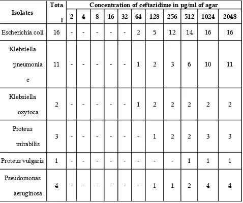
Minimum inhibitory concentration of Ceftazidime for the ESBL producing organisms in the study was between 64 µg/ml of agar
Table – 14
MIC of Ceftazidime for ESBL Producing Organisms in presence of 2 µ g/ml of Clavulanic acid (n = 37)
Isolates Total Concentration of Ceftazidime in µg/ml of agar
0.5 1 2 4 8 16 32 64 128 256 512
Escherichia.coli 16 1 12 14 16 16 16 16 16 16 16 16
Klebsiella
pneumonia
e
11 2 4 11 11 11 11 11 11 11 11 11
Klebsiella
oxytoca 2 2 2 2 2 2 2 2 2 2 2 2
Proteus
mirabilis 3 1 3 3 3 3 3 3 3 3 3 3
Proteus vulgaris 1 - - 1 1 1 1 1 1 1 1 1
Pseudomonas
aeruginosa
4 - 2 4 4 4 4 4 4 4 4 4
Minimum inhibitory concentration of Ceftazidime for the ESBL producing
organisms in the study was between 0.5 µg/ml of agar to 4 µg/ml of agar in the presence of
Table – 15
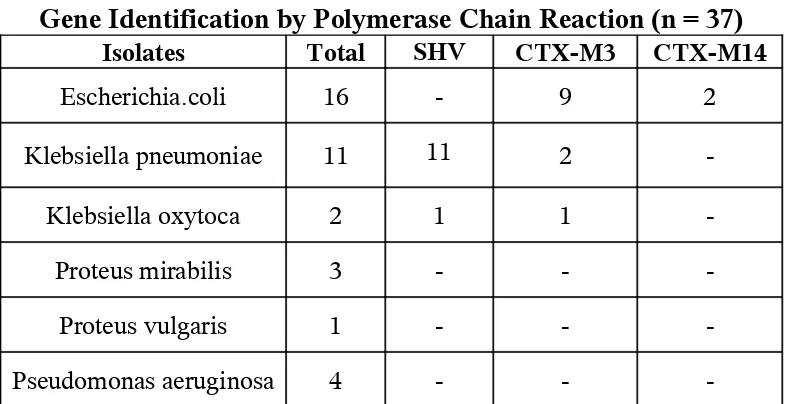
Gene Identification by Polymerase Chain Reaction (n = 37)
Isolates Total SHV CTX-M3 CTX-M14
Escherichia.coli 16 - 9 2
Klebsiella pneumoniae 11 11 2
-Klebsiella oxytoca 2 1 1
-Proteus mirabilis 3 - -
-Proteus vulgaris 1 - -
-Pseudomonas aeruginosa 4 - -
-PCR for determination of SHV, CTX-M-3, and CTX-M-14
identified. SHV gene in all the Klebsiella pneumoniae and one
Klebsiella oxytoca isolates, CTX-M-3 was found in 9 Escherichia.coli, 2
Klebsiella pneumoniae and one Klebsiella oxytoca isolates. CTX-M-14
was found in 2 Escherichia.coli isolates. 2 Klebsiella pneumoniae and
Table – 16
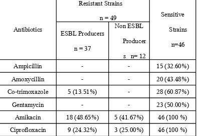
Antibiotic Susceptibility of ESBL producing and non ESBL producing GNB To Various non – β –Lactam Antibiotics (n = 95)
Antibiotics
Resistant Strains
n = 49 Sensitive
Strains
n=46 ESBL Producers
n = 37
Non ESBL
Producer
s n= 12
Ampicillin - - 15 (32.60%)
Amoxycillin - - 20 (43.48%)
Co-trimoxazole 5 (13.51%) - 28 (60.87%)
Gentamycin - - 23 (50.00%)
Amikacin 18 (48.65%) 5 (41.67%) 46 (100 %)
Ciprofloxacin 9 (24.32%) 3 (25.00%) 46 (100 %)
Out of 37 ESBL producers, 5 (13.51%) were sensitive to Co-trimoxazole, 18
DISCUSSION
Emergence of ESBL due to the extensive use of extended
spectrum Cephalosporins since 1980s were a significant evolution in
antimicrobial resistance.
ESBLs are now a problem in hospitalised patients throughout the
world. Incidence among clinical isolates vary greatly world wide and in
Geographic areas and are rapidly changing overtime. Only few studies
have been conducted to find the incidence and prevalence of ESBL
producers in Indian hospitals but ESBL producing Gram negative bacilli
may have evolved in several hospitals all over the country. Since the
incidence from ICU was found to be high from various studies, the
present study was conducted to find the incidence of ESBL producing
Gram negative bacilli from ICU of Stanley medical college hospital
from Urine, Blood, Tracheobronchial aspirate, wound swab and
drainage tube tips.
Double Disc Synergy Test (DDST) being a simple and cost
effective procedure was taken as the method of choice for screening of
ESBL producing organisms in this study.
100 cases of both male and female between 20-60 years of age