0022-538X/96/$04.0010
Copyrightq1996, American Society for Microbiology
Gene Expression and Cytopathic Effect of Vaccinia Virus
Inactivated by Psoralen and Long-Wave UV Light
KANGLA TSUNG,1* JOHN H. YIM,1WALTER MARTI,1R. MARK L. BULLER,2
ANDJEFFREY A. NORTON1
Laboratory of Biological Therapy, Department of Surgery, Washington University School of Medicine, St. Louis, Missouri 63110,1and Department of Molecular Microbiology and Immunology,
St. Louis University School of Medicine, St. Louis, Missouri 631042
Received 28 June 1995/Accepted 12 October 1995
Induction of the cytopathic effect (CPE) in cells infected with poxvirus seems ubiquitous in that it has been associated with all different strains and preparations of poxviruses, regardless of the replicating status of these viruses. The study of the mechanisms by which CPE is induced by nonreplicating poxviruses is hampered by the lack of any noncytopathic mutant strains and preparations. In this paper, we report on the patterns of gene expression and induction of CPE by vaccinia viruses treated by limited cross-linking with psoralen and long-wave UV light (PLWUV). We show that treatment of cell-free virus with PLWUV could inactivate viral replication without abolishing the ability of the virus to infect cells. Viral transcription as indicated by reporter genes was generally enhanced and prolonged under early viral promoters and abolished under late promoters. Furthermore, increasing the levels of cross-linking with PLWUV resulted in a decrease and abolishment of viral expression of a large reporter gene and a concomitant loss of the induction of CPE. Cells infected with such a virus were able to express the reporter genes and proliferate. The generation of nonreplicating and noncytopathic recombinant vaccinia viruses may help in studies of the mechanisms of CPE induction by poxvirus and may facilitate the use of poxviral vectors in broader areas of research and clinical applications.
The broad host range, ability to express multiple genes, and ease of use of vaccinia virus (VV) contribute to its popularity as a eukaryotic gene expression vector in the laboratory and as an immunization vector in the clinic. However, the infectious and cytopathic nature of VV limits its further use in some other applications. Although nonreplicating poxviral vectors have been created to address safety concerns for in vivo use (28, 30, 31), to date, there has been no report on the genera-tion of a noncytopathic poxviral vector. In fact, there is no evidence supporting the feasibility of obtaining a nonreplicat-ing and noncytopathic poxvirus. The cytopathic effect (CPE) of infection with poxvirus in general and VV in particular in-cludes the induction of early cell rounding, damage to the host genome and RNA, inhibition of host protein synthesis, and eventually, death of the infected cells (2). While the inhibition of host protein synthesis is mainly associated with replicating virus (10) or short-wave-UV-inactivated virus used at a very high concentration during infection (24), the induction of early cell rounding and cell death was found to be independent of the replicating status and evident even under conditions for which the multiplicities of infection (MOIs) were low (3, 14). The cause for this type of CPE is not yet clearly identified. The activities of known toxic VV late gene products (12, 22) or synthesis of small polyadenylated RNA (4) cannot account for the induction of CPE in this case, because these components have been found to inhibit protein synthesis and the induction of early cell rounding is dependent on protein synthesis (1). However, such a requirement does not distinguish between the two possibilities that cell rounding is a host response to viral entry or is a result of the function(s) of certain viral proteins, although indirect evidence supports the latter possibility (3). Without an available noncytopathic poxvirus mutant strain or
preparation, it is difficult to elucidate the mechanism(s) re-sponsible for the induction of CPE by nonreplicating poxvirus, thus hampering the effort to develop better viral vectors for broader application. In this study, we were able to generate nonreplicating and noncytopathic VVs by treatment of cell-free VV with psoralen and long-wave UV light (PLWUV), which is known to target nucleic acid preferentially and intro-duce chemical cross-linking in the viral genome (15). The non-cytopathic VVs thus generated were able to infect cells and express small but not large reporter genes under early viral promoters for at least 3 days efficiently. Our results indicate that poxvirus infection-associated early CPE and death of the infected cells are induced by the function of certain early viral proteins, the expression of which could be blocked by treat-ment with PLWUV.
MATERIALS AND METHODS
Cell lines and media.BSC40 cells, derivatives of the monkey kidney cell line BSC-1; mouse B16 melanoma cells and their granulocyte-macrophage colony-stimulating factor (GM-CSF) gene-transferred derivatives (8), generously pro-vided by R. Mulligan (Whitehead Institute, Massachusetts Institute of Technol-ogy, Cambridge, Mass.); mouse L cells; and monkey CV-1 cells were grown in Dulbecco modified Eagle medium (Gibco-BRL, Grand Island, N.Y.) supple-mented with 10% newborn calf serum.
recVV.All recombinant VVs (recVVs) used in this study are derivatives of the WR strain of VV (ATCC VR-119). They were generated by homologous recom-bination (9) with a series of VV sequence-based insertion-expression plasmid vectors which have been developed in our laboratory and which will be described in detail elsewhere. In brief, these plasmids contain (i) VV sequences flanking the insertion sites (A56R, hemagglutinin gene, and I4L, ribonucleotide reductase large subunit gene [17]); (ii) within the flanking sequences, two to three expres-sion cassettes arranged head to tail and composed of a viral promoter followed by multiple cloning sites and then by a VV early transcriptional termination sequence (TTTTTNT); and (iii) the Escherichia coli gpt gene under the P7.5early VV promoter, either within or outside of the flanking sequences. Murine inter-leukin-2 (IL-2) cDNA was cloned into these plasmid vectors under the various VV promoters listed in Table 1. Except for the Pkt13–IL-2 construct (Table 1), all other IL-2 expression constructs targeted the viral A56R locus for integration. The Pkt13–IL-2 construct has the IL-2 gene cloned under the Pkt13promoter in an I4L locus-based insertion-expression vector. The mouse
heat-stable-antigen-* Corresponding author. Phone: (314) 8315. Fax: (314) 362-0196.
165
on November 9, 2019 by guest
http://jvi.asm.org/
(HSA)-expressing recVV was generated with a hemagglutinin locus-based inser-tion-expression plasmid containing the HSA gene (21) (generously provided by Yang Liu, New York University Medical Center, New York, N.Y.) under the Pmj425 promoter (Table 1). The LacZ-expressing recVV, vMJ343, has been described elsewhere (6). Viruses were amplified on BSC40 cells, purified by centrifugation twice through a 35% sucrose cushion as described previously (9), and stored at2708C until use. Viral infectivity was measured as described previously (9) and expressed as PFU per milliliter.
PLWUV inactivation of virus.Psoralen (49-aminomethyl-Trioxsalen; Calbio-chem, La Jolla, Calif.) stock solution (1 mg/ml in H2O) was added to viruses (1
3108
to 103108
PFU/ml) suspended in Hanks balanced salt solution supple-mented with 0.1% bovine serum albumin to defined concentrations. The suspen-sion, usually 1 ml in a 35-mm-diameter well, was incubated at room temperature for 10 min and irradiated in a Stratalinker 1800 UV cross-linking unit (Strat-agene, La Jolla, Calif.) equipped with five 365-nm long-wave UV bulbs. The remaining plaque-forming activities of the inactivated virus were determined on a BSC40 cell monolayer. As much as 108
PFU of the preinactivation equivalent of the PLWUV-treated virus was used to infect one well of BSC40 cells in duplicate in a six-well plate. A titer of 0 was recorded when no plaques formed on the viable BSC40 cell monolayer after 3 days. In cases in which this amount of virus resulted in the killing of all the cells in the monolayer and the next 10-fold dilution did not show cell killing or plaques, the titer was recorded as 0/CPE (see Fig. 6), indicating that the virus was nonreplicating and cytopathic. Inactivation of viral DNA replication was assessed with BSC40 cells infected with PLWUV-treated VV and labeled with [3
H]thymidine for 16 h postinfection (p.i.). BSC40 cells used in the assay were rendered nonreplicating by treatment with 100 ng of psoralen per ml and then 10 min of long-wave UV irradiation.
Characterization of reporter gene expression.Cells were infected with repli-cating or inactivated VVs at MOIs of 5 to 10 PFU per cell in various experiments. The doses of PLWUV used for the inactivation of VV are indicated in each experiment. The infections were carried out either in cell suspensions (107cells per ml) in Hanks balanced salt solution for 1 h on a rocker set in a 378C incubator or on monolayers of cells in six-well plates at an infection volume of 0.25 ml for 30 to 60 min at 378C. Following the initial viral absorption, the cells were washed once in medium and cultured in six-well plates at 106cells per well. At specified time points p.i., the culture medium was changed and the levels of IL-2 or GM-CSF in the collected medium were determined by enzyme-linked immu-nosorbent assay (Pharmingen, San Diego, Calif.), with recombinant proteins being used as standards. In the experiment with B16 GM-CSF cells, the cells were preirradiated with X rays at 3,500 rads to eliminate cell proliferation. The expression of HSA on the surfaces of the infected BSC40 cells was determined 16 h p.i. by staining the cells with fluorescein isothiocyanate-labeled antibody to HSA (Pharmingen) and then subjecting the cells to flow cytometry analysis. Expression of the lacZ gene was measured by assaying the enzymatic activities of b-galactosidase in infected cells against known amounts of LacZ protein, as described previously (6).
Characterization of CPE and cell proliferation.To examine the early cell rounding, a monolayer of cells was infected with the testing VVs at an MOI of 10 PFU per cell for 30 min and cultured in medium after the unabsorbed viruses were removed. The appearance of the infected cells in the cultures was examined at specified time points p.i. and photographed with a microscope. DNA synthesis inhibitor 1-b-D-arabinofuranosylcytosine (araC) (Sigma, St. Louis, Mo.), when used, was added after the 30-min infection period to a final concentration of 50 mg/ml. In the cell survival and proliferation test, B16 cells were infected at an MOI of 10 PFU per cell with either the wild-type VV or a recVV encoding IL-2 and the E. coli guanine-xanthine phosphoribosyl transferase (gpt), both of which were inactivated with PLWUV. A total of 105
uninfected cells or cells infected with PLWUV-treated VV were cultured in a six-well plate with 2 ml of medium in the absence or presence of selection for gpt with mycophenolic acid (MPA), xanthine, and hypoxanthine as described previously (9). The appearances of the
cultures were observed daily with a microscope and photographed. To label cells with [3H]thymidine, 53103cells were cultured in each well of a 96-well plate for 2 days and 1mCi of [3H]thymidine was added to each well for an additional 4 h of incubation. In the test with sorted cells, B16 cells were infected as described above with a recVV encoding a cell surface protein (I-Akor HSA). The cells were cultured for 16 h, stained with an antibody specific to the expressed cell surface protein, and sorted on a magnetic column (Miltenyi Biotec Inc., Sunny-vale, Calif.) according to the manufacturer’s instructions. The positively selected cells were analyzed by flow cytometry to confirm the expression of the surface molecule and were evaluated for growth in culture.
RESULTS
Inactivation of VV replication by psoralen-mediated DNA
cross-linking with long-wave UV. Psoralen-mediated
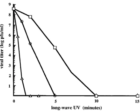
cross-linking has been shown to be an efficient way to inactivate the replication of various DNA and RNA viruses, including VV, with minimal effect on viral proteins, and viruses thus inacti-vated retain surface antigenicity (15). Theoretically, a few cross-links in the large genome of VV should be sufficient to inhibit replication without abolishing the locally confined viral transcription. Therefore, we explored whether limited treat-ment of VV with PLWUV could create a nonreplicating virus capable of expressing inserted foreign genes after it infected cells. We first examined the inactivation of viral replication with various doses of PLWUV. As can be seen in Fig. 1, inactivation of VV replication was achieved with several com-binations of different concentrations of psoralen and various periods of long-wave UV irradiation, with starting viral titers as
high as 4.43108PFU/ml. Consistent with the known effect of
psoralen on the viral genome (33), no viral DNA replication was observed in cells infected with the inactivated VV (data not shown).
Expression of a reporter gene under early or late viral
pro-moters.We next determined whether VV inactivated by
lim-ited treatment with PLWUV would retain the ability to infect cells and to express a reporter gene. We inactivated several recVVs which encode the murine IL-2 gene under various early and late VV promoters, infected BSC40 cells with these inactivated viruses, and measured the production of IL-2 in the culture supernatant of the infected cells 24 h later. As controls, BSC40 cells were also infected with the same untreated,
rep-FIG. 1. Inactivation of VV WR by PLWUV treatment. Wild-type WR virus with a starting titer of 4.43108
[image:2.612.320.554.70.255.2]PFU/ml was treated with 0.3 (triangle), 1 (circle), and 10 (square)mg of psoralen per ml and irradiated with long-wave (365-nm) UV light for various time periods as indicated. The residual viral infectivity was determined by viral plaque-forming assays and is expressed as log10PFU per milliliter.
TABLE 1. VV promoter activities from replicating and inactivated recVVs
Promoter Class
IL-2 production (U/106cells)
Replicating VV Inactivated VV
Pmj425
a Early 80 132
Pkt10b Early 242 536
Pkt13
b Early 450 1,000
P11kc Late 3,025 0
a
This promoter has been characterized by Davison and Moss (7). It was derived from the viral thymidine kinase gene promoter with one point mutation.
b
These promoters were designed and synthesized in our laboratory. The Pkt10 promoter is based on the viral P7.5promoter with multiple up-regulation muta-tions. The Pkt13promoter is based on the viral ribonucleotide reductase small subunit gene with two point mutations.
c
This natural viral promoter has been characterized by Davison and Moss (6).
on November 9, 2019 by guest
http://jvi.asm.org/
licating viruses. As can be seen from Table 1, the IL-2 gene
under well-characterized late viral promoter P11k was
ex-pressed only by the untreated virus and not by the PLWUV-treated virus. In contrast, the IL-2 gene under all early pro-moters tested was expressed by both the replicating and the inactivated recVVs, with expression by the inactivated viruses always being higher than that of their replicating counterparts. This finding indicated that the early phase of viral transcription was not affected by psoralen-mediated DNA cross-linking to the same degree as viral replication and late gene expression. The complete loss of late viral promoter activity after psoralen treatment is consistent with the lack of viral DNA replication, which is required for the activation of late VV promoters (6, 7). The level of IL-2 secretion in the experiment described above did not reflect the proportion of cells infected with PLWUV-inactivated virus. We addressed this issue by exam-ining cell surface expression of the mouse HSA encoded by a
recVV under the Pmj425early viral promoter. As can be seen
from Fig. 2, infection of BSC40 cells with this PLWUV-inac-tivated virus at an MOI of 5 PFU per cell resulted in the expression of HSA by virtually all cells, indicating that the ability of PLWUV-treated VV to infect cells was retained.
PLWUV-inactivated recVV is noncytopathic.During the
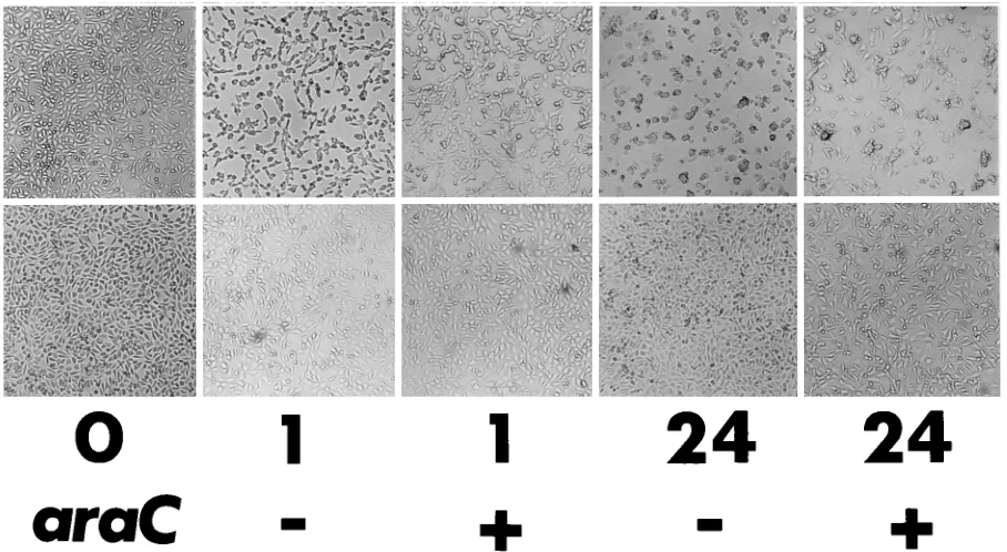
[image:3.612.80.534.443.692.2]ex-periments discussed above, the total destruction of the BSC40 cell monolayer during titer assays was routinely not observed even when PLWUV-inactivated viruses were used at an MOI of as high as 50 PFU per cell. In addition, we repeatedly saw the continued proliferation of bulk-cultured cells infected with PLWUV-inactivated viruses at MOIs of less than 20 PFU per cell. This observation was in contrast to the known CPE asso-ciated with poxvirus infection, even when the infection is due to nonreplicating viruses (14). Previous studies have shown that CPE induced by VV infection of cultured cells includes an early morphological change characterized by cell rounding (1) and a rapid shutoff of host macromolecule synthesis (24, 26). To find out whether the early cell rounding was indeed blocked by PLWUV treatment, we infected a monolayer of mouse L cells with replicating or PLWUV-inactivated VV at an MOI of 10 PFU per cell, cultured the infected cells in the presence and absence of the DNA synthesis inhibitor, araC, and recorded the morphological changes of the cells at various time points following infection (Fig. 3). Almost all the cells infected with replicating virus showed obvious cell rounding as early as 1 h p.i., in both the presence and absence of araC, confirming that infection at this MOI was complete as well as that CPE is a pre-DNA replication event. On the other hand, cells infected at the same MOI with PLWUV-inactivated virus did not show signs of cell rounding either during the early period following infection or for 24 h (Fig. 3). Since L cells have been used previously to show the early CPE induced by nonreplicating
FIG. 2. PLWUV-inactivated VV retains the ability to infect cells efficiently. BSC40 cells were infected with either the wild-type VV (open histogram) or a recVV encoding HSA (solid histogram), both inactivated with 10mg of psoralen per ml and by 2 min of long-wave UV irradiation, at an MOI of 5 PFU per cell. The cell surface expression of HSA at 16 p.i. was determined by antibody staining.
FIG. 3. Early CPE was blocked in cells infected with PLWUV-inactivated VV. A monolayer of mouse L cells was infected at an MOI of 10 PFU per cell with either the replicating WR strain of VV (top panel) or the same virus inactivated with 10mg of psoralen per ml and by 2 min of long-wave UV irradiation (bottom panel). Following infection, the cells were cultured either in the absence (2) or in the presence (1) of araC. The morphological changes of the cells not infected (0) and infected with the two viruses at 1 (1) and 24 (24) h p.i. are shown. Magnification,340.
on November 9, 2019 by guest
http://jvi.asm.org/
VV (3, 14), this lack of early CPE induction by PLWUV-treated VV is unique to PLWUV-inactivated VV.
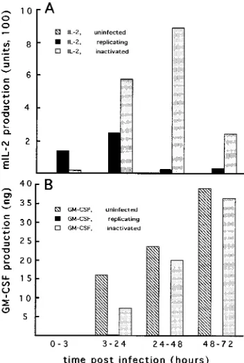
To analyze and compare the fates of host gene expression following infection with replicating or PLWUV-inactivated VV, we examined the coexpression of both virus- and host-encoded cytokines with a GM-CSF-producing cell line derived from mouse B16 melanoma cells (8). PLWUV-inactivated or untreated recVV encoding IL-2 under early viral promoter
Pkt13(Table 1) was used to infect the GM-CSF-producing B16
melanoma cells at an MOI of 10 PFU per cell. To simplify the interpretation of the results of the experiment, the cells were preirradiated with a known dose of X rays which would stop cell proliferation without abolishing GM-CSF expression (8). Following infection, the cells were cultured, the medium was changed at specified time points, and the levels of both IL-2 and GM-CSF in each supernatant were determined. As can be seen from Fig. 4, infection of B16 GM-CSF cells with replicat-ing recVV resulted in the complete shutoff of the cellular expression of GM-CSF between 3 and 24 h p.i. In contrast, GM-CSF production was only transiently lowered by infection with PLWUV-inactivated recVV, and it recovered by 24 h p.i. and persisted at levels similar to those of the uninfected con-trol cells for the rest of the time points (Fig. 4B). Because the cells were preirradiated and no sign of cell proliferation was observed during the experiment, the recovered levels of GM-CSF production could not result from the expansion of a few uninfected cells in the culture. In parallel to GM-CSF produc-tion, the expression of virally encoded IL-2 in these cells was detected up to 3 days p.i., in clear contrast to the lower level
and much shorter period of IL-2 production by the replicating virus-infected cells (Fig. 4A).
To further demonstrate that cells infected with PLWUV-inactivated VV are not killed and are able to continue prolif-eration, we carried out an experiment in which cell prolifera-tion was regulated by MPA, the guanine synthesis inhibitor. Since the inhibition by MPA can be bypassed by expression of the E. coli gpt gene (11, 25), we asked whether cells infected with a recVV encoding gpt under an early viral promoter would grow in the presence of MPA. Mouse B16 melanoma cells were, therefore, infected at an MOI of 10 PFU per cell with either the wild-type VV or a recVV encoding both gpt and the IL-2 gene under separate early viral promoters. Cells infected with these viruses were compared with uninfected cells for growth in culture in the presence and absence of MPA. While active cell growth was observed in all three cultures in the absence of MPA (Fig. 5, row 3), only cells infected with PLWUV-inactivated recVV carrying the gpt marker gene were able to grow in the presence of MPA (Fig. 5, rows 1 and 2). In fact, these were the only cells which continued to proliferate after the MPA was removed following selection with the drug
for 2 days (Fig. 5, row 4). Results from [3H]thymidine labeling
of the cultures also supported the finding, and the presence of recVV in the cells during the 2-day selection period with MPA was confirmed by the detection of IL-2 in the culture super-natant (data not shown). In alternative experiments, we se-lected cells infected with PLWUV-inactivated recVV by sort-ing for a cell surface protein (major histocompatibility complex class II and HSA, for example) encoded by the virus and cultured the positively selected cells. Continued cell growth was observed in all cases when BSC40, B16, and several other murine tumor cell lines were used (data not shown).
The correlation between the lack of CPE and the degree of
inhibition of viral transcription.The lack of CPE in the
pres-ence of early viral transcription is puzzling in that a previous study suggested that early viral protein synthesis is sufficient to induce CPE (3). The experiments using IL-2 as a reporter gene clearly indicated that not only was early viral transcrip-tion preserved after treatment with PLWUV, the levels and duration of the early transcription were actually enhanced by PLWUV-mediated inactivation (Table 1 and Fig. 3). A clue to the explanation for this paradox came from the analysis of the expression patterns of a large reporter gene, lacZ from E. coli. Under cross-linking conditions which allowed IL-2 expression and abolished CPE induction, the expression of lacZ under an early viral promoter was completely inhibited. This observation prompted us to reexamine the patterns of gene expression and the induction of CPE under various degrees of cross-linking by PLWUV by using a recVV, vMJ343, that encodes LacZ (6). As can be seen from Fig. 6, replicating vMJ343 expressed high levels of LacZ, as has been reported previously (6), and caused severe early cell rounding of CV-1 cells by 2 h p.i. Inactivation of the replication of this virus with PLWUV at the minimal
required doses (0.3 mg of psoralen per ml and 10 min of
long-wave UV irradiation) produced a nonreplicating virus which expressed increased levels of LacZ and induced clear but less severe early cell rounding in CV-1 cells. With further increases in the degree of cross-linking, both the expression of LacZ and the severity of CPE decreased. These observations indicate that certain large early viral genes may not be ex-pressed under the cross-linking conditions that abolish CPE induction. Furthermore, we found that the transcription of small reporter genes, such as IL-2, could also be inhibited by treatment with elevated doses of PLWUV. For example, IL-2
expression was abolished when 20mg of psoralen per ml and 8
[image:4.612.89.265.71.333.2]min of long-wave UV irradiation were used to treat the recVV
FIG. 4. Coexpression of cellular and viral genes in cells infected with PLWUV-inactivated recVV. GM-CSF-secreting B16 cells were irradiated with X rays and infected at an MOI of 10 PFU per cell with Pkt13–IL-2-encoding recVV in the form of either replicating or PLWUV-inactivated (10mg of psoralen per ml and 2 min of long-wave UV irradiation) virus. A total of 106
infected or irradiated uninfected cells were cultured in 2 ml of medium in a six-well tissue culture plate. At specified time points following infection, the culture medium was changed and the amounts of IL-2 (A) and GM-CSF (B) in the collected
culture supernatant were determined.
on November 9, 2019 by guest
http://jvi.asm.org/
(data not shown). These observations are consistent with the findings of a study of short-wave-UV-mediated cross-linking that showed that inactivation of a gene by cross-linking is directly proportional to the size of the target gene and the dose of the cross-linking reagents (5).
DISCUSSION
Three findings emerge from the present study. First, limited treatment of VV with PLWUV results in the inactivation of viral replication without the abolition of viral transcription under early but not late viral promoters. Second, compared with that by replicating virus, early viral transcription by PLWUV-inactivated virus is enhanced both in degree and in duration. Third, proper treatment of VV with PLWUV leads to a non-replicating and noncytopathic virus capable of expressing in-serted foreign genes.
The large genome size (about 200 kb) of VV should be a major factor contributing to the differential effects of PLWUV-mediated cross-linking on viral replication and transcription. As we had speculated, limited cross-linking was able to inhibit viral DNA replication, probably by blocking the movement of viral DNA polymerase along the genome. In addition, the size of the viral DNA polymerase gene, which is over 3 kb in size and encodes 1,006 amino acid residues (32), may be an impor-tant factor. This large early viral gene should be among the first of the genes inactivated by PLWUV, which was indicated by results of our analysis with the 3-kb lacZ gene in this study (Fig. 6). On the other hand, small genes, such as IL-2 (0.5 kb), and several other cytokine genes (data not shown) were well ex-pressed by PLWUV-inactivated, noncytopathic recVVs in our experiments. The expression efficiency of midsized genes by PLWUV-inactivated recVVs falls between the efficiencies of large and small genes. In several cases, we observed the
coex-FIG. 5. Proliferation of cells infected with PLWUV-inactivated VV. Mouse B16 melanoma cells were left uninfected (A) or were infected at an MOI of 10 PFU per cell with PLWUV-treated (10mg of psoralen per ml and 2 min of long-wave UV irradiation) VV, either the wild-type WR virus (B) or a recVV encoding the E. coli gpt genes (C). The cells were cultured in the presence (rows 1 and 2) or absence (row 3) of MPA for 2 days. The cultures were observed for cell growth at the ends of days 1 (row 1) and 2 (rows 2 and 3). MPA was removed after 2 days, and cell growth after an additional 2 days in culture (row 4) is shown. Magnification,340.
on November 9, 2019 by guest
http://jvi.asm.org/
pression of two to three foreign genes by PLWUV-inactivated recVVs. For example, the functional mouse major
histocom-patibility complex class II molecule, I-Ak, encoded by two
subunit genes was expressed from recVV carrying these genes. Although expression of the reporter genes was used for this paper to demonstrate viral transcription, there is evidence in-dicating that some of the viral early proteins were also made in cells infected with PLWUV-treated, noncytopathic virus. Cells
infected with PLWUV-inactivated VV and labeled with [35S]
methionine showed the coexpression of both viral and cellular proteins (unpublished observation). Furthermore, the anti-genicity of the PLWUV-inactivated VV was found indistin-guishable from that of the replicating VV, both for restimu-lating the VV-primed cytotoxic T lymphocytes and serving as the cytotoxic T-lymphocyte target in vitro (unpublished re-sult).
The enhancement and prolongation of early viral transcrip-tion by PLWUV treatment are more dramatic than what has been observed in virus rendered nonreplicating by DNA syn-thesis inhibitor (6), which confirms the finding from a previous report that blocking viral DNA replication and late gene ex-pression per se does not lead to increased and prolonged early viral transcription (23). The persistence of the early transcrip-tion is shown here to last at least 3 days (Fig. 4) and has been observed to reach as long as 5 days, albeit at decreasing levels, probably reflecting the lifetime of the viral transcription ma-chinery assembled in the virion during the late phase of the previous infection cycle (13). The patterns of early viral tran-scription suggest that secondary viral uncoating is blocked by treatment with PLWUV, because similar enhancement of early viral transcription has been observed under conditions that blocked uncoating either with protein synthesis inhibitors or by short-wave UV irradiation (19, 34). It should be pointed out, however, that we failed to observe any similar enhancement of early viral transcription by inactivation of recVVs by short-wave UV irradiation (unpublished observation). It seems that inactivation of VV by both short- and long-wave UV irradia-tion may result in the blocking of secondary uncoating and thus enhance early viral transcription, but viral transcription be-comes much more sensitive to the inhibitory effect of increased
short-wave UV treatment (34) than to that of the PLWUV treatment. Joklik has suggested that secondary viral uncoating is dependent on the expression of certain viral inducer proteins and that this expression is blocked by short-wave UV treat-ment (18). In an effort to test this possibility, we attempted to provide the factors responsible for secondary uncoating in
trans by suprainfecting cells preinfected with a
PLWUV-inac-tivated recVV encoding IL-2 with replicating vMJ343 under araC. In spite of the complete reinfection by the second virus, as judged by both the expression of LacZ and the return of early CPE, the elevated expression of IL-2 by the PLWUV-inactivated virus persisted (unpublished result). Therefore, it remains to be determined whether the effect on early viral transcription by PLWUV treatment is mediated by the block-ing of secondary viral uncoatblock-ing.
The demonstration of a noncytopathic VV in this study indicates that CPE induction is not necessarily a host cell response to VV infection and that it can be eliminated without abolishing infection and expression of all virally encoded genes. This possibility could not be eliminated by using heat-inactivated VV, because the treated virus did induce cell death, albeit by a different mechanism (14). On the other hand, all strains and preparations of poxvirus in the past have been cytopathic. While it is clear that a replicating VV shuts off host protein synthesis after viral DNA replication (12, 16, 26, 29), much less is known about the mechanism(s) by which nonrep-licating virus induces CPE. Under the conditions of infection with a nonreplicating VV prepared by short-wave UV
irradi-ation and given at a high MOI (.50 PFU per cell), there is a
[image:6.612.63.297.72.231.2]rapid inhibition of total protein synthesis in the infected cells (24). Even with PLWUV-inactivated VV, early cell death was evident when such a high MOI was used (unpublished obser-vation), suggesting that invading virions contain toxic compo-nents, as has been indicated by previous studies (12, 22, 27). This effect is, however, not evident in cells infected with a low MOI (24) and, therefore, could not explain the CPE observed in these cells. In this study, we have mainly examined the early CPE induced by VV infection at a low MOI which was suffi-cient to infect nearly all cells (Fig. 2). The induction of early cell rounding independent of viral replication was confirmed by the use of araC (Fig. 3). In previous experiments, we have also confirmed this phenomenon by the use of VV inactivated by short-wave UV irradiation (unpublished results). In con-trast to these methods of inactivation, PLWUV treatment was effective in eliminating induction of CPE (Fig. 3), inhibition of host gene expression (Fig. 4), and cell death (Fig. 5). The PLWUV doses required to achieve these were one step beyond those required for inactivation of viral replication. Titration experiments with the recVV encoding LacZ indicate that the gradual loss of CPE correlated with the gradual increase in inhibition of synthesis of full-size viral transcripts. Small poly-adenylated viral RNA produced by short-wave-UV-inactivated VV has been found in previous studies to inhibit the transla-tion of normal cellular RNA and has been proposed to be operational in vivo (4). It is not known whether PLWUV-treated VV in our study produced such groups of small poly-adenylated RNA, although the conditions for the elimination of CPE by PLWUV treatment actually favored the overpro-duction of truncated small viral transcripts. If such viral RNA were produced, it did not seem to be toxic in vivo. What, then, makes the PLWUV treatment different from all other inacti-vation methods which are not effective in eliminating CPE? It seems that more than one factor contributed to the effects of PLWUV treatment. On the one hand, since the induction of early cell rounding has been linked to early viral protein syn-thesis (3) and since it was absent when the transcription of
FIG. 6. Correlation between the expression of lacZ and early CPE. A mono-layer of CV-1 cells was infected at an MOI of 10 PFU per cell with vMJ343 encoding LacZ. The virus was treated with various combinations of psoralen and long-wave UV light, as indicated. The remaining viral titer was determined and shown (see Materials and Methods for an explanation of the titer assignment). The early cell rounding of the infected cells was recorded at 2 h p.i., and the amount of LacZ produced by each culture was measured at 24 h p.i.111, complete CPE;11,.50% of cells in culture show CPE;1,.20% but,50% of cells show CPE;1/2,,10% of cells show CPE;2, no sign of CPE.
on November 9, 2019 by guest
http://jvi.asm.org/
early viral genes was partially inhibited in cells infected with PLWUV-treated VV, our results suggest that either a large early viral protein or the cooperation of several small ones may be responsible for the induction of early CPE. Poxviruses in-activated by host range restriction would be expected to ex-press all early viral proteins, including the putative CPE induc-er(s). On the other hand, this partial inhibition of early viral transcription alone may not be enough, as a similar phenom-enon has been described for the short-wave-UV-mediated in-activation of VV (5). It is currently not clear what is differen-tially modified in VV by PLWUV and short-wave UV treatments. It cannot be ruled out that the structure of the virion is modified directly by psoralen. In any case, identifying the differences between PLWUV- and short-wave-UV-medi-ated inactivation of VV and the early viral genes expressed by untreated and PLWUV-treated virus should help to elucidate the mechanism(s) of CPE induction.
The findings of the present study may facilitate the broader application of VV as a gene delivery and expression vector. Nonreplicating poxviral vectors based on host range restriction have been developed for use in vivo for safe immunizations (28, 31). Recently, the profiles of in vitro gene expression by recombinant fowlpox viruses have been reported (20). The data indicate that cytokine gene (IL-6 and gamma interferon) expression under early promoter by nonreplicating fowlpox viral vector persists in cultured human cells for up to 5 days, with peak levels in the first day and decreasing levels thereaf-ter, a profile similar to what we observed with PLWUV-treated recVVs in the present report. Nevertheless, unlike these other nonreplicating poxviral vectors which require special host cells for manipulation, the generation and amplification of the PLWUV-inactivated recVV are the same as those for a repli-cating virus, except that inactivation is achieved at a time after viral production. Although the expression of foreign genes by PLWUV-inactivated recVVs is transient and at levels much lower than those achievable with late VV promoters, it seems that the use of PLWUV-inactivated VV in the manipulation of immune responses is feasible. For example, recVV encoding both subunits of mouse major histocompatibility complex class
II molecule I-Ak was able to express a functional I-Ak for
presenting antigen peptide to I-Ak-restricted CD4 T cells in
vitro and stimulating a strong anti-I-Akallogeneic response in
vivo (unpublished result). Also, PLWUV-inactivated VV could be used for transient expression of certain cytokines in tumor cells, which would be effective in stimulating a host antitumor response (unpublished result). The noncytopathic feature of recVV prepared by PLWUV treatment provides an additional advantage for use in cases in which the survival and prolifer-ation of the infected cells and transient gene expression are critical. For example, such a nonreplicating and noncytopathic recVV could be used for the expression of small growth factor genes (for epidermal growth factor, platelet-derived growth factor, fibroblast growth factor, etc.) to facilitate wound heal-ing. We are currently exploring these and other potential ap-plications.
REFERENCES
1. Bablanian, R. 1968. The prevention of early vaccinia-virus-induced cyto-pathic effects by inhibition of protein synthesis. J. Gen. Virol. 3:51–61. 2. Bablanian, R. 1975. Structural and functional alterations in cultured cells
infected with cytocidal viruses. Prog. Med. Virol. 19:40–83.
3. Bablanian, R., B. Baxt, J. A. Sonnabend, and M. Esteban. 1978. Studies on the mechanisms of vaccinia virus cytopathic effects. II. Early cell rounding is associated with virus polypeptide synthesis. J. Gen. Virol. 39:403–413. 4. Cacoullos, N., and R. Bablanian. 1991. Polyadenylated RNA sequences
produced in vaccinia virus-infected cells under aberrant conditions inhibit protein synthesis in vitro. Virology 184:747–751.
5. Cooper, J. A., R. Wittek, and B. Moss. 1981. Hybridization selection and
cell-free translation of mRNA’s encoded within the inverted terminal repe-tition of the vaccinia virus genome. J. Virol. 37:284–294.
6. Davison, A. J., and B. Moss. 1989. Structure of vaccinia virus early promot-ers. J. Mol. Biol. 210:749–769.
7. Davison, A. J., and B. Moss. 1989. Structure of vaccinia virus late promoters. J. Mol. Biol. 210:771–784.
8. Dranoff, G., E. Jaffee, A. Lazenby, P. Golumbek, H. Levitsky, K. Brose, V.
Jackson, H. Hamada, D. Pardoll, and R. C. Mulligan.1993. Vaccination with irradiated tumor cells engineered to secrete murine granulocyte-macrophage colony-stimulating factor stimulates potent, specific, and long-lasting anti-tumor immunity. Proc. Natl. Acad. Sci. USA 90:3539–3543.
9. Earl, P. L., and B. Moss. 1991. Protein expression IV: expression of proteins in mammalian cells using vaccinia, p. 16.15.1–16.18.10. In F. M. Ausubel, R. Brent, R. E. Kingston, D. D. Moore, J. G. Seidman, J. A. Smith, and K. Struhl (ed.), Current protocols in molecular biology. Greene Publishing Associates and Wiley-Interscience, New York.
10. Esteban, M., and D. H. Metz. 1973. Early virus protein synthesis in vaccinia virus-infected cells. J. Gen. Virol. 19:201–216.
11. Falkner, F. G., and B. Moss. 1992. Escherichia coli gpt gene provides dom-inant selection for vaccinia virus open reading frame expression vectors. J. Virol. 62:1849–1854.
12. Fernandez, A. P., and G. Beaud. 1986. Purification and characterization of a protein synthesis inhibitor associated with vaccinia virus. J. Biol. Chem.
261:8283–8289.
13. Gershon, P. D., and B. Moss. 1990. Early transcription factor subunits are encoded by vaccinia virus late genes. Proc. Natl. Acad. Sci. USA 87:4401– 4405.
14. Hanafusa, H. 1960. Killing of L-cells by heat- and UV-inactivated vaccinia virus. Biken J. 3:191–199.
15. Hanson, C. V. 1992. Photochemical inactivation of viruses with psoralens: an overview. Blood Cells (New York) 18:7–25.
16. Jefferts, E. R., and J. A. Holowczak. 1971. RNA synthesis in vaccinia-infected L cells: inhibition of ribosome formation and maturation. Virology 46:730– 744.
17. Johnson, G. P., S. J. Goebel, and E. Paoletti. 1993. An update of the vaccinia virus genome. Virology 196:381–401.
18. Joklik, W. K. 1964. The intracellular uncoating of poxvirus DNA. II. The molecular basis of the uncoating process. J. Mol. Biol. 8:277–288. 19. Kates, J. R., and B. R. McAuslan. 1967. Messenger RNA synthesis by a
‘‘coated’’ viral genome. Proc. Natl. Acad. Sci. USA 57:314–320.
20. Leong, K. H., A. J. Ramsay, D. B. Boyle, and I. A. Ramshaw. 1994. Selective induction of immune responses by cytokines coexpressed in recombinant fowlpox virus. J. Virol. 68:8125–8130.
21. Liu, Y., B. Jones, A. Aruffo, K. M. Sullivan, P. S. Linsley, and C. A. Janeway. 1992. Heat-stable antigen is a costimulatory molecule for CD4 T cell growth. J. Exp. Med. 175:437–445.
22. Mbuy, G. N., R. E. Morris, and C. H. Bubel. 1982. Inhibition of cellular protein synthesis by vaccinia virus surface tubules. Virology 116:137–147. 23. McDonald, W. F., V. Crozel-Goudot, and P. Traktman. 1992. Transient
expression of the vaccinia virus DNA polymerase is an intrinsic feature of the early phase of infection and is unlinked to DNA replication and late gene expression. J. Virol. 66:534–547.
24. Moss, B. 1968. Inhibition of HeLa cell protein synthesis by the vaccinia virion. J. Virol. 2:1028–1037.
25. Mulligan, R. C., and P. Berg. 1981. Selection for animal cells that express the Escherichia coli gene coding for xanthine-guanine phosphoribosyltrans-ferase. Proc. Natl. Acad. Sci. USA 78:2072–2076.
26. Pedley, S., and R. J. Cooper. 1984. The inhibition of HeLa cell RNA syn-thesis following infection with vaccinia virus. J. Gen. Virol. 65:1687–1697. 27. Pogo, B. G. T., and S. Dales. 1973. Biogenesis of poxviruses: inactivation of
host DNA polymerase by a component of the invading inoculum particle. Proc. Natl. Acad. Sci. USA 70:1726–1729.
28. Radaelli, A., M. Gimelli, C. Cremonesi, C. Scarpini, and C. D. G. Morghen. 1994. Humoral and cell-mediated immunity in rabbits immunized with live non-replicating avipox recombinants expressing the HIV-1SF2env gene. Vac-cine 12:1110–1117.
29. Rice, A. P., and B. E. Roberts. 1983. Vaccinia virus induces cellular mRNA degradation. J. Virol. 47:529–539.
30. Tartaglia, J., M. E. Perkus, J. Taylor, E. K. Norton, J.-C. Audonnet, W. I.
Cox, S. W. Davis, J. Van Der Hoeven, B. Meignier, M. Riviere, B. Languet, and E. Paoletti.1992. NYVAC: a highly attenuated strain of vaccinia virus. Virology 188:217–232.
31. Taylor, J., C. Trimarchi, R. Weinberg, B. Languet, F. Guillemin, P.
Desmet-tre, and E. Paoletti.1991. Efficacy studies on a canarypox-rabies recombinant virus. Vaccine 9:190–192.
32. Traktman, P. 1990. The enzymes of poxvirus DNA replication, p. 93–124. Springer-Verlag, Berlin.
33. Veith Hanson, C., J. L. Riggs, and E. H. Lennette. 1978. Photochemical inactivation of DNA and RNA viruses by psoralen derivatives. J. Gen. Virol.
40:345–358.
34. Woodson, B. 1967. Vaccinia mRNA synthesis under conditions which pre-vent uncoating. Biochem. Biophys. Res. Commun. 27:169–175.