A COMPARATIVE ANALYSIS OF TENS AND ACTIVE SPINAL EXERCISES VERSES TENS AND TRIGGER POINT RELEASE
TECHNIQUE IN IMPROVING LOW BACK PAIN OF MECHANICAL ORIGIN
A dissertation submitted in partial fulfillment of the requirement for the degree of
MASTER OF PHYSIOTHERAPY
(ELECTIVE –PHYSIOTHERAPY IN ORTHOPEADICS) To
The Tamil Nadu Dr. M.G.R. Medical University Chennai-600032
April 2012
(Reg. No.27101906)
RVS COLLEGE OF PHYSIOTHERAPY
(Affiliated to the Tamil Nadu Dr. M.G.R Medical University, Chennai- 32) SULUR, COIMBATORE – 641 402
CERTIFICATE
Certified that this is the bonafide work of Sujith S., a second year
student of R.V.S. College of Physiotherapy, Sulur, Coimbatore submitted in
partial fulfillment of the requirements for Master of Physiotherapy Degree
course from The Tamil Nadu Dr M.G.R Medical University under the
Registration No:27101906.
ADVISOR PRINCIPAL
Mr. E. Magesh, M.P.T., (Ph.D.), Prof. Mrs. R. Nagarani, MPT, MA, (Ph.D.),
Professor, RVS College Of Physiotherapy,
RVS college of physiotherapy, Sulur,
Sulur, Coimbatore. Coimbatore.
Place:
A COMPARATIVE ANALYSIS OF TENS AND ACTIVE SPINAL EXERCISES VERSES TENS AND TRIGGER POINT RELEASE
TECHNIQUE IN IMPROVING LOW BACK PAIN OF MECHANICAL ORIGIN
INTERNAL EXAMINER EXTERNAL EXAMINER
SUBMITTED IN THE PARTIAL FULFILLMENT OF THE REQUIREMENT FOR DEGREE OF
“MASTER OF PHYSIOTHERAPY” TO
THE TAMIL NADU DR. M.G.R. MEDICAL UNIVERSITY CHENNAI
DECLARATION
I hereby declare and present my project work entitled
“A COMPARATIVE ANALYSIS OF TENS AND ACTIVE SPINAL EXERCISES VERSES TENS AND TRIGGER POINT RELEASE TECHNIQUE IN IMPROVING LOW BACK PAIN OF MECHANICAL ORIGIN” The outcome of the original research work
undertaken and carried out by me, under the guidance of Professor
Mr. E. Magesh, MPT., (Ph.D)., RVS College Of Physiotherapy, Sulur,
Coimbatore.
I also declare that the material of this project work has not formed
in any way the basis for the award of any other degree previously from the
Tamil Nadu Dr. M.G.R Medical University.
Date: SIGNATURE
Place:
ACKNOWLEDGEMENT
I give my thanks to God almighty for providing me the wisdom and
knowledge to complete my study successfully.
This study will be an incomplete one without my gratitude towards my
‘Lovable Parents’ who made me what I am today.
I acknowledge my sincere thanks to Chairman and Secretary of
R.V.S Educational Trust, Sulur, Coimbatore for providing me an opportunity to do this project.
I would like to express my gratitude to our principal
Mrs. R.Nagarani M.P.T., M.A., (PhD)., for providing me constant support and motivation in the form of resources and inputs.
I would like to thank my guide Mr. E. Magesh, MPT, (Ph.D)., offering
me perceptive inputs and guiding me entirely through the course of my work and
without his tired less guidance and support this project would not have come through.
I also thank my friends for their co-operation in completion of this project.
I offer my thanks and gratitude to our librarians for their supports in
providing books to complete my study.
I take this golden opportunity to thank each and every subject who took part in
TABLE OF CONTENT
CHAPTER TITLE PAGE No.
I. INTRODUCTION 1
1.1 Need for the study 5
1.2 Statement of the problem 6
1.3 Hypothesis 6
1.4 Operational Definitions 7
II. REVIEW OF LITERATURE
9
III. MATERIALS AND METHODOLOGY 15
3.1 Study design 15
3.2 Study Setting 15
3.3 Sample Size 15
3.4 Inclusion Criteria 15
3.5 Exclusion Criteria 16
3.6 Study duration 16
3.7 Variables used in the study 16
3.8 Measurement Tool 16
3.9 Treatment Procedure 17
IV. DATA ANALYSIS AND RESULT 22
4.1 Data Analysis 22
4.2 Results 36
V. DISCUSSION 38
Vi. CONCLUSION 40
VII. BIBLIOGRAPHY 42
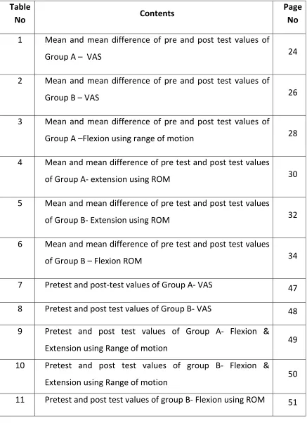
Table
No Contents
Page
No
1 Mean and mean difference of pre and post test values of
Group A – VAS 24
2 Mean and mean difference of pre and post test values of
Group B – VAS 26
3 Mean and mean difference of pre and post test values of
Group A –Flexion using range of motion 28
4 Mean and mean difference of pre test and post test values
of Group A‐ extension using ROM 30
5 Mean and mean difference of pre test and post test values
of Group B‐ Extension using ROM 32
6 Mean and mean difference of pre test and post test values
of Group B – Flexion ROM 34
7 Pretest and post‐test values of Group A‐VAS 47
8 Pretest and post test values of Group B‐ VAS 48
9 Pretest and post test values of Group A‐ Flexion &
Extension using Range of motion 49
10 Pretest and post test values of group B‐ Flexion &
Extension using Range of motion 50
[image:7.612.110.547.107.709.2]
LIST OF GRAPHS
Graph
No
Contents Page
No
1 Group A pre and post test VAS means score 25
2 Group B pre and post test VAS means score 27
3 Group A Flexion ROM pre and post test means Value 29
4 Group A Extension ROM pre and post test means Value
31
5 Group B Flexion ROM pre and post test means Value 33
6 Group B Extension ROM pre and post test means Value 35
12 Pretest and post test values of group B‐ Extension using
1. INTRODUCTION
Pain is a process which can affect the individual physically, emotionally, psychologically, socially, occupationally and in many other
ways.
Knowledge of mechanical low back pain disorders has matured
beyond past that all back pain is from the intervertebral disc or the
zygopophyseal joints or is myofascial in nature, or that we have only an
isolated injury. If we can identify the offending forces, especially during
a patients activities of daily living, and minimize these forces while
allowing the person to stay active, then the healing process will more
readily occur. In effect one of the goals of treatment for any mechanical
injury to provide an optimal healing environment.
The clinician and patient are thus challenged to identify the forces
that are stimulating the nociceptive system and reproducing symptoms
and to control and alter the way that they reach the lumbopelvic region. It
is extremely important that the patient have an active role in management.
Less than 2 percent of his walking time is spent in treatment, the
clinician must convince the patient of the importance of other 98 percent
of his walking time with respect to managing his own syndrome.
professional. Musculoskeletal disorders are the main cause of disability in
the working age population and are among the leading causes of
disability in other age groups.
Transcutaneous Electrical Nerve Stimulation, electrotherapy
modality in which low electrical current is sent through a pad at an injury
site, stimulating the brain to release endorphins Rehabilitation medicine
A modality for controlling pain by delivering low-level electric shocks to
the skin; TENS effect is explained by the 'gate' theory of pain and is used
to relieve pain of the lower back and neck, 'phantom' limb syndrome,
amputation stump pain.
Fascia is the soft tissue component of the connective tissue that
provides support and protection for most structures within the human
body, including muscle. This soft tissue can become restricted due to
psychogenic disease, overuse, trauma, infectious agents, or inactivity,
often resulting in pain, muscle tension, and corresponding diminished
blood flow. Although fascia and its corresponding muscle are the main
targets of myofascial release, other tissue may be affected as well,
including other connective tissue.
the connective tissue, and this thickening causes pain and irritation,
resulting in reflexive muscle tension that causes more inflammation. In
this way, the cycle creates a positive feedback loop and can result in
ischemia and somatic dysfunction even in the absence of the original
offending agent. Myofascial techniques aim to break this cycle through a
variety of methods acting on multiple stages of the cycle.
Myofascial point pain is common painful muscle disorder caused
by myofascial trigger points. Myofascial trigger points are characterized
by pain originating from small circumscribed areas of local hyper
irritability and myofascial structures resulting in local and related pain.
In medical literature, the term myofascial was historically used by
Janet G. Travell, M.D. in the 1940s referring to musculoskeletal pain
syndromes and trigger points. In 1976 Dr. Travell began using the term
"Myofascial Trigger Point" and in 1983 published the reference
"Myofascial Pain & Dysfunction: The Trigger Point Manual". There is no
evidence she actually used what is now termed "myofascial release".
Some practitioners use the term "Myofascial Therapy" or "Myofascial
Trigger Point Therapy" referring to the treatment of trigger points,
usually in medical-clinical sense. The phrase has also been loosely used
for different manual therapy techniques, including soft tissue
mobilization, foam rolling, structural integration, and strain-counter strain
techniques. However, in current medical terminology, myofascial release
refers mainly to the soft tissue manipulation techniques described below.
The trigger point model states that unexplained pain frequently
radiates from these points of local tenderness to broader areas, sometimes
distant from the trigger point itself. Practitioners claim to have identified
reliable referred pain patterns, allowing practitioners to associate pain in
one location with trigger points elsewhere. Many practitioners of
chiropractic and massage therapy find the model useful, but the medical
community at large has not embraced trigger point therapy. There is no
consistent methodology for diagnosis of trigger points and a dearth of
theory to explain how they arise and why they produce specific patterns
of referred pain. Today much treatment of trigger points and their pain
complexes are handled by massage therapist, physical therapist,
occupational therapist, chiropractic and acupuncturist.
The patient treatment given for myofascial pain syndrome include
ultra sound, electric nerve stimulator, heat and stretch technique.
Around 75% of pain clinic patients have trigger point as the sole
Myofascial techniques generally fall under the two main categories
of passive (patient stays completely relaxed) or active (patient provides
resistance as necessary), with direct and indirect techniques used in each.
Myofascial release is a form of soft tissue therapy used to treat
somatic dysfunction and accompanying pain and restriction of motion.
This is accomplished by relaxing contracted muscles, increasing
circulation, increasing venous and lymphatic drainage, and stimulating
the stretch reflex of muscles and overlying fascia.
1.1 NEED FOR THE STUDY
Low back pain is very common in the general population with
reported prevalence of 15 to 25 percent in patients with 40 to 50 years of
age. The highest incidence is in adult aged 30 to 35 years. Women are
affected slightly more frequently than men.
To regain normal function, Physiotherapy treatment like Cryotherapy,
Transcutaneous electrical nerve stimulation, Ultrasound therapy,
Phonophoresis, or IFT and recent advanced techniques like Manual
therapy are used in general practice.
Recent researches show that manual therapy techniques are helpful in
So the need was felt to find the effectiveness of myofascial trigger
point release in improving mechanical low back pain.
1.2 STATEMENT OF THE PROBLEM
A comparative analysis of TENS and Active spinal exercises
versus TENS and Myofascial trigger point release technique to relieve
pain in low back pain of mechanical origin.
1.3 HYPOTHESIS
Null Hypothesis
Ho1 There is no significant improvement on low back pain following
TENS and myofacial trigger point release technique.
Ho2 There is no significant improvement on low back pain following
TENS and active spinal exercises in improving mechanical low back
pain.
Ho3 There is no significant difference between TENS and myofacial
trigger point release and TENS and active spinal exercises.
Alternative Hypothesis
HA1 There is significant improvement on low back pain following with
HA2 There is significant improvement on low back pain following with
TENS and active spinal exercises technique.
HA3 There is significant difference between TENS and myofacial trigger
point release and TENS and active spinal exercises in improving
mechanical low back pain.
1.4 OPERATIONAL DEFINITIONS Pain:
It is an unpleasant sensory or emotional experience which is
usually associated with or described in terms of tissue damage or both.
Pain acts as a warning signal that an injury is immediately impending
such as touching a hot object or has occurred
Myofascial Pain Syndrome:
Myofascial pain is defined as localized musculoskeletal pain
originating from a hyperirritable spot or trigger point with a taut band of
skeletal muscle or muscle fascia.
TENS: Transcutaneous Electrical Nerve Stimulation:
Electrotherapy modality in which low electrical current is sent
through a pad at an injury site, stimulating the brain to release endorphins
low-level electric shocks to the skin; TENS effect is explained by the
'gate' theory of pain and is used to relieve pain of the lower back and
neck, 'phantom' limb syndrome, amputation stump pain.
Trigger Point:
A highly irritable localized spot of exquisite tenderness in a nodule
in a palpable taut band of (skeletal) muscle.
Acute Low Back Pain:
Acute low back pain is a sharp or widespread pain and is often
accompanied by a lack of flexibility and tenderness in the lower back that
lasts for less than three months.
Low Back Pain In Mechanical Origin:
Pain resulting from inherent susceptibility of spine to static load due
to muscle and gravitational force and to kinetic deviation from normal
function.
Functional Ability:
Functional ability refers to the actual or potential capacity to
2. REVIEW OF LITERATURE
2.1. SECTION: A
Studies on Active spinal exercises: 1.Bartelink(1957)
Trunk flexion exercises protect the lumbar disc from excessive
posteroanterior pressure through the development of intra abdominal
pressure.
2.Pauley(1966)
Spinal extensors are the main muscle groups in postural holding
and in the eccentric control of trunk flexion.
3.Kapandji(1979)
Extension exercises promote normal physiologic lumbar curve of
the spine allowing it to withstand axial compression force.
2.2. SECTION: B
Studies on effects of TENS: 1.Melzack and Wall (1965)
Continuous stimulation of cutaneous afferents blocks pain in the
substantia gelatinosa of spinal cord.
2.Bonica(1979)
TENS elevate endogenous opiate levels in the brain and spinal cord
3.Richard A Devo M. D. (1990)
Examined the effectiveness of transcutaneous electrical nerve
stimulation (TENS), a program of stretching exercises, or a combination
of both for low back pain. Patients with chronic low back pain (median
duration, 4.1 years) were randomly assigned to receive daily treatment
with TENS (n = 36), sham TENS (n = 36), TENS plus a program of
exercises (n = 37), or sham TENS plus exercises (n = 36). Result was
concluded that for patients with chronic low back pain, treatment with
TENS is no more effective than treatment with a placebo, and TENS adds
no apparent benefit to that of exercise alone.
4.Ronald Melzack, (1990)
Concluded that compared transcutaneous electrical nerve
stimulation at intense levels and gentle, mechanically administered
massage. Transcutaneous electrical nerve stimulation produced
significantly greater pain relief, based on two measures of the McGill
Pain Questionnaire, and significant improvement in straight leg raising.
The results indicate that pain-relief scores provide valuable information
and can easily be obtained from patients for whom pain is a major
2.3. SECTION: C
Studies on Mechanical low back pain 1.Torill H. Tveito, Mari Hysing (2004)
Low back pain interventions at the workplace: a systematic literature
reviewThe results show that there is good reason to be careful when
considering interventions aiming to prevent LBP among employees. Of
all the workplace interventions only exercise and the comprehensive
multidisciplinary and treatment interventions have a documented effect
on LBP. There is a need for studies employing good methodology
2.Meode.T.W, Dyer.S, Browne.W, Townend.J Frank.A.O (1990)
Low back pain of mechanical origin randomized control trail- showed the effectiveness of chiropractic technique.
3.Biering Sorensen.F (1983)
A prospective study of low back pain in general population occurance and recurrence. Seal.J Rehab Med 1983
4.Craw ford ,Creed F(1990)
About the life events and psychological disturbances in patients with disc prolapse
.
5.Fishbain D,Abdel-Moty(1994)
Measuring residual functional capacity in disc prolepses based on the dictionary of occupational tital
6.T W Meade(2002)
Reported that when chiropractic or hospital therapists treat patients
chiropractic derive more benefit and long term satisfaction than those
treated by hospitals.
2.4. SECTION: D
Studies on the effects of Trigger point release:
1.Chang-Zern Hong (2001)
Compared study on trigger point (TrP) injection between patients
having both myofascial pain syndrome (MPS) caused by active TrPs and
fibromyalgia syndrome (FMS) and patients with MPS due to TrPs but
without FMS
2.TRAVEL et, al (1999)
Trigger point are discrete, focal hyper irritable spot are painful on
compression and can be produced referred pain, referred tenderness,
motor dysfunction and autonomic phenomena.
3.FISCHER AA (1996):
Acute sports injury caused by acute sprain or repetitive stress,
surgical scar and tissue under tension frequently found after spinal
4.HOPWOOD MB et al.,(1994)
Referred pain is an important characteristic of a trigger point. It
differentiates a trigger point from a tender point, which is associated with
pain at the site of palpation only.
5.RACHLIN (1994):
Occupational or recreational activities produced repetitive stress on specific muscle group commonly caused chronic stress in muscle
fiber, leading to trigger point.
6.ROBERT(1992)
Ergonomic stress associated with work, computer operater, labour
and any activities associated with prolonged static position lead to
development of trigger point pain.
7.MENSE s et al., (1977)
The referred pain is felt not at the site of trigger point origin but
remote from it. These often described as spreading or radiating.
2.6. Section : E - Studies on Visual Analogue Scale 1.Boonsta, Anne M, Schiphorst Preuper ( 2008)
Conducted a study to determine the reliability and validity of visual
analogue scale in musculoskeletal pain aged over 18 years. The study
in the validity study. The conclusion of the study was that the validity of
VAS was moderate to good and its reliability was questionable.
2.Olaegun, Mathew, Adedoyin, Rufus (2004)
Conducted a study to determine the intraclass and inter-class
correlation VAS and schematic differential sibe patients with low back
pain. 25 patients with chronic low back pain patients were selected for the
study. Two testers independently rated the pain experienced by the
patient. The results suggested that visual analogue scale is reliable and
3. MATERIALS AND METHODOLOGY
3.1 STUDY DESIGN
The research design of this study is experimental, comparative in
nature.
3.2 STUDY SETTING
This study was carried out in SAI Hospital Palakkad.
3.3. STUDY DURATION
Total no. of session 10
One session perday,30 minutes per session.
3.4 SAMPLING SIZE
20 subjects who fulfilled inclusion and exclusion criteria were
selected by random sampling method, out of them 10 were allotted in
Group “A” and 10 in group “B”
SELECTION CRITERIA
3.5. Inclusion Criteria
- Age 30 -35 Years
- Patients with acute low back pain.
- Both males and females.
3.6. Exclusion criteria
- Patients with chronic low back pain
- Patients with pathological low back pain such as herniated disc
- Patients with renal calculi
- Patients with abdominal aortic aneurysm
- Spondylolisthesis
- Spondylosis
- Sacralization
3.7 VARIABLES. Independent Variables :
- TENS with Active spinal exercises
- TENS with Trigger point release.
Dependent Variables :
- Pain
- Range of motion.
3.8. MEASUREMENT TOOLS: 1. Visual analogue scale(VAS)
Visual Analog Scale :
The VAS is the most commonly known and used for
measurement of pain. The scale consists of a straight line of a specified
length (100mm) with verbal descriptors at each end. The line may be
horizontal or vertical. NO PAIN is on one end of the line and WORST
PAIN is on the other end of the line. The subjects are instructed to place a
mark on the line to report, the intensity of pain experienced at that
moment. Scoring is done by measuring the millimeters from the low end
of the scale to the subjects mark.
Range of motion
Anatomical landmarks (spinous processes) are identified and
marked. A tape measure measurement is made of the distance between
the two points. The patient is asked to flex or extend the spine and the
new distance between the two points were measured. With flexion, the
two points will be further apart, conversely, with extension the two points
will approximate. The difference between the first and second
measurement is an objective assessment of segmental or regional spine
mobility between the initial anatomical landmarks.
3.9. TREATMENT TECHNIQUE Myofascial release
Preparation of patient
Explain about the nature of treatment and examination done for
possible contraindications.
Position of patient
Position of therapist
Therapist should stand at the side of the patient with feet’s apart.
Technique
Pressure is applied over trigger points.
Duration
30 minutes
Repetation 10 times
2.TENS
Mode - Pulsed
Intensity - 30mA
Duration - 10 minutes
Frequency - 1 to 5 HZ
Session - One session per Day
Method of application - By placing electrode
Preparation of the Patient
Explain about the nature of treatment and examination done for the
possible contraindications.
Preparation of the part
Position of the patient
Prone lying on a couch with back support using pillows.
Position of the therapist
Therapist should stand at the side of the patient with feets apart.
Method of application
The four electrodes of the tens apparatus are applied on the patients
skin on the pain area in the lower back applying electrode gel..
ACTIVE SPINAL EXERCISES 1. Knee to chest
Starting position
Patient is instructed to lie on back on a firm surface. Action
Patient is instructed to Clasp his hands behind the thigh and pull it
toward his chest. Keep the opposite leg flat on the surface of the table
maintain the position for 30 sec.
2 .Hip rolling Starting position
Patient is instructed to lie on his back on a firm surface, both knees
bent, feet flat on the table.
Action
Patient is instructed to cross his arms over the chest. Turn head to the
right as turn both knees to the left. Allow knees to relax and go down
without forcing. Bring knees back up, head to center, reverse direction.
3 .Pelvic tilt Starting position
Patient is instructed to lie on back on a table or flat surface. Keep feet
are flat on the surface and knees are bent. Keep legs together cross your
Action
Patient is instructed to tilt his pelvis and push his low back to the floor
as in previous exercises, then slowly lift buttocks off the floor as far as
possible without straining .Tell him to Maintain this position for 5
seconds. Lower the buttocks to the floor, do not hold breath.
4.Spinal extension exercise Starting position
Prone lying Action
Ask the patient to raise the head. Then head with upper chest raised
after that both the upper limbs and lower limbs are raised. Then
alternative arm and leg are raised.
3.10. PROCEDURE
Pre test measurement was taken before starting the treatment procedure and post test was taken 10th day after the intervention.
Group A were given TENS and Active spinal exercise group B
were given myofascial trigger point release technique and TENS.
20 patients who fulfilled the criteria were randomly divided into two
IV. DATA ANALYSIS AND RESULT
The data collected from 20 patients were evaluated statistically.
Descriptive analytical study was done by using paired ‘t’ test and unpaired ‘t’ test.
a) Paired ‘t’ test
t =
Where,
– Difference between pre test and post test values
d – Mean difference
n – Total number of subjects
s – Standard deviation
b) Unpaired ‘t’ test,
Where,
S = Standard deviation
= Number of subject in group-I
= Number of subject in group-II
= Average of the difference in value between pre-test and post test in group-I
= Average of the difference in value between pre-test and post test in group-II
[image:32.612.125.526.246.395.2]
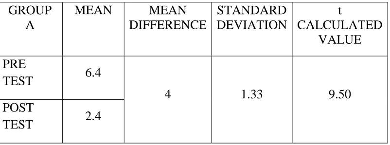
TABLE - 1
MEAN AND MEAN DIFFERENCE OF PRE TEST AND POST TEST VALUES OF GROUP A (VAS)
GROUP A
MEAN MEAN
DIFFERENCE
STANDARD DEVIATION
t
CALCULATED VALUE
PRE
TEST 6.4
4 1.33 9.50 POST
TEST 2.4
For 14 degrees of freedom at 5% level of significance the calculated t
value for VAS for group A was 9.50 and t table value was 2.14 .the t
calculated value was greater than t table value, which states that there is
0 2 4 6 8
PAIN
PRE TEST POST TEST
MEAN
GROUP A PRE AND POST TEST VAS MEAN SCORE
TABLE - 2
MEAN AND MEAN DIFFERENCE OF PRE TEST AND POST TEST VALUES OF GROUP B (VAS)
GROUP B
MEAN MEAN DIFFERENCE
STANDARD DEVIATION
t
CALCULATED VALUE
PRE
TEST 4.7
1.6 1.15 8.24 POST
TEST 3.1
For 14 degrees of freedom at 5% level of significance the calculated t
value for VAS for group A was 8.24 and t table value was 2.145 .the t
calculated value was gre5ater than t table value, which states that there is
0 1 2 3 4 5
cm
PRE TEST POST TEST
ROM
TABLE - 3
MEAN AND MEAN DIFFERENCE OF PRE TEST AND POST TEST VALUES OF GROUP A FLEXION USING RANGE OF MOTION(ROM)
GROUP A
MEAN MEAN DIFFERENCE
STANDARD DEVIATION
t
CALCULATED VALUE
PRE
TEST 4.74
1 .94 5.44 POST
TEST 5.74
For 14 degrees of freedom at 5% level of significance the calculated t
value for ROM for group A was 5.44 and t table value was 2.145 .the t
calculated value was greater than t table value, which states that there is
0 2 4 6
cm
PRE TEST POST TEST
MEAN
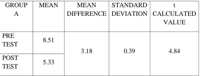
TABLE - 4
MEAN AND MEAN DIFFERENCE OF PRE TEST AND POST TEST VALUESOF GROUP A EXTENSION USING RANGE OF MOTION(ROM)
GROUP A
MEAN MEAN DIFFERENCE
STANDARD DEVIATION
t
CALCULATED VALUE
PRE
TEST 8.51
3.18 0.39 4.84 POST
TEST 5.33
For 14 degrees of freedom at 5% level of significance the calculated t
value for ROM for group A was 4.84 and t table value was2.145 .the t
calculated value was greater than t table value, which states that there is
0 2 4 6 8 10
cm
PRE TEST POST TEST
MEAN
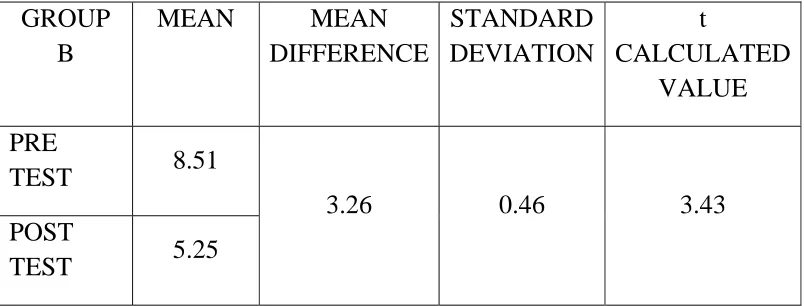
TABLE - 5
MEAN AND MEAN DIFFERENCE OF PRE TEST AND POST TEST VALUES OF GROUP B EXTENSION USING RANGE OF MOTION(ROM)
GROUP B
MEAN MEAN DIFFERENCE
STANDARD DEVIATION
t
CALCULATED VALUE
PRE
TEST 8.51
3.26 0.46 3.43 POST
TEST 5.25
For 14 degrees of freedom at 5% level of significance the calculated t
value for ROM for group A was 3.43 and t table value was2.145 .the t
calculated value was greater than t table value, which states that there is
0 2 4 6 8 10
cm
PRE TEST POST TEST
MEAN
TABLE - 6
MEAN AND MEAN DIFFERENCE OF PRE TEST AND POST TEST VALUES OF GROUP B FLEXION USING RANGE OF MOTION(ROM)
GROUP B
MEAN MEAN DIFFERENCE
STANDARD DEVIATION
t
CALCULATED VALUE
PRE
TEST 4.39
5.98 0.49 4.90 POST
TEST 10.37
14 degrees of freedom at 5% level of significance the calculated t value
for ROM for group A was 4.90 and t table value was 2.145 .the t
calculated value was greater than t table value, which states that there is
0 5 10 15
cm
PRE TEST POST TEST
MEAN
RESULTS
Effectiveness of Group A (VAS) is elicited by comparing the pre
test and post test values of Group A using paired ‘t’ test; the calculated
value is 9.50, whereas the critical value is 2.145. Since the calculated
value is greater than the critical value, there exists a significant difference
between the pretest and post test values of Control group. When
comparing the mean values of both, pre test mean value 64 is greater than
the post test mean value 24 which confirms that there is a significant
improvement in pain and functional activities.
Effectiveness of Group B (VAS) is elicited by comparing the
pretest and post test values of Experimental group using paired ‘t’ test,
the calculated value is 8.24 , whereas the critical value is 2.145. Since the
calculated value is greater than the critical value, there exists a significant
difference between the pretest and post test values of Experimental group.
When comparing the mean values of both, the pre test mean value 47 is
greater than the post test mean value 31, which confirms that there is a
significant improvement in pain and functional activities.
In Group A flexion the mean ROM pre test value was 47.4 and
post test value was 57.4 for 14 degree of freedom 0.05 level of
In Group A, extension the mean ROM pre test value was 85.1
and post test value was 53.3 for 14 degree of freedom 0.05 level of
significance. The t table value is 2.145 and t calculate value is 4.84 which
is greater than t value.
In Group B, flexion the mean ROM pre test value was 43.9 and
post test value was 103.7 for 14 degree of freedom 0.05 level of
significance. The t table value is 2.145 and t calculate value is 4.90 which
is greater than t value.
In Group B extension, the mean ROM pre test value was 85.1
and post test value was 52.5 for 14 degree of freedom 0.05 level of
significance. The t table value is 2.145 and t calculate value is 3.43 which
is greater than t value.
V. DISCUSSION
TRAVEL et, al (1999) stated that Trigger point are discrete, focal
hyper irritable spot painful on compression and can be produced referred
pain, referred tenderness, motor dysfunction and autonomic phenomena.
The project is the documentation of effects of myofascial release
technique on relieving pain in mechanical low back pain patient..
Pre test and post test pain intensities were evaluated ‘t’ value
shows that there was a significant effecting of giving myofascial release
technique. .
Gentle pressure and sustained stretching of myofascial release
believed to free adhesion, softens and lengthens the fascia.
Myofascial release is also set to enhance the body innate
restorative powers by improving circulation and nervous system
transmission (Suman Kuhar)
During myofascial technique ,heat will be elicited as a result the
vasomotor response that increase blood flow to the affected area,
enhances lymphatic drainage of toxic wastes.
This last activity reprogramme the central nervous system, enabling
a normal functional range of motion without eliciting the old pain pattern.
The effect of the trigger point release thus effectively reduce pain
VI. CONCLUSION
In an effort to find out the effectiveness of myofascial trigger point
release technique on relieving pain in mechanical low back pain20
subjects were selected by using non-probability purposive random
sampling technique and assigned into two groups with 10 subjects each.
Group A was treated with TENS and active spinal exercises and group B
was treated with trigger point release technique and tens for a period of
10 days.
The pre test and post test scores are noted and analysis was done
using independent ‘t’ test which favored the alternate hypothesis.
The intra group analysis was done and results were analysed using
paid ‘t’ test, which favored the alternative hypothesis.
The statistical analysis shows there is significant improvement in
pain and functional ability in following TENS and myofascial trigger
point release technique.
It is concluded that combination of myofascial trigger point
releasing technique with TENS was found to be more significant in
improving pain and functional activities in mechanical low back pain
VI. LIMITATION AND SUGGESTIONS
This study has been done with small sample size so further study can
be done with larger samples.
This study was very short term study and there for to make the
results more valid, long term study should be done.
Since the study has been done with very smaller group of subjects ,
further studies should be conducted with larger groups.
This study could be analysed with various other scales like Mc Gill
questionnaire , etc.
This study is done with myofascial trigger point release techniques
further studies can be conducted with taping techniques and heat
modalities.
Variation in calamite, drugs, diet, personal habit, side of involvement,
gender, age could not be controlled.
This study measures one time performance and results were infured.
VII. BIBILOGRAPHY
1. Barnes, John F. 1990. Myofascial Release: 10/e: 126-133(p).
Rehabilitation Services Inc.
2. Berner J.N.J. “Orthopaedics Myofascial Stretching a Guide of self
treatment” 3/e :86-94(p).
3. Brenda Pardy , Sill mortion , Myofascial stretching a guide of self
treatment” 1/e:34-46(p).
4. S. Brent Bratz man, MD, “clinical Orthopaedic Rehabilitation”
4/e:436-458(p).
5. Brotzman brent , MD, et . al. “Clinical Orthopaedic Rehabilitation”
4/e: 421-441(p)
6. Cantu, Robert I. & Grodin, Alan J. 2001. Myofascial Manipulation,
Theory and Clinical Application, 2/e:39-46(p), Aspen Publishers
Inc.
7. Carol J Manhein “The Myofascial Release Manual Books”
8. Crauford Jone Adams “outline of Orthopaedic “ 3/e:111-119(p),
New York , Churchill Living Stone Publication , 1995.
9. Cyriax JH , “Illustration Manual of Orthopaedic Medicine”
10. David J Magee. “Orthopaedic
Assessment”.8/e:123-140,523-536(p)
11. Donatelli Wooden , “Orthopaedic Physical Therapy ,3/e:126-134(p)
12. Jayanth Joshi, “Essential of Orthopaedics and applied
Physiotherapy”. 4/e:96-102(p).
13. John Barnes, Myofascial release Therapy Practitioners – Holistic
Health Therapist”
14. John Crew Ford Adams , “Outline of orthopaedics” 7/e:145-156(p).
15. John Ebenezar “Essential of Orthopaedic for Physiotherapy”
8/e:78-89(p).
16. John F Barnes , “Myofascial Release Approach” 56-75(p)
17. Lealy MP , “ Active Release Technique in Soft Tissue”
18. Lonnie R Mercier , Practical Orthopaedics 1980, 6/e:345-378(p)
19. Lucis Whyte Tengusan , Rodert Gerwin , “Clinical Mastery of the
Myofascial Pain”
20. Maheswari J, Essential Orthopaedics” 1997, 2/e:1451267(p)
21. Manheim, Carol. 2001. The Myofascial Release Manual.
3/e:35-66(p).
22. Michel Stanborough, “Direct Release Myofascial Technique”
23. Myers, Tom. 2004. Structural Integration - developments in Ida
Rolf's 'Recipe'- 1. Journal of Bodywork and Movement Therapies 8,
24. Natarajan M.N., Text Book of Orthopaedics and Traumatology”
3/e: 145-156(p).
25. Polly .E. Bijur , Ph.D. Wen Dy Sliver. “Journal of Orthopaedic and
Sports Physiotherapy”2002,
26. Samuel. L. Turek, “Orthopaedics principles and their Applications”
4/e:243-267(p), 1998.
27. William E, Techniques in Musculo Skeletal
Rehabilitation”2/e:68-79(p).
28. Robert J ,Myofascial Manipulation Theory and Clinical Application
“ 2/e:126-143(p), 2001
29. Prentice , “Techniques in Musculo-skeletal Rehabilitation” 2001
30. Sola AE “Myofascial pPain Syndrome” 1990
31. Simson DG “Myofascial Pain and Dysfunction” 2 /e:67-89(p),1999
32. Stanborough, Michael. 2004. Direct Release Myofascial Technique.
Elsevier.
33. Ward, Robert C. 2003, Integrated Neuromusculoskeletal Release
and Myofascial Release, in Ward RC, 2003, Foundations for
Osteopathic Medicine, 2nd edition, Chapter 60, pp 932–968,
Lippincott, Williams and Wilkins, Philadelphia
34. Yunus MB “Fibromyalgia Syndrome and Myofascial pain
36. Bella T may,exercise for low back pain therapeutic
exercisep-312-315
37. Karolyn kisner,the spine therapeutic foundation and technique
p588-590
VIII. APPENDIX-I
CASE ASSESSMENT PROFORMA
CASE SHEET NO :
NAME :
AGE :
SEX :
ADDRESS :
CHIEF COMPLIANT :
PAST MEDICAL HISTORY :
PRESENT MEDICAL HISTORY :
PERSONAL HISTORY :
ON OBSERVATION :
ON EXAMIATION :
DIAGNOSIS :
MODE OF EXERCISE :
MEASUREMENT TOOL :
(VAS)
APPENDIX-II
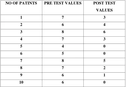
TABLE 7
PRETEST AND POST TEST VALUES OF CONTROL GROUP USING VISUAL ANALOGUE SCALE (VAS)
NO OF PATINTS PRE TEST VALUES POST TEST VALUES
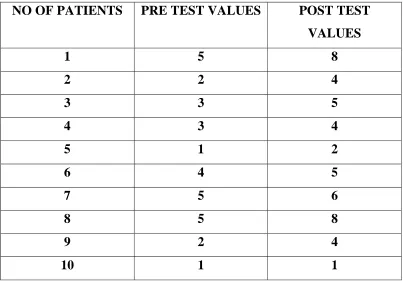
TABLE 8
PRE TEST AND POST TEST VALUES OF EXPERIMENTAL GROUP
USING VISUAL ANALOGUE SCALE (VAS)
NO OF PATIENTS PRE TEST VALUES POST TEST
VALUES
1 5 8 2 2 4 3 3 5 4 3 4 5 1 2 6 4 5 7 5 6
8 5 8
9 2 4 10 1 1
TABLE 9
PRE TEST AND POST TEST VALUES OF CONTROL GROUP FOR FLEXION USING RANGE OF MOTION(ROM)
NO OF PATIENTS PRE TEST VALUES POST TEST VALUES
TABLE 10
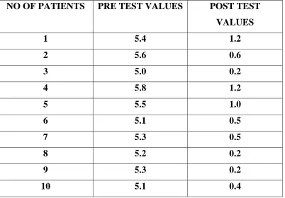
PRE TEST AND POST TEST VALUES OF CONTROL GROUP FOR EXTENSION USING RANGE OF MOTION(ROM)
NO OF PATIENTS PRE TEST VALUES POST TEST
VALUES
TABLE 11
PRE TEST AND POST TEST VALUES OF EXPERIMENTAL GROUPFOR FLEXION USING RANGE OF MOTION(ROM) NO OF PATIENTS PRE TEST VALUES POST TEST
VALUES
TABLE 12
PRE TEST AND POST TEST VALUES OF EXPERIMENTAL GROUP FOR EXTENSION USING RANGE OF MOTION(ROM)
NO OF PATIENTS PRE TEST VALUES POST TEST VALUES
APPENDIX IV
PATIENT CONSENT FORM Participant Identification Number:
Title of project: “A Comparative Analysis Of Tens And Active Spinal Exercises Versus Tens And Trigger Point Release Technique To Relieve
Pain In Low Back Pain Of Mechanical Origin”
Name of Researcher: Name of advisor:
Please tick where appropriate:
1. I confirm that I have read the information sheet for the above study and
had the opportunity to ask questions.
2. I understand that my participation is voluntary and that I am free to
withdraw at any time, without giving any reason.
3. I agree to take part in the above study.
4. I would like to receive a summary of the results.
5. Please send a summary of the results to ……….
Name of the participant:
Signature: Date:
Researcher: