A CLINICAL STUDY OF PENETRATING EYE
INJURIES
Dissertation Submitted for
M.S.Degree(Branch III) Ophthalmology
April 2013
THE TAMILNADU DR.M.G.R.MEDICAL UNIVERSITY
Dept. Of Ophthalmology Govt. Rajaji Hospital Madurai
CERTIFICATE
This is to certify that this dissertation entitled “A CLINICAL STUDY OF PENETRATING EYE INJURIES ” has been done under my guidance in the Department of OPHTHALMOLOGY, MADURAI
MEDICAL COLLEGE, MADURAI.
I Certify regarding the authenticity of the work done to prepare this
dissertation.
Dr.P.THIYAGARJAN , M.S.,D.O., PROFESSOR & H.O.D
Dept. Of Ophthalmology GOVT. RAJAJI HOSPITAL MADURAI MEDICAL
COLLEGE,
DECLARATION
I, Dr. JULIANA ROSITTA STEPHEN, Solemnly declare that the dissertation titled, “A CLINICAL STUDY OF PENETRATING EYE INJURIES” has been prepared by me.
This is submitted to the “THE TAMILNADU
DR.M.G.R.MEDICAL UNIVERSITY, CHENNAI, In partial fulfillment of the requirement for the award of M.S., (Ophthalmology) Branch-III
degree examination to be held in APRIL 2013.
Place: Madurai
Date: Dr. JULIANA ROSITTA
ACKNOWLEDGEMENT
I am grateful to the Dean, Madurai Medical College and Govt.Rajaji
Hospital, Madurai for permitting me to utilize the clinical materials of this
hospital.
I am extremely grateful to Dr.P.THIYAGARAJAN, M.S.D.O.,
Professor and Head of the Department of Ophthalmology, Madurai Medical
College, Madurai, for his guidance and help for executing my study.
. I am extremely indebted to my beloved guide Dr.G.S.SRINIVASAN,
M.S.,D.O., Professor of ophthalmology, Madurai Medical College, Madurai
for his constant encouragement and guidance throughout this dissertation.
I am grateful to Dr.K.KAVITHA , M.S.,Assistant Professor of
ophthalmology for her valuable guidance, support and encouragement
I am grateful to Dr.S.V.CHANDRAKUMAR , M.S.,D.O., Assistant
Professor of ophthalmology for his valuable guidance, support and
encouragement rendered to me during the study.
My sincere thanks to all my Assistant Professors for their
valuable suggestions in carrying out this study.
I thank my study subjects, who formed the back bone of this
study and without whom this work would not have been possible.
Last but not the least, I thank “God the Almighty”, for being my
CONTENTS
PART – I
1. INTRODUCTION
1
2. ANATOMY AND PHYSIOLOGY
3
3. CLASSIFICATION OF EYE INJURIES
20
4. CAUSATIVE AGENTS AND RISK FACTORS OF EYE INJURIES
23
5. EVALUATION OF PENETRATING EYE INJURIES
26
6. DIAGNOSTIC TESTS 33
7. COMPLICAITONS OF PENETRATING OCULAR TRAUMA
36
8. MANAGEMENT OF PENETRATING EYE INJURIES
39
9. VISUAL PROGNOSIS OF PENETRATING OCULAR TRAUMA
53
10.PREVENTION OF EYE INJURIES 54
PART – II
12.REVIEW OF LITERATURE
56
13.AIMS AND OBJECTIVES
63
14.MATERIALS AND METHODS
64
15.OBSERVATION AND RESULTS
68
16.SUMMARY
88
17. DISCUSSION 91
18. CONCLUSION
96
a. BIBLIOGRAPHY
b. PROFORMA
c. ANNEXURE
INTRODUCTION
Ocular trauma is an important cause of preventable morbidity
worldwide1. It is a major cause of acquired unilateral blindness and visual impairment. It constitutes about 1.5% of all the causes of blindness, 7% of
all bodily injuries and 10-15% of all eye diseases. It is common in young
males especially young adults. Since ocular injuries affect mostly the
productive population, it causes a major socioeconomic loss.
Recently ocular trauma has gained more importance because of its
increased incidence and advancement in the therapeutic approach. These
have improved the prognosis. Ocular injuries can occur in any setting like
recreational, sports activities, home, agricultural activities, at workplace and
road traffic accidents. Penetrating ocular trauma caused by sharp objects and
foreign bodies are considered as emergency. The availability of diagnostic
modalities like CT, MRI and ultrasound have improved the assessment and
management of trauma. The advent of microsurgical techniques and
vitreoretinal surgeries has greatly improved the visual prognosis of the
Hence ocular trauma should be given greater importance. Nearly 90%
of the eye injuries are preventable. Preventive measures should be taken in
sports, agriculture and work related activities. People should be educated
regarding the prevention of ocular injuries. Children should be supervised
and taught about the dangers of sharp objects like pens ,pencils, scissors,
compasses, etc. Glasses and sharp metals should be kept out of reach of
children. Protective eye wears should be provided for people working with
high speed grinders, cutters and in activities where there is risk of flying
objects. Ophthalmologists play an important role in the management as well
as prevention of ocular trauma. The adage ‘PREVENTION IS BETTER
ANATOMY OF CORNEA
Dimensions
• The anterior surface of cornea is elliptical with an average horizontal
diameter of 11.75mm and vertical diameter of 11mm
• The posterior surface of cornea is circular with an average diameter of
11.75mm
• Thickness of cornea in centre is 0.52mm and periphery is 0.67mm • Anterior and posterior radii of curvature of central part of cornea are
7.8 and 6.5mm respectively.
• Refractive power of cornea is +43D and refractive index is 1.37
Histology
• The cornea consists of five distinct layers, from anterior to posterior
are:
1. Epithelium
2. Bowman’s membrane
3. Stroma (substantia propia)
4. Descemet’s membrane
Epithelium
Corneal epithelium is of stratified squamous type about 50-90µm
thick consisting of 5-6 layers The deepest is the basal layer comprising of
tall columnar cells arranged in palisade manner. It forms the germinal layer.
The cells are firmly joined together by desmosomes and maculae occludens
which accounts for the transparency and its barrier function. The wing cells
forms 2-3 layers of polyhedral cells. The most superficial two layers
consists of flattened cells. The anterior wall of these cells has many
microvilli which play an important role in tear film stability.
Bowman’s membrane
This layer consists of acellular mass of condensed collagen fibrils about
8-14µm thick. It shows considerable resistance to infection and injury. It
does not regenerate.
Stroma
This layer is about 0.5mm thick and constitutes 90% of corneal thickness.
It consists of collagen fibrils and cells embedded in hydrated matrix of
The lamellae are arranged in many layers. They have oblique orientation
in anterior one third. In the posterior two thirds, the alternating layers are at
right angles to each other. The fibrils are mainly of type 1 collagen.
The cells present are keratocytes, lymphocytes and wandering
macrophages, and histiocytes.
Descemet’s membrane
It is a strong homogenous layer which binds the stroma posteriorly. It
is made of collagen and glycoprotein with no elastic fibres. It can regenerate.
In the periphery it terminates at the anterior limit of trabecular meshwork as
Schwalbe’s line.It is made up of type 1B collagen fibres arranged in a
hexagonal pattern and embedded in matrix.
Endothelium
It consists of single layer of flat polygonal cells. The cell density of
endothelium is 2400-3000cells/sq.mm. The endothelial cells are attached to
Descemet’s membrane by hemidesmosomes and laterally to each other by
important role in barrier function. It has abundant mitochondria, free
ribosomes, golgi complexes and smooth endoplasmic reticulum.
Blood supply
Cornea being avascular derives its nourishment from anterior ciliary
vessels which invades its peripheral 1mm and diffusion from the aqueous
humour. It gets oxygen supply from air in the central part whereas from the
anterior ciliary vessels in the peripheral part.
Nerve supply
Cornea has rich sensory supply derived from long ciliary nerves,
branches of nasociliary nerve-branch of ophthalmic division of trigeminal
nerve. The long ciliary nerves run in the suprachoroidal space and pierce the
sclera a short distance posterior to limbus to form annular plexus from which
branches run radially to enter corneal stroma. The nerve fibres after losing
their myelin sheaths form subepithelial plexus. Its fine terminal branches
then pierce Bowman’s membrane and between the epithelial cells they form
intraepithelial plexus. They do not have specialized nerve endings or sensory
Corneal transparency
The main function of cornea is to act as a major refracting medium to
form a clear retinal image.
Anatomical factors
1. Arrangement of stromal lamellae
2. Avascularity and nonmyelination of nerve fibres
3. Regular and uniform arrangement of epithelium and precorneal tear
film.
Physiological factors
1. Stromal imbibition pressure
2. Barrier function of epithelium and endothelium
3. Hydration control by active metabolic pump
4. Evaporation from surface of cornea
Epithelium
The transparency of corneal epithelium is due to the homogenecity of
the refractive index throughout the cellular layer. Normal precorneal tear
Avascularity and non myelination of nerve fibres
The cornea is normally avascular except for capillary palisade at
limbal margin. Corneal vascularisation is always pathological. The cornea
gets a rich sensory supply through long ciliary nerves which lose their
myelin sheath within 1-2mm from limbus.
Arrangement of stromal lamellae
Maurice theory:
Collagen fibres are arranged in a uniform and regular lattice so that
scattered light is destroyed by mutual interference. Cornea remains
transparent as long as the fibres are regularly arranged and separated by less
than a wavelength of light.
Goldmann theory
Corneal transparency is because fibrils are small in relation to light
and do not interfere unless they are larger than one half of the wavelength of
Stromal imbibition pressure
It is the pressure exerted by glycosoaminoglycans of corneal stroma.
The electrostatic repulsion of glycosoaminoglycans expands the tissue
sucking in fluid called imbibition pressure.
Barrier function of epithelium and endothelium
The epithelium and endothelium are semipermeable and acts as a
barrier to diffusion of sodium chloride and urea and to the flow of water.
The barrier function of endothelium is calcium dependent.
Hydration control by active pump: Corneal endothelium plays an important role in controlling fluid transport due to severe enzyme pumps.
The pump mechanisms require energy and are Na-K ATPase pump,
bicarbonate dependent ATPase, carbonic anhydrase enzyme and Na/H
Evaporation of water from the corneal surface: The evaporation of water from the precorneal tear film concentrates this fluid and increases its
osmolarity. This hypertonicity draws water from the cornea.2
CORNEAL WOUND HEALING
CORNEAL EPITHELIAL WOUND HEALING
Latent phase (4-6 hours): Epithelial debridement incites polymorphonuclear leucocyte invasion which removes the necrotic debris.
This causes retraction of epithelial cells, reduction of hemidesmosomal
attachments in turn commencing lamellipodial and filopodial extensions.
Cell migration and adhesion ( 24-36 hours): Migration of epithelial cells is by changes in cytoskeletal and cell shape which involves redistribution of
actin-myosin filaments. Actin filaments accumulates at the leading edges of
lamellipodial and filopodial extensions. Migration is also dependent on
matrix induced intracellular signaling through components like fibronectin,
leading epithelial cells across stromal surface thus completing the epithelial
monolayer covering the wound area. This is followed by disappearance of
fibronectin. Adhesion of epithelium to basement membrane and Bowman’s
membrane is via hemidesmosomes, lamina densa and type 7 collagen fibres.
Cell proliferation (36 hours to several months): The mitotic activity in the epithelium takes place at the limbus. The limbal stem cells produce
transient amplifying cells which give rise to post mitotic cells. Post mitotic
cells give rise to terminally differentiated cells which causes establishment
of hemidesmosomes and possible epithelial hyperplasia.
STROMAL WOUND HEALING
Stromal wounds take longer time to heal because of its avascularity.
The immediate effect of an incisional injury to the stromal matrix is wound
gaping and imbibition of water to become opaque. This is followed by a
series of events like fibrin deposition, rapid epithelization and activation of
keratocytes undergo fibroblast transformation. Fibroblasts produce collagen,
matrix. The newly formed collagen fibres are larger in diameter than normal
because of high concentration of chondroitin sulphate and dermatan
sulphate. This contributes to disruption of corneal transparency and scarring.
Corneal stromal remodelling is controlled by various metalloproteinases.
ENDOTHELIAL WOUND HEALING
Endothelial cells have very minimal capacity to undergo mitosis. It is
dependent on enlargement and movement of surrounding cells to cover the
wound site. The direct response to injury is cell slide. If sufficient number of
cells are lost then its pump fails and cornea imbibes water and becomes
opaque.
The various growth factors playing important role in regulation are
epidermal growth factor, fibroblast derived growth factor, platelet derived
growth factor, insulin derived growth factor and transforming growth
ANATOMY OF LIMBUS
Anatomically Limbus refers to circumcorneal transition zone of
conjunctivocorneal and corneoscleral junction.
At the conjunctivocorneal junction, the bulbar conjunctiva is firmly
adhered to the underlying structures. The epithelium becomes several layers
thick and are irregularly arranged at the limbus. At the sclerocorneal
junction transparent corneal fibres becomes continuous with oblique,
circular and opaque scleral fibres.
SURGICAL LIMBUS:
It is 2mm zone characterized by the following landmarks:
The anterior limbal border: It is the anterior boundary and marked by a prominent ridge which is created by insertion of conjunctiva and Tenon’s
Blue limbal zone: It is bluish transparent zone of variable width due to the position of insertion of conjunctiva and Tenon’s capsule. It is 1mm
superiorly, 0.8mm inferiorly and 0.4mm nasally and temporally.
Midlimbal zone: It is junction of blue zone with white area and overlies Schwalbe’s line.
Posterior limbal border: It lies about 1mm posterior to midlimbal line. It overlies scleral spur.
White limbal zone: Lies between mid limbal line and posterior limbal border. It overlies trabecular meshwork.2
ANATOMY OF SCLERA
Sclera is the tough opaque white coloured outer covering of posterior
five-sixth of the eyeball. Its outer surface is covered by Tenon’s capsule and
bulbar conjunctiva on its anterior part. Its inner surface is in contact with
choroid with suprachoroidal space in between. It is thickest posteriorly and
thinnest just behind the insertion of recti. The sclera consists of three ill
defined layers, sclera proper with episclera on the outer side and lamina
fusca inferiorly. The special regions of sclera are scleral sulcus which houses
the Schlemm’s canal, scleral spur and lamina cribrosa. The sclera has three
sets of apertures:
1. Posterior apertures transmitting long and short ciliary nerves and
vessels.
2. Middle apertures through which four vortex veins pass.
3. Anterior apertures transmitting anterior ciliary vessels, perivascular
The optic nerve fibres pass through lamina cribrosa of sclera. The
avascularity of sclera and lack of reaction of its fibrous tissue to any insult
make diseases of the sclera to be relatively rare and when they occur they
are chronic and respond slowly to treatment.2
Anterior chamber:
The anterior chamber is the space filled with aqueous humour. It is
bounded in front by cornea, behind by iris and part of anterior lens surface.
Its peripheral recess is called angle of anterior chamber which is bounded by
root of iris and ciliary body and anteriorly by corneosclera. Inner to this is
Schlemm’s canal which drains aqueous humour. At the angle is the
trabecular meshwork. The anterior chamber is about 2.5mm deep in the
centre.3
Iris:
The iris is composed of stroma with a rich blood supply. The anterior
surface of stroma is lined by two layers of pigmented epithelium. There are
two unstriped muscles, sphincter pupillae and dilator pupillae. Iris is
by oculomotor nerve and dilator pupillae is supplied by fibres from the
cervical sympathetic chain.3
Ciliary Body:
The ciliary body is composed of ciliary muscle. The inner surface of
ciliary body is divide into two areas: the anterior part -pars plicata and
posterior part - pars plana. Ciliary processes are seen between the plications
which secrete the aqueous humour. The ciliary body extends upto ora
serrata.3
Choroid:
The choroid is an extremely vascular structure and is separated from
the sclera by epichoroidal or suprachoroidal space. The inner surface of the
choroid is covered by Bruch’s membrane. The choroid is supplied by the
choriocapillaries.3
Retina :
The retina consists of ten layers which are:
1. Retinal pigment epithelium
3. External limiting membrane
4. Outer nuclear layer
5. Outer plexiform layer
6. Inner nuclear layer
7. Inner plexiform layer
8. Ganglion cell layer
9. Nerve fibre layer
10. Internal limiting membrane
Retina is formed by three strata of cells and their synapses which includes
the visual cells externally, bipolar cells intermedially and ganglion cells
internally.
Vitreous:
Vitreous humour is a jelly like substance with few cells and wandering
leucocytes. It is attached to the posterior surface of lens, vitreous base, the
Optic nerve:
Optic nerve consists of about 1.2 million axons of second order neurons. It is
divided into:
i. Intraocular part(1 mm)
ii. Intraorbital part(10-16 mm)
iii. Intracanalicular part(5-9 mm)
iv. Intracranial part(10-16 mm)
CLASSIFICATION OF EYE
Ocular Trauma Classification group has introduced a new
classification system based on following variables:
1. Type of injury
2. Grade of injury based on visual acuity at initial examination
3. Presence of relative afferent pupillary
4. Zone of injury based on location
TYPE OF INJURY
CLOSED GLOBE
CONTUSION
CLASSIFICATION OF EYE INJURIES4
Ocular Trauma Classification group has introduced a new
classification system based on following variables:
Grade of injury based on visual acuity at initial examination
Presence of relative afferent pupillary defect in involved eye
Zone of injury based on location
INJURY
CLOSED GLOBE
LAMELLAR LACERATION
OPEN GLOBE
LACERATION
PENETRATING IOFB PERFORATING RUPTURE
Ocular Trauma Classification group has introduced a new
Grade of injury based on visual acuity at initial examination
defect in involved eye
Brimingham eye trauma terminology (BETT):5
Eyewall : Sclera and cornea
Closed globe injury: No full thickness wound of eyewall
Open globe injury : Full thickness wound of eyewall
Contusion : No full thickness wound
Lamellar laceration: Partial thickness wound of eyewall
Rupture : Full thickness wound of eyewall caused by blunt
object
Laceration : Full thickness wound of eyewall caused by a
sharp object
Penetrating injury : Entrance wound
GRADE OF INJURY
Grade of injury is based upon visual acuity of the involved eye at the
time of presentation.
Grade 1 : greater or equal to 6/12
Grade 2 : 6/18 to 6/36
Grade 3 : 6/60 to 2/60
Grade 4 : no perception of light
PRESENCE OR ABSENCE OF RELATIVE AFFERENT PUPILLARY DEFECT
ZONE OF INJURY BASED ON LOCATION
In open globe injury
Zone 1 : isolated to cornea
Zone 2 : limbus to a point 5mm posterior in to sclera
In closed globe injury
Zone 1 : external (limbus to bulbar conjunctiva,sclera and cornea)
Zone 2 : anterior segment (including posterior lens capsule and
pars plicata)
Zone 3 : posterior segment (all internal structures posterior to
posterior lens capsule)4
CAUSATIVE AGENTS FOR EYE INJURIES
The following are the agents which cause potentially dangerous eye injuries:
1. Stick 10. Sharp metal
2. Stone 11. Finger nail
3. Needle 12. Twig
4. Nail 13. Arrow
5. Wooden splinter 14. Thorn
6. Shattered glass 15. Pen and pencil
7. Pin 16. Wire
8. Chisel 17. Drill
RISK FACTORS
1. Age: Risk due to age has a bimodal curve with first peak between 5-25
years and second peak over 70 years.
2. Gender: In younger ages, the male-female ratio may vary from 1.8:1 to
8:1
3. Socioeconomic status: People of low economic status are at more risk
because of the workspace and domestic setting.
4. Type of activity:
1. At home: Most of the accidents occur at home ie, while doing
cooking, household work, gardening etc. Women are prone to develop
such injuries in developing countries.
2. Workplace: The following occupation are more vulnerable to injury
Mechanic, blacksmith, electrician, plumber, sand blaster, rock blaster,
construction worker, carpenter, military personnel, stone mason,
agricultural workers.
In India where farmers and industry workers form the majority, eye
injuries with metallic particles, thorn and stone are very common.
3. At school and playground: Children are more vulnerable for injury
Broomstick injury: This is common among children. The tip of the
broomsticks are sharp and highly contaminated. It can cause mechanical
EVALUATION OF PENETRATING EYE
INJURIES
If a patient presents with both ocular and systemic trauma, treatment
of life threatening conditions takes priority. Once the patient is found to be
systemically stable more detailed history and ocular examination should be
carried out. The events preceding and leading to injury, a complete
description about the mechanism of injury should be obtained.
Penetrating ocular injury history:
• Visual acuity prior to injury • Nature of injury
o Associated life threatening injury
o Time and circumstance of the injury
o Suspected IOFB composition( brass, copper, iron, vegetable,
glass, soil)
o Use of any protective eyewear
Past ocular history:
• Ocular diseases • Refractive history
• Current ophthalmic medication • Previous surgery
Medical history:
• Diabetes mellitus, immunosuppression, vitamin or protein deficiency • Medications
• Drug allergy
• Status of tetanus immunization
The timing of an injury is also important.
Examination Visual acuity:
Initial visual acuity is the best predictor of final visual acuity. It is
External examination: Initial examination should focus on the orbit and periorbital tissues. These areas should be thoroughly inspected and palpated
under bright illumination looking for asymmetry, laceration, ecchymoses,
edema, lid abnormalities, bone deformities, fractures, exophthalmos,
enophthalmos, hypoesthesia, crepitus and foreign bodies.
Ocular motility:
Extraocular motility abnormalities are most likely occur with orbital
injuries. Binocular testing of ocular motility should be done. Patients with
limited ductions should be further evaluated with forced duction test to
differentiate between muscle paresis and entrapment. This test as well as
testing with lid retractors is avoided in known or suspected open globe
injuries.5
Pupillary Examination:
The presence or absence of normal pupillary function is important as a
part of ocular examination. Size, shape symmetry, direct and consensual
reflex and presence or absence of afferent pupillary defect should be
Anterior segment evaluation:
The eyelids should be evaluated for the presence of laceration. The
conjunctival surface is inspected for evidence of laceration and rupture like
uveal prolapse or sclera defects. In ocular rupture, a topical anesthetic is
applied and the conjunctiva is gently manipulated to look for foreign bodies
in the subconjunctival space.
The corneal epithelial defects should be examined for the presence of
staining defects, foreign bodies or corneal laceration. Full thickness corneal
laceration is followed by swelling of cut edges of the stroma leading to
partial or complete sealing of the laceration. Siedel test should be done. This
is done by the application of 2% fluorescein to the area of suspected
laceration and observed with cobalt blue filter. Aqueous flowing from the
laceration and diluting the stain indicates a full thickness wound.
Anterior chamber depth should be examined. An increase in depth is
seen in cases of posterior scleral rupture and posterior dislocation or
subluxation of lens. Shallow anterior chamber occur in corneal laceration,
misdirection, anterior dislocation or subluxation of lens. The presence of
cells and blood should be noted. In patients with intact globe gonioscopy
should be done to look for foreign body at the angle.
The iris should be evaluated for tears in the stroma or sphincter, iritis,
iridodialysis and cyclodialysis. In intact globe, gonioscopy should be done to
look for angle recession. Iris with transillumination defects may indicate a
penetrating injury.
Lens capsule should be inspected for areas of decreased lucency and
signs of penetrating injury like cataract, foreign body, or disruption. Lens
stability and position should be noted. Pigment dispersion on the anterior
lens capsule-vossius ring should be noted. Injury can cause rupture of the
zonules resulting in lens subluxation, dislocation or phacodonesis.
The anterior vitreous should be examined for the presence of IOFB
Posterior segment examination:
In extensive ocular rupture, examination of posterior segment should
be deferred until wound is appropriately treated. Slit lamp biomicroscopy
and a fundus lens should be used to evaluate the integrity of the posterior
pole. Optic nerve head is examined for colour, size of the cup and the
presence of edema. The appearance of the macula is noted. The presence of
any subretinal hemorrhage may indicate choroidal rupture. Any disruption of
retinal perfusion is noted. Any evidence of pigmented debris or cells in the
vitreous is to be noted. Presence of cells is an early indicator of traumatic
endophthalmitis. Indirect ophthalmoscopy should be done to examine the
retinal periphery to look for retinal defects or detachments. A thorough
inspection for intraocular foreign body should be done and its location is
noted. Tobacco dust in vitreous is suggestive of retinal break. Choroidal
rupture if present is noted. They are crescent shaped concentric to the optic
disc and can lead to profound visual loss if it occur through fovea.5
Intraocular pressure:
It is deferred in eyes with open globe injuries. IOP may be abnormally
may be elevated in hyphema, inflammatory debris blocking the trabeculum,
foreign body, aqueous misdirection, suprachoroidal hemoorhage or pupillary
DIAGNOSTIC TESTS
Plain X-ray:
It is a valuable tool for the evaluation of orbital fractures and
intraocular or intraorbital foreign bodies. It is cost effective. Disadvantages
are it cannot identify & localize radiolucent foreign bodies and fail to show
the presence or extension of penetrating orbitocranial injuries.
Ultrasonography:
It is useful in eyes with open globe injuries. It is useful for diagnosing
retinal detachment, vitreous hemorrhage, intraocular foreign bodies (both
radiolucent and radioopaque), choroidal detachment(can differentiate serous
from hemorrhagic), posterior vitreous separation, vitreous incarceration,
areas of vitreoretinal adhesion, and presence of any intraocular mass. It is
not useful in the evaluation of orbital pathology.
Computed tomography:
It allows detection of exact extension of orbital wall fractures and
Clearly defines soft tissue of orbit, retrorbital space, allows
recognition of intraorbital and intraocular air which can occur in penetrating
trauma.
Defines exact location of foreign bodies.
It can detect cerebral edema, hematoma and pneumocephalous(signs
of intracranial injury)
Contrast enhanced CT is useful in suspected vascular injuries like
carotico cavernous fistula and dural arteriovenous malformation.
The disadvantages are:
Thick slice CT may miss very small metallic foreign body.
May fail to detect wooden foreign body.
Magnetic resonance imaging:
It is better in evaluating soft tissues than CT, shows better resolution and detection of any optic nerve laceration or avulsion, can provide better
resolution of low density objects like vegetable matter and wooden foreign
cannot be used in patients with pacemakers or metallic implants and in
COMPLICATIONS OF PENETRATING OCULAR
TRAUMA
ANTERIOR SEGMENT TRAUMA: Iris :
Can cause direct iris injury as:
1. Iris prolapse
2. Incarceration of iris in the wound
3. Tear in the sphincter and pupillary frill causing irregular non
reactive pupil
4. Iridodialysis resulting in acute hyphema.
Traumatic glaucoma:
Acute rise in intraocular pressure may be due to obstruction of
trabecular meshwork by red blood cells, platelets, fibrin and inflammatory
debris or due to direct damage to outflow system. Large hyphema can cause
pupillary block and acute angle closure. Glaucoma can also be caused after
several weeks after vitreous hemorrhage due to ghost cells clogging the
hemosiderosis, formation of peripheral anterior synechiae and posterior
synechiae and angle recession.
Zonular injury:
Can cause lens subluxation and dislocation. Diagnosis is done by
maximal dilatation to look for separation of zonules from the lens.
Lens injury:
Direct and indirect forces can damage lens epithelium and capsule
causing cataract formation. Lens capsule rupture leads to rapid opacification
of lens. Lens induced inflammation may occur secondary to release of lens
proteins due to traumatic rupture of lens capsule. Phacoanaphylactic uveitis
or phacolytic glaucoma may occur after injury to lens. Zonular rupture may
allow percolation of vitreous into anterior chamber.
Post traumatic uveitis:
It can occur following direct or indirect penetrating or non
penetrating injury. Primary traumatic uveitis is the inflammation secondary
to trauma without any underlying disease. Secondary traumatic uveitis is the
infection worsened by trauma. Penetrating eye injuries can cause bacterial or
fungal endophthalmitis.
Intraocular foreign bodies:
They are most common after any activity which has involved
striking metal on metal. Foreign bodies can be lodged at the cornea, iris,
angle, anterior chamber, lens, vitreous, retina, or orbit.
Foreign bodies can cause infection, chemical reaction and
mechanical effect.
Infected perforating wound:
Can occur as a primary infection at the time of injury, as a secondary
infection before wound healing or as a later infection resulting in fistula or
sloughing.
Sympathetic ophthalmia:
It results from penetrating injury involving ciliary body and its
incarceration. Incarceration of iris or lens capsule also can cause
Post traumatic astigmatism is a common sequelae following corneal
injury.
An occult scleral rupture may occur. Chemosis or subconjunctival
hemorrhage suggests presence of occult rupture.
MANAGEMENT:
CORNEOSCLERAL WOUNDS:
Broad spectrum systemic antibiotics should be started immediately.
Self sealing corneal corneal wounds (3-4mm) without retained foreign
bodies with maintenance of anterior chamber can be conservatively managed
with antibiotic drops or ointment, cycloplegics and therapeutic contact lens.
Wounds with spontaneous leakage or flat anterior chamber may be
managed with cyanoacrylate glue after filling the anterior chamber with
viscoelastic material. After reconstruction of anterior chamber, the wound is
dried and the glue is applied with a needle or sterile brush. It is necessary to
material is removed from the anterior chamber and a therapeutic contact lens
is applied to avoid irritation of the tarsal conjunctiva and to prevent removal
of the glue.6
Corneal wounds – surgical approach:
The aims of surgical approach are
1. To restore globe integrity.
2. To restore anatomy of the eye
3. To avoid future complications
Any remnants of tissue preventing apposition of wound borders
should be removed. Uveal tissues should be replaced and the necrotic parts
are removed. As a rule those tissue that have been exposed for less than
24-36 hours can be replaced, those exposed for longer duration, showing colour
Corneal suturing:
Corneal sutures are essential in children, large laceration, displaced
wounds, wounds with loss of tissue and laceration with incarceration of
tissue.
Unsutured corneal incision flatten cornea whereas sutured incision
both radial and circumferential flatten cornea under the suture but steepens it
closer to visual axis or corneal centre.
The principles to be followed during corneal suturing are:
Zone of tissue compression along the incision is approximately equal
to the length of the suture. The zones of compression caused by adjacent
suture are to be in contact or overlap slightly to avoid wound leakage.
The sutures should be equidistant and parallel to each other.
The depth of the sutures depend on the amount of tissue lost. The suture
needle passes through the entrance and exit points are made of 90% depth
for correct apposition. In oblique wounds the intrastromal length should be
Corneal laceration should be sutured from periphery to centre, first
stabilizing the limbus. As the corneal centre approaches suture should be
shorter with sufficient tension and length to allow border confrontation and
to avoid formation of fish mouth openings, which can cause aqueous
leakage.
Monofilament 10-0 nylon suture material is preferred because it
causes least astigmatism.
Full thickness bites are usually avoided because the can introduce
microbes from the ocular surface. The anterior chamber is then deepened
and the wound is checked for leakage. All knots are timed and buried
superficially away from the visual axis for minimal scarring, inflammation
and neovascularisation.
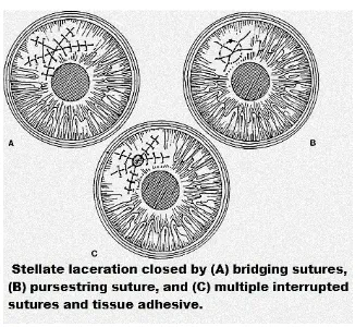
In cases of stellate corneal laceration, multiple interrupted, bridging
or purse string sutures are useful.
A peritomy near the damaged area should be performed when both
cornea and sclera are involved to improve wound visibility. Then wound is
sutured from the more distal end and continued anteriorly. Once the wound
is closed, peritomy may be enlarged for the exploration of more posterior
areas. The orbital tissues might seal them.
Globe reconstruction may not be a better option when corneoscleral
wounds are wide with loss of large amount of intraocular contents.
Enucleation can be done in such cases also to prevent sympathetic
ophthalmia.6
Corneal laceration with involvement of lens:
The indications for primary lens removal with corneal tear repair are:
1. Disrupted capsule and lens material in anterior chamber.
2. Cataractous lens
A separate limbal incision is made for extraction of the lens and
should never be done through the wound. If the zonules and posterior
capsule are intact, standard ECCE can be done. Intraocular lens implantation
can be of individual choice.7
Corneal laceration with involvement of vitreous:
The primary goal in such cases is to release any vitreous
incarceration to prevent chronic inflammation, cystoid macular edema,
vitreous fibrosis, retinal detachment, infection due to vitreous wick
syndrome and corneal endothelial damage due to vitreous touch.6
Traumatic hyphema:
It is important to exclude systemic disorders like sickle cell anemia
and hemophilia. Steroids should be started to control inflammation. If IOP is
high, it is controlled initially using beta blockers and acetazolamide if there
are no contraindications. Surgical intervention is done when there is blood
staining of cornea, if the intraocular pressure is greater than 60mm of Hg, if
there is eight-ball hemorrhage or if the IOP is above 35mm of Hg for seven
Traumatic glaucoma:
Topical steroids are used in minimal inflammation. They reduce the
formation of anterior and posterior synechiae. Acute rise in IOP is treated
with beta blockers, alpha agonists and carbonic anhydrase inhibitors. In
pupillary block, laser peripheral iridotomy should be done. Any factors
leading to glaucoma are to be treated. Chronic glaucoma is treated with
medical therapy. Laser trabeculoplasty can be done. Filtering surgery should
be done if the above measures fail.
Intraocular foreign body:
Management of an intraocular foreign body injury require immediate
closure of the globe and removal of the IOFB. The patient should be started
on broad spectrum antibiotics. Tetanus prophylaxis is necessary.
The appropriate technique for IOFB extraction depends on the
location, composition, size, shape number of IOFB, and associated ocular
abnormalities. Foreign bodies at the angle, iris, anterior chamber or lens can
be extracted through a limbal incision placed over the object or across
Magnets are commonly employed for the majority of magnetic IOFB.
Their ability to automatically align the IOFB in the long axis of the magnetic
field and deliver the smallest diameter through the sclerotomy makes them
ideal. Magnetic intraretinal or subretinal IOFB located anterior to equator
may be removed through a scleral cut down. IOFB localization is done using
indirect ophthalmoscopy and diathermy should be applied to the uveal bed
prior to IOFB delivery to limit hemorrhage during transuveal passage.
Surrounding laser photocoagulation is applied if the foreign body is
intraretinal prior to its removal. After removal a scleral buckle may be
placed if necessary.
Visible magnetic foreign body in the vitreous or on retinal surface
should be removed with an external magnet through a pars plana incision.
Posterior segment IOFB that are obscured by opaque media,
composed of non magnetic material or embedded in the posterior retina,
Posterior segment complication:
Penetrating eye injury can cause retinal breaks which are mostly
seen along the anterior or posterior border of vitreous base. Non
rhegmatogenous retinal detachment can occur due to traction to the retina
from dense vitreous membranes that are formed after injury. Penetrating eye
injury can cause proliferative vitreoretinopathy.
Penetrating injury with vitreous loss:
Vitreous incarceration is common and detachment occurs. Traction
is exerted by vitreous gel at its area of attachment. Retinal detachment is
caused by retinal tear located far from penetration site.15
Traumatic endophthalmitis:
This occurs in 2-7%of all penetrating ocular injury. Injuries along
with foreign bodies contaminated by soil or vegetable matter are at increased
risk in developing endophthalmitis. The organisms commonly causing
endophthalmitis are staphylococcus, streptococcus and bacillus species. The
delayed antibiotic therapy, presence of intraocular foreign body, injury to
lens and wound length of more than 5mm.16,17,18,19,20
Optic nerve involvement:
There can be partial or complete transection of optic nerve following
penetrating trauma or by the intruding object.
Management of posterior segment complication:
Posterior segment evaluation would be difficult due to the presence
of traumatic cataract in most of the cases. IOP should be checked digitally. If
IOP is low, then retinal detachment should be suspected.
The primary aim of surgery is to prevent secondary complications
like retinal detachment, endophthalmitis, cyclitic membrane and damage
caused by retained foreign body.
Posterior segment repair is divided into primary and secondary
Primary repair is immediate action to restore external anatomic
globe integrity.
Secondary repair is taken to restore intraocular anatomical globe
integrity like:
Cataract extraction
Vitrectomy
Removal of intraocular foreign body
Drainage of hemorrhagic choroidal detachment
Intravitreal antibiotic injection
Scleral buckling
Indications for vitrectomy following penetrating ocular trauma:
1. Vitreous incarceration into wound
2. Vitreous hemorrhage with retinal detachment
3. Vitreous hemorrhage with retinal tear
4. Retained foreign body
5. Vitreous hemorrhage with posterior lens capsular rupture
6. Retinal detachment
7. Posterior scleral perforation
Vitrectomy is done to remove the injured vitreous gel completely. It
can be done: early (within 72 hours) , late(3-14 days) or delayed(>3 weeks).
Indications for early vitrectomy are:
1. Endophthalmitis
2. Intraocular foreign body contaminated with soil
3. Retinal detachment
4. Non magnetic and copper containing foreign body
5. Reactive foreign body
Indications for late vitrectomy (3-14 days):
1. Lens vitreous injury
2. Severe vitreous hemorrhage
3. Retinal detachment
4. Intraocular foreign body
Indications for delayed vitrectomy (>3 weeks):
1. Epiretinal membrane formation
2. Vitreous opacification
3. Ghost cell glaucoma
4. Dislocated lens
5. Retinal detachment with proliferative vitreoretinopathy
Sequelae following primary wound repair are:
1. Corneal scarring, fibrovascular pannus and astigmatism
2. Pupillary or cyclitic membrane
3. Secondary glaucoma
4. Conjunctival scarring and symblepharon
5. Vitreous incarceration into the wound and associated inflammation,
Techniques of secondary reconstruction are:
• Penetrating keratoplasty • Lens removal
• Anterior vitrectomy
• Removal of organized fibrovascular and hyaloid membrane • Goniosynechiolysis
• Iridoplasty
Lid injury:
Penetrating eye injury can be associated with injury to lid and
canthus. The injured eyelid tissue should be handled with delicate
instruments and precision. After cleaning the tissues, the laceration should
be placed in its anatomical location. Simple laceration involving only the
skin and orbicularis is sutured with small caliber sutures. When lacerations
run across normal skin tension lines, a vertical mattress suture may be
placed. Full thickness eyelid lacerations should be repaired so that lid
VISUAL PROGNOSIS OF PENETRATING OCULAR INJURY
Depends on the following factors:
1. Initial visual acuity:
Visual acuity at the time of presentation is the most important
factor in predicting final visual acuity.
2. Afferent pupillary defect:
This indicates optic nerve or retinal dysfunction and is usually
associated with poor visual outcome.
3. Size of laceration:
Corneal laceration >9mm carries poor prognosis.
4. Location :
Posterior segment injuries involving vitreous, retina, ciliary body and
those causing vitreous hemorrhage carry poor prognosis.
5. Site :
Lacerations along the visual axis carries poor prognosis.
6. Endophthalmitis:
Traumatic endophthalmitis carries a poor prognosis though prompt
7. Hyphema:
Nearly 75% of eyes with hyphema have a visual outcome of 20/50 or
better but in cases with half to near total hyphema and total eight ball
hyphema only 25% to 50% regain vision better than 20/50.
8. Presence of intraocular foreign body:
Large foreign bodies have poor prognosis. Because of the newer
techniques it is achieving a better prognosis now.
PREVENTION OF EYE INJURIES:
Health promotion includes health protection, health education and
prevention of diseases. Vision is a powerful determinant of health and well
being. So its preservation is a health promoting activity. Eye health
promotion stresses the responsibility of the society both at the government
Preventive measures:
1. Protective wears made of polycarbonate lens are usually preferred.
They are particularly needed for:
a. One eyed patients
b. Persons with thin retina, weak sclera, bleeding tendencies
c. Work activities with risk of ocular injury
d. Sports activities with risk of ocular injury
2. Static shielding equipments- established safety barriers when high
speed grinders, cutters are being used.
3. Hazardous toys should not be given to children.
4. Glasses and sharp metals should be out of reach of children.
5. Stick and tools used in garden should be handled properly.
6. Defective machinery are to be corrected.
7. Children should be supervised.
8. Children should be taught regarding the dangers of pen, pencil,
scissors, etc.
REVIEW OF LITERATURE
1. David et al had reported that 80% of the penetrating eye injuries
occurred in males and the mean age was 29 years. The most common
initial physical findings in his study included hyphema (76%),
abnormality of pupil and uvea (94%). The initial visual acuity was
worse than 20/200 in 77% of the patients. Complications occurred in
about 25% of the cases, most commonly traumatic cataract and
infection. Final visual outcome was 28% with enucleation, no
perception of light in 10% of the patients, light perception to 20/200
in 24%, 20/200 or better in 36% of the patients. Complications were
present in 25% of the cases, majority were traumatic cataract or
infection.21
2. Hany E El Mekawey et al reported that open globe injuries were the
most prevalent comprising 33.45% of the emergencies. Most patients
were male (69%). The age group 6-16 years accounted for 24% of the
injuries and patients over 45 years accounted for 26.8%. The most
common ocular hemorrhage was hyphema. The most common type of
occupational injuries(26.3%) and motor accidents(24.4%) were the
main causes resulting open globe injuries in the region of upper
Egypt.22
3. Thompson et al reported that the most common cause of penetrating
ocular injury was fencing wire in the region of rural South Wales
(18.2%) then followed by hammering metal (16.9%). The mean age
was 32.6 years. 88% of them were males. The commonest location of
injury was at home accounting for 38%. The location of the injury
was corneal in 57%, sclera in 19% and corneoscleral in 23% of the
patients. Final visual acuity of 6/12 or more was attained in 61% of
the patients. Visual prognosis was best for injuries which involved the
cornea only.23
4. Patel et al reported that of the total penetrating ocular injuries 34%
were involved in children below the age of 15 years. Dart, knife and
airgun injuries constituted 41% of the trauma. Of them 54% attained a
visual acuity of 6/12 or better. 12% had undergone enucleation. From
the analysis of activities that caused injury it was considered that most
5. Caroline et al reported that 69.9% of the penetrating eye injuries
occurred during work, 18.3% while leisure and domestic activities,
2.3% occurred during sports activities, 1.9% of the injuries due to
assaults, 2.3% of them due to contact lens injury and in 5.3% of the
patients the cause was not known. Children below 10 years
constituted 4%. In 98.3% of the individuals periorbital and superficial
ocular structures were involved. The intraocular structures were
involved in remaining cases. Only one case of intraocular foreign
body was present. 36.3% of the patients underwent surgery. Most of
the injuries did not threaten sight in the adult population. But injuries
in children were sight threatening.25
6. Fasina et al reported that 58% of the injuries occurred at home in a
domestic setting. The commonest mode of injury was projectile
missiles. 80% of the patients were males and male to female ratio was
4:1.The mean age affected was 18 years.41.5% of them were below
15 years of age. The right eye was involved in 45.9% of the patients.
The commonest agent of injury was metallic piece or vegetative
corneoscleral injury was present in 41.5%, 68.1% of them had uveal
prolapse, hyphema was present in 47.4% and 28.1% had cataract. 1
7. Gyasi et al reported that males constituted 75% of the injured patients.
Patients below 30 years accounted for 82.3%. Right eye was affected
in 44.8% of the patients. Visual impairment at the initial presentation
was present in 89.5% of the patients. At the time of discharge, 69.3%
of the patients had visual impairment.26
8. Usha et al reported that 33.33% of the open globe injuries were
related to occupation. Among the patients 95.35% were males and
females constituted 4.65%. Patients in the age group between 16 to 45
years accounted for 79.06%. 37.5% of the injuries were caused by
sickle and stick. 12.5% of the injury was due to bullgore injury. 63.7%
of the patients were injured while working on lathe machine. 36.84%
were injured due to grinding machine. 68.42% of the injured patients
were not wearing protective eye wear. 55.81% required primary
wound repair, lens extraction was done in 23.26% of the patients.
were administered in 6.97% of the patients. Extracapsular cataract
extraction with PCIOL implantation was done as a secondary
procedure in 27.91% of the patients.27
9. Malla et al reported in his study that males were commonly involved
accounting for 71.9% and females accounted for 28.1%. majority of
the injuries occurred at home. Students constituted 32.8%, farmers
were 17.2%, labourers were 14.1% and housewives were 3% among
injured. The commonest agent of injury was mechanical objects like
wooden particles, metallic pieces and stone constituting 84.3%.
Agricultural agents accounted upto 11.7%. 52.4% of the patients had
hyphema.28
10. Mukherjee et al reported that males were more commonly affected
accounting for 73.17%. Patients below 30 years accounted for
44.91%. Metallic injuries were the most common form of injury
accounting for 33% which reflected the increased incidence of
industrial accidents. Corneal perforation was present in 62.21% of the
injury was present in 8.53% of the patients. Lens was involved in
56.10% of the patients and hyphema was present in 39.02% of the
patients. Posterior segment involvement was present in 34.14% of the
patients.29
11. Wykes et al reported that penetrating eye injuries were more common
among the male population with a ratio of 7:1. In 48% of the patients
right eye was involved and in 52% of the patients left eye was injured.
Sports and play injury was the most common (79.2%) among
children. Among the adults, 53% were due to industrial accidents.
Final visual acuity of 6/12 and more was attained in 43.3% of the
patients.30
12.Michael et al reported that majority of the patients with penetrating
eye injury were in the age group 21 to 30 years. Males were more
affected than females. Majority of the patients were injured in
domestic settings. Traumatic cataract and corneal perforation were the
commonest manifestations. 47.6% of the patients attained a final
13.Barry et al reported in a survey conducted on ocular trauma that
36.3% of the ocular trauma is constituted by penetrating corneal
injury. He also reported that intraocular foreign bodies were
associated with 73% of the penetrating injuries.32
14. Jazy et al reported that occupational injuries accounted upto 15.6%.
the mean age was 33.8 years. Majority of the injured were males.
35.7% of the injuries were due to repair and maintenance work.
Corneal injury accounted to 57.1%, 28.6% were scleral, 14.3% were
corneoscleral. 28.6% of the injuries were associated with intraocular
foreign body. 25% of the patients showed improvement in final visual
acuity, 50% did not improve after treatment.33
AIMS AND OBJECTIVES
The following were the aims and objectives of this prospective study:
1. To determine the risk factors associated with penetrating eye
injuries.
2. To study the different causative agents and to analyse the visual
MATERIALS AND METHODS
This is prospective study of 52 patients with penetrating eye injury
admitted at government Rajaji Hospital, Madurai from March -2012 to
November – 2012. A total of 60 patients were enrolled but 8 of them were
excluded from the study because of poor follow up.
All patients who presented with penetrating eye injuries were selected
for this study. Various patients were enrolled and categorized according to
many criteria like age, sex, place at which the injury occurred and causative
factors.
All patients were examined and been followed up for a minimum of
INCLUSION CRITERIA:
1. Patients with history of injury having slit lamp evidence with or
without the presence of foreign body.
2. All age group
3. Minimum follow up period of 3 months.
EXCLUSION CRITERIA:
1. Poor follow up of less than 3 months.
2. Pre existing corneal pathology like previous scar, prior ocular trauma
The following parameters were noted for all the patients:
1. Name, age, sex of the patient
2. Occupation and the place at which injury occurred
3. Agents involved in causing injury
4. Any prior treatment and the time elapsed since trauma
5. Visual acuity with pinhole at presentation
6. Slit lamp examination to evaluate the extent of injury.
7. Investigations like plain X-ray in suspected metallic foreign body.
Ultrasonogram was done in all cases to evaluate the posterior
segment, to see the integrity of the posterior capsule and to see the
presence of any foreign body. CT scan was also done to find out the
exact location of foreign bodies and in the case of associated
fractures.
8. Treatment: Informed consent was taken from all patients. The
patients were started on broad spectrum antibiotics. Further treatment
was done based on whether the wound was self-sealing or not. In self
sealing injury with normal anterior chamber without any uveal
prolapse, medical line of management was done. Except for pediatric
primary repair of the wound along with abscission of prolapsed uveal
tissue and anterior chamber reformation was done. In cases of
traumatic cataract, cataract extraction and IOL implantation was
done as a secondary procedure in most of the cases. Vitrectomy was
done in a case with intraocular foreign body.The treatment given,
medical or surgical was analyzed.
9. Final best corrected visual acuity was recorded at the end of three
months.
OBSERVATION AND RESULTS
1. Age pattern
Age group Frequency Percent (%)
<15 years 10 19.2
16-45 years 34 65.3
>45 years 8 15.3
All age group were included in this study. The minimum age
was 6 years and maximum age was 70 years. Majority of the patients
were in the age group of 16-45 years accounting for 65.3% followed
by patients less than 15 years.
0-15
16-45
Male
Female
Most of the patients were male (76.9%). Male: female ratio was 3.3: 1
2. Gender
Frequency Percent
40 76.9
12 23.1
atients were male (76.9%). Male: female ratio was 3.3: 1 Percent (%)
76.9
23.1
atients were male (76.9%). Male: female ratio was 3.3: 1
MALE
RE
LE
All the patients had unilateral injury. There was no significant
preponderance.
3. Laterality
Frequency Percent
27 51.9
25 48.1
All the patients had unilateral injury. There was no significant Percent (%)
51.9
48.1
All the patients had unilateral injury. There was no significant
RE
Agent Stick Stone Thorn Metallic object Glass Pencil Others
The commonest agent of injury was stick (34.3%) followed by
thorn (19.2%). 0 5 10 15 20 25 30 35 Stick Stone 34.6 13.4
4. Agent involved
Frequency Percent
18 34.6
7 13.4
10 19.2
Metallic object 8 15.3
2 3.8
3 5.7
4 7.6
The commonest agent of injury was stick (34.3%) followed by
Stone Thorn Metallic object Glass Pencil Others 19.2 15.3 3.8 5.7 7.6 Percent (%) 34.6 13.4 19.2 15.3 3.8 5.7 7.6
The commonest agent of injury was stick (34.3%) followed by
Domestic Field Factory/Work place Playground Road School
Most of the injury was at home (38.4%) followed by field (26.9%).
0 10 20 30 40
5. Place of injury
Frequency Percent
20 38.4
14 26.9
Factory/Work place 8 15.4
3 5.7
4 7.7
3 5.7
Most of the injury was at home (38.4%) followed by field (26.9%). Percent (%) 38.4 26.9 15.4 5.7 7.7 5.7
Student Mechanic/Factory worker Farmer Housewife Others
30.7% of the patients were farmers. This could be attributed to the fact
that most of the people visiting here are from rural areas whose
occupation is agriculture.
0 10 20 30 40 19.2 6. Occupation
Frequency Percent
10 19.2
Mechanic/Factory
12 23.0
16 30.7
Housewife 8 15.3
6 11.5
30.7% of the patients were farmers. This could be attributed to the fact
that most of the people visiting here are from rural areas whose
agriculture. 19.2 23 30.7 15.3 Percent (%) 19.2 23.0 30.7 15.3 11.5
30.7% of the patients were farmers. This could be attributed to the fact
that most of the people visiting here are from rural areas whose
Sealed
Leaking
Most of the cases had self sealed corneal injury(
0 10 20 30 40 50 60
Sealed 55.7
7. Corneal perforation
Frequency Percent
29 55.7
23 44.2
Most of the cases had self sealed corneal injury(55.7%).
Leaking 44.2
Percent
55.7
≤ 6 mm
>6 mm
69.2% of the injuries were less
0 10 20 30 40 50 60 70
8. Corneal wound size
Frequency Percent
36 69.2
16 30.8
69.2% of the injuries were less than or equal to 6 mm.
≤ 6 mm >6 mm 69.2
30.8
Percent (%)
69.2
I
II
III
80.7% of the injuries were in zone I
0 10 20 30 40 50 60 70 80 90 I 80.7
9. Zone of injury
Frequency Percent
42 80.7
10 19.2
-
80.7% of the injuries were in zone I
Corneal
Corneoscleral
Majority of the injury involved the
0 Corneal Corneoscleral
10.Wound site
Frequency Percent
43 82.7
Corneoscleral 9 17.3
Majority of the injury involved the cornea (82.7%)
17.3
10 20 30 40 50 60 70
Percent (%)
82.7
17.3
82.7