To determine whether m
ycophenolic acid Area under
the Curve Correlates with disease activity in pediatric
lupus nephritis patients treated With Mycophenolate
Mofetil
A DISSERTATION SUBMITTED IN PARTIAL
FULFILLMENT OF THE RULES AND REGULATIONS FOR
THE MD BRANCH II (PEDIATRICS) DEGREE
CERTIFICATE
This is to certify that the dissertation entitled “To determine whether mycophenolic acid area under the curve correlates with disease activity in pediatric lupus patients treated with mycophenolate mofetil” is the original work of Dr. Kirubakaran Navamani towards the M.D branch II (Pediatrics) degree examination of The Tamil Nadu Dr. M.G.R Medical University, Chennai to be held in April 2013.
Signatures
Guide: Co-Guide: Co-Guide: Dr. T. Sathish Kumar MD, DCH Dr. Indira Agarwal MD, FISN Dr.Ratna Prabha MD Professor Professor & Head Asst Professor
Dept of Pediatrics –II Dept of Pediatrics-II Dept of clinical Pharmacology Christian Medical College Christian Medical College Christian Medical College Vellore-632004 Vellore- 632004 Vellore-632004
Head of the department Dr. Anna Simon MD, DCH Professor and head
Department of Paediatrics Christian Medical College Vellore-632004
ACKNOWLEDGEMENTS
It is with great pleasure that I express my gratitude to my respected teacher and guide Dr. T. Sathish Kumar and also to my respected teacher and co-guide Dr. Indira Agarwal for their valuable suggestions, expert guidance, support and encouragement in doing this study.
I am also grateful to Dr. Anna Simon and the entire department of Child health for all the support received in preparing this dissertation and throughout my two year course in Pediatrics
I would like to thank Mrs. Visalakshi, Department of Biostatistics who helped me analyze the data for this dissertation.
I am grateful to Dr. Binu Susan, Dr. Ratna Prabha and all the staff in Clinical pharmacology for their invaluable support by working over hours for the patient’s convenience and providing timely results.
I am extremely grateful to my family and friends for their moral support and encouragement throughout my studies.
I thank my patients for their co-operation and willingness to be a part of the study
TABLE OF CONTENTS
1. Introduction 5
2. Literature Review 8
3. Aims of the study 29
4. Materials and Methods 31
5. Results 38
6. Discussion 54
7. Limitations 63
8. Summary and Conclusions 65
9. Bibliography 67
INTRODUCTION
Systemic lupus erythematosus (SLE) is an autoimmune disorder with multisystem involvement.
It predominantly involves adolescent girls. 20% of all SLE begin in childhood after 5 years of
life. It is more predominant in females with a female to male ratio of 8:1. The ratio lowers down
to 2:1 in the pediatric age group(1,2).
The Etiology of systemic lupus erythematosus is unknown. Environmental factors (UV light,
drugs, toxins and infection) may play a role in the genetically predisposed population. The
pathogenesis is complex. The autoantibodies, polyclonal B cell activation and the T cell
dysfunction contributes significantly to the disease activity (3). Lupus nephritis is very common
among the pediatric population. It is reported that about 40-75% of patients diagnosed to have
SLE, develop lupus nephritis within 5 years of diagnosis and almost all patients have a degree of
glomerular abnormality. The risk of development of end stage renal disease is 18-50%(44,5).
High dose corticosteroids have improved the course of lupus nephritis. However in the last 20
years additional treatment including the use of cytotoxic therapy has increased the 10 year
survival to 80% (6).
Mycophenolate Mofetil (MMF) is an immunosuppressant that is routinely given for treatment of
acute graft rejection and for prophylaxis following solid organ transplant. MMF is the
Lymphocytes are selectively inhibited because lymphocytes rely on de novo synthesis of purines
(7). MMF also depletes the lymphocytes and monocytes of guanosine triphosphate by selectively
inhibiting inosine monophosphate dehydrogenase(8). Successful use of MMF in lupus nephritis
has been described in various studies(7,9). MMF is also known to produce lower rates of
infection and cytopenia compared to patient who are treated with cyclophosphamide(10).
However the optimum dose of MMF in lupus nephritis has not yet been defined. The pediatric
post organ dose of 30mg/kg twice daily or 600mg/m2 twice daily is being followed for the
treatment of lupus nephritis(11). A fixed dose of 0.5 g to 1.5 g bid adjusted to the tolerance of
the individual is also being followed in the treatment of lupus nephritis(12). A target
concentration of MPA that is aimed at is 30-60 mg h/L. Also there is an inter-individual
variability of MPA concentrations for a given dose of MMF and the MPA concentrations also
depend on whether the drug is given along with other drugs like cyclosporine or tacrolimus(13).
We try to demonstrate the inter-individual variability of MPA concentrations for a given dose of
drugs and also co-relate the association between the disease activity as measured by the SLE
LITERATURE REVIEW
:HISTORY
:The history of lupus dates back to the 13th century when the physician Rogerius described the lesions as wolf bite. Lupus in Latin means wolf. Incidentally that is when the term lupus was
coined(14). It was then considered to be a cutaneous disease. Only in the latter half of the 19th century was it recognized as a multisystem disease after the works of Kaposi.
DISEASE BURDEN
:
Systemic lupus erythematosus is an autoimmune disorder with multisystem involvement and is
potentially fatal. The prevalence of lupus ranges from 40 per 100,000 persons among the
Europeans. Among blacks the prevalence is as much as 200 per 10,000 persons(15). In a study
from Asia 15 to 20% of all lupus is diagnosed in the pediatric age group (16 years and less).The
prevalence of lupus in the pediatric age group of the Asian population was found to be 6.3 to
ETIOLOGY:
The etiology of lupus is unclear. Around 90 % of lupus is females. Hence the contributory role of
female sex hormones and the protective role of male sex hormones are possible. Buyon et al
concluded that, the post menopausal women with SLE who received hormonal replacement with
conjugated estrogen and progesterone were more prone for disease flare than the patients
receiving placebo(17). But clinical trials which involves administration of
dihydroepiandrosterone for the treatment of lupus has not been promising (18).
Genetic Factors
There is a possible genetic role in the etiology of SLE. It tends to occur in families. However
there is no clear cut Mendelian inheritance. There is more occurrence of the disease in families.
There is 2% increased risk of disease in a sibling with SLE. However in even monozygotic twins
there is only 25% increased risk of disease and in dizygotic twins the risk is even lower(19).
These rates mean that there is not enough evidence for a genetic role. Many genes have been
identified after genome-wide genetic association studies in families with multiple lupus patients
which possibly contribute to the disease(20). Certain genes for the major histocompatibility
Early complement component( C1q, C2 or C4) deficiency is strongly associated with lupus (22).
Family studies have identified many genes in patients with lupus. Their function is to code for
components of the Immune system. A Scandinavian study showed that a single nucleotide
polymorphism in two interferon-related genes coding for tyrosine kinase 2 and interferon
regulatory factor 5(23). In mice studies Wakeland identified three genetic loci that are associated
with lupus namely sle1, sle2 and sle3. Sle1 gene mediates the loss of immunological tolerance to
nucleolar autoantigens. Sle 2 and Sle3 mediates B cell hyperactivity and T cell dysregulation
respectively(20).
Environmental factors
Drugs like hydralazine, procainamide and quinidine are known to cause drug-induced lupus. The
drug induced lupus presents with rash and joint pain and generally do not cause nephritis and
CNS manifestation (24). Ultraviolet radiations has been identified as the most important
environmental factor in causing lupus(25,26). Preceding viral infections are noted before the
presentation of prior to a flare. Particular causative virus has not been described yet. However
Ebstein-Barr(EBV) virus is possibly associated as disease presentation and the infection
occurring simultaneously has been reported(27). A case control study showed that EBV DNA
Auto-antibodies in lupus
Autoantibodies play a major role in the pathogenesis of lupus. Kidneys of patients with lupus
nephritis were demonstrated to have antibodies which bound to native double stranded
DNA(Ds-DNA) (28). Anti-Ds-DNA is present in as many as 70% of patients with lupus. It is also present
in about 0.5% of healthy individuals and in individuals with other connective tissue like
rheumatoid arthritis(29). It is strongly associated with the disease activity(30).Patients with
anti-ds-DNA positivity and clinically asymptomatic have a 80% chance of having active disease in 5
years(31). In other post mortem renal biopsy studies by Mannik et al there was evidence of auto
antibodies against non DNA proteins like Ro (ribonucleoprotein complex), La (RNA binding
protein), Sm (nuclear particles) and C1q( a subunit of C1) (32). Anti-ribosomal P antibodies are
more common in childhood nephritis and is positive in severe nephritis(33). The presence of
anti-RO or anti-LA or both during pregnancy is associated with 1 to 2 % risk of fetal heart
block(34) .
Anti-N-methyl d-aspartate (NMDA) may be important in CNS lupus. Kawal showed that
antibodies against NMDA and DNA produced cognitive impairment in patients with lupus and
hippocampal damage in mice models (35). Anti Ro and anti-nucleosome antibodies are seen in
active skin lesion but active nephritis(36). Hemolytic anemia and thrombocytopenia in lupus
occurs secondary to antibody mediated destruction of platelets and red blood cells.(37)
There are two theories of damage caused by autoantibodies. Berden et al suggest that auto
antibodies against the double stranded DNA (anti ds-DNA) bind to the nucleosome which are
released into the cytoplasm and this compound deposits in the glomerular basement membrane
thereby causing glomerulonephritis(38). In animal models this nucleosome- autoantibody
complex is studies to initiate complement activation(39). The second theory states that both
anti-nucleosome antibody and anti-ds-DNA both cross react with proteins in the kidney and thereby
Clinical manifestations:
Systemic lupus erythematosus presents in children acutely and usually with multisystem
involvement. Fever, musculoskeletal symptoms, fatigue and anorexia are the common
presentation.
Arthritis is usually found in 50-75% of the individuals. The arthritis in lupus is characteristically
non deforming and non erosive (41). Arthritis may be symmetrical, painful polyarthritis which
affects both small and large joints. The ultrasound study in the joints affected showed
tenosynovitis with thinning of the tendon(42). 20-30% of children have myalgia. Although
myositis is rare when present makes it difficult to differentiate it from dermatomyositis.
Mucocutaneous involvement is seen in 60-80% of patients with pediatric SLE at the time of
presentation. The rash associated with SLE is the malar rash a maculopapular rash which
involves the malar area and the bridge of the nose. It is usually photosensitive and resolves
without any residual scarring. A discoid lesion is uncommonly seen in lupus. It is photosensitive
associated with the disease is not very frequently seen in Indian children. The presence is also
not pathognomonic of the disease(44).The oral mucosal lesions are hyperemia and ulcerations
usually seen in the hard palate and are characteristically painless. Mild alopecia may be the
presenting feature. It can also be severe (45).
Hematological manifestations are seen in about 100% of patients. The ethnic background plays a
large role in the incidence of hematological manifestation. Anemia is seen in 75% of the Indian
children. Anemia of chronic disease is most commonly seen in SLE. The anemia is initially
normocytic normochromic and later microcytic hypochromic. 30-40% has Coomb positivity.
However only10-15% of them have overt hemolysis (46). 15 to 45% of patients have
thrombocytopenia. It can be the presenting feature in 15% of the patients. Children who present
with autoimmune thrombocytopenia should be evaluated routinely for lupus in view the high
incidence(47). Leucopenia ( both lymphopenia and granulocytopenia) is found in 20-40% of
individuals(48). Coagulation abnormalities are seen. 20% of patients with pediatric SLE have
lupus anticoagulant positivity. They usually have thromboembolic events. They have 20-30
times increased chance of have a thromboembolic event (49). The commonest cause for
secondary antiphospholipid syndrome is systemic lupus erythematosus in children. Lupus may
also manifest many years after presentation of primary antiphospholipid syndrome(50). It present
Among the cardiovascular manifestations Pericarditis with pericardial effusion is the most
common. Mild pericardial effusion which is picked up by echocardiography is seen in1/3rd of the patients. However overt pericardial effusion is seen in 25-40% of the individuals(41,44).
Pleuropumonary involvement is seen in 25 – 75% of patients (51).Pleuritis is the most common
manifestation. Acute lupus pneumonitis may present similar to infective pneumonia or
pulmonary hypertension(52).5-10% of lung manifestations re pulmonary hemorrhages (53).
Pulmonary hypertension secondary to pulmonary vascular disease is a rare but potentially fatal
complication(54)
Involvement of the central nervous system is reported in a wide range of patients. It may occur in
as low as 20% to about 90% of patients(55). The reason for this wide variation in the incidence
of central nervous system complications is because of the discrepancy in the definition of certain
central nervous system symptoms. The commonest neuropsychiatric manifestation is Lupus
headache. Lupus headache is defined as an unremitting headache which requires narcotic
treatment (56). 30-50% of neuropsychiatric manifestations are contributed by psychosis, They
more commonly have visual hallucinations and less commonly auditory and tactile
hallucinations in that order. They characteristically have preserved insight(57). 12 to 30% of
patients with neuropsychiatric manifestations have cerebrovascular disease(58). 10-20% of
children with CNS lupus have cerebral venous thrombosis and are almost universally associated
Abdominal pain and diarrhea are the most common gastro intestinal manifestations(59). When
associated with vasculitis if the gastrointestinal system the patients are at risk of developing an
intestinal perforation(60). Pancreatitis is a rare complication presenting in about <5% of the
cases. Patients usually present with vomiting and diffuse abdominal pain(61).
Ocular finding that is most associated with lupus is cytoid bodies. This is secondary to retinal
vasculitis(62).
Infectious complications are extremely common and are a major cause of death. They create
diagnostic and therapeutic challenges. In the presence of fever along with respiratory symptoms
or central nervous system symptoms like seizures, altered behavior infection needs to be ruled
out. If investigations show high counts with neutrophilic predominance which is unlike SLE
sepsis needs to be ruled out. However counts may not be necessarily high in the setting of
infection as patients with SLE may have apparently normal counts during infection as their
baseline counts may be low. An elevated CRP also suggests infection. Opportunistic infections
Lupus nephritis is very common among individuals with SLE. Almost all patients are reported to
have some degree of glomerular abnormality. About 40-75% of patients with SLE develop
clinical nephritis within 5 years of diagnosis(63). The risk of progression to end stage renal
disease is 18-50% (4,5). The presentation of lupus nephritis can vary extensively from
individual to individual. It can range anything between mild nephrologic abnormality to rapidly
progressing and nephrotic syndrome. Hematuria and proteinuria are the most common.
Hematuria is present in 67-100% of individuals with lupus nephritis whereas nephritic syndrome
is present in about 50% at diagnosis. Hypertension and renal insufficiency is seen in 50% of
affected individuals(5). Age related disease manifestations were studied. Children were found to
have higher incidence of hypertension, proteinuria, hematuria, cellular casts and Creatinine(64).
Patients with hematuria and/or proteinuria may have any class of glomerulonephritis. The
prognosis depends on the stage of renal involvement and it is of utmost importance in deciding
the treatment (65). Children with silent disease may have major histopathological abnormalities.
Hence the clinical picture always does not correlate with the renal histopathology(6). WHO
developed a classification for lupus nephritis in 1973 which helps in prognosis and deciding on
further therapy. The classification uses light microscopy, immunofluorescence and electron
WHO has classified lupus nephritis as follows (66)
Class I : Normal glomeruli a) Normal by light microscopy b) immunological deposits by
electron microscopy
Class II: Mesangiopathy. a) Pure mesangial widening with mild hypercellularity b) moderate
hypercellularity
Class III: Focal and segmental glomerulonephritis a) active and necrotic lesions b) active and
sclerosing lesion c) sclerosing lesions
Class IV: Diffuse proliferating glomerulonephritis (severe mesangial, mesagiocapillary or
endocapillary and /or extensive subendothelial deposits) without segmental lesions b) with
Class V: Diffuse membranous glomerulonephritis a) pure membranous glomerulonephritis b)
associated with lesions of category IIa or IIb. C) Associated with lesions of category III (a-c) d)
associated with lesions of category IV (a-d)
Class VI: Chronic sclerosing glomerulopathy
Treatment of lupus nephritis:
Clinical trials in the pursuit of treatment for SLE started as early as 1894 when Payne reported
the beneficial effects of quinine in SLE. Brief period later salicylates were proved beneficial. But
the break-through in the treatment of lupus was not attained until the 1950s when Hench reported
the efficacy of cortisone in the treatment of rheumatological conditions like SLE (67) and ever
since steroids have been the primary therapy in the treatment of SLE.
However the ideal treatment for SLE nephritis is unclear despite years of research primarily
because of the basic pathophysiology. It is not clear whether the excess B cell activity or
defective T cell suppressor activity or excess helper T cell activity is being dealt with(68).
With the advent of the recent treatment options the outcome of pediatric SLE has improved
dramatically over the past years. The 10 year survival rate in the 60’s was as low as 30% and in
the early 90’s it has improved to excess of 90%(69). Non renal causes like infections has
replaced renal failure the common cause of death in Lupus(70). The treatment needs to be
therapy aimed at minimal aide effects. Delayed onset of treatment is associated with poorer
outcome(71).
Therapy of class I, II nephritis:
Patients with class I nephritis are very rare and no specific treatment has been described. There is
no specific treatment described for the treatment of class II nephritis either. Corticosteroid, as a
long term treatment of lupus nephritis is not indicated. It is rather aimed at associated extra-renal
manifestations of lupus(64). However there is need for long term follow up for the progression
of disease
Therapy of class III, IV nephritis:
The course of the disease in class III lupus nephritis is the same as that of class IV when more
than 40% of glomeruli are involved and hence the same aggressive therapy is needed(6).
However if less than 20% of glomeruli are involved , the prognosis is quite good with <5% of
patients progressing to end stage renal disease at the end of 5 years(72).
Patients with Class IV lupus nephritis (Diffuse proliferative lupus nephritis) is prone for
hypertension , nephrotic syndrome and end stage renal failure. Prompt treatment is warranted in
class IV lupus nephritis. Corticosteroids have dramatically changed the course of the disease.
Recent studies have shown that low dose corticosteroids are as equal in efficacy as high dose
Sterinberg et al assessed 111 children and concluded that the patients in the study arm which
involved treatment with a cytotoxic drug plus low dose prednisolone had significantly better
preservation of renal function compared to those treated only with high dose steroids(74).
Unlike adults treatment with corticosteroids and immunosuppressants are required in the
treatment of Child hood SLE(75). Treatments that are currently used are antimalarials,
corticosteroids, cyclophosphamide, azathioprine and mycophenolate mofetil.
Corticosteroids:
For over two decades the teaching was to treat diffuse proliferative glomerulonephritis with high
dose corticosteroids. Recent studies show that high dose steroids are no better than low dose
steroids and are associated with unwarranted side effects. In his randomized control trial
Steinberg demonstrated that patients randomized to the groups treated with cytotoxic therapy in
addition to steroids had better preservation of renal function that did the group treated with
prednisolone alone(74). Pulse methylprednisolone administered intravenously as pulse doses
leads to dramatic improvement in patients with acute deterioration of renal function(76).
However the long term effect of this regimen in preserving renal function matched only
prednisolone. Also intravenous methylprednisolone is associated with side effects like cardiac
arrhythmias and cardiac arrest. Other side effects like flushing sensation, acute hypertension and
Cyclophosphamide:
There is much evidence that combination of cyclophosphamide with steroids has better results in
preserving the renal function than steroids alone55. Cyclophosphamide is metabolized in the liver to its active metabolite. The active metabolite alkylates the macromolecules(78). Initially
oral regimens in the dose of 1-3mg/kg/day for 8-12 weeks were described. Currently monthly
boluses at a starting dose of 750mg/m2 is suggested to be less toxic than oral daily doses at 2mg/kg(79). The dose of cyclophosphamide may be increased to 100mg/m2 if the WBC count remains more than 3000/mm3. The duration of therapy after the initial control of disease is not well described. In his study Lehman used cyclophosphamide for 3 years and reported
improvement in hemoglobin C3, C4 and Creatinine clearance(80)
Cyclophosphamide therapy is associated with the risk of toxicity which includes alopecia, bone
Azathioprine:
Azathioprine is an antimetabolite that interferes with protein synthesis(78). It has been proved to
be safe when given in the log-term. It is given in the doses of 2-2.5mg/kg per 24 hours. It may be
used with prednisolone in the initial treatment of lupus nephritis(82) and later substituted for iv
cyclophosphamide after 6 months if the disease is well controlled. It can also be given after
completing 8-12 weeks of oral cyclophosphamide(64). When Azathioprine is given in
combination with steroids the steroid dose has to be changed in the event of withdrawing
Azathioprine as the disease may relapse. This is because of the steroid sparing effect of
azathioprine(83) . Azathioprine is relatively safe, however long term administration causes bone
Cyclosporine A
It is a relatively new drug which is used in the event of steroid resistance or in severe steroid
toxicity. It acts by interfering with the production of lymphokines produced by the
lymphocytes(85). Cytotoxic T call recruitment is stopped by inhibiting production of
interleukin-2 and thereby decreasing inflammation(64). Cyclosporin used along with steroids is
known to decrease proteinuria and improve renal function with better growth rate as compared to
patients treated with prednisolone and cyclophosphamide combination and its use alone(85).
Side effects are minimal. Hypertension, transient elevation of serum Creatinine, hypertrichosis
Mycophenolate Mofetil:
Mycophenolate mofetil is a relatively new drug which has been used routinely in the prophylaxis
against graft rejection and in its treatment in the post renal transplant patients. Mycophenolate
mofetil (MMF) is immunosuppressive drug which acts by irreversibly inhibiting the enzyme
inosine monophosphate dehydrogenase (IMPDH) and hence selectively inhibiting proliferation
of T- cells and B cells as they require de-novo synthesis of purines(86). Mycophenolic acid
(MPA) is the active form of the inactive prodrug mycophenolate mofetil (MMF). MMF is
converted to MPA by liver, plasma and intestinal esterases (87). There are many recent studies
which show that mycophenolate mofetil is as efficacious or atleast comparable with
cyclophosphamide in the induction treatment of childhood systemic lupus erythematosus(88,89).
MMF is also known to produce lower rates of infection and cytopenia compared to patient who
are treated with cyclophosphamide(10).
The dose recommended in clinical practice is based on clinical trials for renal transplantation. It
is a fixed dose of 2 to 3 grams per day in divided doses. Other evidences based on
in children. The dosage is lower than in patients who have undergone solid organ transplantation
(1200 to 2400 mg/m2) who simultaneously receive calcineurin inhibitors. In renal transplantation
therapeutic drug monitoring has been developed for individualization of doses. MPA area under
the plasma concentration time curve from 0-12(MPA AUC0-12 hrs) has been successfully
correlated in the outcome of patients who have undergone solid organ transplantation(90).
The MPA pharmacokinetics in patients with systemic lupus erythematosus is different from the
patients who have undergone solid organ transplants who are on Cyclosporin A (91,92) .
Patients with SLE often have a third peak in the AUC due to the absence of calcineurin
inhibitors. Plasma concentration of MPA are reduced by cyclosporine as it inhibits the
enterohepatic circulation of MPA(93,94). Target concentration of AUC over 12h of 30-60 mg h/l
when measured with high performance liquid chromatography or 35 to 70 mg h/l when measured
by enzyme multiplied immunotechnique is advised for patient undergoing renal transplant (95).
(12).
A target concentration of MPA that is aimed at for the therapy of lupus nephritis is also 30-60
mg h/L. Also there is an inter-individual variability of MPA concentrations for a given dose of
MMF and the MPA concentrations also depend on whether the drug is given along with other
The target concentrations of MPA in children treated with MMF for SLE is very important as
this reflects the clinical outcome and the association with side effects. There are very few studies
which have reported the inter-individual variability of MPA concentration and even fewer in
children. Filler et al studied 5 children with autoimmune diseases and compared the
pharmacokinetics of mycophenolate sodium (EC-MPS) with pharmacokinetics of mycophenolate
mofetil (MMF). Factors affecting inter-individual variability are studied in transplanted
individuals. Patients with renal insufficiency (creatinine clearance of <25ml/min) and low
albumin (32g/l) have low MPA exposure because MPA clearance depends on its non protein
bound fraction(96). Both hypoalbuminemia and renal insufficiency results in a high free fraction
of MPA and hence increased clearance of MPA(92,97).To our knowledge the target
AIMS:
1) To determine whether MPA AUC 0-12 correlates with disease activity in children with
lupus nephritis on mycophenolate mofetil
2) To demonstrate the inter individual variability of the MPA concentration in children
taking MMF
Materials and Methods:
a. Study setting:
The recruitment for this study took place at the pediatric rheumatology clinic of Christian
Medical College, Vellore. The OPD functions on 3 week days (Wednesdays, Fridays and
Saturdays) a week. The OPD serves on an average about 40 patients which includes 5 -10
patients with systemic lupus erythematosus.
Children whose parents are willing to take part in the study are recruited after an
informed written consent. Children were recruited over a period of 6 months from March
b. Participants:
Inclusion criteria:
*Children diagnosed to have systemic lupus erythematosus as per American College of
Rheumatology criteria with proliferative lupus nephritis (Class III and IV) and treated with
similar doses of prednisolone
*Children aged between 8 and 18 years
*Children who have received stable doses of MMF for atleast 1 month.
Exclusion Criteria:
*Children with SLE aged >18 years
*Children with SLE who had Class II or Class V Lupus nephritis
*Children taking MMF not on empty stomach or taking not on regular intervals
*Children who have developed significant side effects to MMF like diarrhoea and marrow
suppression
*Children with lupus nephritis with concomitant drugs like cyclophosphamide, cyclosporine,
c. Variables:
Exposure: All children with proliferative lupus nephritis on MMF for at least one month
(standard dose 50 mg/kg/day)
Outcome: MPA AUC correlates with disease activity as measured by SLEDAI
Predictors: Dose of MMF, Disease activity, C3, C4, Albumin, Creatinine
d. Data Sources/measurement:
Complete blood count: Sample is sent in an EDTA tube and sent to the clinical
pathology lab. Blood is processed in the Beckwith coulter machine and the validated
results are considered.
Serum creatinine: 2ml blood is collected in a clotted tube and sent to the biochemistry
lab. Creatinine is calculated by the picric acid method. The values are measured in
Total complements/C3, C4: 2ml of blood is collected in a clotted tube and sent to
microbiology lab. Serum complements (C3 and C4) are measured by the nephelometry
method.
Ds-DNA: Blood is processed in the autoimmune lab by the ELISA method.
Urine protein creatinine ratio: Spot urine sample is processed in the biochemistry lab
e. Sample size: A total of 25 children who fulfilled the inclusion criteria were recruited
f. Statistical methods:
The quantitative variables were presented using the mean and standard deviation.
All categorical variables were presented using the frequencies and percentages
. The mean MPA values were compared across male and female using independent t test.
The comparison of MPA across BSA values was compared using Mann Whitney U test.
Spearman Rank correlation was used to find the relationship between MPA and all
continuous variables.
.All the variables that were significant from the bivariable analysis were included for
multivariable regression analysis
Methodology:
Children with systemic lupus erythematosus with lupus nephritis who have been on MMF for
the induction or maintenance therapy were inducted for the study. Most importantly children
should have been on the drug for at least 1 month and the intake of drugs should be organized
and appropriately spaced through the day. An informed, written consent was obtained for each
patient after detailed one to one discussion with the parents/patient. After the consent, the child
was examined.
After getting the informed written consent from the patient/ guardians the patient is asked to
give blood for complete blood count, serum Creatinine, serum albumin, complements and
anti-ds DNA.. The patient is also asked to give urine for routine analysis and for spot protein
Creatinine ratio. After the results are available the disease activity is assessed with the SLE
disease activity index scoring (SLEDAI) system. The patient is instructed to take the tablet at
the usual dose and given instructions to come fasting and not to take the next day’s morning
dose. The patient is instructed to come to the clinical pharmacology unit the next day on an
empty stomach. In the clinical pharmacology lab an IV cannula is inserted in situ and blood for
trough MPA level is taken after which the line is flushed with heparin saline. The child is then
instructed to take the drug. After the baseline (trough) sample, samples are taken at 0.5, 1, 1.5,
2, 2.5, 3, 4, 8 and 12 hours after MMF administration. The specimen will be centrifuged and
Plasma MPA concentrations will be determined by high performance liquid chromatography
(HPLC). Plasma concentration data will be used to estimate the concentration time curve
(AUC0-12). The patients’ AUC 0-12 concentration is determined and compared with the clinical
status and the serological status. It is postulated that a high drug concentration level will
correspond with a lower disease status viz normal complements and a negative ds-DNA as well
as clinical remission(86). It will also be tried to ascertain the optimal drug concentration to
induce or maintain remission. To my knowledge there has not been any such study on the
Indian population and very few studies overseas have been done on children with SLE.
This study is approved by Institutional Review Board (IRB) of Christian Medical College,
RESULTS
Patient characteristics:
Twenty-five outpatients who fulfilled the inclusion criteria were recruited for the study. Out of
25 children, 20 were girls and 5 were boys (Fig1). Out of the twenty five, 16 children received
2gms/day, 7 received 1.5gms/day and 2 received 1gm/day. Indications for administering MMF
were lupus nephritis class III (n=8), lupus nephritis class IV (n=17) (Fig2). On the day of study,
13 patients were receiving concomitant prednisolone and 22 children were receiving
Hydroxychloroquine. SLEDAI scores done on all 25 children showed that 5 patients (21%) had
active disease (SLEDAI score ≥6) and 20 patients (79%) had inactive disease (SLEDAI score<6)
Table 1. Baseline characteristics of patients with active and inactive SLE
MPA AUC0-12:= Mycophenolic acid Area under the curve 0-12hours, Ds-DNA:=double stranded
deoxyribonucleic acid
The active and inactive disease groups were similar in age (mean age was 15.0 ±3.3 in the active
disease group and was 14.75±1.7 in the inactive disease group).The two groups were also similar
in weight, height and serum albumin levels. Hemoglobin, C3 and C4 levels were statistically
significant between the two groups (p < 0.05) Parameters (Normal range)
Active SLE (SLEDAI ≥6) n=5 Inactive SLE (SLEDAI <6) n=20 p value
Age 15.0±3.3 14.75 ±1.7 0.272
Height (Cms) 151.8±12.3 152.9±8.8 0.974
Weight ( Kgs) 48.6±12.39 48.35±12.21 0.767
Creatinine (0.7 to 1.1 mg/dl) 0.8±.18 0.78±.11 0.92
Serum Albumin (3.5 to 5 gm/dl) 4.08±.5 4.4±.36 0.272
Anti-ds-DNA (<100 IU/ml) 240±62 156.9±227 0.060
Total count /Cu.mm 8740±2967 8900±3078 1.000
Hemoglobin ( gm%) 10.2±1.9 12.3±0.72 0.007
C3 level (90-180 mg/dl) 66.12±13.29 99.69±15.59 0.001
Figure1: Showing the sex distribution among the participants
80% of our patients were girls (n=22) and 20% were boys (n=3)
Female
n=20,
(80%)
Figure 2. Lupus nephritis class among the patients
Our participants predominantly had class IV nephritis.
Class III
8
(32%)
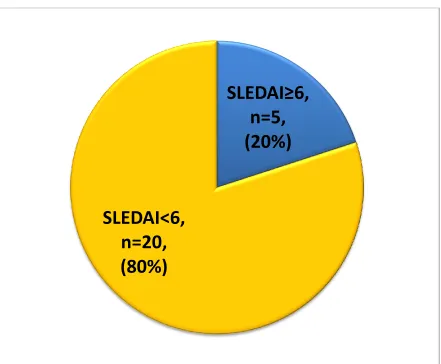
Fig3: Distribution of disease activity among the participants
80% of the participants (n=20) had inactive disease(SLEDAI score<6) on the day of
examination.20% (n=5) had active disease (SLEDAI score≥6).
SLEDAI≥6,
n=5,
(20%)
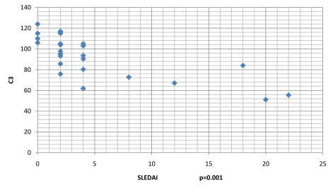
Figure 4. Correlation of C3 levels with SLEDAI
SLEDAI:=Systemic Lupus Erythematosus Disease Activity Index
Low C3 was significantly correlated with high disease activity (P=0.001) 0
20 40 60 80 100 120 140
0 5 10 15 20 25
C3
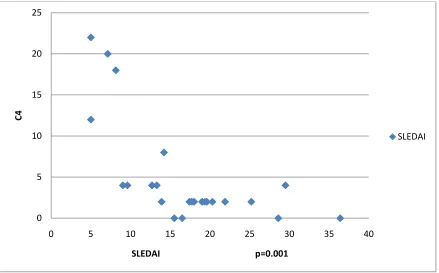
Figure 5. Correlation of C4 with SLE disease activity
Low C4 was significantly correlated with high disease activity (P=0.001) 0
5 10 15 20 25
0 5 10 15 20 25 30 35 40
C4
SLEDAI p=0.001
Figure 6: Correlation of hemoglobin and SLEDAI
Hemoglobin correlated significantly with disease activity (SLEDAI scores) 0
5 10 15 20 25
0 2 4 6 8 10 12 14 16
SLE
D
A
I
Haemoglobin p=0.007
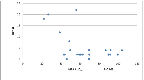
Fig7: Correlation of MPA AUC0-12 with SLE disease activity
MPA AUC0-12:= Mycophenolic acid Area under the curve 0-12hours
High SLEDAI scores correlated with low MPA AUC 0-12 on the day of examination(P=0.003)
0 5 10 15 20 25
0 20 40 60 80 100 120
SLE
D
A
I
[image:47.612.72.566.146.421.2]Inter-Individual variability:
Fig8.Between-patient variability of MPA AUC0-12 /gm of MMF
MPA:-Mycophenolic acid
MPA AUC0-12 showed wide variability (Fig8) with lowest level of 22.1 and a highest level of
104µg.hr/ml. The mean ±SD MPA AUC0-12 of the active disease group (38.46±14.3µg.hour/ml)
was significantly lower than that of the inactive group (69±19.24µg.hour/ml) with a p value of
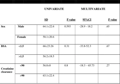
Table 2Parameters influencing MPA AUC0-12 levels
BSA: =Body Surface Area
A univariate and multivariate regression was done to predict the factors influencing MPA AUC
0-12 levels. Factors included were sex, body surface area, dose of MMF, albumin and C3 levels.
Only MMF dose was found to be significantly influencing the MPA AUC0-12.
UNIVARIATE
MULTIVARIATE
SD P value 95%CI P value
Sex Male 64.1±22.6 0.593 -28.9 - 18.2 .65
Female 58.1±20.6
BSA <1.5 66±23.26 0.31 -33.8-52.3 .67
>1.5 56.2±18.5
Creatinine clearance
<90 56.0±0 0.8 -18.3 – 65.73 .27
Figure 9 Correlation of MPA AUC-0-12 with dose of MMF
Higher the dose of MMF correlated to better MPA levels(Fig9). 0
20 40 60 80 100 120
0 0.5 1 1.5 2 2.5
M
PA
A
UC0
-12
Fig10. Correlation of albumin with MPA AUC 0-12
Figure 11. Showing correlation of C3 with MPA AUC 0-12
C3 did not correlate significantly with MPA AUC0-12 (fig 11) .
0 20 40 60 80 100 120
0 20 40 60 80 100 120 140
C3
MPA AUC0-12 P=0.43
Figure 12. Showing correlation of age with MPA AUC 0-12
DISCUSSION
This is the first study to evaluate MMF pharmacokinetics in children with Lupus nephritis. There
has been a dramatic improvement in the survival rates of patients with systemic lupus
erythematosus. The 4 year survival rates in the 1950s was 50% and has currently the 15 year
survival rate has improved to 80% with the novel treatment options(98). Among them is
mycophenolate mofetil which has been routinely used in the solid organ transplant patients.
MMF is as effective and has fewer side effects than steroids and cyclophosphamide(12).
However the ideal dose of MMF in children with lupus nephritis is not described. Traditionally a
dose 2 to 3 grams per day in divided doses is given. A dose of 900mg/m2 has been recommended in children as against a dosage of 1200 to 2400 mg/m2 in patients with solid organ transplants.
These dosing are derived from that recommended for solid organ transplants and there are no
randomized control trial derived doses available for the treatment of lupus nephritis(90). There
are a number of literatures suggesting that weight or body surface area dosing of MMF is not
advisable as it does not predict MPA pharmacokinetics and MPA Pharmacodynamics (99). In
renal transplantation therapeutic drug monitoring has been developed for individualization of
doses(90). The inter-individual variability of drug concentration of MMF is a characteristic
feature of the drug. There are a number of factors that are described to affect the drug
concentration like concomitant drug intake namely cyclosporine, tacrolimus and associated co
In solid organ transplanted patients MPA area under the plasma concentration time curve from
0-12 hrs has been successfully correlated in the outcome (90). Notably the pharmacokinetics in
SLE is different from the MPA pharmacokinetics of post solid organ transplants who are on
Cyclosporin A (91,92) . There is a third peak in patients with SLE who have a third peak in the
AUC due to the absence of calcineurin inhibitors(93,94). Individualized doses aiming at a Target
concentration of AUC over 12h of 30-60 mg h/l when measured with high performance liquid
chromatography or 35 to 70 mg h/l when measured by enzyme multiplied immunotechnique is
advised for patient undergoing renal transplant (95). Target concentration of MPA that is aimed
at for the therapy of lupus nephritis in adults is 30-60 mg h/L. The target concentrations of MPA
in children treated with MMF for SLE is very important as this reflects the clinical outcome and
the association with side effects. To our knowledge the target concentration of MPA in children
with Lupus nephritis has not been discussed till date. Hence this study was done to demonstrate
the inter-individual variability of MPA AUC0-12 levels in children taking MMF for lupus. There
are no studies to our knowledge done in the pediatric age group in our country.
The setting of the study was at the pediatric rheumatology clinic which runs twice a week. It
caters to an average of 40 patients per day among which systemic lupus erythematosus accounts
for about 30% of the cases. The recruitment started in March 2012. Children with lupus nephritis
who were on MMF for at least 1 month were included. Also the time spacing of the drugs were
given importance. Children who had not been taking drugs equally spaced through the day were
asked to come the next month after changing the drug timing. The reason for this is irregular
25 patients who fulfilled the criteria were recruited over 8 months. 20 patients were girls which
constituted 80% of the sample size and 5 were boys and contributed to 20% of the sample size.
This was in concordance with the data about the global sex distribution of the disease although in
children the female to male preponderance is not as significant as in adults. The female to male
ratio is about 4.5:1 as against 8 -13:1 in adult onset patients(77). The peak onset of child hood
SLE occurs during puberty(100). Our patient profile is in accordance with this finding. The mean
age of our series of patient was 14.8 years.
The patients’ height, weight and body surface areas were also recorded on the day of
examination. Mycophenolate mofetil dose for treatment of lupus nephritis is derived from doses
for solid organ transplants. Optimum doses for treating lupus nephritis patients are not well
established. A dose of 2 to 3 grams per day in 2 divided doses is usually given. The pediatric
transplant dose of 30mg/kg twice daily or 600mg/m2 are being traditionally used(59,60). We used a dose of 600mg/m2 for our patients. 64% of our patients had body surface area between 1-1.5 m2. 32 % of our patients had a body surface area of more than 1.5 m2 and 4% of the patients had body surface area less than 1 m2.
All our patients who are on MMF have had lupus nephritis diagnosed and staged after a renal
biopsy. 68% of the patients had class IV nephritis and 32% of them had class III nephritis. The
indications for use of MMF among our patients were exclusively for lupus nephritis. Many
studies report the successful use of MMF in the treatment of lupus nephritis(103). MMF is
indicated in lupus nephritis, dermatitis and other manifestations of SLE(104,105).Among the
studies the majority were on treatment of lupus nephritis with MMF after failed cycles of
cyclophosphamide and has reported dramatic outcome. The corticosteroid sparing effect is of
Concomitant medications that the child is on along with that of MMF were recorded. 56% of our
patients were on prednisolone, 16% were on deflazocort, 84% were on hydroxychloroquine
(HCQ) and 52% were on calcium and vitamin D supplements. None of them were on
cyclosporine or tacrolimus. Plasma concentration of MPA is decreased by concomitant use of
cyclosporine. Cyclosporine use inhibits the biliary excretion of MPA 7-0 – glucuronide which is
the inactive MPA metabolite(106). MMF is often given in combination with other
immunosuppressants and corticosteroids. Steroids contribute to the MPA metabolism and hence
decreased the exposure over time(107).
On the day of examination the children are examined and the disease activity is assessed with the
Systemic Lupus Erythematosus Disease Activity Index (SLEDAI) . Recently, there are many
disease activity indices that are validated(108). Among these are the British Isles Lupus
assessment group (BILAG), the European Consensus Lupus activity measurement (ECLAM),
the systemic lupus activity measure (SLAM) and the SLE disease activity index(SLEDAI) and
the safety of estrogen in lupus erythematosus national assessment (SELENA) SLEDAI. Each of
these indices are designed for longitudinal studies(109,110). In our study we decided to use the
SLEDAI scoring system for the following reasons. The SLEDAI scoring system unlike BILAG
is a global index and not organ specific and fewer number of variables (24 Vs 86)(111). Zahr et
al in their study used both SLEDAI and BILAG index to assess disease activity and successfully
correlated the MPA AUC0-12 with disease activity(86). In our study the patients were examined
using the SLEDAI scoring system and then asked to come to the clinical pharmacology unit the
In the clinical pharmacology unit, the child will have the mycophenolic acid assay. The child is
then instructed to take the drug in the lab after the baseline (trough) sample. Later samples are
taken at 0.5, 1, 1.5, 2, 2.5, 3, 4, 8 and 12 hours after MMF administration. The specimen will be
centrifuged and plasma separated into a clean eppendorf tube
.
All specimens will be stored at – 20oC until analysis. We used the high performance liquid chromatography method (HPLC) foranalysis of MPA AUC 0-12. HPLC is currently the preferred method for acute monitoring of the
Mycophenolic acid assays(95). There are other methods to determine MPA levels namely the
EMIT method. In a study done by Weber et al EMIT technique was found to be comparable to
the HPLC method in determining the MPA levels. However there is cross reactivity with the
metabolites of mycophenolic acid namely phenolic MPA glucuronide (MPAG) 7-o-MPAG, acyl
glucuronide (AcMPAG) and phenolic glucoside of MPA(95).
Markers for active nephritis that we included were urine microscopy for blood and protein. For
quantification of protein we used urine spot protein/Creatinine ratio done by pyrogall indicator
method. We chose the spot protein Creatinine ratio as against the 24 hour collection mainly for
the patient’s convenience and the time it saves. Urine for spot protein Creatinine ratio is an
alternative to the 24 hour urine protein method(112). In a study done in Asia, Absar et al
concluded that single voided protein/creatinine ratio is an alternative to the 24 hour collection
method at all levels of GFR(113). Any ratio > 2 was considered nephritic range of proteinuria.
Proteinuria and hematuria are the most commonly associated findings in lupus nephritis(100).
Hematuria is almost universal. Nephrotic range of proteinuria is found in 50% of patients with
Creatinine was done for all patients prior to the MPA analysis. None of the 25 patients had renal
insufficiency. Severe renal insufficiency is known to interfere with the MPA AUC 0-12(97).
Creatinine was measured in the picric acid method and expressed in mg/dl. Albumin,
complement levels were included to be independent variables that might influence the MPA
AUC 0-12.
Complements were done for all the patients by the In the study done by Zahr et al multivariate
analysis showed that Creatinine clearance dose of MMF and albumin was significantly
associated with the MPA AUC0-12. Complements (C3 and C4) were done by the nephelometry
method. All co morbidities in the patient on the day of examination were looked for. None of our
patients had any co morbidities.
In our study we have clearly shown a strong association between MPA AUC 0-12 and the disease
activity as assessed by scoring the disease by the SLEDAI scoring system. In Zahr et al’s study
the MPA AUC0-12 correlated with disease activity as assessed by both the SLEDAI and BILAG
scoring(86). In the study by Rolland a similar finding was observed but however unlike the
current study disease activity was not assessed by validated indices(114).
We found that the mean MPA AUC0-12 was significantly lower in patients with active disease
than the patients with inactive disease. It is notable that the two groups (active disease with
SLEDAI>6 and inactive disease with SLEDAI <6) were similar in mean age, sex distribution,
We have demonstrated that MPA AUC 0-12 was correlated with SLEDAI, complement level (C3,
C4) and hemoglobin. Complement level being one of the two main biological markers of SLE
activity. Zahr et al found that complements, MPA AUC 0-12 along with ds-DNA were associated
with SLE disease activity.(86)
In our multivariate analysis we found that daily MMF doses were recognized as independent
variables influencing the MPA AUC 0-12. It is very unlikely that other factors like duration of
treatment with MMF or prior treatment with other drugs could have influenced the results since
all the patients were on MMF for at least more than 8 weeks. The doses were not modified in the
past 8 weeks. Moreover the frequencies of concomitant drug intake among the two groups were
similar.
Therapeutic compliance is a critical issue. This may contribute to variability in the drug levels
and disease activity(115). This was addressed at the time of recruitment itself. Only patients who
were on regular medications and dosing were recruited.
Hence it can be said that the dosing of MMF needs to be based on the MPA AUC 0-12 levels.
Body surface area and weight dosing might not attain adequate drug concentration and thereby
We have also demonstrated the inter-individual variation of drug concentration per gram of
MMF which substantiates that fixed daily doses are not recommended.
Limitations of the study:
Since it is a cross sectional study the definitive conclusion regarding the association between
MPA AUC0-12 levels and the SLE activity cannot be established. It can be done through regular
Conclusion:
In conclusion, there is a strong correlation between the disease activity and the MPA AUC0-12 in
children with lupus nephritis taking MMF. We have also demonstrated an inter-individual
variability among patients taking a standard dose of MMF. In light of this the inclusion of MMF
AUC0-12 as parameter in treating children with lupus nephritis patients with MMF is
recommended. Also prospective pharmacokinetic study needs to be done to evaluate the target
Bibliography:
1. Platt JL, Burke BA, Fish AJ, Kim Y, Michael AF. Systemic lupus erythematosus in the first two decades of life. Am. J. Kidney Dis. 1982;2:212–222.
2. Cameron JS. Lupus nephritis. J. Am. Soc. Nephrol. 1999;10:413–424.
3. Al Salloum AA. Lupus nephritis in childhood. Saudi J Kidney Dis Transpl. 2003;14:43–56.
4. Bartosh SM, Fine RN, Sullivan EK. Outcome after transplantation of young patients with systemic lupus erythematosus: a report of the North American pediatric renal transplant cooperative study. Transplantation. 2001;72:973–978.
5. Cameron JS. Lupus nephritis in childhood and adolescence. Pediatr. Nephrol. 1994;8:230–249.
6. Niaudet P. Treatment of lupus nephritis in children. Pediatr. Nephrol. 2000;14:158–166.
7. Fu YF, Liu GL. Mycophenolate mofetil therapy for children with lupus nephritis refractory to both intravenous cyclosphosphamide and cyclosporine. Clin. Nephrol. 2001;55:318–321.
8. Chaigne-Delalande B, Guidicelli G, Couzi L, et al. The immunosuppressor mycophenolic acid kills activated lymphocytes by inducing a nonclassical actin-dependent necrotic signal. J. Immunol. 2008;181:7630–7638.
9. Wallman L, Stewart G, Chapman J, O’Connell P, Fulcher D. Mycophenolate mofetil for treatment of refractory lupus nephritis: four pilot cases. Aust N Z J Med. 2000;30:712–715.
11. Ettenger R, Cohen A, Nast C, et al. Mycophenolate mofetil as maintenance immunosuppression in pediatric renal transplantation. Transplant. Proc. 1997;29:340–341.
12. Chan TM, Li FK, Tang CS, et al. Efficacy of mycophenolate mofetil in patients with diffuse proliferative lupus nephritis. Hong Kong-Guangzhou Nephrology Study Group. N. Engl. J. Med. 2000;343:1156–1162.
13. Cattaneo D, Merlini S, Zenoni S, et al. Influence of co-medication with sirolimus or cyclosporine on mycophenolic acid pharmacokinetics in kidney transplantation. Am. J. Transplant. 2005;5:2937– 2944.
14. Blotzer JW. Systemic lupus erythematosus I: historical aspects. Md State Med J. 1983;32:439–441.
15. Johnson AE, Gordon C, Palmer RG, Bacon PA. The prevalence and incidence of systemic lupus erythematosus in Birmingham, England. Relationship to ethnicity and country of birth. Arthritis Rheum. 1995;38:551–558.
16. Huang JL, Yeh KW, Yao TC, et al. Pediatric lupus in Asia. Lupus. 2010;19:1414–1418.
17. Buyon JP, Petri MA, Kim MY, et al. The effect of combined estrogen and progesterone hormone replacement therapy on disease activity in systemic lupus erythematosus: a randomized trial. Ann. Intern. Med. 2005;142:953–962.
18. Chang D-M, Lan J-L, Lin H-Y, Luo S-F. Dehydroepiandrosterone treatment of women with mild-to-moderate systemic lupus erythematosus: a multicenter randomized, double-blind, placebo-controlled trial. Arthritis Rheum. 2002;46:2924–2927.
20. Wakeland EK, Liu K, Graham RR, Behrens TW. Delineating the genetic basis of systemic lupus erythematosus. Immunity. 2001;15:397–408.
21. Walport MJ, Black CM, Batchelor JR. The immunogenetics of SLE. Clin Rheum Dis. 1982;8:3–21.
22. Walport MJ. Complement and systemic lupus erythematosus. Arthritis Res. 2002;4 :S279–293.
23. Cunninghame Graham DS, Akil M, Vyse TJ. Association of polymorphisms across the tyrosine kinase gene, TYK2 in UK SLE families. Rheumatology (Oxford). 2007;46:927–930.
24. James JA, Kaufman KM, Farris AD, et al. An increased prevalence of Epstein-Barr virus infection in young patients suggests a possible etiology for systemic lupus erythematosus. J. Clin. Invest. 1997;100:3019–3026.
25. Tan EM, Cohen AS, Fries JF, et al. The 1982 revised criteria for the classification of systemic lupus erythematosus. Arthritis Rheum. 1982;25:1271–1277.
26. Hochberg MC. Updating the American College of Rheumatology revised criteria for the classification of systemic lupus erythematosus. Arthritis Rheum. 1997;40:1725.
27. James JA, Kaufman KM, Farris AD, et al. An increased prevalence of Epstein-Barr virus infection in young patients suggests a possible etiology for systemic lupus erythematosus. J. Clin. Invest. 1997;100:3019–3026.
28. Isenberg DA, Manson JJ, Ehrenstein MR, Rahman A. Fifty years of anti-ds DNA antibodies: are we approaching journey’s end? Rheumatology (Oxford). 2007;46:1052–1056.
30. Ter Borg EJ, Horst G, Hummel EJ, Limburg PC, Kallenberg CG. Measurement of increases in anti-double-stranded DNA antibody levels as a predictor of disease exacerbation in systemic lupus erythematosus. A long-term, prospective study. Arthritis Rheum. 1990;33:634–643.
31. Ng KP, Manson JJ, Rahman A, Isenberg DA. Association of antinucleosome antibodies with disease flare in serologically active clinically quiescent patients with systemic lupus erythematosus. Arthritis Rheum. 2006;55:900–904.
32. Mannik M, Merrill CE, Stamps LD, Wener MH. Multiple autoantibodies form the glomerular immune deposits in patients with systemic lupus erythematosus. J. Rheumatol. 2003;30:1495–1504.
33. Reichlin M, Wolfson-Reichlin M. Evidence for the participation of anti-ribosomal P antibodies in lupus nephritis. Arthritis Rheum. 1999;42:2728–2729.
34. Buyon JP, Clancy RM. Maternal autoantibodies and congenital heart block: mediators, markers, and therapeutic approach. Semin. Arthritis Rheum. 2003;33:140–154.
35. Kowal C, Degiorgio LA, Lee JY, et al. Human lupus autoantibodies against NMDA receptors mediate cognitive impairment. Proc. Natl. Acad. Sci. U.S.A. 2006;103:19854–19859.
36. Grootscholten C, Van Bruggen MCJ, Van der Pijl JW, et al. Deposition of nucleosomal antigens (histones and DNA) in the epidermal basement membrane in human lupus nephritis. Arthritis Rheum. 2003;48:1355–1362.
37. Pujol M, Ribera A, Vilardell M, Ordi J, Feliu E. High prevalence of platelet autoantibodies in patients with systemic lupus erythematosus. Br. J. Haematol. 1995;89:137–141.
39. Kramers C, Hylkema MN, Van Bruggen MC, et al. Anti-nucleosome antibodies complexed to nucleosomal antigens show anti-DNA reactivity and bind to rat glomerular basement membrane in vivo. J. Clin. Invest. 1994;94:568–577.
40. Michaud J-L, Lemieux LI, Dubé M, et al. Focal and segmental glomerulosclerosis in mice with podocyte-specific expression of mutant alpha-actinin-4. J. Am. Soc. Nephrol. 2003;14:1200–1211.
41. Platt JL, Burke BA, Fish AJ, Kim Y, Michael AF. Systemic lupus erythematosus in the first two decades of life. Am. J. Kidney Dis. 1982;2:212–222.
42. Demirkaya E, Ozçakar L, Türker T, et al. Musculoskeletal sonography in juvenile systemic lupus erythematosus. Arthritis Rheum. 2009;61:58–60.
43. Sampaio MC de A, De Oliveira ZNP, Machado MC da MR, Dos Reis VMS, Vilela MAC. Discoid lupus erythematosus in children--a retrospective study of 34 patients. Pediatr Dermatol. 2008;25:163–167.
44. Ali US, Dalvi RB, Merchant RH, et al. Systemic lupus erythematosus in Indian children. Indian Pediatr. 1989;26:868–873.
45. Al-Refu K, Goodfield M. Scar classification in cutaneous lupus erythematosus: morphological description. Br. J. Dermatol. 2009;161:1052–1058.
46. Al-Mosawi Z, Al-Hermi BE, Al-Saad KK, Farid EM, Makki HA. Juvenile systemic lupus erythematosus in Bahrain. A tertiary referral center experience. Saudi Med J. 2009;30:667–672.
48. Liu H, Ozaki K, Matsuzaki Y, et al. Suppression of haematopoiesis by IgG autoantibodies from patients with systemic lupus erythematosus (SLE). Clin. Exp. Immunol. 1995;100:480–485.
49. Levy DM, Massicotte MP, Harvey E, Hebert D, Silverman ED. Thromboembolism in paediatric lupus patients. Lupus. 2003;12:741–746.
50. Avcin T, Cimaz R, Silverman ED, et al. Pediatric antiphospholipid syndrome: clinical and immunologic features of 121 patients in an international registry. Pediatrics. 2008;122:e1100–1107.
51. Sant SM, Doran M, Fenelon HM, Breatnach ES. Pleuropulmonary abnormalities in patients with systemic lupus erythematosus: assessment with high resolution computed tomography, chest radiography and pulmonary function tests. Clin. Exp. Rheumatol. 1997;15:507–513.
52. Leslie KO, Trahan S, Gruden J. Pulmonary pathology of the rheumatic diseases. Semin Respir Crit Care Med. 2007;28:369–378.
53. Godfrey S. Pulmonary hemorrhage/hemoptysis in children. Pediatr. Pulmonol. 2004;37:476–484.
54. Gonzalez-Lopez L, Cardona-Muñoz EG, Celis A, et al. Therapy with intermittent pulse cyclophosphamide for pulmonary hypertension associated with systemic lupus erythematosus. Lupus. 2004;13:105–112.
55. Siamopoulou-Mavridou A, Mavridis AK, Dimou G, Drosos AA. Clinical and serological spectrum of systemic lupus erythematosus in Greek children. Clin. Rheumatol. 1991;10:264–268.
57. Steinlin MI, Blaser SI, Gilday DL, et al. Neurologic manifestations of pediatric systemic lupus erythematosus. Pediatr. Neurol. 1995;13:191–197.
58. Ainiala H, Dastidar P, Loukkola J, et al. Cerebral MRI abnormalities and their association with neuropsychiatric manifestations in SLE: a population-based study. Scand. J. Rheumatol. 2005;34:376–382.
59. Eberhard A, Shore A, Silverman E, Laxer R. Bowel perforation and interstitial cystitis in childhood systemic lupus erythematosus. J. Rheumatol. 1991;18:746–747.
60. Tu Y-L, Chen L-C, Ou L-H, Huang J-L. Mesenteric vasculitis as the initial presentation in children with systemic lupus erythematosus. J. Pediatr. Gastroenterol. Nutr. 2009;49:251–253.
61. Rose W, Puliyel MM, Moses PD, Danda D. Acute pancreatitis as the initial presentation in pediatric systemic lupus erythematosus. Indian J Pediatr. 2009;76:846–847.
62. Au A, O’Day J. Review of severe vaso-occlusive retinopathy in systemic lupus erythematosus and the antiphospholipid syndrome: associations, visual outcomes, complications and treatment. Clin. Experiment. Ophthalmol. 2004;32:87–100.
63. Sorof JM, Perez MD, Brewer ED, Hawkins EP, Warren RW. Increasing incidence of childhood class V lupus nephritis. J. Rheumatol. 1998;25:1413–1418.
64. Gloor JM. Lupus nephritis in children. Lupus. 1998;7:639–643.
65. Ponticelli C, Moroni G. Renal biopsy in lupus nephritis--what for, when and how often? Nephrol. Dial. Transplant. 1998;13:2452–2454.