0022-538X/09/$08.00⫹0 doi:10.1128/JVI.02112-08
A Conserved Domain in the Leader Proteinase of Foot-and-Mouth
Disease Virus Is Required for Proper Subcellular Localization
and Function
䌤
Teresa de los Santos,* Fayna Diaz-San Segundo, James Zhu, Marla Koster,
Camila C. A. Dias, and Marvin J. Grubman
Plum Island Animal Disease Center, North Atlantic Area, Agricultural Research Service, U.S. Department of Agriculture, Greenport, New York 11944
Received 7 October 2008/Accepted 24 November 2008
The leader proteinase (Lpro
) of foot-and-mouth disease virus (FMDV) is involved in antagonizing the innate immune response by blocking the expression of interferon (IFN) and by reducing the
immediate-early induction of IFN- mRNA and IFN-stimulated genes. In addition to its role in shutting off
cap-dependent host mRNA translation, Lpro
is associated with the degradation of the p65/RelA subunit of
nuclear factor B (NF-B). Bioinformatics analysis suggests that Lpro
contains a SAP (for SAF-A/B, Acinus, and PIAS) domain, a protein structure associated in some cases with the nuclear retention of molecules involved in transcriptional control. We have introduced a single or a double mutation in
conserved amino acid residues contained within this domain of Lpro
. Although three stable mutant viruses were obtained, only the double mutant displayed an attenuated phenotype in cell culture. Indirect
immunofluorescence analysis showed that Lpro
subcellular distribution is altered in cells infected with the double mutant virus. Interestingly, nuclear p65/RelA staining disappeared from wild-type (WT)
FMDV-infected cells but not from double mutant virus-FMDV-infected cells. Consistent with these results, NF-
B-dependent transcription was not inhibited in cells infected with double mutant virus in contrast to cells infected with WT virus. However, degradation of the translation initiation factor eIF-4G was very similar
for both the WT and the double mutant viruses. Since Lpro
catalytic activity was demonstrated to be a requirement for p65/RelA degradation, our results indicate that mutation of the SAP domain reveals a
novel separation-of-function activity for FMDV Lpro
.
Foot-and-mouth disease virus (FMDV) is the etiologic agent of FMD, a highly contagious disease that affects wild and domestic cloven-hoofed animals, including swine and cattle (19). The virus is the prototype member of the aphthovirus genus of thePicornaviridaefamily and consists of a positive-strand RNA genome of about 8 kb surrounded by an icosahe-dral capsid containing 60 copies each of four structural pro-teins. Upon infection, the viral RNA is translated as a single polyprotein that is concurrently processed by three virus-en-coded proteinases, leader (Lpro), 2A, and 3Cprointo precursors
and mature structural (VP1, VP2, VP3, and VP4) and non-structural (Lpro, 2A, 2B, 2C, 3A, 3B, 3Cpro, and 3Dpol) proteins
(44). Translation of the polyprotein is initiated at two different AUGs which are separated by 84 nucleotides (nt), yielding two alternative forms of Lpro. Initiation at the first AUG results in
Lab, an Lpro form of 201 amino acids, and initiation at the
second AUG results in Lb, an Lproform of 173 amino acids
that is predominantly produced (6, 40). Lprois a
well-charac-terized papainlike proteinase (27, 40, 43) that self-cleaves from the nascent polyprotein precursor and also cleaves the host translation initiation factor eIF-4G, an event that results in the shutoff of host cap-dependent mRNA translation, a hallmark
of picornaviruses infection (15, 26, 35). Determination of the Lprocrystal structure has provided insights into the mechanism
of action for both these scissions (21).
Studies in our laboratory have demonstrated that Lpro
plays a critical role in the pathogenesis of FMDV. Viruses lacking the Lprocoding region (leaderless) are attenuated in
vitro and in vivo (5, 8, 39). One of the reasons for this attenuation is the inability of the leaderless virus to block host cell translation, in particular, translation of type I alpha/beta interferon (IFN-␣/) (9). In most cell types, ex-pression of IFN is induced in response to viral infection. Subsequently, IFN protein is secreted and binds to specific cell surface receptors acting in an autocrine or paracrine manner. The interaction between IFN and its receptor in-duces a series of signal transduction events that lead to the expression of IFN-stimulated genes (ISGs) which have an-tiviral and/or antiproliferative properties (22, 23). Among the ISGs, the IFN-induced double-stranded RNA-depen-dent protein kinase (PKR) and the IFN-induced RNase L (RNase L) have been shown to inhibit FMDV replication (10, 13). Therefore, the general block in cap-dependent cellular translation induced by Lpro results in the limited
synthesis of IFN protein and IFN-triggered antiviral effects. Recent data have demonstrated that Lpro also inhibits the
induction of transcription of various cellular genes including IFN-, regulated upon activation, normal T-cell expressed, and secreted (RANTES) and tumor necrosis factor alpha (TNF-␣) (13). In uninfected cells, transcription of IFN-is not
* Corresponding author. Mailing address: Plum Island Animal Dis-ease Center, USDA, ARS, NAA, P.O. Box 848, Greenport, NY 11944. Phone: (631) 323-3020. Fax: (631) 323-3006. E-mail: teresa.delossantos @ars.usda.gov.
䌤Published ahead of print on 3 December 2008.
1800
on November 8, 2019 by guest
http://jvi.asm.org/
detectable, but upon viral infection latent transcription factors, including nuclear factorB (NF-B), IFN regulatory factors 3 and 7 (IRF3 and IRF7) and the activating transcription factor 2/cellular Jun protein complex (ATF2/c-Jun, also named AP-1) are activated and translocated from the cytoplasm to the nu-cleus, where they bind to their respective IFN- enhancer elements, thereby inducing gene expression (23). Several stud-ies have shown that one of the mechanisms used by different viruses to antagonize the innate immune response is the inhi-bition of the induction of IFN-transcription (11, 22). Among picornaviruses, it has been reported that poliovirus causes the degradation of several proteins, including the p65/RelA sub-unit of NF-B and the RNA helicase MDA-5, resulting in reduced IFN-transcription (2, 38). Furthermore, for Cardio-virus, the only other genus of the Picornaviridaefamily that contains an L protein at the beginning of its open reading frame, and also for poliovirus, the induction of nucleocytoplas-mic traffic disorder is associated with an inhibitory effect on host translation and IFN responses (4, 12, 30, 49).
Our group has shown that during FMDV infection down-regulation of IFN- transcription is associated with Lpro
-dependent degradation of p65/RelA (13, 14). Interestingly, our studies showed that Lprotranslocates to the nucleus of infected
cells, and there is a correlation between the translocation of Lpro and the decrease in the amount of nuclear p65/RelA.
However, it still remains unclear how FMDV Lpro induces
p65/RelA degradation since highly conserved Lpro cleavage
sites have not been found in the protein primary sequence, nor have defined p65/RelA degradation products been detected during FMDV infection (14).
Bioinformatics analysis of the protein sequence of FMDV Lproreveals that there is a putative SAP (for SAF-A/B, Acinus,
and PIAS) domain between amino acids 47 and 83 (following
the numbering from the Lb form of Lpro). SAP domains are
usually present in eukaryotic proteins that bind DNA and are involved in multiple steps of DNA metabolism, including rep-lication, transcription, repair, etc. (1). Embedded within the Lproputative SAP domain, the IQKL amino acid sequence is
related to the LXXLL signature motif that is found in most members of the protein inhibitor of activated STAT (PIAS) protein family (46). We mutated conserved Lpro residues
within the putative SAP domain and found that a double mu-tation abolished the retention of Lproin the nuclei of
FMDV-infected cells. Interestingly, this mutant virus was unable to induce degradation of nuclear p65/RelA. In contrast to infec-tion with wild-type (WT) virus, infecinfec-tion with the SAP double mutant virus did not prevent the induction of transcription of NF-B-dependent mRNAs. Since processing of the translation initiation factor eIF-4G was not significantly affected by the LproSAP mutation, our results suggest that, in addition to the
proteinase activity, subcellular localization is another impor-tant determinant for Lproinhibition of the early innate immune
response.
MATERIALS AND METHODS
Domain annotation.A consensus sequence of Lprowas obtained from the alignment of all available FMDV amino acid sequences in GenBank. The con-sensus sequence was used in the annotation of protein domains with the simple modular architecture research tool (SMART) (29).
[image:2.585.52.539.81.317.2]Cells.Bovine kidney (LF-BK) (48) and porcine kidney (IBRS-2) cell lines were obtained from the Foreign Animal Disease Diagnostic Laboratory at the Plum Island Animal Disease Center. Secondary porcine kidney (PK) cells and primary bovine embryonic kidney cells (EBK) were provided by the Animal, Plant, and Health Inspection Service, National Veterinary Service Laboratory, Ames, IA. These cells were maintained in minimal essential medium (MEM; Gibco-BRL/Invitrogen, Carlsbad, CA) containing 10% fetal bovine serum and supplemented with 1% antibiotics and nonessential amino acids. BHK-21 cells (baby hamster kidney cells strain 21, clone 13, ATCC CL10) obtained from the TABLE 1. Oligonucleotide primer and probe sequences for real-time RT-PCR
Gene Primer and probe setsa Sequence NCBI GenBank
accession no.
GAPDH Porcine GAPDH-327F CGTCCCTGAGACACGATGGT AF017079
Porcine GAPDH-380R CCCGATGCGGCCAAAT
Porcine GAPDH-348T AAGGTCGGAGTGAACG
IFN- Porcine IFN--11F AGTGCATCCTCCAAATCGCT M86762
Porcine IFN--69R GCTCATGGAAAGAGCTGTGGT
Porcine IFN--32T TCCTGATGTGTTTCTC
IRF7 Porcine IRF7–418F CTGCGATGGCTGGATGAA TC224060
Porcine IRF7–511R TAAAGATGCGCGAGTCGGA
Porcine IRF7–450T CCGCGTGCCCTGGAAGCACTT
Mx1 Porcine Mx1–803F GAGGTGGACCCCGAAGGA M65087
Porcine Mx1–859R CACCAGATCCGGCTTCGT
Porcine Mx1–824T AGGACCATCGGGATC
RANTES Porcine RANTES-54F TGGCAGCAGTCGTCTTTATCA F14636
Porcine RANTES-125R CCCGCACCCATTTCTTCTC
Porcine RANTES-101T TGGCACACACCTGGCGGTTCTTTC
TNF-␣ Porcine TNF-␣-338F TGGCCCCTTGAGCATCA NM214022
Porcine TNF-␣-405R CGGGCTTATCTGAGGTTTGAGA
Porcine TNF-␣-356T CCCTCTGGCCCAAGGACTCAGATCA
a
As reported by Moraes et al. (37). Numbering in the primer name refers to the nucleotide positions in the specific gene coding region. Suffix letters: F, forward primer; R, reverse primer; T, TaqMan FAM-MGB probe.
on November 8, 2019 by guest
http://jvi.asm.org/
American Type Culture Collection (Rockville, MD) were used to propagate virus stocks and to measure virus titers. BHK-21 cells were maintained in MEM containing 10% calf serum and 10% tryptose phosphate broth supplemented with 1% antibiotics and nonessential amino acids. Cell cultures were incubated at 37°C in 5% CO2.
Viruses.FMDV A12-WT was generated from the full-length serotype A12 infectious clone, pRMC35 (42), and A12-LLV2 (leaderless virus) was derived from the infectious clone lacking the Lb coding region, pRM-LLV2 (39). The A12#47, A12#48, and A12#49 mutant viruses were derivatives of A12-WT constructed by site-directed mutagenesis as described below. Theiler’s murine encephalomyelitis virus (TMEV) and a chimeric TMEV containing the Lb cod-ing region of FMDV A12 (TMEV-Lb) were previously described (41). Mutant TMEV-LbC23A was derived from TMEV-Lb and was constructed as described below. Viruses were propagated in BHK-21 cells and were concentrated by polyethylene glycol precipitation, titrated on BHK-21 cells, and stored at⫺70°C. Construction of mutant viruses.Mutant FMDV and TMEV-Lb viruses were constructed by introducing specific nucleotide changes in the cDNA of the respective infectious clones utilizing a QuikChange mutagenesis kit (Stratagene, La Jolla, CA) according to the manufacturer’s directions. For FMDV mutants, plasmid pRMC35 and oligonucleotide pairs that annealed to nt 147 to 188, considering the AUG start codon of Lb as nt 1, were used as follows: I55A_FW (5⬘-CTCACACTAG CAGCCGCCAAACAGCTGGAGGAACTCACAGGG) and I55A_RW (5⬘-CCC TGTGAGTTCCTCCAGCTGTTTGGCGGCTGCTAGTGTGAG) for A12#47, L58A_FW (5⬘-CTCACACTAGCAGCCATCAAACAGGCGGAGGAACTCAC AGGG) and L58A_RW (5⬘-CCCTGTGAGTTCCTCCGCCTGTTTGATGGCTG CTAGTGTGAG) for A12#48, and I55A,L58A_FW (5⬘-CTCACACTAGCAGCC GCCAAACAGGCGGAGGAACTCACAGGG) and I55A,L58A_RW (5⬘-CCCT GTGAGTTCCTCCGCCTGTTTGGCGGCTGCTAGTGTGAG) for A12#49. For the TMEV-LbC23A mutant, plasmid pDAFSCC1-Lb (41) and the oligonucle-otide pair that anneals between nt 47 and 90 of FMDV-Lb, LbC23A_FW (5⬘ -GGCCCAACAACCACGACAACGCTTGGTTGAACACCATCCTCCAG) and LbC23A_RW (5⬘-CTGGAGGATGGTGTTCAACCAAGCGTTGTCGTGGTTG TTGGGCC), were used.
FMDV cell infections.Cultured cell monolayers were infected with FMDV or TMEV at the indicated multiplicity of infection (MOI) for 1 h at 37°C. After adsorption, cells were rinsed and incubated with MEM at 37°C. For kinetics of growth or indirect immunofluorescence analyses of FMDV-infected cells, unab-sorbed virus was removed by washing the cells with a solution containing 150 mM NaCl in 20 mM morpholineethanesulfonic acid (MES; pH 6.0), before adding MEM and proceeding with the incubation. When indicated, 200 nM leptomycin B (LMB; Sigma Aldrich, St. Louis, MO) was added throughout the infection.
Analysis of mRNA.A quantitative real-time reverse transcription-PCR (RT-PCR) assay was used to evaluate the mRNA levels of porcine IFN-, IRF7, Mx1, RANTES, and TNF-␣, as previously described (14, 37). Primers and probes sequences are listed in Table 1.
Protein analysis. (i) Western blotting.Cytoplasmic and nuclear cell fractions were prepared as described previously (14). Proteins were resolved in 4 to 20% NuPAGE Novex Tris-acetate gels (Invitrogen), transferred to polyvinylidene difluoride membranes, and detected by Western blotting using an Immun-Star HRP chemiluminescent kit (Bio-Rad, Hercules, CA) according to the manufac-turer’s directions. eIF-4G (also named p220) and NF-B–p65/RelA, were de-tected with rabbit polyclonal antibodies (Ab) anti-p220 (15) and Ab-1 RB-1638 (NeoMarkers; Lab Vision, Freemont, CA), respectively. FMDV VP1 and tubu-lin-␣were detected with monoclonal Ab (MAb) 6HC4 (3) and Ab-2 MS-581 (clone DM1A; NeoMarkers; Lab Vision), respectively.
(ii) Analysis of IFN protein.A porcine IFN-␣(pIFN␣) double-capture en-zyme-linked immunosorbent assay (ELISA) previously developed in our labora-tory was used to quantitate IFN-␣protein in the supernatants of infected cells (36). pIFN␣MAb K9 and F17 were purchased from R&D Systems (Minneap-olis, MN). MAb K9 (1g/ml) was used for antigen capture, and biotinylated MAb F17 (0.35g/ml) in conjunction with horseradish-peroxidase-conjugated streptavidin (KPL, Gaithersburg, MD) were used for detection. pIFN␣ concen-trations were determined by extrapolation on a standard curve prepared with recombinant pIFN␣(PBL Biomedical Laboratories, Piscataway, NJ).
(iii) Radioimmunoprecipitation of FMDV-infected cell lysates. [35 S]methi-onine-labeled FMDV proteins from LF-BK-infected cells (250,000 cpm/sample) were immunoprecipitated with bovine convalescent-phase serum. The samples were resolved by sodium dodecyl sulfate-polyacrylamide gel electrophoresis on a 15% gel and visualized by autoradiography.
Indirect immunofluorescence analyses (IFA).Subconfluent cell monolayers prepared in 12-mm glass coverslips were infected with FMDV or TMEV at an MOI of 10 or treated with 25g of poly[IC] and Lipofectamine 2000 (Invitro-gen)/ml for the indicated time. The cells were fixed in 4% paraformaldehyde,
permeabilized with 0.5% Triton X-100 (Sigma) in phosphate-buffered saline (PBS), blocked with blocking buffer (PBS, 2% bovine serum albumin, 5% normal goat serum, 10 mM glycine), and then incubated overnight at 4°C with the respective primary Abs. FMDV VP1 was detected with mouse MAb 6HC4 (3), Lpro
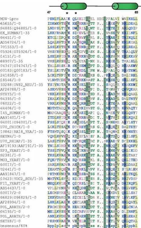
[image:3.585.304.538.66.444.2]was detected with a rabbit polyclonal Ab elicited against bacterially ex-pressed recombinant protein (39), TMEV VP1 was detected with MAb DAmAb2 (28), and NF-B–p65/RelA was detected with rabbit polyclonal Ab-1 RB-1638 (NeoMarkers; Lab Vision). Alexa Fluor 488 and Alexa Fluor 594 (Molecular Probes, Invitrogen)-conjugated secondary Abs were used for detec-tion. Nuclei were visualized by DAPI (4⬘,6⬘-diamidino-2-phenylindole) staining included in ProLong Gold Antifade mounting medium (Invitrogen). Cells were examined in an Olympus BX40 fluorescence microscope, and the images were FIG. 1. Alignment of FMDV Lpro partial amino acid sequence.
Lproprotein sequence was aligned to all available sequences utilizing
SMART software. Depicted are sequences in single letters between amino acids 47 and 88, and a schematic diagram displays the approx-imate location of predicted␣-helices (green ovals). Asterisks mark the location of amino acids targeted by mutagenesis. Summary of color coding including consensus sequence:ⴙ(positive sign in blue), posi-tive charged amino acids (H, K, and R); b (lowercase letter “b” high-lighted in yellow), amino acids with a large or bulky side chain (E, F, H, I, K, L, M, Q, R, W, and Y); c (lowercase letter “c” in pink font), charged amino acids (D, E, H, K, and R); l (lowercase letter “l” highlighted in yellow), aliphatic amino acids (I, L, and V); p (lowercase letter “p” in blue font), polar amino acids (C, D, E, H, K, N, Q, R, S, and T); s (lowercase letter “s” in green font), amino acids with a small side chain (A, C, D, G, N, P, S, T, and V); and gray highlighted, amino acids withⱖ60% of the homology in the alignment.
on November 8, 2019 by guest
http://jvi.asm.org/
taken with a DP-70 digital camera using DP-BSW v2.2 software (Olympus America, Central Valley, PA).
RESULTS
FMDV Lpro
coding region contains a putative SAP domain. Analysis of the Lprocoding sequence utilizing SMART showed
that from amino acids 47 to 83 (following the numbering of Lb) there is a conserved sequence motif that resembles a previously defined SAP domain (1) (Fig. 1). Although the homology with any specific sequence of SAP domains is lower than 25% per-cent, the homology with the consensus SAP domain sequence is greater than 60%. When the Lprosequence was compared to
the sequence profile of the SAP domains, more than 80% the of the Lproamino acids within this region were found in the
profile. In addition, the three-dimensional structure of the Lpro
sequence (21) shared the same␣-helix-turn-␣-helix structure found in SAP domains (1). Despite the presence of a two-amino-acid insertion between the two ␣-helices in Lpro, the
data support the presence of a SAP domain within Lpro.
Mutations of Lpro
SAP domain partially affect virus growth. In order to determine whether the putative SAP domain is important for Lprofunction, we mutated two residues at
posi-tions 55 or 58, individually (A12#47 and A12#48) or in com-bination (A12#49). We selected these amino acids based on previous studies with PIAS3, a SAP-containing protein, where it was reported that mutation of a similar region altered PIAS3 nuclear localization and retention (16). Viruses derived from transfected cells were passaged four times in BHK-21 cells, and the Lprocoding region of the resulting viruses was sequenced
to confirm that the only changes were at the mutated sites. Figure 2 shows the kinetics of growth of the mutant viruses in
two different cell types. In BHK-21 cells all viruses grew with similar kinetics, reaching final titers with differences of less than a half log with respect to the WT. In EBK cells the growth differences between WT and some of the mutant viruses were more pronounced. As previously reported, A12-LLV2 grew to a final titer ⬃50-fold lower than that of the WT virus (9). Interestingly, A12#49 (the double SAP mutant) grew to a final titer of⬃5-fold lower than WT virus, whereas A12#47 and A12#48 (single SAP mutants) grew to titers similar to that of the WT virus. Regarding plaque size, A12#47 and A12#48 resembled A12-WT virus, and A12#49 was more related to the small-plaque phenotype of A12-LLV2. These results indicated that disruption of the predicted signature motif of the SAP domain requires at least mutations at two sites and affects the growth characteristics of FMDV, resulting in a partially atten-uated phenotype.
Mutation of Lpro
SAP domain affects nuclear retention.We
have previously reported that during FMDV infection Lpro
progressively translocates to the nucleus of infected cells (14). We analyzed the subcellular localization of Lpro in the SAP
mutants. In LF-BK cells infected with the single mutant viruses (A12#47 and A12#48) the translocation of Lprointo the
nu-cleus was indistinguishable from A12-WT (data not shown). In contrast, the double SAP mutant (A12#49) displayed a distinct phenotype. At the beginning of the infection and up to 4 h postinfection (hpi), Lproof A12#49 was observed in the
cyto-plasm and then progressively appeared in the nucleus of the infected cells (Fig. 3, panels 3, 6, and 9) similarly to A12-WT (Fig. 3, panels 1, 4, and 7). We observed that nuclear translo-cation of Lprowas slightly delayed for A12#49. However, by 6
hpi almost no Lpro nuclear staining was detected in cells
in-FIG. 2. Kinetics of growth and plaque morphology. (A) Growth curves. BHK-21 or EBK cells were infected with the indicated viruses and, after 1 h, unabsorbed virus was removed by washing with 150 mM NaCl–20 mM MES (pH 6.0), followed by the addition of complete medium. Samples were taken at 1, 3, 6, and 24 hpi, and virus titers were determined by plaque assay on BHK-21 cells. (Reported values display one out of three representative experiments with similar results.) (B) BHK-21 cells were infected with similar amounts of viruses and treated as described in panel A, but medium with a gum tragacanth overlay was added and the plaques were stained at 40 hpi.
on November 8, 2019 by guest
http://jvi.asm.org/
[image:4.585.120.461.68.307.2]fected with A12#49 in contrast to cells infected with A12-WT (Fig. 3, panels 12 and 10, respectively). Most of Lpro was
distributed throughout the cytoplasm concentrated in granules, as eventually observed for several other FMDV viral proteins when infection is well established. As a control, infection with A12-LLV2 did not display any Lprostaining (Fig. 3, panels 2, 5,
8, and 11). These observations led us to conclude that mutation of the predicted SAP domain prevented Lpronuclear
accumu-lation during the course of infection.
p65/RelA is not degraded after infection with mutant
FMDV. In our previous studies we demonstrated that p65/
RelA degradation correlated with nuclear accumulation of Lpro (14). Figure 4 shows the IFA results of p65/RelA in
LF-BK cells infected with A12-WT, A12-LLV2, and A12#49. As seen previously in infected PK cells, by 6 hpi the p65/RelA signal was almost absent from the nucleus of WT-infected cells (Fig. 4, panels 1 and 3) and accumulated in the nucleus of
A12-LLV2-infected cells (Fig. 4 panels 4 and 6). Interestingly, the pattern for A12#49 (Fig. 4, panels 7 and 9) exactly resem-bled the pattern for A12-LLV2, with a bright p65/RelA stain-ing concentrated in the nuclei of infected cells. Western blot analysis of cytoplasmic and nuclear extracts of infected LF-BK cells showed similar results (data not shown). Since there is a correlation between p65/RelA degradation and Lpro nuclear
accumulation, we decided to add LMB, an inhibitor of the CRM1-dependent pathway of active nuclear export (18). We hypothesized that p65/RelA would be degraded if the exit of A12#49 Lprofrom the nucleus was blocked. However, there
was no nuclear accumulation of Lproafter the addition of LMB
to A12-WT- or A12#49-infected cells (Fig. 5, panels 3 and 4). The addition of LMB did increase the number of cells display-ing nuclear p65/RelA staindisplay-ing (Fig. 5, panels 5 to 7 compared to panels 6 to 8) as previously described (33).
[image:5.585.132.451.63.479.2]These results suggested that nuclear accumulation of Lprois
FIG. 3. IFA of Lproduring FMDV infection. LF-BK cells were infected at an MOI of 10 with FMDV A12-WT (panels 1, 4, 7, and 10),
A12-LLV2 (panels 2, 5, 8, and 11), or double SAP mutant A12#49 (panels 3, 6, 9, and 12) and were fixed at different times postinfection. Viral protein Lprowas detected using a rabbit polyclonal Ab and an Alexa Fluor 488-conjugated secondary Ab. Viral protein VP1 was detected using
mouse MAb 6HC4 and an Alexa Fluor 594-conjugated secondary Ab.
on November 8, 2019 by guest
http://jvi.asm.org/
required for p65/RelA degradation and that the CRM1-depen-dent nuclear export pathway is not involved in the exit of Lpro
from the nuclei of A12#49-infected cells.
Mutation of Lpro
SAP domain prevents Lpro
inhibition of
IFN expression.We earlier reported that Lproantagonizes the
innate immune response by blocking the expression of IFN (9, 10, 13, 14). To test whether the SAP double mutant had a similar effect, we analyzed the expression of IFN-, the proin-flammatory cytokine TNF-␣, the chemokine RANTES, and the ISGs Mx1 and IRF7 in virus-infected cells. We used
[image:6.585.97.483.66.545.2]sec-ondary PK cells in this assay, rather than secsec-ondary EBK or LF-BK cells, because we have previously optimized standard procedures of real-time PCR in PK cells (13, 14) and FMDV SAP mutants behave consistently in these cell types (Fig. 2 to 4 and data not shown). By 4 hpi, there was no significant difference in the induction of any of the analyzed genes after infection with A12-WT, A12-LLV2, or A12#49 (Fig. 6A). For IFN-we observed at most a twofold difference for A12#49 or A12-LLV2 relative to A12-WT. However, by 8 hpi, there was an increase in IFN-expression of⬃10-fold for A12#49 and
FIG. 4. IFA of p65/RelA during FMDV infection. LF-BK cells were infected at an MOI of 10 with WT (panels 1 to 3), A12-LLV2 (panels 4 to 6), and the double SAP mutant A12#49 (panels 7 to 9). As a control, cells were mock infected (panels 10 and 11) or treated with synthetic dsRNA poly[IC] (25g/ml) and Lipofectamine (panels 12 and 13). At 6 hpi, cells were fixed and stained. p65/RelA was detected using a rabbit polyclonal Ab (Abcam RB-1638) and an Alexa Fluor 488-conjugated secondary Ab. Viral protein VP1 was detected using mouse MAb 6HC4 and an Alexa Fluor 594-conjugated secondary Ab. Nuclei were stained with DAPI (panels 11 and 13).
on November 8, 2019 by guest
http://jvi.asm.org/
14-fold for A12-LLV2 compared to A12-WT. A similar pattern was observed for all of the analyzed genes, although the dif-ferences were slightly lower, varying from 3- to 10-fold higher for the mutants compared to WT. ELISA quantitation of se-creted IFN-␣protein in the supernatants of infected PK cells followed similar kinetics (Fig. 6B). By 24 hpi, we detected 6- to 12-fold-higher amounts of IFN-␣ protein for A12-LLV2 and A12#49, respectively, compared to A12-WT.
These results indicated that mutations of the SAP domain prevented the inhibitory effect of Lpro on NF-B-dependent
transcriptional activity and IFN protein expression.
Cleavage of translation initiation factor eIF-4G is not
af-fected by mutation of the Lpro
SAP domain.Lprois responsible
for cleaving the translation initiation factor eIF-4G and shut-ting off host cell translation, a hallmark of picornavirus infec-tion (44). We examined the kinetics of eIF-4G cleavage in LF-BK-infected cells by Western blot analysis. Figure 7A shows that, as expected, eIF-4G (p220) was completely pro-cessed in A12-WT-infected cells by 4 hpi. Interestingly, only a minor delay, 1 to 2 h, on eIF-4G processing was detected for double SAP mutant (A12#49). Comparable amounts of viral VP1 suggested that the stage of infection was similar for A12-WT and A12#49. Although complete eIF-4G cleavage was achieved for A12#49 by 5 to 6 hpi, low-molecular-weight cleavage products persisted throughout the course of infection. No eIF-4G processing was observed in mock-infected or A12-LLV2-infected cells. By 4 hpi the p65/RelA signal was signif-icantly reduced in the cytoplasm of cells infected with A12-WT. In contrast, the p65/RelA signal did not decrease in extracts of A12-LLV2- or double SAP mutant A12#49-in-fected cells.
Analysis of viral polyprotein processing using a radioimmu-noprecipitation assay revealed no major differences between A12-WT and A12#49 (Fig. 7B). Processing into mature prod-ucts, L, VP0, VP1,VP3, 2B, 2C, and 3D proceeded almost equivalently by 4 hpi. As previously reported, viral protein synthesis in A12-LLV2-infected cells was significantly delayed (39).
These results indicated that disruption of the LproSAP
do-main selectively prevented p65/RelA processing without affect-ing the ability of Lproto cleave eIF-4G.
Lpro
catalytic activity is required for p65/RelA processing.In
our previous studies, using a recombinant Theiler’s virus that expresses Lpro in the absence of any other FMDV protein
(TMEV-Lb), we demonstrated that the presence of Lpro is
necessary and sufficient for p65/RelA degradation (14). Using the same recombinant virus, we examined whether the catalytic activity of Lprowas a requirement for p65/RelA disappearance.
For this purpose, we mutated the catalytic cysteine residue of Lpro (C23) to alanine, creating TMEV-LbC23A. Mutation of
Lpro C23 did not affect the pattern of localization previously
observed for WT Lpro (Fig. 8, panels 4 and 6 compared to
panels 1 and 3). Interestingly, this mutation abolished the ability of FMDV Lproto cause p65/RelA degradation (Fig. 8,
panels 10, 11, and 12 compared to panels 7, 8, and 9), indicat-ing that the protease activity of Lprois essential for this
func-tion.
DISCUSSION
We have earlier reported that Lpro antagonizes the innate
immune response by blocking the expression of IFN. At least two mechanisms are involved in this function: (i) the shutoff of host cell translation resulting in lower levels of IFN protein expression and (ii) the interference in the induction of IFN- transcription. Lprocleaves the eukaryotic translation initiation
factor eIF-4G, which is required for cap-dependent mRNA translation without affecting the internal ribosome entry site-dependent translation of viral RNA, and thus the virus takes advantage of decreased levels of IFN protein to establish a productive infection (9, 20, 26). In addition, Lproinduces the
FIG. 5. Addition of LMB does not block the nuclear export of FMDV Lpro. LF-BK cells were infected at an MOI of 10 with A12-WT
(panels 1, 3, 5, and 7) and the double SAP mutant A12#49 (panels 2, 4, 6, and 8). Where indicated, 200 nM LMB was added to the culture medium. At 6 hpi, cells were fixed and stained. For panels 1 to 4, viral protein Lprowas detected using a rabbit polyclonal Ab and an Alexa
Fluor 488-conjugated secondary Ab. Viral protein VP1 was de-tected using mouse MAb 6HC4 and an Alexa Fluor 594-conjugated secondary Ab. For panels 5 to 8, p65/RelA was detected using rabbit polyclonal Ab Abcam RB-1638 and an Alexa Fluor 488-conjugated secondary Ab.
on November 8, 2019 by guest
http://jvi.asm.org/
degradation of the p65/RelA subunit of the transcription factor NF-B, and this degradation is associated with Lpro nuclear
localization (14). A block in the upregulation of IFN- tran-scription also results in lower levels of IFN protein (13).
The availability of multiple FMDV protein sequences (7), the high-resolution crystal structure of Lpro(21), and powerful
software tools (29) have allowed us to predict that a conserved SAP domain is situated between amino acids 47 and 83 of Lb. In the present study we demonstrated that this domain is important for Lprofunction. Double mutation of the SAP
do-main resulted in an attenuated virus phenotype yielding lower titers and a smaller plaque size. Although the phenotype was not as clear-cut as in the case of the Lprodeletion in leaderless
virus, it was indicative of a role of this domain in FMDV virulence. Early translocation of mutant Lpro from the
cyto-plasm to the nucleus of infected cells was only slightly delayed. However, by 6 hpi, mutant Lpro, in contrast to WT Lpro, was
absent from the nuclei of infected cells. Failure in nuclear retention has been reported for another SAP-containing pro-tein, PIAS3L, when this domain was mutated (16). PIAS3L requires an intact SAP box, in conjunction with a RING and a PINIT domain, for proper nuclear localization and retention (16). Perhaps FMDV Lprodepends on an intact SAP domain
[image:8.585.113.473.70.265.2]for docking in the nucleus of infected cells, allowing for inter-actions with host proteins that might be involved in regulating an antiviral response.
FIG. 6. Analysis of IFN expression. (A) The expression of IFN, TNF-␣, RANTES, Mx1, and IRF7 mRNAs was measured by real-time RT-PCR in secondary PK cells infected with A12-WT, A12-LLV2, or A12#49 FMDV at an MOI of 2 for the indicated times. Porcine GAPDH (glyceraldehyde-3-phosphate dehydrogenase) was used as an internal control. The results are expressed as the fold increase in gene expression for virus-infected with respect to mock-infected cells. (Reported values display the findings for one out of three representative experiments with similar results.) (B) pIFN␣expression in the supernatants of PK-infected cells determined by ELISA. The values are presented as the mean⫾the standard deviation of three independent determinations.
FIG. 7. Processing of cellular proteins during infection with WT and mutant FMDV. (A) LF-BK cells were infected with WT (A12-WT), leaderless (A12-LLV2), and SAP mutant (A12#49) at MOIs of 10 for 6 h. At the indicated times, cytoplasmic extracts were prepared and analyzed by Western blotting using rabbit polyclonal Ab anti-eIF-4G (p220), rabbit polyclonal Ab anti-p65/RelA (RB-1638), mouse MAb 6HC4 (VP1), and mouse MAb anti-tubulin-␣(Ab-2 MS-581). CP, p220 cleavage products. (B) Radioimmunoprecipitation of FMDV-infected cell lysates at different times postinfection. [35S]methionine-labeled cell lysates from FMDV-infected LF-BK cells were immunoprecipitated with serum from a
conva-lescent bovine. Samples were resolved by sodium dodecyl sulfate-polyacrylamide gel electrophoresis and developed by autoradiography.
on November 8, 2019 by guest
http://jvi.asm.org/
[image:8.585.126.463.514.667.2]One of the most interesting observations in the present study was the absence of NF-B degradation upon infection with an FMDV double SAP mutant even though these mutations did not affect the catalytic activity of Lpro. With the exception of
leaderless virus, no other viable FMDV Lpromutant has been
previously reported. In vitro studies using mutant recombinant protein or plasmid transient transfection have been very infor-mative, demonstrating that residue C23 is required for the protease enzymatic activity and is utilized by Lpro for
self-cleavage from the viral polyprotein and self-cleavage of the trans-lation initiation factor eIF-4G (15, 34, 40, 43). Recently, Mayer et al. (34), using rabbit reticulocyte lysates, have shown that Lpro residue L115 is also a determinant of self-cleavage and
eIF-4G cleavage specificity. Utilizing a recombinant cardiovi-rus expressing Lproin the absence of any other FMDV protein,
we also demonstrated that the catalytic activity of Lpro is
re-quired for NF-B degradation; mutation of the catalytic
resi-due C23 prevented degradation. Unfortunately, the mecha-nism used by Lproto cause NF-B degradation is still unclear.
As mentioned above, LproSAP mutations did not affect Lpro
enzymatic activity; self-processing and eIF-4G processing pro-ceeded almost normally. We did observe that degradation of the eIF-4G cleavage products was delayed in cells infected with the double mutant. Mutation of the SAP domain may partially affect the interaction between Lpro and eIF-4G. Quantitative
kinetics studies should be performed to verify this hypothesis. It has been reported that FMDV 3C can also cleave eIF-4G at later times after FMDV infection (47); thus, it is possible that the Lpro SAP mutant interferes with the 3C/eIF-4G
interac-tion. Our results, however, suggest that 3C from double mutant SAP virus behaves normally since processing of the viral polyprotein proceeded similarly for the mutant and WT vi-ruses.
The levels of several transcripts, including cytokines,
chemo-FIG. 8. FMDV Lprocatalytic activity is required for p65/RelA degradation. IBRS-2 cells were infected with TMEV-Lb-containing WT or
mutant C23A Lproat an MOI of 10. At 24 hpi, p65/RelA and viral proteins were visualized by IFA. p65/RelA was detected with rabbit polyclonal
Ab (RB-1638) and Alexa Fluor 488-conjugated secondary Ab. Lprowas visualized a rabbit polyclonal Ab and Alexa Fluor 488-conjugated secondary
Ab. TMEV VP1 was detected with MAb DAmAb2 (28) and Alexa Fluor 594-conjugated secondary Ab.
on November 8, 2019 by guest
http://jvi.asm.org/
[image:9.585.132.449.66.473.2]kines, and ISGs, were significantly higher after infection with FMDV Lpro SAP mutant #49 compared to WT infection,
indicating that disruption of the SAP domain prevented Lpro
inhibition of NF-B-dependent transcription. SAP domains are involved in protein-protein interactions. This motif is re-quired for the repressive activity of PIASy on STAT1-mediated gene activation (31), and it has been proposed that several members of the PIAS protein family negatively regulate NF-B and STAT signaling, affecting the expression of more than 60 genes (45). Furthermore, the specific role of PIAS proteins in the regulation of NF-B activity has been examined in vivo utilizing PIAS1-null mice (32). These studies demon-strated that in the absence of PIAS only a subset of NF- B-regulated genes is affected (ca. 48%), suggesting that there might be alternative mechanisms, independent of PIAS1, for NF-B regulation.
More interestingly, Jang et al. (24) have provided evidence that the N-terminal region of PIAS3, which contains a SAP domain, is necessary for binding to the p65/RelA subunit of NF-B, thereby blocking the transcriptional activation. Fur-thermore, an LXXLL signature motif of PIAS3 is involved in this physical interaction. Although not identical, this motif resembles the IQKL sequence present in FMDV Lpro. Our
results suggest that this putative interaction may be involved in docking Lproin the nucleus of infected cells where Lpro
-depen-dent p65/RelA degradation takes place during FMDV infec-tion. We are currently testing this hypothesis.
SAP domains are also found in several proteins displaying DNA-binding activity, and the contact with defined A/T rich sequences found in matrix attachment regions (MARs) of chromatin is mediated by the predicted␣-helices delimited by the SAP box (25). Protein interactions with MARs regions determine the chromatin architecture in zones of interactions with the nuclear matrix. Interestingly, several viral proteins have been shown to localize to these regions, leading to the proposal that viral protein interaction with MARs regions may have a role in blocking host antiviral activities (17). The pres-ence of a SAP domain may allow FMDV Lproto localize to
similar nuclear regions globally affecting the function of tran-scription factors situated in close proximity during viral infec-tion.
Our results provide new insights into the mechanism used by FMDV to escape the immune response. Structure-function analysis of Lprohas demonstrated that, in addition to the
pro-teinase activity, an intact protein motif, SAP, is required for FMDV virulence. A more detailed understanding of the inter-actions between FMDV Lpro and/or other viral proteins and
the host at the molecular level should help in the development of specific antiviral strategies that could limit virus spread.
ACKNOWLEDGMENTS
We thank Z. Lu for technical assistance with DNA sequencing. This study was supported in part by the Plum Island Animal Disease Research Participation Program administered by the Oak Ridge Insti-tute for Science and Education through an interagency agreement between the U.S. Department of Energy and the U.S. Department of Agriculture (appointment of F.D.-S.S. and C.C.A.D.) and by CRIS Project no. 1940-32000-052-00D, ARS, USDA (T.D.L.S., J.Z., and M.J.G.).
REFERENCES
1.Aravind, L., and E. V. Koonin.2000. SAP: a putative DNA-binding motif involved in chromosomal organization. Trends Biochem. Sci.25:112–114. 2.Barral, P. M., J. M. Morrison, J. Drahos, P. Gupta, D. Sarkar, P. B. Fisher,
and V. R. Racaniello.2007. MDA-5 is cleaved in poliovirus-infected cells. J. Virol.81:3677–3684.
3.Baxt, B., D. O. Morgan, B. H. Robertson, and C. A. Timpone.1984. Epitopes on foot-and-mouth disease virus outer capsid protein VP1 involved in neu-tralization and cell attachment. J. Virol.51:298–305.
4.Belov, G. A., Lidsky, P. V., Mikitas, O. V., Egger, D., Lukyanov, K. A., Bienz, K., and V. I. Agol.2004. Bidirectional increase in permeability of nuclear envelope upon poliovirus infection and accompanying alterations of nuclear pores. J. Virol.78:10166–10177.
5.Brown, C. C., M. E. Piccone, P. W. Mason, T. S. McKenna, and M. J. Grubman.1996. Pathogenesis of wild-type and leaderless foot-and-mouth disease virus in cattle. J. Virol.70:5638–5641.
6.Cao, X., I. E. Bergmann, R. Fullkrug, and E. Beck.1995. Functional analysis of the two alternative translation initiation sites of foot-and-mouth disease virus. J. Virol.69:560–563.
7.Carrillo, C., E. R. Tulman, G. Delhon, Z. Lu, A. Carreno, A. Vagnozzi, G. F. Kutish, and D. L. Rock.2005. Comparative genomics of foot-and-mouth disease virus. J. Virol.79:6487–6504.
8.Chinsangaram, J., P. W. Mason, and M. J. Grubman.1998. Protection of swine by live and inactivated vaccines prepared from a leader proteinase-deficient serotype A12 foot-and-mouth disease virus. Vaccine16:1516–1522. 9.Chinsangaram, J., M. E. Piccone, and M. J. Grubman.1999. Ability of foot-and-mouth disease virus to form plaques in cell culture is associated with suppression of alpha/beta interferon. J. Virol.73:9891–9898. 10.Chinsangaram, J., M. Koster, and M. J. Grubman. 2001. Inhibition of
L-deleted foot-and-mouth disease virus replication by alpha/beta interferon involves double-stranded RNA-dependent protein kinase. J. Virol.75:5498– 5503.
11.Conzelmann, K.-K.2005. Transcriptional activation of alpha/beta interferon genes: interference by nonsegmented negative-strand RNA viruses. J. Virol. 79:5241–5248.
12.Delhaye, S., V. van Pesch, and T. Michiels.2004. The leader protein of Theiler’s virus interferes with nucleocytoplasmic trafficking of cellular pro-teins. J. Virol.78:4357–4362.
13.de los Santos, T., S. de Avila Botton, R. Weiblen, and M. J. Grubman.2006. The leader proteinase of foot-and-mouth disease virus inhibits the induction of beta interferon mRNA and blocks the host innate immune response. J. Virol.80:1906–1914.
14.de los Santos, T., F. Diaz-San Segundo, and M. J. Grubman.2007. Degra-dation of nuclear factorB during foot-and-mouth disease virus infection. J. Virol.81:12803–12815.
15.Devaney, M. A., V. N. Vakharia, R. E. Lloyd, E. Ehrenfeld, and M. J. Grubman.1988. Leader protein of foot-and-mouth disease virus is required for cleavage of the p220 component of the cap-binding protein complex. J. Virol.62:4407–4409.
16.Duval, D., G. Duval, C. Kedingerc, O. Pocha, and H. Boeufa.2003. The PINIT motif, of a newly identified conserved domain of the PIAS protein family, is essential for nuclear retention of PIAS3L. FEBS Lett.554:111–118. 17.Everett, R. D., and M. K. Chelbi-Alix.2007. PML and PML nuclear bodies:
implications in antiviral defence. Biochimie89:819–830.
18.Fukuda, M., S. Asano, T. Nakamura, M. Adachi, M. Yoshida, M. Yanagida, and E. Nishida.1997. CRM1 is responsible for intracellular transport me-diated by the nuclear export signal. Nature390:308–311.
19.Grubman, M. J., and B. Baxt.2004. Foot-and-mouth disease. Clin. Micro-biol. Rev.17:465–493.
20.Grubman, M. J., M. P. Moraes, F. Diaz-San Segundo, L. Pena, and T. de los Santos.2008. Evading the host immune response: how foot-and-mouth dis-ease virus has become an effective pathogen. FEMS Immunol. Med. Micro-biol.53:8–17.
21.Guarne´, A., J. Tormo, R. Kirchweger, D. Pfistermueller, I. Fita, and T. Skern.1998. Structure of the foot-and-mouth disease virus leader protease: a papain-like fold adapted for self-processing and eIF4G recognition EMBO J.17:7469–7479.
22.Haller, O., G. Kochs, and F. Weber.2006. The interferon response circuit: induction and suppression by pathogenic viruses. Virology344:119–130. 23.Honda, K., H. Yanai, A. Takaoka, and T. Taniguchi.2006. Regulation of the
type I IFN induction: a current view. Int. Immunol.17:1367–1378. 24.Jang, H. D., K. Yoon, Y. J. Shin, J. Kim, and S. Y. Lee.2004. PIAS3
suppresses NF-B mediated transcription by interacting with the p65/RelA subunit. J. Biol. Chem.279:24873–24880.
25.Kipp, M., F. Go¨hring, T. Ostendorp, C. M. van Drunen, R. van Driel, M. Przybylski, and F. O. Fackelmayer.2000. SAF-Box, a conserved protein domain that specifically recognizes scaffold attachment region DNA. Mol. Cell. Biol.20:7480–7489.
26.Kirchweger, R., E. Ziegler, B. J. Lamphear, D. Waters, H. D. Liebig, W. Sommergruber, F. Sobrino, C. Hohenadl, D. Blaas, R. E. Rhoads, and T. Skern.1994. Foot-and-mouth disease virus leader proteinase: purification of
on November 8, 2019 by guest
http://jvi.asm.org/
the Lb form and determination of its cleavage site on eIF-4 gamma. J. Virol. 68:5677–5684.
27.Kleina, L. G., and M. J. Grubman.1992. Antiviral effects of a thiol protease inhibitor on foot-and-mouth disease virus. J. Virol.66:7168–7175. 28.Kong, W. P., G. D. Ghadge, and R. P. Roos.1994. Involvement of cardiovirus
leader in host cell-restricted virus expression. Proc. Natl. Acad. Sci. USA 91:1796–1800.
29.Letunic, I., R. R. Copley, B. Pils, S. Pinkert, J. Schultz, and P. Bork.2006. SMART 5: domains in the context of genomes and networks. Nucleic Acids Res.34:257–260.
30.Lidsky P. V., S. Hato, M. V. Bardina, A. G. Aminev, A. C. Palmenberg, E. V. Sheval, V. Y. Polyakov, F. J. van Kuppeveld, and V. I. Agol.2006. Nucleo-cytoplasmic traffic disorder induced by cardioviruses. J. Virol.80:2705–2717. 31.Liu, B., M. Gross, J. ten Hoeve, and K. Shuai.2001. A transcriptional corepressor of STAT1 with an essential LXXLL signature motif. Proc. Natl. Acad. Sci. USA98:3203–3207.
32.Liu, B., R. Yang, K. A. Wong, C. Getman, N. Stein, M. A. Teitell, G. Cheng, H. Wu, and K. Shuai. 2005. Negative regulation of NF-B signaling by PIAS1. Mol. Cell. Biol.25:1113–1123.
33.Loewe, R., W. Holnthoner, M. Gro¨ger, M. Pillinger, F. Gruber, D. Mechtch-eriakova, E. Hofer, K. Wolff, and P. Petzelbauer.2002. Dimethylfumarate inhibits TNF-induced nuclear entry of NF-B/p65 in human endothelial cells. J. Immunol.168:4781–4787.
34.Mayer, C., D. Neubauer, A. T. Nchinda, R. Cencic, K. Trompf, and T. Skern. 2008. Residue L143 of the foot-and-mouth disease virus leader proteinase is a determinant of cleavage specificity. J. Virol.82:4656–4659.
35.Medina, M., E. Domingo, J. K. Brangwyn, and G. J. Belsham.1993. The two species of the foot-and-mouth disease virus leader protein, expressed indi-vidually, exhibit the same activities. Virology194:55–359.
36.Moraes, M. P., J. Chinsangaram, M. C. S. Brum, and M. J. Grubman.2003. Immediate protection of swine from foot-and-mouth disease: a combination of adenoviruses expressing interferon alpha and a foot-and-mouth disease virus subunit vaccine. Vaccine22:268–279.
37.Moraes, M. P., T. de los Santos, M. Koster, T. Turecek, H. Wang, V. G. Andreyev, and M. J. Grubman.2007. Enhanced antiviral activity against foot-and-mouth disease virus by a combination of type I and II porcine interferons. J. Virol.81:7124–7135.
38.Neznanov, N., K. M. Chumakov, L. Neznanova, A. Almasan, A. K. Banerjee, and A. V. Gudkov.2005. Proteolytic cleavage of the p65-RelA subunit of NF-B during poliovirus infection. J. Biol. Chem.280:24153–24158. 39.Piccone, M. E., E. Rieder, P. W. Mason, and M. J. Grubman.1995. The
foot-and-mouth disease virus leader proteinase gene is not required for viral replication. J. Virol.69:5376–5382.
40.Piccone, M. E., M. Zellner, T. F. Kumosinski, P. W. Mason, and M. J. Grubman.1995. Identification of the active-site residues of the L proteinase of foot-and-mouth disease virus. J. Virol.69:4950–4956.
41.Piccone, M. E., H.-H. Chen, R. R. Roos, and M. J. Grubman.1996. Con-struction of a chimeric Theiler’s murine encephalomyelitis virus containing the leader gene of foot-and-mouth disease virus. Virology226:135–139. 42.Rieder, E., T. Bunch, F. Brown, and P. W. Mason.1993. Genetically
engi-neered foot-and-mouth disease viruses with poly(C) tracts of two nucleotides are virulent in mice. J. Virol.67:5139–5145.
43.Roberts, P. J., and G. J. Belsham.1995. Identification of critical amino acids within the foot-and-mouth disease virus leader protein, a cysteine protease. Virology213:140–146.
44.Rueckert, R. R.2007.Picornaviridae:the viruses and their replication, p. 795–948.InD. M. Knipe, P. M. Howley, D. E. Griffin, R. A. Lamb, M. A. Martin, B. Roizman, and S. E. Straus (ed.), Fields virology, 5th ed. Lippin-cott-Raven Publishers, Philadelphia, PA.
45.Shuai, K., and B. Liu.2005. Regulation of gene-activation pathways by PIAS proteins in the immune system. Nat. Rev. Immunol.5:593–605.
46.Shuai, K.2006. Regulation of cytokine signaling pathways by PIAS proteins. Cell Res.16:196–202.
47.Strong, R., and G. J. Belsham.2004. Sequential modification of translation initiation factor eIF4GI by two different foot-and-mouth disease virus pro-teases within infected baby hamster kidney cells: identification of the 3Cpro cleavage site. J. Gen. Virol.85:2953–2962.
48.Swaney, L. M.1988. A continuous bovine kidney cell line for routine assays of foot-and-mouth disease virus. Vet. Microbiol.18:1–14.
49.Zoll, J., W. J. Melchers, J. M. Galama, and F. J. van Kuppeveld.2002. The mengovirus leader protein suppresses alpha/beta interferon production by inhibition of the iron/ferritin-mediated activation of NF-B. J. Virol.76: 9664–9672.