0022-538X/08/$08.00
⫹
0
doi:10.1128/JVI.01716-07
Copyright © 2008, American Society for Microbiology. All Rights Reserved.
Intracellular Localization Map of Human Herpesvirus 8 Proteins
䌤
Gaby Sander,
1† Andreas Konrad,
1† Mathias Thurau,
1Effi Wies,
2Rene Leubert,
3Elisabeth Kremmer,
4Holger Dinkel,
5Thomas Schulz,
6Frank Neipel,
2and Michael Stu
¨rzl
1*
Division of Molecular and Experimental Surgery, Department of Surgery, University of Erlangen-Nuremberg, Schwabachanlage 10,
D-91054 Erlangen, Germany
1; Institute of Clinical and Molecular Virology, University of Erlangen-Nuremberg, Schlossgarten 4,
D-91054 Erlangen, Germany
2; Department of Virus-Induced Vasculopathy, GSF-National Research Center for Environment and
Health, Ingolsta
¨dter Landstrasse 1, D-85764 Neuherberg, Germany
3; GSF-Service Unit Monoclonal Antibodies and Cell Sorting,
GSF-National Research Center for Environment and Health, Marchioninistrasse 25, D-81377 Munich, Germany
4;
Division of Bioinformatics, Institute of Biochemistry, University of Erlangen-Nuremberg, Fahrstrasse 17,
D-91054 Erlangen, Germany
5; and Medical School Hannover, Department of Virology,
Carl-Neubergstrasse 1, D-30625 Hannover, Germany
6Received 7 August 2007/Accepted 21 November 2007
Human herpesvirus 8 (HHV-8) is the etiological agent of Kaposi’s sarcoma. We present a localization
map of 85 HHV-8-encoded proteins in mammalian cells. Viral open reading frames were cloned with a Myc
tag in expression plasmids, confirmed by full-length sequencing, and expressed in HeLa cells. Protein
localizations were analyzed by immunofluorescence microscopy. Fifty-one percent of all proteins were
localized in the cytoplasm, 22% were in the nucleus, and 27% were found in both compartments.
Surpris-ingly, we detected viral FLIP (v-FLIP) in the nucleus and in the cytoplasm, whereas cellular FLIPs are
generally localized exclusively in the cytoplasm. This suggested that v-FLIP may exert additional or
alternative functions compared to cellular FLIPs. In addition, it has been shown recently that the K10
protein can bind to at least 15 different HHV-8 proteins. We noticed that K10 and only five of its 15
putative binding factors were localized in the nucleus when the proteins were expressed in HeLa cells
individually. Interestingly, in coexpression experiments K10 colocalized with 87% (13 of 15) of its putative
binding partners. Colocalization was induced by translocation of either K10 alone or both proteins. These
results indicate active intracellular translocation processes in virus-infected cells. Specifically in this
framework, the localization map may provide a useful reference to further elucidate the function of
HHV-8-encoded genes in human diseases.
Human herpesvirus 8 (HHV-8) belongs to the family of
gammaherpesviruses. HHV-8 infection is associated with
sev-eral severe human diseases such as multicentric Castleman’s
disease, primary effusion lymphoma, and Kaposi’s sarcoma (7,
9, 18, 45, 81).
The HHV-8 genome consists of 165 kbp. To date, 86
differ-ent open reading frames (ORFs) have been iddiffer-entified (68).
The absolute number of HHV-8-encoded genes is still under
investigation due to the detection of differentially spliced gene
products in different types of infected cells (68, 80).
Previously, the pathogenic activity of HHV-8 was
preferen-tially analyzed in studies with single genes. More
comprehen-sive analyses may be required to understand the complexity of
the HHV-8 pathogenic repertoire. Systems biology approaches
are a new powerful tool for the analysis of complex biological
processes. However, these methods have been preferentially
applied to study the cell biology of yeast (30, 53, 70) and only
in a very limited way to study pathogenic activities of infectious
agents. Only recently, the first proteome-wide protein
interac-tion study of HHV-8 and varicella-zoster virus was published
(82). In this study the K10 protein of HHV-8 was identified as
a key interacting protein, binding to at least 15 different
HHV-8-encoded proteins (82).
In addition to protein interactions, subcellular localization
of proteins is closely associated with protein function. This is
generally appreciated, and it is underscored by the rapid
growth of localization databases, such as Organelle DB (85).
The subcellular localization of most HHV-8-encoded proteins
is not known yet. Therefore, we generated a complete
local-ization map of all known HHV-8-encoded genes in
mamma-lian cells. Several unexpected findings were obtained clearly
documenting the usefulness of systems biology approaches to
study HHV-8.
MATERIALS AND METHODS
Cloning of HHV-8 genes.Specific primers with suitable overhanging restric-tion enzyme motifs were used to amplify the ORFs of interest via PCR from DNA derived from BCBL-1 cells (67) or from phages containing large
frag-ments of HHV-8 DNA (52). A mixture of Platinum Taq (Invitrogen,
Karlsruhe, Germany) andPfuUltra (Stratagene, La Jolla, CA) DNA
poly-merase was used (16:1 U) for PCR. By using this combination, the constructs of the spliced K8, K10, ORF40/41, and ORF57 genes contained the intron sequences. In addition, the spliced K8.1, K10.5, K11, K15, ORF29, and ORF50 genes were cloned from cDNA isolated from HHV-8-infected cells (83). After digestion with the appropriate restriction enzymes and purifica-tion via agarose gel extracpurifica-tion (QIAquick gel extracpurifica-tion kit; Qiagen, Hilden, Germany), the PCR products were cloned in the expression plasmids
* Corresponding author. Mailing address: University of
Erlangen-Nuremberg, Department of Surgery, Division of Molecular and
Ex-perimental Surgery, Schwabachanlage 10, D-91054 Erlangen,
Ger-many. Phone: 49 9131 85 33109. Fax: 49 9131 85 32077. E-mail:
michael.stuerzl@uk-erlangen.de.
† G.S. and A.K. contributed equally to this study.
䌤
Published ahead of print on 12 December 2007.
1908
on November 8, 2019 by guest
http://jvi.asm.org/
pcDNA3.1 and pcDNA4-Myc/His in frame with a Myc/His tag at the 3⬘end. The plasmids containing K15 and LANA-1 were provided by T. Schulz (6, 66).
LANA-1 was cloned in pcDNA3 with a His tag at its 5⬘end. K10 was also
cloned with a Flag tag at its 3⬘end in order to allow simultaneous detection
of K10 and different HHV-8 proteins in the same cell using anti-Flag and anti-Myc antibodies.
All cloned constructs were confirmed by full-length sequencing. The sequences were aligned with the U93872 (52), U75698 (71), U86667 (38), or AF148805 (25, 68) sequences. When isolated DNA sequences varied from those of the pub-lished sequences, the respective reading frames were analyzed to ensure that they were open in full length, and the sequences of three independent clones were determined. When identical sequences were obtained, the isolated sequence was considered as a natural variant of the respective gene. This was the case for the genes of the following proteins: K10, K12, K14, K15, ORF9, ORF16, ORF19, ORF22, ORF40/41, ORF45, ORF48, ORF49, ORF50, ORF52, ORF64, ORF65, ORF66, ORF72, ORF73, and ORF75.
For construction of K13-green fluorescent protein (GFP), the Myc tag was replaced by insertion of a GFP-coding sequence in frame with the K13 sequence. To generate the untagged K2, K8.1, K10.5, and K13, the respective ORFs were amplified via PCR from cDNA derived from BCBL-1 cells with specific primers containing suitable overhanging restriction enzyme motifs (67). After digestion with restriction enzymes and purification via agarose gel extraction (QIAquick gel extraction kit; Qiagen), the PCR products were cloned in the pcDNA4
expression plasmids without a Myc/His tag at the 3⬘end.
Cloning of other plasmids.The luciferase reporter plasmid NF-B-Luc was constructed by inserting a promoter with four tandem repeats of the consensus
NF-B binding site and the thymidine kinase minimal promoter (51) in the
luciferase reporter plasmid pGL3-Basic (Promega, Mannheim, Germany).
Cell culture.HeLa cells were grown in Dulbecco’s modified Eagle’s medium
(PAA, Co¨lbe, Germany) supplemented with 10% fetal calf serum (Biochrom,
Berlin, Germany), 2 mML-glutamine (PAA), and 50 U/ml penicillin G and 50
g/ml streptomycin (PAA).
Antibodies and blocking solution.Fluorescence-labeled secondary antibodies were purchased from Invitrogen. The following primary antibodies were used: polyclonal rabbit antibodies against Flag tag (working dilution, 1:500; Affinity-BioReagents, Golden, CO), Myc tag (1:500; CellSignaling, Danvers, MA), and calnexin (1:100; Abcam, Cambridge, United Kingdom). Mouse monoclonal an-tibodies against Myc tag (clone 9B11; 1:5,000) (CellSignaling), GFP (clones 7.1 and 13.1; 1:1,000) (Roche, Penzberg, Germany), and the Golgi marker GM130 (1:1,000; BD Bioscience, Erembodegem, Belgium) were also used along with rat monoclonal antibodies against LANA-1 (1:500; Tebu-Bio, Columbia, MD) and K8.1 (1:5,000; Tebu-Bio). The K13 (clone 4C1) and the K10.5 (clone 3G7) monoclonal antibodies were produced by immunization of LOU/C rats with His-tagged purified recombinant K13 (full-length) protein or K10.5 (N-terminal
298-amino-acid fragment) protein (50g each) according to a previously
de-scribed procedure (41). Goat normal serum was purchased from Dianova (Ham-burg, Germany).
Indirect immunofluorescence microscopy.HeLa cells were plated on cham-ber slides (Nunc, Roskilde, Denmark) the day before transfection. Transfec-tion was performed using the calcium phosphate precipitaTransfec-tion procedure. At 48 h posttransfection, chamber slides were washed once with phosphate-buffered saline and fixed for 20 min with 100% ethanol at 4°C. For rehydra-tion, cells were incubated in graded ethanol solutions (100%, 96%, 85%, and 70%) two times for 2 min at room temperature. Cells were then washed in Tris-buffered saline (TBS) for 5 min. Permeabilization was carried out by incubating the cells for 20 min in 0.1% saponin (Sigma-Aldrich, Hamburg, Germany) in TBS or 0.1% Triton X-100 (Sigma-Aldrich) in TBS. After permeabilization, the cells were blocked with 10% goat normal serum for 10 min and incubated with anti-Myc tag mouse monoclonal antibody diluted 1:5,000 in 5% goat normal serum for 2.5 h. After two washes in TBS, cells were incubated for 45 min at room temperature with the secondary antibody (goat mouse immunoglobulin G [IgG]-Alexa Fluor 488-conjugated anti-body), diluted 1:500 in 5% goat normal serum. Nuclei were counterstained
with DAPI (4⬘,6⬘-diamidino-2-phenylindole). Finally, cells were washed two
times with TBS, and then the slides were mounted with fluorescence mount-ing medium (DAKO, Glostrup, Denmark) and analyzed by usmount-ing an
immu-nofluorescence microscope at a magnification of⫻1,000 (Leica DMRBE;
Bensheim, Germany). Classification of subcellular localization of the proteins was determined by four researchers independently, and categorization was discussed until consensus was reached.
Double staining procedure.For detection of endoplasmic reticulum (ER) and Golgi localization, HHV-8-encoded proteins were stained as described above; in addition, the ER was stained with a polyclonal rabbit antibody
against calnexin, which was detected with a goat anti-rabbit IgG-Alexa Fluor 546-conjugated antibody (1:500; Invitrogen). To determine Golgi-associated localization, HHV-8-encoded proteins were stained with a polyclonal rabbit antibody against the Myc tag and a goat anti-rabbit IgG-Alexa Fluor 488-conjugated secondary antibody. Subsequently, the Golgi was stained with a mouse monoclonal antibody against GM130 (BD Bioscience), and detection was carried out with a goat anti-mouse IgG-Alexa Fluor 546-conjugated secondary antibody (1:500; Invitrogen). Colocalization was analyzed with a Zeiss Axiovert 100 M confocal laser scanning microscope (Oberkochen, Ger-many).
For detection of Flag-tagged K10 and Myc-tagged interaction partners, K10 interaction partners were stained as described above; in addition Flag-tagged K10 was stained with a polyclonal antibody against Flag (1:250), which was
FIG. 1. Influence of Myc tag and cell permeabilization on
sub-cellular localization and immunocytochemical accessibility of
HHV-8-encoded proteins. (A) Myc-tagged and untagged HHV-HHV-8-encoded
proteins were expressed in HeLa cells and detected with either
specific antibodies against the different HHV-8 proteins (nontagged
proteins) or against the Myc tag. As a negative control, cells were
transfected with the vector control (pcDNA4-Myc/His) and stained
with antibodies against the viral proteins. Secondary antibodies
were conjugated with Alexa Fluor 488. Cells permeabilized by
Tri-ton X-100 (B) and saponin (C) were subjected to
immunocytochem-ical analysis of Myc-tagged K8, ORF20, and ORF54. For staining an
antibody against the Myc tag was used. Secondary antibodies were
conjugated with Alexa Fluor 488. No differences in localization and
staining sensitivity were observed under the conditions used.
Pic-tures were obtained using an epifluorescence microscope. The bar
in K2 represents 10
m. The same magnification was used in all
panels.
on November 8, 2019 by guest
http://jvi.asm.org/
[image:2.585.300.543.66.429.2]detected with a goat anti-rabbit IgG-Alexa Fluor 546-conjugated antibody (1: 500; Invitrogen).
Nuclear/cytosol fractionation.Fractionation was done with a nuclear/cytosol fractionation kit from BioVision (Wiesbaden, Germany). The protein concen-tration was determined with a Bio-Rad detergent-compatible (DC) protein assay
kit in a microplate reader (Mu¨nchen, Germany) at 750 nm.
Luciferase reporter gene assay.Cells were harvested with 200l of 1⫻
passive lysis buffer (luciferase reporter assay system; Promega) according to the manufacturer’s instructions. Expression of firefly luciferase was deter-mined quantitatively using a luminometer (Luminoskan Ascent; Ther-moFisher, Langenselbold, Germany) employing the luciferase assay reagent (Promega) as a substrate. Obtained values were normalized according to their total protein content as determined by the DC protein assay (Bio-Rad).
Computer-assisted nuclear localization signal (NLS) prediction. For each HHV-8 protein, the presence of a possible nuclear localization was predicted using the PredictNLS server (15).
RESULTS
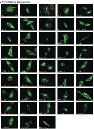
[image:3.585.141.451.68.492.2]Cellular localization map of all HHV-8 proteins.
The coding
sequences of all HHV-8-encoded genes except the ORF17.5
gene, which is a splice-variant of ORF17, were isolated, and an
immunological tag (Myc tag)-encoding sequence was fused in
frame at the 3
⬘
end of the coding sequences to allow
immu-nochemical detection of the respective proteins. The amplified
sequences were cloned into expression plasmids, confirmed by
full-length sequencing, and expressed in HeLa cells. HeLa cells
were used as a cell system for these studies because they exhibit
a larger cytoplasm than HEK 293 cells and are easier to
trans-fect than endothelial cells, which are the more commonly used
cell types of HHV-8 research.
FIG. 2. Subcellular localization of all HHV-8-encoded proteins. Myc-tagged HHV-8 proteins were expressed in HeLa cells, detected with a
specific antibody against the Myc tag, and categorized as cytoplasmic (A), nuclear (B), and both nuclear and cytoplasmic (C). Localization of
ORF73 (LANA-1) was detected with a specific antibody against LANA-1. As a negative control cells were transfected with the vector control
(pcDNA4-Myc/His) and also stained with an antibody against the Myc tag. The secondary antibody was conjugated with Alexa Fluor 488. Pictures
were obtained using an epifluorescence microscope. The bar in K1 represents 10
m. The same magnification was used in all panels.
on November 8, 2019 by guest
http://jvi.asm.org/
In order to determine whether the Myc tag affected cellular
localization, two cytoplasmic (K2 and K8.1) and one nuclear
(K10.5) HHV-8 proteins against which specific antibodies were
available were expressed with and without a Myc tag (Fig. 1A).
Subsequently, the different proteins were detected
immunocy-tochemically either with specific antibodies directed against the
different HHV-8 proteins or with an antibody against the Myc
tag. In all three cases untagged and Myc-tagged proteins
showed identical localizations, suggesting that the Myc tag
does not have significant effects on cellular localization of
HHV-8 proteins.
In addition, it has been reported that immunocytochemical
detection of nuclear proteins is critically dependent on the
method used for cell permeabilization (22, 59). For this reason
we compared the performance of two different
permeabiliza-tion procedures (Triton X-100 and saponin) for the detecpermeabiliza-tion
of three different nuclear HHV-8 proteins (Fig. 1B and C).
Under both conditions each protein could be detected with
identical sensitivity regardless of which method was used for
permeabilization (Fig. 1B and C). In all further
immunocyto-chemical stainings, saponin treatment was used for
permeabi-lization.
[image:4.585.135.452.67.517.2]To investigate subcellular localization, expression
plas-mids encoding all 85 HHV-8 genes were transfected into
HeLa cells, and the proteins were detected by
immunocy-tochemical staining (Fig. 2). In order to obtain a general
overview of the subcellular localization of each protein,
epifluorescence microscopy was used. With this approach all
HHV-8 proteins could be clearly detected in numerous
transfected cells.
FIG. 2—Continued.
on November 8, 2019 by guest
http://jvi.asm.org/
TABLE 1. Subcellular localization of HHV8 encoded protein
aGeneb Alternative description or
product (designation)c
Position in the
genomed Reference Subcellular localization
Golgi or ER
localizatione
K1
fSignaling molecule K ITAM-signaling (KIS)
105–944
39, 72
Cytoplasmic heterogeneously
ORF4
Complement control protein (KCP)
1112–2764
78
Cytoplasmic heterogeneously
ORF6
Major single-stranded DNA binding protein
3179–6577
86
Nuclear
ORF7
Processing and transport protein (ICP18.5)
6594–8681
Nuclear and cytoplasmic
ORF8
Glycoprotein B (gB)
8665–11202
4, 63
Cytoplasmic heterogeneously
ER
ORF9
DNA polymerase
111329–14367
86
Cytoplasmic diffuse
ORF10
HVS homologue
14485–15741
Nuclear and cytoplasmic
ORF11
HVS homologue
15756–16979
Nuclear and cytoplasmic
K2*
Viral interleukin-6 (vIL-6)
c17227–17841
3, 62
Cytoplasmic heterogeneously
ORF2
Dihydrofolate reductase (DHFR)
c17887–18159
Nuclear and cytoplasmic
K3
fModulator of immune recognition 1 (vMIR1)
c18574–19542
16, 31, 73
Cytoplasmic granular
ORF70
Thymidylate synthase (TS)
c20023–21036
Cytoplasmic heterogeneously
K4
Macrophage inflammatory protein-II
(vMIP-II; vCCL-2)
c21480–21764
49
Cytoplasmic perinuclear, focally
enriched
Golgi
K4.1
vMIP-III; vCCL-3
c22117–22461
Cytoplasmic perinuclear, focally
enriched
Golgi
K4.2
fc22530–23078
31
Cytoplasmic diffuse
K5
vMIR2
c25865–26635
29, 31
Cytoplasmic heterogeneously
ER
K6
vMIP-I; vCCL-1
c27289–27576
49
Cytoplasmic perinuclear, focally
enriched
K7
Viral inhibitor of apoptosis (vIAP)
28774–29154
20, 84
Cytoplasmic heterogeneously
ORF16
Viral B-cell-lymphoma 2 (v-Bcl-2)
30242–30769
56
Nuclear and cytoplasmic
ORF17
Capsid assembly protein, protease
30857–32524
Nuclear
ORF18
HVS homologue
32523–33296
Cytoplasmic granular
ORF19
Virion/tegument protein
c33293–34942
Nuclear and cytoplasmic
ORF20
HVS homologous, fusion protein
c34710–35672
Nuclear
ORF21
Thymidine kinase (TK)
35482–37224
23
Cytoplasmic perinuclear focally
enriched
Golgi
ORF22
Glycoprotein H (gH)
37212–39404
50
Cytoplasmic heterogeneously
ER
ORF23
HVS homologue
c39401–40615
Cytoplasmic perinuclear, focally
enriched
ORF24
HVS homologue
c40619–42877
Nuclear and cytoplasmic
ORF25
Major capsid protein
42876–47006
Cytoplasmic diffuse
ORF26
fMinor capsid protein
47032–47949
34, 58
Nuclear and cytoplasmic
ORF27
HVS homologue
47973–48845
Cytoplasmic heterogeneously
ORF28
HVS homologous glycoprotein
49091–49399
Cytoplasmic perinuclear, focally
enriched
Golgi
ORF29
gDNA packaging protein, terminase
c49462–50604
⫹
53855–54775
Nuclear and cytoplasmic
ORF30
HVS homologue
50723–50956
Cytoplasmic diffuse
ORF31
HVS homologue
50863–51537
Cytoplasmic granular
ORF32
HVS homologue
51504–52868
Nuclear and cytoplasmic
ORF33
HVS homologue
52861–53865
Nuclear
ORF34
HVS homologue
54774–55757
Nuclear
ORF35
HVS homologue
55738–56190
Nuclear and cytoplasmic
ORF36
Serine protein kinase, phosphotransferase
56075–57409
60
Nuclear
ORF37
Alkaline DNA-exonuclease shutoff and
exonuclease (SOX)
57372–58832
24
Nuclear
ORF38
Myristylated tegument protein EHV-2
homologue
58787–58972
Cytoplasmic granular
ORF39
Glycoprotein M (gM), integral membrane
protein
59072–60274
Cytoplasmic perinuclear, focally
enriched
ER
ORF40/41
hDNA helicase-primase complex component
60407–61756
⫹
61884–62543
86
Cuclear and cytoplasmic
ORF42
HVS homologue
c62535–63371
Nuclear and cytoplasmic
ORF43
Minor capsid protein
c63235–65052
Cytoplasmic granular
Golgi
ORF44
DNA replication protein (helicase/primase
subunit)
64991–67357
86
Cytoplasmic diffuse
ORF45
KSHV-immediate-early-2 (KIE-2)
c67452–68675
89, 90
Cytoplasmic diffuse
ORF46
Uracil DNA glucosidase
c68736–69503
Nuclear and cytoplasmic
ORF47
Glycoprotein L (gL)
c69511–70014
Cytoplasmic heterogeneously
ER
ORF48
HVS homologue
c70272–71480
Nuclear and cytoplasmic
ORF49
HVS homologue
c71728–72637
27
Nuclear
ORF50
gReplication and transcription activator
(RTA)
71695–71712
⫹
72671–74728
11, 28, 42, 57
Nuclear
K8
hK-basic leucin zipper/replication-associated
protein (K-bZIP/RAP)
74949–75662
⫹
75744–75890
14, 34, 65, 86
Nuclear
Continued on following page
on November 8, 2019 by guest
http://jvi.asm.org/
TABLE 1—Continued
Geneb Alternative description or
product (designation)c
Position in the
genomed Reference Subcellular localization
Golgi or ER
localizatione
K8.1 beta
g*
Glycoprotein (gp35-37)
76014–76437
⫹
76532–76794
34, 40, 42, 46,
87, 91, 92
Cytoplasmic heterogeneously
ORF52
HVS homologue
c76901–77296
Cytoplasmic perinuclear, focally
enriched
ORF53
HVS homologue
c77432–77764
Cytoplasmic perinuclear, focally
enriched
Golgi
ORF54
dUTPase homologue
77835–78722
Nuclear
ORF55
HVS homologue
c78864–79547
Cytoplasmic perinuclear, focally
enriched
Golgi
ORF56
DNA replication protein (helicase/primase
subunit)
79535–82066
86
Cytoplasmic diffuse
ORF57
hImmediate-early protein (MTA)
82169–82217
⫹
82326–83644
5, 36, 43, 55,
86
Nuclear
K9
vIRF-1
c83960–85309
61, 75
Cuclear and cytoplasmic
K10
hvIRF-4
c86174–88442,
88544–89010
32, 34
Nuclear
K10.5
g*
vIRF-3; latency-associated nuclear antigen-2
(LANA-2)
c89700–90945,
91042–91496
1, 47, 69
Nuclear
K11
f,gvIRF-2
c92066–93620,
93742–94229
34
Nuclear and cytoplasmic
ORF58
HVS homologue
c94577–95650
Cytoplasmic perinuclear, focally
enriched
Golgi
ORF59
DNA polymerase processivity factor (PF-8)
c95655–96845
8, 21, 33, 34,
46, 86, 92
Nuclear
ORF60
Ribonucleotide reductase small subunit
homologue
c96976–97893
Nuclear and cytoplasmic
ORF61
Ribonucleotide reductase large subunit
homologue
c97922–100300
Cytoplasmic granular
ORF62
Capsid assembly and DNA maturation
protein
c100305–101300
Nuclear
ORF63
Tegument protein
101314–104100
Nuclear and cytoplasmic
ORF64
Large tegument protein
104106–112013
Cytoplasmic heterogeneously
ORF65
fCapsid protein
c112037–112549
34
Nuclear and cytoplasmic
ORF66
HVS homologue
c112576–113865
Nuclear and cytoplasmic
ORF67
Tegument protein
c113799–114614
Cytoplasmic perinucelar, focally
enriched
Golgi
ORF67.5
EHV-2 ORF67A homologue
c114669–114911
Cytoplasmic granular
ORF68
Major envelope glycoprotein
114874–116511
Cytoplasmic diffuse
ORF69
HVS homologue
116544–117452
Nuclear
K12
Kaposin (virus structure protein, T0.7)
c118025–118207
48
Cytoplasmic heterogeneously
K13
FLICE-inhibitory protein cellular
homologue (vFLIP)
c122393–122959
Nuclear and cytoplasmic
ORF72
Viral cyclin (v-cyc)
c123042–123815
26
Nuclear
ORF73**
Latency associated nuclear antigen 1
(LANA-1)
c124057–127446
17, 34, 35, 64,
66
Nuclear
K14
OX-2 membrane-glycoprotein homologue
(vOX-2)
128264–129079
Cytoplasmic diffuse
ORF74
Viral G protein coupled receptor (vGPCR)
129520–130548
12
Cytoplasmic heterogeneously
Golgi
ORF75
Tegument protein,
phosphoribosylfor-mylglycineamide amidotransferase
homologue (FGARAT)
c130699–134589
Nuclear and cytoplasmic
K15
f,gLatency-associated membrane protein
(LAMP)
c134824–135287,
135373–135474,
135557–135664,
135747–135889,
135977–136066,
136155–136397,
136481–136573,
136683–136899
13, 25, 76
Cytoplasmic perinuclear, focally
enriched
a
Subcellular localization of all proteins was determined with an antibody against the Myc tag except where indicated.
b
Order according to the 5⬘–3⬘appearance in the HHV-8 genome. *, subcellular localization of proteins was detected with an antibody against the Myc tag and in
addition with a specific antibody directed against the protein; **, subcellular localization of LANA-1 was determined with a specific antibody against LANA-1.
c
HVS, herpesvirus samiri; EHV-2, equine herpesvirus 2; vIRF, viral interferon regulatory factor.
d
Position numbers are according to GenBank accession number AF148805 (25, 68). c, coding sequence complementary.
e
Colocalization with the marker GM130 or calnexin for the Golgi or ER, respectively.
f
There were slight differences between our findings and previous reports. ORF26 was detected only in the cytoplasm, and K11 and ORF65 were detected only in the nucleus by others. K1, K3, and K4.2 were found in the ER or Golgi by other investigators.
g
Spliced genes isolated from cDNA.
h
Spliced genes expressed via isolated genomic DNA.
on November 8, 2019 by guest
http://jvi.asm.org/
According to their localization, the proteins could be
classi-fied into three groups: those with cytoplasmic localization
(51%) (Fig. 2A) or nuclear localization (22%) (Fig. 2B) and
those which were localized in both the cytoplasm and the
nucleus (27%) (Fig. 2C). Proteins with purely cytoplasmic
lo-calization were further subcategorized into four groups:
cyto-plasmic granular (16%); cytocyto-plasmic perinuclear, focally
en-riched (30%); cytoplasmic diffuse (21%); and cytoplasmic
heterogeneous (33%). A summary of all results is presented in
Table 1 and Fig. 3. Graphical depiction showed that nuclear
proteins are mainly encoded by genes in the second half of the
viral genome (Fig. 3, red).
[image:7.585.43.542.68.479.2]We further analyzed whether nuclear proteins (Fig. 2B and C)
exhibit an NLS using the prediction algorithm PredictNLS (15).
In this study an NLS was detected in K11 in addition to the
proteins K10.5, ORF37, ORF50, ORF57, and ORF73, in which
an NLS has been detected previously by other investigators (Table
2). All of these proteins were detected in the nucleus in our study.
[image:7.585.42.284.614.707.2]FIG. 3. Gene map and intracellular localization of HHV-8 proteins. Protein coding regions are indicated by colored arrows, and gene names
are given. Orientations of the arrows indicate the transcriptional orientations. Genes are color coded as shown on the figure.
TABLE 2. Nuclear localization sequences of
HHV-8-encoded proteins
Protein NLS(s)a Amino acid
position Reference
K10.5 RRHERPTTRRIRHRKLRS 367–384 47
K11 KHREKALRRSLRKK 146–159 PredictNLS
ORF37 PRKKRKL 315–320 24
ORF50 KRKQRSKERSSKKRK 515–529 11
ORF57 RYGKKIK 101–107 43
KRPRRRPRDR 121–130
RAAPKRATRR 143–152
ORF73 RKRNRSP 24–30 64
aAmino acids are given in the single-letter code; ORF57 encodes three
dif-ferent NLSs.
on November 8, 2019 by guest
http://jvi.asm.org/
Golgi and ER localization of HHV-8 proteins.
Cytoplasmic
proteins with heterogeneous distribution, granular staining
patterns, or perinuclear enrichment may be associated with the
ER or the Golgi. In order to confirm the putative association
with these intracellular organelles, double staining
experi-ments of the HHV-8 proteins and the ER marker calnexin
(Fig. 4) or the Golgi marker GM130 (Fig. 5) were carried out
and analyzed by laser scanning microscopy. Colocalization with
calnexin confirmed ER localization of five proteins (K5,
ORF8, ORF22, ORF39, and ORF47) (Fig. 4 and Table 1).
Enrichment in the Golgi was observed for 10 proteins (K4,
K4.1, ORF21, ORF28, ORF43, ORF53, ORF55, ORF58,
ORF67, and ORF74) (Fig. 5 and Table 1), all of which
colo-calized with GM130. Of note, ER-associated proteins were
exclusively encoded in the first half of the viral genome (Fig. 3,
yellow), whereas the genes of Golgi-associated proteins were
randomly distributed (Fig. 3, green).
Nuclear localization of v-FLIP.
A surprising observation was
obtained with the K13 gene product. K13 encodes a viral
Fas-associated death domain-like interleukin-1

-converting
en-zyme-inhibitory protein (v-FLIP). Cellular FLIPs are
exclu-sively localized in the cytoplasm (44, 54). Unexpectedly, the
protein encoded by the K13 gene was localized in the
cyto-plasm and the nucleus (Fig. 6A, arrow). In order to determine
whether nuclear localization was due to the Myc tag, a
GFP-tagged K13 protein (K13-GFP) was expressed in HeLa cells
and detected by direct fluorescence analysis (Fig. 6B). In
ad-dition, a rat monoclonal K13-specific antibody was generated
and used to detect the localization of an untagged K13 protein
(Fig. 6C). All of these controls showed concordantly that K13
is resident in both the cytoplasm and the nucleus of the cell
(Fig. 6A to C, arrows). No signal was observed in a control
staining with only the secondary antibody (Fig. 6D). To
con-firm these results, we isolated nuclear and cytoplasmic
frac-tions of HeLa cells that expressed K13 with a Myc and a GFP
tag (Fig. 6E). Western blot analyses of the isolated cell
frac-tions clearly confirmed that both Myc-tagged (Fig. 6E, upper
panels) and GFP-tagged (Fig. 6E, lower panels) K13 proteins
are clearly present in the cytoplasm and in the nucleus. To
exclude the possibility that the Myc tag may affect the function
of K13, we compared a Myc-tagged and an untagged K13 in a
functional test. A major function of K13 is its capability to
activate the NF-
B pathway (10). In an NF-
B reporter test,
the Myc-tagged and the untagged K13 activated NF-
B at
comparable levels (Fig. 7).
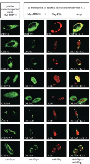
Effects of K10 binding partners on subcellular localization
of K10.
Recently it has been shown that K10 interacts with at
least 15 different HHV-8 proteins (K12, ORF2, ORF9,
ORF28, ORF29b, ORF31, ORF37, ORF39, ORF41, ORF47,
ORF59, ORF60, ORF61, ORF67.5, and ORF68) (82).
Ac-cording to Rezaee et al. (68) truncated forms of ORF29 and
ORF40/41 were used by Uetz et al. (82) in an interaction study
of all HHV-8 genes. In order to allow comparison of our
results with those of Uetz and colleagues, we also used the
truncated forms of ORF29 and ORF40/41 (ORF29b and
ORF41, respectively) for this study. We noticed that K10
lo-calized in the nucleus. In contrast, only five (ORF2, ORF37,
ORF41, ORF59, and ORF60) of the potential K10 interacting
proteins were also detected in the nucleus when they were
expressed alone in HeLa cells (Table 1 and Fig. 2). However,
coexpression of K10 with the putative interacting proteins
re-sulted in a clear colocalization in 13 cases (Table 3 and Fig. 8)
(K12, ORF2, ORF9, ORF29b, ORF31, ORF37, ORF39,
ORF41, ORF47, ORF59, ORF60, ORF67.5, and ORF68), as
detected in an analysis with the laser scanning microscope.
Colocalization was induced either by a change of K10
subcel-lular localization (K12, ORF9, ORF29b, ORF31, ORF39,
ORF47, and ORF67.5) or by a change of the localization of
both K10 and the putative binding protein (ORF41, ORF60,
and ORF68) (Fig. 8). K10 did not colocalize with ORF28 and
ORF61 in our experiments. To demonstrate that the
relocal-ization of K10 is not simply an artifact of overexpression, we
tested six different HHV-8 proteins (K3, K4, K5, K8, ORF38,
and ORF54) that did not interact with K10 (82). No
relocal-ization of any of the proteins was observed when they were
coexpressed with K10 (data not shown).
DISCUSSION
[image:8.585.44.284.71.419.2]We determined the intracellular localization of all
HHV-8-encoded proteins in mammalian cells. At present, antibodies
FIG. 4. ER localization of HHV-8 proteins. ER localization of
HHV-8 proteins was determined by costaining with an antibody
against calnexin. Colocalization was analyzed by confocal laser
scan-ning microscopy. The bar in K5 represents 10
m. The same
magni-fication was used in all panels.
on November 8, 2019 by guest
http://jvi.asm.org/
are available against only a few HHV-8-encoded proteins. In
order to allow immunocytochemical detection of the different
proteins, a tag was fused in frame at the 3
⬘
end of each coding
sequence. Several observations indicated that the tag did not
exert significant effects on subcellular localization of HHV-8
proteins. First, the cellular localization of three proteins (K2,
K8.1, and K10.5) against which specific antibodies were
avail-able was not affected by the tag. Second, the great majority
(82%) of the available published results on localization of
HHV-8 proteins were in clear agreement with our findings. Of
the 85 different HHV-8 proteins, 38 have been investigated by
others to our knowledge. Only in seven cases were slight
differences between previous findings and our study results
observed (Table 1). Specifically, ORF26 was detected only
in the cytoplasm, and K11 and ORF65 were found only in
the nucleus by other investigators, whereas in our study all
three proteins were detected in both the cytoplasm and the
nucleus (34, 58). In addition, K1, K3, K4.2, and K15 were
found in the ER or Golgi by others (16, 31, 39, 76). We also
detected each of these four proteins in the cytoplasm but
could not confirm colocalization with the respective
com-partment markers calnexin and GM130 (Table 1).
Alto-gether, a high concordance with the available published
results was observed, which clearly supported the validity of
the localization map described here.
It is of interest that 22% of the HHV-8-encoded proteins
were detected in the nucleus, whereas only 12% of randomly
selected cellular proteins showed nuclear localization (77).
Nu-clear preponderance of HHV-8-encoded proteins is in good
agreement with the viral life cycle, which is preferentially
as-sociated with the nucleus. Comparing protein localization with
the expression state during the viral life cycle, we noticed that
all latency-associated proteins showed a nuclear staining
pat-tern, whereas only 47% of primary lytic, 43% of secondary
lytic, and 43% of tertiary lytic proteins showed a nuclear
stain-ing pattern (37). This apparently is in agreement with the fact
that latency is a regulatory state, which may predominantly
depend on nuclear proteins to control host cell and viral
tran-scription.
ER and Golgi localization was observed for 17% of the
HHV-8 proteins. It is in clear agreement with these results that
many of the proteins were suspected to associate with these
organelles. The putative Golgi and/or ER proteins were the
following: (i) glycoproteins (gB/ORF8, gH/ORF22, gM/
ORF39, and gL/ORF47) (4, 50, 63), which are known to be
incorporated into the viral envelope, as well as ORF28, which
FIG. 5. Golgi localization of HHV-8 proteins. Golgi localization of HHV-8 proteins was determined by costaining with an antibody against
GM130. Colocalization was analyzed by confocal laser scanning microscopy. The bar in K4 represents 10
m. The same magnification was used
in all panels.
on November 8, 2019 by guest
http://jvi.asm.org/
reveals similarities to a glycoprotein from saimiriine
herpesvi-ruses (19); (ii) membrane-associated proteins such as viral
inter-leukin-8 receptor-like G protein-coupled receptor homolog
(vGPCR; ORF74), vMIR2, K5 (29, 31), and the tegument
pro-tein ORF67 with homology to the membrane-associated
phos-phoprotein LF2 from HHV-6; and (iii) chemokine-like
pro-teins (vMIP-II/K4 and vMIPIII/K4.1) which may be secreted
(79). Unexpected Golgi localization was observed with
thymi-dine kinase (ORF21) and the minor capsid protein (ORF43).
It remains to be determined in future studies whether this may
indicate membrane-associated and/or secretory functions of
these proteins.
Among the most surprising findings of our study was the
partial nuclear localization of v-FLIP/K13. Nuclear
localiza-tion is not observed for cellular FLIPs, which are resident
exclusively in the cytoplasm (44, 54). This indicates that
HHV-8-encoded v-FLIP may exert different and/or
addi-tional functions compared to cellular FLIPs. It is in line with
our finding that nuclear localization of other death effector
domain (DED)-containing molecules such as DEDD,
DEDD2, partially processed caspase-8, and the N-terminal
DED of caspase-8 (DEDa) has been detected recently and
associated with their regulatory function in apoptosis (2, 74,
88).
[image:10.585.42.282.71.219.2]We noticed that the nuclear K10 protein was identified
re-cently as a major interacting protein of HHV-8, which can bind
to at least 15 different HHV-8 proteins (82). Interestingly, only
33% (5 of 15) of the potential binding factors of K10 were
detected also in the nucleus when the proteins were expressed
alone. However, when K10 was coexpressed with its putative
binding factors, colocalization was observed in 87%. These
findings demonstrate that mutual protein interactions in an
infected cell affect subcellular localization. In agreement with
this, other investigators have described significant relocations
of HHV-8 proteins in latently rather than lytically infected
primary effusion lymphoma cells (34, 86). The localization map
of individually expressed HHV-8 proteins described here may
provide a useful reference to detect positional effects of other
HHV-8 proteins in virus-infected cells. In addition, the
intra-cellular localization map may provide a valuable platform to
further elucidate the function of HHV-8-encoded genes in
human diseases.
FIG. 6. Nuclear localization of v-FLIP/K13. K13 was expressed in
HeLa cells with a Myc tag (A), in fusion with GFP (B), and without a
tag (C). Recombinantly expressed K13 was detected with antibodies
against the Myc tag or against K13 or by direct epifluorescence. As a
control cells were transfected with pcDNA4-Myc/His and subjected to
immunocytochemical staining using the anti-K13 antibody (D).
Pic-tures were obtained using an epifluorescence microscope. The bar
shown in K13-Myc represents 10
m. The same magnification was
used in all panels. For cell fractionation experiments HeLa cells were
transfected with Myc-tagged or GFP-tagged K13, and nuclear and
cytoplasmic fractions were isolated and analyzed by Western blotting
(E). K13 proteins were detected with antibodies against the Myc or the
GFP tag.
[image:10.585.301.541.88.277.2]FIG. 7. NF-
B activation with Myc-tagged and untagged K13.
HeLa cells were cotransfected with reporter plasmid (pNF-
B-Luc; 1
g) and increasing concentrations of effector plasmids encoding
Myc-tagged or unMyc-tagged K13. Activation of the NF-
B promoter was
ana-lyzed by luciferase measurement 48 h after transfection. Values were
adjusted to total protein content, and the results are expressed in terms
of the relative increase in induction in comparison with the negative
control (0
g).
TABLE 3. Subcellular localization of K10 and putative K10
interacting proteins
Putative K10 interaction
partner
Localization change at
coexpressiona
Colocalization Interaction
partner K10
K12
⫺
⫹
Yes
ORF2
⫺
⫺
Yes
ORF9
⫺
⫹
Yes
ORF28
⫺
⫹
No
ORF29b
⫺
⫹
Yes
ORF31
⫺
⫹
Yes
ORF37
⫺
⫺
Yes
ORF39
⫺
⫹
Yes
ORF41
⫹
⫹
Yes
ORF47
⫺
⫹
Yes
ORF59
⫺
⫺
Yes
ORF60
⫹
⫹
Yes
ORF61
⫺
⫺
No
ORF67.5
⫺
⫹
Yes
ORF68
⫹
⫹
Yes
a⫹
, change in localization was observed;⫺, no change in localization was
observed.
on November 8, 2019 by guest
http://jvi.asm.org/
[image:10.585.54.275.468.650.2]FIG. 8. Colocalization of K10 and its putative interaction partners. A Flag-tagged K10 protein (Flag-K10) and different Myc-tagged HHV-8
proteins (Myc-HHV-8) were expressed in HeLa cells either alone or in combination (Myc-HHV-8
⫹
Flag-K10). The different proteins were
detected with antibodies directed against either the Myc tag or the Flag tag or against both epitopes simultaneously (bottom line). Colocalization
was analyzed by confocal laser scanning microscopy. The bar shown in K12 represents 10
m. The same magnification was used in all panels.
on November 8, 2019 by guest
http://jvi.asm.org/
ACKNOWLEDGMENTS
We thank Michael Bauer and Elisabeth Naschberger for help with
cloning, Mahimaidos Manoharan for excellent technical assistance,
and Susanne Reed for help in writing the report (all from the Division
of Molecular and Experimental Surgery). The generous support of
Werner Hohenberger (Director of the Department of Surgery) is
gratefully acknowledged.
This work was supported by grants from the Deutsche
Forschungs-gemeinschaft (DFG-SPP 1130, DFG-GK 1071, and DFG 317/2-1),
German Cancer Aid (Deutsche Krebshilfe,
Apoptose-Schwerpunkt-programm) and the Interdisciplinary Center for Clinical Research
(IZKF, project D8) of the University Hospital of the University of
Erlangen-Nuremberg to M.S., and a tandem-project grant of the IZKF
(project B11) to M.S. and F.N.
REFERENCES
1.Ablashi, D. V., L. G. Chatlynne, J. E. Whitman, Jr., and E. Cesarman.2002. Spectrum of Kaposi’s sarcoma-associated herpesvirus, or human herpesvirus
[image:12.585.144.442.63.604.2]8, diseases. Clin. Microbiol. Rev.15:439–464.
FIG. 8—Continued.
on November 8, 2019 by guest
http://jvi.asm.org/
2.Alcivar, A., S. Hu, J. Tang, and X. Yang.2003. DEDD and DEDD2 associate
with caspase-8/10 and signal cell death. Oncogene22:291–297.
3.Aoki, Y., E. S. Jaffe, Y. Chang, K. Jones, J. Teruya-Feldstein, P. S. Moore, and G. Tosato.1999. Angiogenesis and hematopoiesis induced by Kaposi’s
sarcoma-associated herpesvirus-encoded interleukin-6. Blood93:4034–4043.
4.Baghian, A., M. Luftig, J. B. Black, Y. X. Meng, C. P. Pau, T. Voss, P. E. Pellett, and K. G. Kousoulas.2000. Glycoprotein B of human herpesvirus 8 is a component of the virion in a cleaved form composed of amino- and
carboxyl-terminal fragments. Virology269:18–25.
5.Bello, L. J., A. J. Davison, M. A. Glenn, A. Whitehouse, N. Rethmeier, T. F. Schulz, and J. Barklie Clements.1999. The human herpesvirus-8 ORF 57
gene and its properties. J. Gen. Virol.80:3207–3215.
6.Brinkmann, M. M., M. Glenn, L. Rainbow, A. Kieser, C. Henke-Gendo, and T. F. Schulz. 2003. Activation of mitogen-activated protein kinase and
NF-B pathways by a Kaposi’s sarcoma-associated herpesvirus K15
mem-brane protein. J. Virol.77:9346–9358.
7.Cesarman, E., Y. Chang, P. S. Moore, J. W. Said, and D. M. Knowles.1995. Kaposi’s sarcoma-associated herpesvirus-like DNA sequences in
AIDS-re-lated body-cavity-based lymphomas. N. Engl. J. Med.332:1186–1191.
8.Chan, S. R., C. Bloomer, and B. Chandran.1998. Identification and char-acterization of human herpesvirus-8 lytic cycle-associated ORF 59 protein
and the encoding cDNA by monoclonal antibody. Virology240:118–126.
9.Chang, Y., E. Cesarman, M. S. Pessin, F. Lee, J. Culpepper, D. M. Knowles, and P. S. Moore.1994. Identification of herpesvirus-like DNA sequences in
AIDS-associated Kaposi’s sarcoma. Science266:1865–1869.
10.Chaudhary, P. M., A. Jasmin, M. T. Eby, and L. Hood.1999. Modulation of the NF-kappa B pathway by virally encoded death effector
domains-contain-ing proteins. Oncogene18:5738–5746.
11.Chen, J., K. Ueda, S. Sakakibara, T. Okuno, and K. Yamanishi.2000. Transcriptional regulation of the Kaposi’s sarcoma-associated herpesvirus
viral interferon regulatory factor gene. J. Virol.74:8623–8634.
12.Chiou, C. J., L. J. Poole, P. S. Kim, D. M. Ciufo, J. S. Cannon, C. M. ap Rhys, D. J. Alcendor, J. C. Zong, R. F. Ambinder, and G. S. Hayward.2002. Patterns of gene expression and a transactivation function exhibited by the vGCR (ORF74) chemokine receptor protein of Kaposi’s sarcoma-associated
herpesvirus. J. Virol.76:3421–3439.
13.Choi, J. K., B. S. Lee, S. N. Shim, M. Li, and J. U. Jung.2000. Identification of the novel K15 gene at the rightmost end of the Kaposi’s
sarcoma-associ-ated herpesvirus genome. J. Virol.74:436–446.
14.Ciufo, D. M., J. S. Cannon, L. J. Poole, F. Y. Wu, P. Murray, R. F. Ambinder, and G. S. Hayward.2001. Spindle cell conversion by Kaposi’s sarcoma-associated herpesvirus: formation of colonies and plaques with mixed lytic and latent gene expression in infected primary dermal microvascular
endo-thelial cell cultures. J. Virol.75:5614–5626.
15.Cokol, M., R. Nair, and B. Rost.2000. Finding nuclear localization signals.
EMBO Rep.1:411–415.
16.Coscoy, L., and D. Ganem.2000. Kaposi’s sarcoma-associated herpesvirus encodes two proteins that block cell surface display of MHC class I chains by
enhancing their endocytosis. Proc. Natl. Acad. Sci. USA97:8051–8056.
17.Dupin, N., C. Fisher, P. Kellam, S. Ariad, M. Tulliez, N. Franck, E. van Marck, D. Salmon, I. Gorin, J. P. Escande, R. A. Weiss, K. Alitalo, and C. Boshoff.1999. Distribution of human herpesvirus-8 latently infected cells in Kaposi’s sarcoma, multicentric Castleman’s disease, and primary effusion
lymphoma. Proc. Natl. Acad. Sci. USA96:4546–4551.
18.Ensoli, B., M. Stu¨rzl, and P. Monini.2001. Reactivation and role of HHV-8
in Kaposi’s sarcoma initiation. Adv. Cancer Res.81:161–200.
19.Ensser, A., M. Thurau, S. Wittmann, and H. Fickenscher.2003. The genome of herpesvirus saimiri C488 which is capable of transforming human T cells.
Virology314:471–487.
20.Feng, P., J. Park, B. S. Lee, S. H. Lee, R. J. Bram, and J. U. Jung.2002. Kaposi’s sarcoma-associated herpesvirus mitochondrial K7 protein targets a cellular calcium-modulating cyclophilin ligand to modulate intracellular
cal-cium concentration and inhibit apoptosis. J. Virol.76:11491–11504.
21.Flore, O., S. Rafii, S. Ely, J. J. O’Leary, E. M. Hyjek, and E. Cesarman.1998. Transformation of primary human endothelial cells by Kaposi’s
sarcoma-associated herpesvirus. Nature394:588–592.
22.Frisch, S.2004. Nuclear localization of FADD protein. Cell Death Differ.
11:1361–1362; author reply 1362–1364.
23.Gill, M. B., J. E. Murphy, and J. D. Fingeroth.2005. Functional divergence of Kaposi’s sarcoma-associated herpesvirus and related gamma-2 herpesvi-rus thymidine kinases: novel cytoplasmic phosphoproteins that alter cellular
morphology and disrupt adhesion. J. Virol.79:14647–14659.
24.Glaunsinger, B., L. Chavez, and D. Ganem.2005. The exonuclease and host shutoff functions of the SOX protein of Kaposi’s sarcoma-associated
her-pesvirus are genetically separable. J. Virol.79:7396–7401.
25.Glenn, M., L. Rainbow, F. Aurade, A. Davison, and T. F. Schulz.1999. Identification of a spliced gene from Kaposi’s sarcoma-associated herpesvi-rus encoding a protein with similarities to latent membrane proteins 1 and
2A of Epstein-Barr virus. J. Virol.73:6953–6963.
26.Godden-Kent, D., S. J. Talbot, C. Boshoff, Y. Chang, P. Moore, R. A. Weiss, and S. Mittnacht.1997. The cyclin encoded by Kaposi’s sarcoma-associated
herpesvirus stimulates cdk6 to phosphorylate the retinoblastoma protein and
histone H1. J. Virol.71:4193–4198.
27.Gonzalez, C. M., E. L. Wong, B. S. Bowser, G. K. Hong, S. Kenney, and B. Damania.2006. Identification and characterization of the Orf49 protein of
Kaposi’s sarcoma-associated herpesvirus. J. Virol.80:3062–3070.
28.Gwack, Y., H. Byun, S. Hwang, C. Lim, and J. Choe.2001. CREB-binding protein and histone deacetylase regulate the transcriptional activity of Kaposi’s sarcoma-associated herpesvirus open reading frame 50. J. Virol.
75:1909–1917.
29.Haque, M., J. Chen, K. Ueda, Y. Mori, K. Nakano, Y. Hirata, S. Kanamori, Y. Uchiyama, R. Inagi, T. Okuno, and K. Yamanishi.2000. Identification and analysis of the K5 gene of Kaposi’s sarcoma-associated herpesvirus. J. Virol.
74:2867–2875.
30.Huh, W. K., J. V. Falvo, L. C. Gerke, A. S. Carroll, R. W. Howson, J. S. Weissman, and E. K. O’Shea.2003. Global analysis of protein localization in
budding yeast. Nature425:686–691.
31.Ishido, S., C. Wang, B. S. Lee, G. B. Cohen, and J. U. Jung.2000. Down-regulation of major histocompatibility complex class I molecules by Kaposi’s
sarcoma-associated herpesvirus K3 and K5 proteins. J. Virol.74:5300–5309.
32.Kanno, T., Y. Sato, T. Sata, and H. Katano.2006. Expression of Kaposi’s sarcoma-associated herpesvirus-encoded K10/10.1 protein in tissues and its
interaction with poly(A)-binding protein. Virology352:100–109.
33.Katano, H., T. Sata, T. Suda, T. Nakamura, N. Tachikawa, H. Nishizumi, S. Sakurada, Y. Hayashi, M. Koike, A. Iwamoto, T. Kurata, and S. Mori.1999. Expression and antigenicity of human herpesvirus 8 encoded ORF59 protein
in AIDS-associated Kaposi’s sarcoma. J. Med. Virol.59:346–355.
34.Katano, H., Y. Sato, T. Kurata, S. Mori, and T. Sata.2000. Expression and localization of human herpesvirus 8-encoded proteins in primary effusion lymphoma, Kaposi’s sarcoma, and multicentric Castleman’s disease.
Virol-ogy269:335–344.
35.Kedes, D. H., M. Lagunoff, R. Renne, and D. Ganem.1997. Identification of the gene encoding the major latency-associated nuclear antigen of the
Kaposi’s sarcoma-associated herpesvirus. J. Clin. Investig.100:2606–2610.
36.Kirshner, J. R., D. M. Lukac, J. Chang, and D. Ganem.2000. Kaposi’s sarcoma-associated herpesvirus open reading frame 57 encodes a
post-transcriptional regulator with multiple distinct activities. J. Virol. 74:
3586–3597.
37.Krishnan, H. H., P. P. Naranatt, M. S. Smith, L. Zeng, C. Bloomer, and B. Chandran.2004. Concurrent expression of latent and a limited number of lytic genes with immune modulation and antiapoptotic function by Kaposi’s sarcoma-associated herpesvirus early during infection of primary endothelial and fibroblast cells and subsequent decline of lytic gene expression. J. Virol.
78:3601–3620.
38.Lagunoff, M., and D. Ganem.1997. The structure and coding organization of the genomic termini of Kaposi’s sarcoma-associated herpesvirus. Virology
236:147–154.
39.Lee, B. S., X. Alvarez, S. Ishido, A. A. Lackner, and J. U. Jung.2000. Inhibition of intracellular transport of B cell antigen receptor complexes by
Kaposi’s sarcoma-associated herpesvirus K1. J. Exp. Med.192:11–21.
40.Li, M., J. MacKey, S. C. Czajak, R. C. Desrosiers, A. A. Lackner, and J. U. Jung.1999. Identification and characterization of Kaposi’s
sarcoma-associ-ated herpesvirus K8.1 virion glycoprotein. J. Virol.73:1341–1349.
41.Lubeseder-Martellato, C., E. Guenzi, A. Jorg, K. Topolt, E. Naschberger, E. Kremmer, C. Zietz, E. Tschachler, P. Hutzler, M. Schwemmle, K. Matzen, T. Grimm, B. Ensoli, and M. Sturzl.2002. Guanylate-binding protein-1 expres-sion is selectively induced by inflammatory cytokines and is an activation marker of endothelial cells during inflammatory diseases. Am. J. Pathol.
161:1749–1759.
42.Lukac, D. M., R. Renne, J. R. Kirshner, and D. Ganem.1998. Reactivation of Kaposi’s sarcoma-associated herpesvirus infection from latency by expres-sion of the ORF 50 transactivator, a homolog of the EBV R protein.
Virol-ogy252:304–312.
43.Majerciak, V., K. Yamanegi, S. H. Nie, and Z. M. Zheng.2006. Structural and functional analyses of Kaposi sarcoma-associated herpesvirus ORF57
nuclear localization signals in living cells. J. Biol. Chem. 281:28365–
28378.
44.Mathas, S., A. Lietz, I. Anagnostopoulos, F. Hummel, B. Wiesner, M. Janz, F. Jundt, B. Hirsch, K. Johrens-Leder, H. P. Vornlocher, K. Bommert, H. Stein, and B. Dorken.2004. c-FLIP mediates resistance of
Hodgkin/Reed-Sternberg cells to death receptor-induced apoptosis. J. Exp. Med.199:1041–
1052.
45.Moore, P. S., and Y. Chang.1995. Detection of herpesvirus-like DNA se-quences in Kaposi’s sarcoma in patients with and without HIV infection.
N. Engl. J. Med.332:1181–1185.
46.Moses, A. V., K. N. Fish, R. Ruhl, P. P. Smith, J. G. Strussenberg, L. Zhu, B. Chandran, and J. A. Nelson.1999. Long-term infection and transforma-tion of dermal microvascular endothelial cells by human herpesvirus 8. J.
Vi-rol.73:6892–6902.
47.Munoz-Fontela, C., M. Collado, E. Rodriguez, M. A. Garcia, A. Alvarez-Barrientos, J. Arroyo, C. Nombela, and C. Rivas.2005. Identification of a nuclear export signal in the KSHV latent protein LANA2 mediating its
export from the nucleus. Exp. Cell Res.311:96–105.
on November 8, 2019 by guest
http://jvi.asm.org/
48.Muralidhar, S., A. M. Pumfery, M. Hassani, M. R. Sadaie, M. Kishishita, J. N. Brady, J. Doniger, P. Medveczky, and L. J. Rosenthal.1998. Identifi-cation of kaposin (open reading frame K12) as a human herpesvirus 8
(Kaposi’s sarcoma-associated herpesvirus) transforming gene. J. Virol.72:
4980–4988.
49.Nakano, K., Y. Isegawa, P. Zou, K. Tadagaki, R. Inagi, and K. Yamanishi.
2003. Kaposi’s sarcoma-associated herpesvirus (KSHV)-encoded vMIP-I and vMIP-II induce signal transduction and chemotaxis in monocytic cells.
Arch. Virol.148:871–890.
50.Naranatt, P. P., S. M. Akula, and B. Chandran.2002. Characterization of
gamma2-human herpesvirus-8 glycoproteins gH and gL. Arch. Virol.147:
1349–1370.
51.Naschberger, E., T. Werner, A. B. Vicente, E. Guenzi, K. Topolt, R. Leubert, C. Lubeseder-Martellato, P. J. Nelson, and M. Sturzl.2004. Nuclear factor-kappaB motif and interferon-alpha-stimulated response element co-operate in the activation of guanylate-binding protein-1 expression by inflammatory
cytokines in endothelial cells. Biochem. J.379:409–420.
52.Neipel, F., J. C. Albrecht, A. Ensser, Y. Q. Huang, J. J. Li, A. E. Friedman-Kien, and B. Fleckenstein.1997. Human herpesvirus 8 encodes a homolog of
interleukin-6. J. Virol.71:839–842.
53.Niedenthal, R. K., L. Riles, M. Johnston, and J. H. Hegemann.1996. Green fluorescent protein as a marker for gene expression and subcellular
localization in budding yeast. Yeast12:773–786.
54.Niikura, Y., T. Nonaka, and S. Imajoh-Ohmi.2002. Monitoring of caspase-8/ FLICE processing and activation upon Fas stimulation with novel antibodies directed against a cleavage site for caspase-8 and its substrate, FLICE-like
inhibitory protein (FLIP). J. Biochem. (Tokyo)132:53–62.
55.Nishimura, K., K. Ueda, E. Guwanan, S. Sakakibara, E. Do, E. Osaki, K. Yada, T. Okuno, and K. Yamanishi.2004. A posttranscriptional regulator of Kaposi’s sarcoma-associated herpesvirus interacts with RNA-binding protein
PCBP1 and controls gene expression through the IRES. Virology325:364–
378.
56.Ojala, P. M., K. Yamamoto, E. Castanos-Velez, P. Biberfeld, S. J. Korsmeyer, and T. P. Makela.2000. The apoptotic v-cyclin-CDK6 complex
phosphorylates and inactivates Bcl-2. Nat. Cell Biol.2:819–825.
57.Okuno, T., Y. B. Jiang, K. Ueda, K. Nishimura, T. Tamura, and K. Yamanishi.
2002. Activation of human herpesvirus 8 open reading frame K5
indepen-dent of ORF50 expression. Virus Res.90:77–89.
58.O’Neill, E., J. L. Douglas, M. L. Chien, and J. V. Garcia.1997. Open reading frame 26 of human herpesvirus 8 encodes a tetradecanoyl phorbol acetate-and butyrate-inducible 32-kilodalton protein expressed in a body
cavity-based lymphoma cell line. J. Virol.71:4791–4797.
59.O’Reilly, L. A., U. Divisekera, K. Newton, K. Scalzo, T. Kataoka, H. Puthalakath, M. Ito, D. C. Huang, and A. Strasser.2004. Modifications and intracellular trafficking of FADD/MORT1 and caspase-8 after stimulation of
T lymphocytes. Cell Death Differ.11:724–736.
60.Park, J., D. Lee, T. Seo, J. Chung, and J. Choe.2000. Kaposi’s sarcoma-associated herpesvirus (human herpesvirus-8) open reading frame 36 protein
is a serine protein kinase. J. Gen. Virol.81:1067–1071.
61.Parravicini, C., B. Chandran, M. Corbellino, E. Berti, M. Paulli, P. S. Moore, and Y. Chang.2000. Differential viral protein expression in Kaposi’s sarcoma-associated herpesvirus-infected diseases: Kaposi’s sarcoma, primary effusion lymphoma, and multicentric Castleman’s disease. Am. J. Pathol.
156:743–749.
62.Parravicini, C., M. Corbellino, M. Paulli, U. Magrini, M. Lazzarino, P. S. Moore, and Y. Chang.1997. Expression of a virus-derived cytokine, KSHV
vIL-6, in HIV-seronegative Castleman’s disease. Am. J. Pathol.151:1517–
1522.
63.Pertel, P. E., P. G. Spear, and R. Longnecker.1998. Human herpesvirus-8 glycoprotein B interacts with Epstein-Barr virus (EBV) glycoprotein 110
but fails to complement the infectivity of EBV mutants. Virology251:
402–413.
64.Piolot, T., M. Tramier, M. Coppey, J. C. Nicolas, and V. Marechal.2001. Close but distinct regions of human herpesvirus 8 latency-associated nuclear antigen 1 are responsible for nuclear targeting and binding to human mitotic
chromosomes. J. Virol.75:3948–3959.
65.Portes-Sentis, S., E. Manet, G. Gourru, A. Sergeant, and H. Gruffat.2001. Identification of a short amino acid sequence essential for efficient nuclear targeting of the Kaposi’s sarcoma-associated herpesvirus/human
herpesvi-rus-8 K8 protein. J. Gen. Virol.82:507–512.
66.Rainbow, L., G. M. Platt, G. R. Simpson, R. Sarid, S. J. Gao, H. Stoiber, C. S. Herrington, P. S. Moore, and T. F. Schulz. 1997. The 222- to 234-kilodalton latent nuclear protein (LNA) of Kaposi’s sarcoma-associ-ated herpesvirus (human herpesvirus 8) is encoded by orf73 and is a
component of the latency-associated nuclear antigen. J. Virol.71:5915–
5921.
67.Renne, R., M. Lagunoff, W. Zhong, and D. Ganem.1996. The size and conformation of Kaposi’s sarcoma-associated herpesvirus (human
herpesvi-rus 8) DNA in infected cells and virions. J. Virol.70:8151–8154.
68.Rezaee, S. A., C. Cunningham, A. J. Davison, and D. J. Blackbourn.2006. Kaposi’s sarcoma-associated herpesvirus immune modulation: an overview.
J. Gen. Virol.87:1781–1804.
69.Rivas, C., A. E. Thlick, C. Parravicini, P. S. Moore, and Y. Chang.2001. Kaposi’s sarcoma-associated herpesvirus LANA2 is a B-cell-specific latent
viral protein that inhibits p53. J. Virol.75:429–438.
70.Ross-Macdonald, P., P. S. Coelho, T. Roemer, S. Agarwal, A. Kumar, R. Jansen, K. H. Cheung, A. Sheehan, D. Symoniatis, L. Umansky, M. Heidtman, F. K. Nelson, H. Iwasaki, K. Hager, M. Gerstein, P. Miller, G. S. Roeder, and M. Snyder.1999. Large-scale analysis of the yeast genome by
transposon tagging and gene disruption. Nature402:413–418.
71.Russo, J. J., R. A. Bohenzky, M. C. Chien, J. Chen, M. Yan, D. Maddalena, J. P. Parry, D. Peruzzi, I. S. Edelman, Y. Chang, and P. S. Moore.1996. Nucleotide sequence of the Kaposi sarcoma-associated herpesvirus (HHV8).
Proc. Natl. Acad. Sci. USA93:14862–14867.
72.Samaniego, F., S. Pati, J. E. Karp, O. Prakash, and D. Bose.2001. Human herpesvirus 8 K1-associated nuclear factor-kappa B-dependent promoter activity: role in Kaposi’s sarcoma inflammation? J. Natl. Cancer Inst.
Monogr.2001:15–23.
73.Sanchez, D. J., L. Coscoy, and D. Ganem.2002. Functional organization of
MIR2, a novel viral regulator of selective endocytosis. J. Biol. Chem.277:
6124–6130.
74.Schickling, O., A. H. Stegh, J. Byrd, and M. E. Peter.2001. Nuclear local-ization of DEDD leads to caspase-6 activation through its death effector domain and inhibition of RNA polymerase I dependent transcription. Cell
Death Differ.8:1157–1168.
75.Seo, T., D. Lee, Y. S. Shim, J. E. Angell, N. V. Chidambaram, D. V. Kalva-kolanu, and J. Choe.2002. Viral interferon regulatory factor 1 of Kaposi’s sarcoma-associated herpesvirus interacts with a cell death regulator, GRIM19, and inhibits interferon/retinoic acid-induced cell death. J. Virol.
76:8797–8807.
76.Sharp, T. V., H. W. Wang, A. Koumi, D. Hollyman, Y. Endo, H. Ye, M. Q. Du, and C. Boshoff.2002. K15 protein of Kaposi’s sarcoma-associated herpesvi-rus is latently expressed and binds to HAX-1, a protein with antiapoptotic
function. J. Virol.76:802–816.
77.Simpson, J. C., R. Wellenreuther, A. Poustka, R. Pepperkok, and S. Wiemann.2000. Systematic subcellular localization of novel proteins
iden-tified by large-scale cDNA sequencing. EMBO Rep.1:287–292.
78.Spiller, O. B., M. Robinson, E. O’Donnell, S. Milligan, B. P. Morgan, A. J. Davison, and D. J. Blackbourn.2003. Complement regulation by Kaposi’s
sarcoma-associated herpesvirus ORF4 protein. J. Virol.77:592–599.
79.Stine, J. T., C. Wood, M. Hill, A. Epp, C. J. Raport, V. L. Schweickart, Y. Endo, T. Sasaki, G. Simmons, C. Boshoff, P. Clapham, Y. Chang, P. Moore, P. W. Gray, and D. Chantry.2000. KSHV-encoded CC chemokine vMIP-III is a CCR4 agonist, stimulates angiogenesis, and selectively chemoattracts
TH2 cells. Blood95:1151–1157.
80.Stu¨rzl, M., C. Hohenadl, C. Zietz, E. Castanos-Velez, A. Wunderlich, G. Ascherl, P. Biberfeld, P. Monini, P. J. Browning, and B. Ensoli.1999. Expression of K13/v-FLIP gene of human herpesvirus 8 and apoptosis in
Kaposi’s sarcoma spindle cells. J. Natl. Cancer Inst.91:1725–1733.
81.Stu¨rzl, M., C. Zietz, P. Monini, and B. Ensoli.2001. Human herpesvirus-8 and Kaposi’s sarcoma: relationship with the multistep concept of
tumorigen-esis. Adv. Cancer Res.81:125–159.
82.Uetz, P., Y. A. Dong, C. Zeretzke, C. Atzler, A. Baiker, B. Berger, S. V. Rajagopala, M. Roupelieva, D. Rose, E. Fossum, and J. Haas.2006. Her-pesviral protein networks and their interaction with the human proteome.
Science311:239–242.
83.Vieira, J., and P. M. O’Hearn.2004. Use of the red fluorescent protein as a marker of Kaposi’s sarcoma-associated herpesvirus lytic gene expression.
Virology325:225–240.
84.Wang, H. W., T. V. Sharp, A. Koumi, G. Koentges, and C. Boshoff.2002. Characterization of an anti-apoptotic glycoprotein encoded by Kaposi’s sar-coma-associated herpesvirus which resembles a spliced variant of human
survivin. EMBO J.21:2602–2615.
85.Wiwatwattana, N., and A. Kumar.2005. Organelle DB: a cross-species
da-tabase of protein localization and function. Nucleic Acids Res.33:D598–
D604.
86.Wu, F. Y., J. H. Ahn, D. J. Alcendor, W. J. Jang, J. Xiao, S. D. Hayward, and G. S. Hayward.2001. Origin-independent assembly of Kaposi’s sarcoma-associated herpesvirus DNA replication compartments in transient cotrans-fection assays and association with the ORF-K8 protein and cellular PML.
J. Virol.75:1487–1506.
87.Wu, L., R. Renne, D. Ganem, and B. Forghani.2000. Human herpesvirus 8 glycoprotein K8.1: expression, post-translational modification and
localiza-tion analyzed by monoclonal antibody. J. Clin. Virol.17:127–136.
88.Yao, Z., S. Duan, D. Hou, K. Heese, and M. Wu.2007. Death effector domain DEDa, a self-cleaved product of caspase-8/Mch5, translocates to the nucleus by binding to ERK1/2 and upregulates procaspase-8 expression via a
p53-dependent mechanism. EMBO J.26:1068–1080.
89.Zhu, F. X., S. M. King, E. J. Smith, D. E. Levy, and Y. Yuan.2002. A Kaposi’s
on November 8, 2019 by guest
http://jvi.asm.org/
sarcoma-associated herpesviral protein inhibits virus-mediated induction of type I interferon by blocking IRF-7 phosphorylation and nuclear accumulation.
Proc. Natl. Acad. Sci. USA99:5573–5578.
90.Zhu, F. X., and Y. Yuan.2003. The ORF45 protein of Kaposi’s
sarcoma-associated herpesvirus is sarcoma-associated with purified virions. J. Virol.77:4221–
4230.
91.Zhu, L., V. Puri, and B. Chandran.1999. Characterization of human
her-pesvirus-8 K8.1A/B glycoproteins by monoclonal antibodies. Virology262:
237–249.
92.Zoeteweij, J. P., S. T. Eyes, J. M. Orenstein, T. Kawamura, L. Wu, B. Chandran, B. Forghani, and A. Blauvelt.1999. Identification and rapid quantification of early- and late-lytic human herpesvirus 8 infection in single cells by flow cytometric analysis: characterization of antiherpesvirus agents.
J. Virol.73:5894–5902.
on November 8, 2019 by guest
http://jvi.asm.org/