in the Processing of
vpr
mRNA
Steffen Erkelenz,aGereon Poschmann,bStephan Theiss,aAnja Stefanski,bFrank Hillebrand,aMarianne Otte,aKai Stühler,b
Heiner Schaala
Institute for Virology, Medical Faculty, Heinrich Heine University Düsseldorf, Düsseldorf, Germanya
; Molecular Proteomics Laboratory, BMFZ, Universitätsklinikum Düsseldorf, Düsseldorf, Germanyb
Small noncoding HIV-1 leader exon 3 is defined by its splice sites A2 and D3. While 3
=
splice site (3
=
ss) A2 needs to be activated
for
vpr
mRNA formation, the location of the
vpr
start codon within downstream intron 3 requires silencing of splicing at 5
=
ss
D3. Here we show that the inclusion of both HIV-1 exon 3 and
vpr
mRNA processing is promoted by an exonic splicing enhancer
(ESE
vpr) localized between exonic splicing silencer ESSV and 5
=
ss D3. The ESE
vprsequence was found to be bound by members of
the Transformer 2 (Tra2) protein family. Coexpression of these proteins in provirus-transfected cells led to an increase in the
levels of exon 3 inclusion, confirming that they act through ESE
vpr. Further analyses revealed that ESE
vprsupports the binding of
U1 snRNA at 5
=
ss D3, allowing bridging interactions across the upstream exon with 3
=
ss A2. In line with this, an increase or
de-crease in the complementarity of 5
=
ss D3 to the 5
=
end of U1 snRNA was accompanied by a higher or lower
vpr
expression level.
Activation of 3
=
ss A2 through the proposed bridging interactions, however, was not dependent on the splicing competence of
5
=
ss D3 because rendering it splicing defective but still competent for efficient U1 snRNA binding maintained the enhancing
function of D3. Therefore, we propose that splicing at 3
=
ss A2 occurs temporally between the binding of U1 snRNA and splicing
at D3.
D
uring human immunodeficiency virus type 1 (HIV-1) long
terminal repeat (LTR)-driven transcription, RNA
polymer-ase II generates a pre-mRNA that encodes at least 15 viral proteins
(
1
). The preformed 43S ribosomal subunit recognizes the CAP
structure moving along its template until it encounters a
transla-tional start codon, defined by its surrounding sequence (
2
). Thus,
the position of the Gag and Gag/Pol open reading frames (ORFs)
proximal to the 5
=
end of the unspliced viral mRNA ensured their
efficient recognition. However, proper HIV-1 replication is
inti-mately connected to the expression of seven other ORFs located
distal to CAP that encode the viral proteins Vif, Vpr, Tat, Rev, Nef,
Vpu, and Env. Alternative splicing removes inhibitory upstream
AUGs, thereby placing downstream ORFs near the CAP structure
and allowing their efficient translation by the scanning ribosome.
The particular HIV-1 protein encoded by a spliced mRNA is
al-most always specified by the ORF that is immediately downstream
of the 3
=
splice site (3
=
ss) used to create the mRNA. The sole
exception is the
env
ORF within the bicistronic
vpu
-
env
mRNAs,
whose translation is dependent on a minimal upstream ORF
within the HIV-1
vpu
leader (
3
,
4
).
On the basis of their intron contents, three different-sized viral
mRNA classes can be defined: the unspliced (9-kb),
intron-con-taining (4-kb), and intronless (1.8-kb) viral RNAs (
Fig. 1A
) (
5
; for
a recent review, see reference
6
). The accumulation of these viral
mRNA classes occurs in a temporal order (
7
,
8
). In the early phase
of viral gene expression, the HIV-1 pre-mRNA is extensively
spliced, leading to intronless 1.8-kb mRNA species such as the
tat
,
rev
, and
nef
mRNAs. Rev is necessary for the onset of the late phase
of viral gene expression that is characterized by a shift within the
cytoplasmic mRNA pool toward isoforms with increased intron
content and in which the normal nuclear retention mechanisms
are bypassed (
9
). Rev recognizes an RNA secondary structure,
called the Rev-responsive element (RRE), within the
env
coding
sequence. Rev-RRE interactions target the intron-containing
(4-kb) and unspliced (9-(4-kb) viral mRNAs for CRM1 export receptor
pathway-mediated transport into the cytoplasm, which essentially
relies on the multimerization capacity of Rev (
10
). During the late
phase of viral gene expression, the accessory and structural
pro-teins Vif, Vpr, Vpu, and Env are translated from the respective
intron-containing viral mRNAs (4 kb). In addition, the unspliced
viral mRNA (9 kb) is used for translation of the structural and
enzymatic components or enclosed as genomic RNA in progeny
virions. Viral mRNA diversity is further increased by the
alterna-tive inclusion of either one or both of the two noncoding leader
exons, 2 and 3. Noncoding leader exon 3 is flanked by 3
=
ss A2 and
5
=
ss D3. The formation of intron-containing
vpr
mRNA, however,
requires the activation of 3
=
ss A2 but silencing of 5
=
ss D3, since the
ORF of Vpr starts within the downstream intron of exon 3. Thus,
vpr
mRNA processing and exon 3 inclusion are mutually
exclu-sive. Nevertheless, both splicing patterns are negatively regulated
by an exonic splicing silencer (ESSV) within exon 3 (
Fig. 1B
) (
11
–
13
). ESSV contains three (pyrimidine)UAG motifs which
pro-mote the binding of members of the hnRNP A/B protein family to
the viral mRNA, inhibiting splicing at the upstream 3
=
ss A2. In the
absence of functional ESSV, the levels of exon 3 and
vpr
mRNA
splicing are excessively increased, This leads to a severe
perturba-tion of the balance between spliced and unspliced viral mRNAs
that is detrimental to virus particle production (
11
,
13
).
In this work, we identified an exonic splicing enhancer
(termed ESE
vpr) between the repressing ESSV and the 5
=
ss D3
Received5 October 2012Accepted13 December 2012 Published ahead of print19 December 2012
Address correspondence to Heiner Schaal, [email protected]. Copyright © 2013, American Society for Microbiology. All Rights Reserved. doi:10.1128/JVI.02756-12
on November 7, 2019 by guest
http://jvi.asm.org/
that acts positively on its recognition by the U1 snRNP. We
show that the defect in virus particle production seen in the
context of ESSV-negative provirus was efficiently rescued by
additional inactivation of this ESE but that
vpr
mRNA
process-ing critically depended on the presence of intact ESE
vpr.
Fur-thermore, we identified Tra2-alpha and Tra2-beta as the
splicing regulatory proteins by mass spectrometry and
overex-pression analyses. Finally, replacement of 5
=
ss D3 with a
splic-ing-incompetent but U1 binding-competent 5
=
ss-like
se-quence revealed that the ESE
vpr-mediated U1 snRNP
stabilization to HIV-1 exon 3 is essential for
vpr
mRNA
expres-sion. This argues for a function for U1 snRNP binding to 5
=
ss
D3, unrelated to splicing, that resulted in the activation of 3
=
ss A2,
potentially via the formation of an exon definition complex.
A4cab tat
rev
nef env
vif
vpr vpu gag
pol LTR
LTR
D2
A1 D3
A2 D4
A3 A5
D1
A7 TAR
RRE
A
E1 E4 I4 E7
9kb (late) 4kb (late)
Vif 1/2E
Vpr 1/[2]/3E
Tat 1/[2]/[3]/4E
Env/Vpu 1/[2]/[3]/4cabE
Env/Vpu 1/[2]/[3]/5E 1.8kb (early)
Tat 1/[2]/[3]/4/7
Rev 1/[2]/[3]/4cab/7
Nef 1/[2]/[3]/5/7
Nef 1/[2]/[3]/7 Vpr 1/[2]/3E/7
B
D2(10.7)
A1 (6.4)
D3 (14.0)
A2 (8.0)
D4 (15.7)
A3 (9.7)
A4cab (3.7,-1.8,-3,4)
A5 (4.0) D1
(17.5)
A7 (7.2)
5‘ss
3‘ss
D3
A2 A3
A1 A5
A4cab D4
A7 silencer
enhancer
ESE3/ ESS3 ISS ESE2/ESS2
ESS2p
GAR ESSV
ESEM
D2 D1
Ld-2
E42 ESEvpr
GI2-1
ESE-Vif G4
FIG 1Alternative splicing of the HIV-1 pre-mRNA. (A) Schematic of the HIV-1 genome. The ORFs are indicated by open boxes. The LTRs are located at both ends of the provirus. All HIV-1 proteins are encoded in a single primary RNA. Alternative splicing allows all viral proteins to be efficiently translated within the host cell. The 5=(SD) and 3=(SA) splice sites are depicted. Alternatively spliced noncoding exons 2 and 3 within Rev-independent (1.8-kb size class) and Rev-dependent (4-kb size class) spliced mRNAs are shown as boxes (exon 2, dark gray; exon 3, light gray). The positions of the primers used in RT-PCRs for the analyses of viral mRNA splicing are indicated by arrows (E1 [fwd], exon1; E4 [rev], exon 4; I4 [rev], intron 4; E7 [rev], exon 7). (B) Intrinsic strength of the 5=ss (D1 to D4) and 3=ss (A1 to A7) distributed along the HIV-1 pre-mRNA. Each value in parentheses reflects the predicted intrinsic strength (5=ss, HBond score [www.uni-duesseldorf.de/rna]; 3=ss, MaxEnt score [http://genes.mit.edu/burgelab/maxent/Xmaxentscan_scoreseq_acc.html]). The nomenclature of the viral splice sites is from reference9. Positions of known enhancer (white) and silencer (black) sequences within the HIV-1 pre-mRNA are shown. Exon 3 is flanked by 3=ss A2 and 5=ss D3. The positions of the Ld-2 (35), ESE-Vif (36), ESEM (14), G4 (36), GI2-1 (M. Widera, and H. Schaal, submitted for publication), ESSV
(11–13), ESS2p (37), ESE2 (38,39), ESS2 (40–42), GAR, guanosine-adenosine rich (GAR) ESE (16,17,27), E42 (27), ISS (15), ESE3 (43), and ESS3 (43–45; adapted from references27and46) sequences are shown.
on November 7, 2019 by guest
http://jvi.asm.org/
[image:2.585.123.460.65.510.2]MATERIALS AND METHODS
Oligonucleotides.The oligonucleotides used in this study were obtained
from Metabion GmbH (Martinsried, Germany).
Primers for site-directed mutagenesis.The oligonucleotide primers
used for site-directed mutagenesis are described inTable 1.
Primers used for semiquantitative and quantitative real-time RT-PCR.The oligonucleotide primers used for semiquantitative and quanti-tative real-time reverse transcription (RT)-PCR are described inTable 2.
HIV-1-based subgenomic splicing reporter.The HIV-1 NL4-3
(GenBank accession no.M19921)-derived parental plasmid LTR ex2 ex3
contains the two small noncoding leader exons 2 and 3 and the 5=part of tatexon 1 interspersed with their authentic intronic sequences.
[image:3.585.43.548.90.595.2]LTR ex2 ex3 was constructed as follows. First, the EcoRI/PstI fragment of the previously described LTR SD SA tatCAT minigene (14) was re-placed with a PCR product obtained with primer pair 1814/1817 and pNLA1 (15)—a cDNA derivate of pNL4-3—as the template, leading to LTR SD ex2 ex3 SA. Subsequently, viral splice site D1 was inserted via BssHII/EcoRI restriction sites by using an amplicon obtained by PCR with primer pair 2346/2347 and SV-1-env (16) as the template, generating LTR D1 ex2 ex3 SA. In the next step, the NdeI/SalI fragment of LTR D1 ex2 ex3
TABLE 1DNA oligonucleotides used in this work
Plasmid Primer Primer sequence
LTR ex2 ex3 1814 5=GCG CGC ACG GCA AGA 3=
1817 5=CTT TAC GAT GCC ATT GGG A 3=
2346 5=GAA GCG CGC ACG GCA AGA GGC GAG 3=
2347 5=CGC GAA TTC AGG CCT CTC TC 3=
2381 5=GGG CTC GAG ACT AGT GGC TGA CTT CCT GGA TG 3=
2384 5=GGG ACT AGT CAA GAA ATG GAG AAA AAA A 3=
2385 5=GTA CCC GGG CAC CAA TAA CTG CCT TA 3=
2386 5=GGG CAT ATG TAT GTT TCA GGG AAA GCT AGG GGA 3=
LTR ex2 ex3 D3down 3817 5=TTT TCA GAA TCT GCT ATA AGA AAT ACC ATA TTA GGA CGT ATA GTT AGT CCT AGG TGT GAA TAT CAA GCA GGA CAT AAC AAG GTT GGT TCT CTA CAG TAC TTG GCA CTA G 3=
LTR ex2 ex3 ESE⫺25T⬎C, ⫺16A⬎G D3down
3818 5=TTT TTC AGA ATC TGC TAT AAG AAA TAC CAT ATT AGG ACG TAT AGT TAG TCC TAG GTG CGA ATA TCA GGC AGG ACA TAA CAA GGT TGG TTC TCT ACA GTA CTT GGC ACT AG 3=
LTR ex2 ex3 ESSV⫺D3down 3819 5=TTT TTC AGA ATC TGC TAT AAG AAA TAC CAT ATT AGG ACG TAT AGT ATC GCC ACG TTG TGA ATA TCA AGC AGG ACA TAA CAA GGT TGG TTC TCT ACA GTA CTT GGC ACT AG 3=
LTR ex2 ex3 ESSV⫺ESE⫺25T⬎C, ⫺16A⬎G D3down
3820 5=TTT TTC AGA ATC TGC TAT AAG AAA TAC CAT ATT AGG ACG TAT AGT ATC GCC ACG TTG CGA ATA TCA GGC AGG ACA TAA CAA GGT TGG TTC TCT ACA GTA CTT GGC ACT AG 3=
LTR ex2 ex3 D3up 3813 5=TTT TCA GAA TCT GCT ATA AGA AAT ACC ATA TTA GGA CGT ATA GTT AGT CCT AGG TGT GAA TAT CAA GCA GGA CAT AAC AAG GTA GGT AGT CTA CAG TAC TTG GCA CTA GCA G 3=
LTR ex2 ex3 ESE⫺25T⬎C, ⫺16A⬎G D3up
3814 5=TTT TTC AGA ATC TGC TAT AAG AAA TAC CAT ATT AGG ACG TAT AGT TAG TCC TAG GTG CGA ATA TCA GGC AGG ACA TAA CAA GGT AGG TAG TCT ACA GTA CTT GGC ACT AGC AG 3=
LTR ex2 ex3 ESSV⫺D3up 3815 5=TTT TTC AGA ATC TGC TAT AAG AAA TAC CAT ATT AGG ACG TAT AGT ATC GCC ACG TTG TGA ATA TCA AGC AGG ACA TAA CAA GGT AGG TAG TCT ACA GTA CTT GGC ACT AGC AG 3=
LTR ex2 ex3 ESSV⫺ESE⫺25T⬎C, ⫺16A⬎G D3up
3816 5=TTT TTC AGA ATC TGC TAT AAG AAA TAC CAT ATT AGG ACG TAT AGT ATC GCC ACG TTG CGA ATA TCA GGC AGG ACA TAA CAA GGT AGG TAG TCT ACA GTA CTT GGC ACT AGC AG 3=
LTR ex2 ex3 ESSV⫺D3⫹1G⬎C 3889 5=TTT TTC AGA ATC TGC TAT AAG AAA TAC CAT ATT AGG ACG TAT AGT ATC GCC ACG TTG TGA ATA TCA AGC AGG ACA TAA CAA GCT TGG TTC TCT ACA GTA CTT GGC ACT AG 3=
LTR ex2 ex3 ESSV⫺ESE⫺25T⬎C, ⫺16A⬎G D3⫹1G⬎C
3890 5=TTT TTC AGA ATC TGC TAT AAG AAA TAC CAT ATT AGG ACG TAT AGT ATC GCC ACG TTG CGA ATA TCA GGC AGG ACA TAA CAA GCT TGG TTC TCT ACA GTA CTT GGC ACT AG 3=
LTR ex2 ex3 ESSV⫺GTV 3885 5=TTT TTC AGA ATC TGC TAT AAG AAA TAC CAT ATT AGG ACG TAT AGT ATC GCC ACG TTG TGA ATA TCA AGC AGG ACA TAA CCA GCT AAG TAT TCT ACA GTA CTT GGC ACT AGC AG 3=
LTR ex2 ex3 ESSV⫺ESE⫺25T⬎C, ⫺16A⬎G GTV
3886 5=TTT TTC AGA ATC TGC TAT AAG AAA TAC CAT ATT AGG ACG TAT AGT ATC GCC ACG TTG CGA ATA TCA GGC AGG ACA TAA CCA GCT AAG TAT TCT ACA GTA CTT GGC ACT AGC AG 3=
2588 5=CTT TAC GAT GCC ATT GGG A 3=
pUCB U1␣D3 3924 5=GCC CGA AGA TCT CGA TCC TAG CTT GCA GGG GAG ATA CCA TGA TC 3=
3926 5=TTT TCA CTC GAG CCT CCA CTG TAG 3=
pUCB U1␣D3 (⫹1G⬎C) 3925 5=GCC CGA AGA TCT CGA TCC TAC CTT GCA GGG GAG ATA CCA TGA TC 3=
3926 5=TTT TCA CTC GAG CCT CCA CTG TAG 3=
on November 7, 2019 by guest
http://jvi.asm.org/
SA was replaced with an NdeI/XhoI-digested PCR product amplified with primer pair 2386/2381 by using pNLA-1 as the template. This fragment contained viral splice site A3, an SpeI restriction site inserted via reverse primer 2381 and duplicated sequences downstream of 3=ss A3, generating LTR D1 ex2 ex3 A3 dupl. To remove the duplicated sequences obtained during this cloning step, the SpeI/XmaI fragment of LTR D1 ex2 ex3 A3 dubl was replaced with a PCR product obtained with primer pair 2384/ 2385 and LTR SD SAtatCAT as the template, leading to LTR ex2 ex3/ pNLA1. Finally, the LTR ex2 ex3 splicing reporter, whose viral nucleotide sequences are identical to those of pNL4-3, was cloned by insertion of the NdeI/EcoRI fragment from pNL4-3 into LTR ex2 ex3/pNLA1.
LTR ex2 ex3 ESSV and ESEvprmutants were constructed by PCR
mu-tagenesis. For construction, the AlwNI/SpeI fragment of LTR ex2 ex3 was replaced with the respective PCR products using an appropriate forward mutagenesis primer and 2588 as the reverse PCR primer containing Al-wNI and SpeI restriction sites. All plasmid sequences can be obtained on request.
Proviral HIV-1 plasmids.pNL4-3 mutants were constructed by
re-placing the region between PflMI and EcoRI of proviral clone pNL4-3 with the respective mutated LTR ex2 ex3 minigene fragments, which were generated as described above.
U1 snRNA expression plasmids.pUCBU1␣D3 and pUCBU1␣D3
(⫹1G⬎C) were constructed by the insertion of a PCR product amplified with primer pairs 3924/3926 and 3925/3926, respectively, containing Bg-lII and XhoI restriction sites into the template pUCBU1 (kindly provided by Alan M. Weiner) into pUCB⌬U1 (17).
Cell culture and RT-PCR analysis.HeLa and HEK 293T cells were
maintained in Dulbecco’s high-glucose modified Eagle’s medium (Invit-rogen) supplemented with 10% fetal calf serum and 50g/ml each peni-cillin and streptomycin (Invitrogen). Transfections were done in six-well plates with 2.5⫻105cells per plate using FuGENE6 reagent (Roche)
according to the manufacturer’s instructions. Total-RNA samples were collected 30 h after transfection from either HeLa or HEK 293T cells transfected with subgenomic or proviral constructs and pXGH1 as a con-trol. For RT, 4g of RNA was subjected to DNA digestion with 10 U of
DNase I (Roche). DNase I was heat inactivated at 70°C for 5 min, and cDNA synthesis was allowed to occur for 1 h at 50°C and 15 min at 72°C by using 200 U Superscript III RNase H⫺reverse transcriptase (Invitrogen), 7.5 pmol oligo(dT)12-18(Invitrogen) as the primer, 20 U of RNasin
(Pro-mega), and 10 mM each deoxynucleoside triphosphate (Qiagen). For semiquantitative analysis of minigene mRNAs, cDNA was used as the template for a PCR with forward primer 1544 and reverse primer 3632. For a transfection control, a PCR was performed with primers 1224 and 1225 to specifically detect GH1 mRNA. For analysis of exon 3 inclusion in viraltatmRNAs andvprmRNA splicing, a PCR was performed using primers 1544 (E1) and 3632 (E4). For analysis of 1.8-kb HIV-1 mRNAs, a PCR was carried out with forward primer 1544 (E1) and reverse primer 3392 (E7). Partially spliced 4.0-kb HIV-1 mRNAs were detected with primers 1544 (E1) and 640 (I4). PCR products were separated on 8% nondenaturing polyacrylamide gels, stained with ethidium bromide, and visualized with a Lumi-Imager (Roche).
Quantitative real-time PCR assays for the detection of single viral mRNA species were done with primer pairs 3389/3390 for unspliced mRNA, 3391/3392 for multiple spliced mRNA, 3395/3396 forvifmRNA, 3397/3398 forvprmRNA, 3397/3636 for exon 3 inclusion, and 3629/3637 for exon exclusion. For normalization, primers 3387 and 3388 were used and the level of overall viral mRNAs present in each sample was moni-tored. Fluorescence emission was read by a LightCycler 1.5 (Roche). Data are presented as the average of three independent RT-PCR experiments.
Antibodies.The following primary antibodies were used for
immu-noblot analysis. A mouse antibody against␣-actin (A2228) was obtained from Sigma-Aldrich. A mouse antibody against hnRNP A1 (9H10) was purchased from Santa Cruz Biotechnology. A rabbit antibody against Tra2-beta (ab50846) was obtained from Abcam. A sheep antibody against HIV-1 p24 was purchased from Biochrom AG. Rabbit antiserum against Vif and rabbit antiserum against Vpr were obtained through the NIH AIDS Research and Reference Reagent Program from Dana Gabuzda (18) and Jeffrey Kopp, respectively. For detection, we used a horseradish per-oxidase (HRP)-conjugated anti-rabbit antibody (A6154) from Sigma-Al-drich, an HRP-conjugated anti-mouse antibody (NA931) from GE Healthcare (Munich, Germany), and an HRP-conjugated anti-sheep an-tibody from Jackson ImmunoResearch Laboratories Inc. (West Grove, PA).
Protein analysis.Transfected cells were lysed in
radioimmunopre-cipitation assay buffer (25 mM Tris HCl [pH 7.6], 150 mM NaCl, 1% NP-40, 1% sodium deoxycholate, 0.1% SDS, protease inhibitor cocktail [Roche]). Proteins were separated by SDS-polyacrylamide gel electropho-resis (SDS-PAGE), transferred to nitrocellulose membranes, and sub-jected to an immunoblotting procedure. The membranes were probed with the respective primary and secondary antibodies and developed with ECL chemiluminescence reagents (GE Healthcare).
Protein isolation by RNA affinity chromatography.Short RNA
oli-gonucleotides were obtained from Metabion. The RNA olioli-gonucleotides were covalently coupled to agarose beads (Sigma). Immobilized RNAs were incubated in HeLa nuclear extract (Cilbiotech) diluted to a concen-tration of 40% with buffer D (20 mM HEPES-KOH [pH 7.9], 5% [vol/vol] glycerol, 0.1 M KCl, 0.2 mM EDTA, 0.5 mM dithiothreitol). To remove unspecific bound proteins, samples were washed five times with 1 ml buffer D containing 4 mM magnesium chloride (800 rpm, 2 to 3 min, Eppendorf microcentrifuge). Precipitated proteins were eluted from the RNA by heating to 95°C for 10 min in protein sample buffer. Protein samples were subjected to mass spectrometry or loaded onto an SDS-polyacrylamide gel for Western blot analysis.
Mass spectrometry and mass spectrometric data analysis.Protein
[image:4.585.39.286.86.341.2]samples from RNA affinity purification experiments were loaded onto an SDS-polyacrylamide gel, concentrated in the stacking gel, stained with silver, reduced, alkylated, and digested with trypsin. Peptides were ex-tracted from the gel and subjected to liquid chromatography in 0.1% trifluoroacetic acid. For peptide separation over a 140-min gradient, an Ultimate 3000 Rapid Separation liquid chromatography system (Dionex/
TABLE 2Primers used for semiquantitative and quantitative real-time RT-PCR
Viral mRNA
type Primer Primer sequence Classes:
E1 1544 (E1) 5=CTT GAA AGC GAA AGT AAA GC 3=
E7 3392 (E7) 5=CGT CCC AGA TAA GTG CTA AGG 3=
I4 640 (I4) 5=CAA TAC TAC TTC TTG TGG GTT GG 3=
E4 3632 (E4) 5=TGG ATG CTT CCA GGG CTC 3=
All classes 3387 5=TTG CTC AAT GCC ACA GCC AT 3=
3388 5=TTT GAC CAC TTG CCA CCC AT 3=
Unspliced 3389 5=TTC TTC AGA GCA GAC CAG AGC 3=
3390 5=GCT GCC AAA GAG TGA TCT GA 3=
Multiply spliced 3391 5=TCT ATC AAA GCA ACC CAC CTC 3=
3392 5=CGT CCC AGA TAA GTG CTA AGG 3=
vif 3395 5=GGC GAC TGG GAC AGC A 3=
3396 5=CCT GTC TAC TTG CCA CAC 3=
vpr 3397 5=CGG CGA CTG AAT CTG CTA T 3=
3398 5=CCT AAC ACT AGG CAA AGG TG 3=
Exon 3 inclusion 3397 5=CGG CGA CTG AAT CTG CTA T 3=
3636 5=CCG CTT CTT CCT TGT TAT GTC 3=
Exon 3 exclusion 3629 5=GGC GGC GAC TGG AAG AAG C 3=
3637 5=GAG AAG CTT GAT GAG TCT GAC 3=
GH1a 1224 5=TCT TCC AGC CTC CCA TCA GCG TTT GG 3=
1225 5=CAA CAG AAA TCC AAC CTA GAG CTG CT 3= aTransfection control.
on November 7, 2019 by guest
http://jvi.asm.org/
Thermo Scientific, Idstein, Germany) equipped with an Acclaim PepMap 100 C18column (75-m inside diameter, 50-cm length, 2-m particle
size; Dionex/Thermo Scientific, Idstein, Germany) was used. Mass spec-trometry was carried out with an Orbitrap Elite high-resolution instru-ment (Thermo Scientific, Bremen, Germany) operated in positive mode and equipped with a nanoelectrospray ionization source. The capillary temperature was set to 275°C, and the source voltage was set to 1.5 kV. Survey scans were carried out with the Orbitrap analyzer over a mass range of 350 to 1,700m/zat a resolution of 60,000 (at 400m/z). The target value for the automatic gain control was 1,000,000, and the maximum fill time was 200 ms. The 20 most intense doubly and triply charged peptide ions (minimal signal intensity, 500) were isolated, transferred to the linear ion trap (LTQ) part of the instrument, and fragmented by collision-in-duced dissociation. Peptide fragments were analyzed by using a maximal fill time of 200 ms and an automatic gain control target value of 100,000. The available mass range was 200 to 2,000m/zat a resolution of 5,400 (at 400m/z). Two fragment spectra were summed, and already fragmented ions were excluded from fragmentation for 45 s.
Raw files were further processed for protein and peptide identification and quantification using MaxQuant software suite version 1.3.0.5 (Max Planck Institute of Biochemistry, Planegg, Germany). Within the software suite, database searches were carried out by using 86,875 human se-quences from the UniProtKB/SwissProt database, including the Trembl part (release 06.2012), with the following parameters: mass tolerance Fou-rier-transformed mass spectra (Orbitrap) first/second search, 20 ppm/6 ppm; mass tolerance fragment spectra (linear ion trap), 0.4 Da; fixed modification, carbamidomethyl; variable modification, methionine oxi-dation and acetylation at protein N termini. Label-free quantification was done by using the “match between runs” option with a 2-min time win-dow. Peptides and proteins were accepted at a false-discovery rate (FDR) of 1%, and proteins identified with a minimum of two peptides and quan-titative information available for all 10 measured samples were subjected to subsequent statistical analysis. To discriminate selective from nonse-lective binding protein besides calculating conventional Studentttests on log-transformed data, the significance analysis of microarrays (SAM) al-gorithm (19) implemented in Perseus version 1.2.7.4 (Max Planck Insti-tute of Biochemistry, Planegg, Germany) was used (FDR threshold, 0.05; constant S0, 1.2). The algorithm accounts both for the change in protein abundance and standard deviation of measurements.
RESULTS
ESE
vpris necessary for
vpr
mRNA processing.
Using an
enhanc-er-dependent splicing reporter (
16
), we systematically screened
exon 3 for splicing regulatory elements and found the
25-nucleo-tide-long fragment between ESSV and D3 to contain an enhancer
sequence (
20
). On the basis of hexamer score changes, we singled
out two point mutations suspected to impair ESE
vprenhancer
function (
Fig. 2A
) (S. Theiss, S. Erkelenz, and H. Schaal,
unpub-lished data) (
21
).
In order to confirm their relevance for exon 3 splice site
acti-vation and
vpr
mRNA formation, we used proviral clone pNL4-3
(GenBank accession no.
M19921
) and mutant forms thereof to
transfect HEK 293T cells. Semiquantitative RT-PCRs were set up
with different primer pairs to detect exon 3 inclusion and
vpr
mRNA processing within intron-containing and intronless viral
RNAs (
Fig. 2B
). In the presence of the repressing ESSV, the ESE
vprsingle mutation
⫺
16A
⬎
G, as well as the
⫺
25T
⬎
C
⫺
16A
⬎
G
dou-ble mutation, led to nearly undetectadou-ble levels of exon 3 inclusion
in the
tat
,
nef
, and
env
mRNAs (Tat3, Env8, Nef4, Rev7
⫹
8)
(
Fig. 2B
, lanes 1 to 4). Additionally, it was impossible to detect
vpr
mRNAs (
Fig. 2B
, lanes 1 to 4), indicating that ESE
vpris also
re-quired for the activation of 3
=
ss A2. In line with previous
observa-tions (
13
), inactivation of ESSV resulted in a shift from exon 3-less
to exon 3-containing
nef
,
rev
,
tat
, and
env
mRNAs. Additionally,
we observed a considerable increase in the expression of
vpr
mRNAs (
Fig. 2B
, cf. lanes 1 and 5). However, the inclusion of exon
3 in the
nef
,
rev
,
tat
, and
env
mRNA species could be gradually
reduced to near-wild-type levels, starting from
⫺
25T
⬎
C,
fol-lowed by
⫺
16A
⬎
G and then the double mutation (
Fig. 2B
, lanes 6
to 8). Taken together, these results demonstrated that ESE
vprcon-tributed to the regulation of exon 3 inclusion in each of the viral
mRNA species. To thoroughly examine ESE
vprfor its impact on
the regulation of HIV-1 exon 3 splicing, quantitative RT-PCR
analyses were performed. Different primer pairs were used to
spe-cifically quantitate the relative levels of viral unspliced, spliced,
vpr
,
vif
, and exon 3-containing mRNAs. Quantitative RT-PCR
assays showed that ESE
vprmutations did not significantly alter the
levels of unspliced, spliced, and
vif
mRNAs in the context of the
ESSV-positive virus (
Fig. 2C
, parts a to c, bars 1 to 4). However,
the single point mutation
⫺
16A
⬎
G alone was able to
down-modulate the relative amount of
vpr
mRNA, indicating that ESE
vprwas necessary for the activation of 3
=
ss A2 even in the presence of
ESSV (
Fig. 2C
, part d, bars 1 to 4). Furthermore, the levels of exon
3-containing mRNA species were greatly reduced (
Fig. 2C
, part e,
bars 3 and 4). Consistent with previous work (
13
), disruption of
ESSV resulted in a large reduction (
⬃
10- to 20-fold) in the level of
unspliced mRNA (
Fig. 2C
, part a, cf. bars 1 and 5). Moreover, the
relative amount of multiply spliced mRNAs was upregulated
ap-proximately 10-fold (
Fig. 2C
, part b, cf. bars 1 and 5). In addition,
loss of ESSV function because of mutagenesis induced a strong
decrease in
vif
mRNA levels of up to 20-fold (
Fig. 2C
, part c, cf.
bars 1 and 5). In contrast, expression of
vpr
and exon 3-containing
viral mRNAs was detected at highly elevated levels (
Fig. 2C
, parts
d and e, cf. bars 1 and 5). These results were consistent with recent
studies showing that disruption of ESSV causes a dramatic
dereg-ulation of viral splicing. However, second-site mutations within
the ESE
vprelement could compensate for the lack of ESSV activity
(
Fig. 2C
, parts a to e, bars 6 to 8). ESE
vprdouble mutations restored
at least normal levels of unspliced, spliced, and
vif
mRNAs
(
Fig. 2C
, parts a to c, cf. bars 1 and 8).
vpr
mRNA levels were also
decreased in the case of the ESE
vprdouble mutation both with and
without ESSV (
Fig. 2C
, part d, cf. bars 1 and 4, and 5 and 8), albeit
they did not completely return to wild-type levels (
Fig. 2C
, part d,
cf. bars 1 and 8), possibly because of residual enhancer activity.
This notion was supported by the finding that the expression of
exon 3-including mRNAs also did not entirely return to normal
levels (
Fig. 2C
, part e, cf. bars 1 and 8).
Furthermore, we performed Western blot analyses to evaluate
the levels of both intracellular viral proteins and virus particles
released into the supernatant (
Fig. 2D
). In agreement with the
data obtained from real-time PCR assays, the levels of Gag and Vif
proteins were mostly unaffected by the ESE
vprmutants in the
con-text of the intact ESSV (
Fig. 2D
, lanes 2 to 5). Moreover, similar
viral capsid (CA, p24
gag) levels within the supernatant samples
indicated that virus particle production was not significantly
changed (
Fig. 2D
, lanes 2 to 5). However, in the absence of ESSV,
the levels of the Gag and Vif proteins were strongly reduced
(
Fig. 2D
, lane 6). Moreover, we observed a striking defect in Gag
processing, characterized by loss of the Gag precursor p55
cleav-age products p41 and p24 (
Fig. 2D
, lane 6). As expected from the
RT-PCR results, Vpr protein expression was drastically increased
in the ESSV mutant (
Fig. 2D
, lane 6). As anticipated on the basis of
earlier studies (
13
), mutation of ESSV led to a defect in the
on November 7, 2019 by guest
http://jvi.asm.org/
A
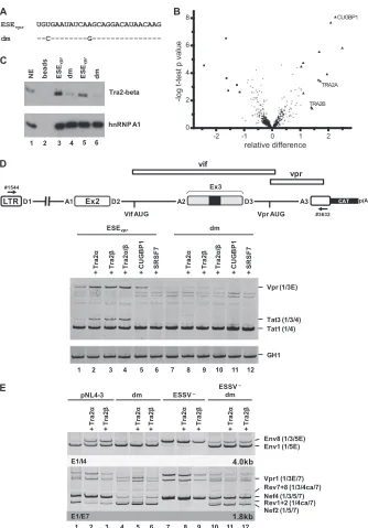
ESEvpr
+1
A2 D3
Ex3
133 134 135 136 137 138 139 140 141 142 143 144
Cys Glu Tyr Gln Ala Gly His Asn Lys Val Gly Ser
TGT GAA TAT CAA GCA GGA CAT AAC AAG/gta gga tct
Cys
--C /
---Gln
--G /
---Cys Gln
--C --G / ----25T>C -16A>G dm Vif ESSV E1/E4 Vpr (1/3E) Tat3 (1/3/4) Tat2 (1/2/4) Tat1 (1/4) pN L4 -3 -25T > C -16A > G dm ESSV – -25T > C -16A > G dm
ESSV –
1 2 3 4 5 6 7 8
E1/E7 Nef2 (1/5/7) Nef4 (1/3/5/7) Rev1+2 (1/4ca/7) Rev7+8 (1/3/4ca/7) 1.8kb E1/I4 Vpr3 (1/3E) Env8 (1/3/5E) Env1 (1/5E) Vif2 (1/2E) 4.0kb
D
B
p41gag p24gag Actin p55gag (sn) p24gag (ly)1 2 3 4 5 6 7 8 9
pN L4 -3 -25T > C -16A > G dm ESSV – -25T > C -16A > G dm
ESSV –
Mo c k Vpr Vif
C
(a) unspliced %
100 101 102 103 104
1 2 3 4 5 6 7 8
R e la ti v e Sp li c in g Effi c ie n c y (b) 100 101 102 103 104
multiply spliced %
1 2 3 4 5 6 7 8
R e la ti v e Sp li c in g Effi c ie n c y (d) 100 101 102 103
104 Vpr %
1 2 3 4 5 6 7 8
R e la ti v e Sp li c in g Effi c ie n c y (c) 100 101 102 103
104 Vif %
1 2 3 4 5 6 7 8
R e la ti v e Sp li c in g Effi c ie n c y (e)
1 2 3 4 5 6 7 8
exon 3 %
100 101 102 103 104 105 106 R e la ti v e Sp li c in g Effi c ie n c y 1: pNL4-3
2: ESE -25T>C
3: ESE -16A>G
4: ESE dm
5: ESSV–
6: ESSV–ESE -25T>C
7: ESSV–ESE -16A>G
8: ESSV–ESE dm
FIG 2ESEvpris necessary for exon 3 inclusion andvprmRNA processing. (A) The wild-type ESEvprsequence and the amino acid sequence encoded by the
overlappingvifORF are shown below exon 3. Mutated ESEvprnucleotide residues are denoted by their positions relative to the GT dinucleotide of viral 5=ss D3.
The black box represents the upstream ESSV. Uppercase letters represent exonic positions, and lowercase letters represent intronic positions. (B) HEK 293T cells (2.5⫻105) were transiently transfected with 1g of each of the proviral plasmids. At 30 h after transfection, total-RNA samples were collected and used for
RT-PCR analyses with different sets of primer pairs. HIV-1 mRNA species are indicated to the right of the gels in accordance with the nomenclature published previously (5). (C) cDNA samples were prepared as described for panel B and used in real-time PCR assays to specifically quantitate the relative abundances of unspliced (a), multiply spliced (b), Vif (c), and Vpr (d) mRNA species and exon 3 inclusion ratios (e). For normalization, primers 3387 and 3388 were used to
on November 7, 2019 by guest
http://jvi.asm.org/
[image:6.585.100.483.67.657.2]ation of virus particles into the cell supernatant, as indicated by the
detection of only small amounts of p24
gag(
Fig. 2D
, lane 6). This
has been hypothesized to result from insufficient amounts of
in-tracellular Gag, which is needed to drive virus assembly at the
cellular plasma membrane. The expression and normal processing
of structural proteins and Vif within the cells were reinstated
fol-lowing the double mutation of ESE
vpr(
Fig. 2D
, lane 9).
Further-more, the failure to efficiently produce virus particles of
ESSV-negative clones could be rescued by the
⫺
16A
⬎
G mutation and
the double mutation (
Fig. 2D
, lanes 8 and 9). Finally, Vpr protein
amounts were strongly reduced following the insertion of the
⫺
25T
⬎
C
⫺
16A
⬎
G double mutation in the absence of ESSV,
demonstrating again that ESE
vpris required for 3
=
ss A2 activation.
In summary, ESE
vprappears to promote the use of splice sites
A2 and D3. Additionally, these observations demonstrate that a
functional enhancer is critical for the expression of
vpr
mRNA,
noteworthy even when ESSV is active, indicating a delicate
inter-play between ESSV and ESE
vprin the regulation of viral HIV-1
exon 3 splicing.
The splicing factors Tra2-alpha and Tra2-beta bind to the
ESE
vprsequence.
To identify cellular factors that bind to the
ESE
vprsequence, RNA affinity purification experiments were
per-formed. Therefore, we incubated short,
in vitro
-synthesized RNA
substrates of either the wild-type or the double-mutated
(
⫺
25T
⬎
C
⫺
16A
⬎
G) ESE
vprsequence (each
n
⫽
5) in HeLa cell
nuclear extracts (
Fig. 3A
). After SDS-PAGE purification, proteins
were in-gel digested with trypsin. The peptides obtained were
sep-arated by liquid chromatography and mass spectrometry for
label-free quantitative analysis. This allowed us to quantify 602 RNA
affinity-purified proteins in each of the 10 samples analyzed. To
discriminate unspecific binding proteins from proteins bound to
the ESE
vprsequence specifically affected by the double mutation,
FDR-controlled statistical analysis based on the SAM method (
19
)
was used. This algorithm assigns a score based on the change in
protein abundance relative to the standard deviation of repeated
measurements and estimates the FDR by using permutations.
This approach revealed 6 proteins in the double-mutated ESE
vprgroup, as well as 12 proteins in the wild-type group, to be
signifi-cantly enriched (
Table 3
;
Fig. 3B
). Eight of those 12 proteins could
be assigned to the gene ontology biological process term mRNA
processing, representing a significant enrichment of this
biologi-cal process (FDR-adjusted
P
value, 0.01). Besides binding to
sev-eral members of the cleavage stimulation factor complexes, as well
as the cleavage polyadenylation-stimulating factor complexes, we
found a significant increase in the proteins Tra2-alpha, Tra2-beta,
and CUGBP1 in the wild-type ESE
vprsequence. Because of their
known role in regulating alternative splicing, we chose them for
further validation (
22
–
25
).
Western blot analyses confirmed that while the levels of
hnRNP A1 were not changed by the double mutation, Tra2-beta
was precipitated with significantly reduced efficiency by the
mu-tated ESE
vprsequence (
Fig. 3C
, cf. lanes 3 and 4 and lanes 5 and 6).
To unravel whether the splicing factors identified are functionally
involved in exon 3 splice site activation, we performed
coexpres-sion experiments and analyzed their effects on HIV-1 exon 3 and
vpr
mRNA splicing. In the context of an HIV-1-based minigene
(
Fig. 3D
), coexpression of Tra2-alpha, -beta, and both increased
exon 3 splice site activation in the presence of the wild-type ESE
vprsequence (
Fig. 3D
, cf. lanes 1 to 4), while it failed to promote exon
3 inclusion following inactivation of the enhancer (
Fig. 3D
, cf.
lanes 7 to 10). Coexpression of CUGBP1 or SRSF7, however, did
not increase exon 3 splice site activation in either in the context of
wild-type ESE
vpr(
Fig. 3D
, cf. lanes 1, 5, and 6) or that of double
mutant ESE
vpr(
Fig. 3D
, cf. lanes 7, 11, and 12). Tra2 proteins were
also coexpressed together with pNL4-3 and the derived mutants
(
Fig. 3E
), reiterating their role in ESE
vpr-controlled exon 3 splice
site activation. Once again, it was found that Tra2-alpha and -beta
overexpression increased the inclusion of exon 3 in the case of
wild-type ESE
vprbut not that of double mutant ESE
vpr(
Fig. 3E
, cf.
lanes 1 to 6). Therefore, we concluded that Tra2 proteins bind to
newly found ESE
vprand are thereby involved in the activation of
exon 3 inclusion and
vpr
mRNA splicing.
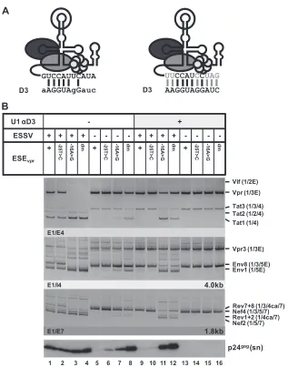
A modified U1 snRNA fully complementary to 5
=
ss D3
strongly activates exon 3 inclusion and
vpr
mRNA expression.
To determine the importance of ESE
vprfor exon 3 splice site use
under conditions of optimal 5
=
ss D3 recognition, we generated a
5
=
-end-mutated U1 snRNA matching all 11 nucleotides of 5
=
ss D3
(
Fig. 4A
). HEK 293T cells were transiently cotransfected with this
modified U1 snRNA expression vector and proviral DNA
con-taining either wild-type or mutant exon 3 sequences. In general,
RT-PCR analysis of RNA isolated from the transfected cells
re-vealed a dramatic shift toward
vpr
and/or exon 3 spliced mRNAs
upon the coexpression of the mutated U1 snRNA, indicated by
larger amounts of
vpr
mRNA species (
Fig. 4B
,
e
.g., Vpr3, cf. lanes
1 to 8 and 9 to 16) and the increased levels of exon 3-containing
mRNA species (
Fig. 4B
,
e
.g., Tat3 [cf. lanes 1 to 8 and 9 to 16] or
Nef4 [cf. lanes 1 to 4 and 9 to 12]). On the basis of these results, we
concluded that the coexpressed U1 snRNA seemed to assemble
correctly into mature snRNPs and that U1—as expected and
an-ticipated by a recent publication (
26
)—increased 5
=
ss D3
recog-nition. Interestingly, the strong increase in exon 3 splice site
acti-vation appeared to suppress the inclusion of exon 2 in the viral
mRNA species (
Fig. 4B
, Tat2, cf. lanes 1 and 9), indicating that a
balanced exon 3 splicing activity is also necessary to permit the use
of exon 2 splice sites A1 and D2. However, in the absence of
func-tional ESE
vpr, U1 snRNA coexpression predominantly activated
vpr
mRNA splicing, while only a minor influence on exon 3
inclu-sion was observed (
Fig. 4B
, Vpr and to a lesser extent Tat3, cf. lanes
3 to 4 and 11 to 12), indicating not only that ESE
vprmay enhance
the early recognition of 5
=
ss D3 but also supports its use later in the
splicing reaction. It is worth noting that inactivation of both ESSV
and ESE
vprallowed efficient
vpr
mRNA splicing and exon 3
inclu-sion upon the coexpresinclu-sion of modified U1 snRNA (
Fig. 4B
, Vpr3
and Tat3, lanes 8 and 16), rather arguing for a repressive activity of
detect the total viral mRNA content of each sample. Data represent expression ratios relative to that of wild-type pNL4-3 (bar 1), which was set to 100%. Values and error bars show the average⫾standard deviation of three independent transfection experiments. Bars correspond to lanes in panel B. (D) HEK 293T cells (2.5⫻105) were transiently transfected with 1g of each of the proviral plasmids. At 48 h posttransfection, viral supernatants were collected, layered onto 20%
sucrose solution, and centrifuged at 28,000 rpm for 90 min at 4°C to pellet the released virions. In addition, cells were harvested and resuspended in lysis buffer. Supernatants and cellular lysates were resolved by 12% SDS-PAGE and electroblotted onto nitrocellulose membranes. To determine virus particle production and the expression of viral proteins, samples were probed with primary antibodies specifically detecting structural p24gag(CA) and the viral infectivity factors Vif
and Vpr. Equal amounts of cell lysates were controlled for by the detection of␣-actin. E, extended exon; dm, double mutation; sn, supernatant; ly, lysate.
on November 7, 2019 by guest
http://jvi.asm.org/
ESSV that addresses splicing after initial 5
=
ss recognition and that
is counteracted by active ESE
vpr. Western blot analysis of the p24
levels within the supernatant suggested that the coexpression of
the U1 snRNA could induce excessive exon 3 splicing even in the
presence of ESSV, thereby dramatically reducing virus particle
production (
Fig. 4B
, cf. lanes 1 and 2 and lanes 9 and 10).
How-ever, binding of the coexpressed U1 snRNA relied on the presence
of ESE
vprto elicit excessive activation of the exon 3 splice sites,
ESE UGUGAAUAUCAAGCAGGACAUAACAAG
dm --C---G---A
-2 -1 0 1 2
0 2 4 6 8
TRA2A CUGBP1
-log t-test p value
relative difference
TRA2B
B
C
D
Ex2 A2 D3 p(A)
Vif AUG Vpr AUG D2
A1
vif
vpr
A3 CAT
LTR
Ex3 D1
#1544
#3632
+ T
ra
2
α
+ T
ra
2
β
+ T
ra
2
α
/β
+
CUG
BP
1
+ S
R
S
F
7
+ T
ra
2
α
+ T
ra
2
β
+ T
ra
2
α
/β
+
CUG
BP
1
+ S
R
S
F
7
dm
Tat3 (1/3/4) Tat1 (1/4)
GH1 Vpr (1/3E)
1 2 3 4 5 6 7 8 9 10 11 12
E
E1/I4
E1/E7 1.8kb Nef2 (1/5/7)
Nef4 (1/3/5/7) Rev1+2 (1/4ca/7) Rev7+8 (1/3/4ca/7) Vpr1 (1/3E/7)
+ T
ra
2
α
+ T
ra
2
β
+ T
ra
2
α
+ T
ra
2
β
+ T
ra
2
α
+ T
ra
2
β
+ T
ra
2
α
+ T
ra
2
β
Env8 (1/3/5E) Env1 (1/5E)
4.0kb
pNL4-3 dm ESSV –
ESSV –
dm
1 2 3 4 5 6 7 8 9 10 11 12 Tra2-beta
hnRNP A1 1 2 3 4 5 6
vpr
NE bead
s
ESE
vp
r
dm ESE
vp
r
dm
ESEvpr
FIG 3ESEvpris bound by the splicing factors Tra2-alpha and Tra2-beta. (A)In vitro-transcribed RNA substrates used for RNA pulldown experiments (dm, double
mutation). (B) Volcano plot of RNA binding proteins purified by RNA pulldown with a nonmutated or a mutated ESEvprsequence with HeLa cell nuclear extract. The
precipitated proteins were digested with trypsin and subjected to quantitative mass spectrometry analysis. Thexaxis of the volcano plot shows the relative difference in protein abundance as calculated by the SAM method, whereas theyaxis shows the⫺logt-testPvalue of the groupwise comparison of protein abundances. Besides the majority of probably unspecifically binding proteins (circles), some proteins preferably bound to the wild-type ESEvprsequence (triangles) or the mutated ESEvprvariant
(squares). The proteinsTra2-alpha and Tra2-beta were selected for validation experiments. (C) Immunoblot analysis with an antibody specific for Tra2-beta and hnRNPA1 confirmed significantly smaller amounts of Tra2-beta for the double mutant. (D) HeLa cells (2.5⫻105) were transiently cotransfected with 1g of each of
the HIV-1-based LTR ex2 ex3 splicing reporters, 0.2g of SVctat (47); 1g of pXGH5 (GH1) as a transfection control, and 1g of pcDNA3.1(⫹), an expression plasmid for Tra2-alpha, Tra2-beta, CUGBP1, and SRSF7. At 30 h posttransfection, total RNA was isolated and subjected to semiquantitative RT-PCR analyses with primers 1544 and 3632. For measurement of equal transfection efficiencies, a separate PCR was carried out with a primer pair (1224/1225) specific for human growth hormone 1 (GH1). (E) RT-PCR analyses of intronless (2-kb) and intron-containing (4-kb) viral mRNA species following the transient transfection of HEK 293T cells with 1g of the respective proviral construct and 1g of pcDNA3.1(⫹), an expression plasmid for either Tra2-alpha or Tra2-beta. E, extended exon; dm, double mutation.
on November 7, 2019 by guest
http://jvi.asm.org/
[image:8.585.125.459.62.541.2]causing severely reduced viral particle production (
Fig. 4B
, cf.
lanes 9 and 12). When ESSV was disrupted, U1 coexpression
effi-ciently inhibited viral particle production independent of ESE
vpractivity (
Fig. 4B
, cf. lanes 5 to 8 and 13 to 16). These findings
emphasize that the complementarity between the 5
=
ss D3 and
the U1 snRNA is not the sole determinant of exon 3 inclusion.
However, the absence of functional ESE
vprcould be (at least
partially) bypassed by increasing base pairing between U1
snRNA and 5
=
ss D3.
vpr
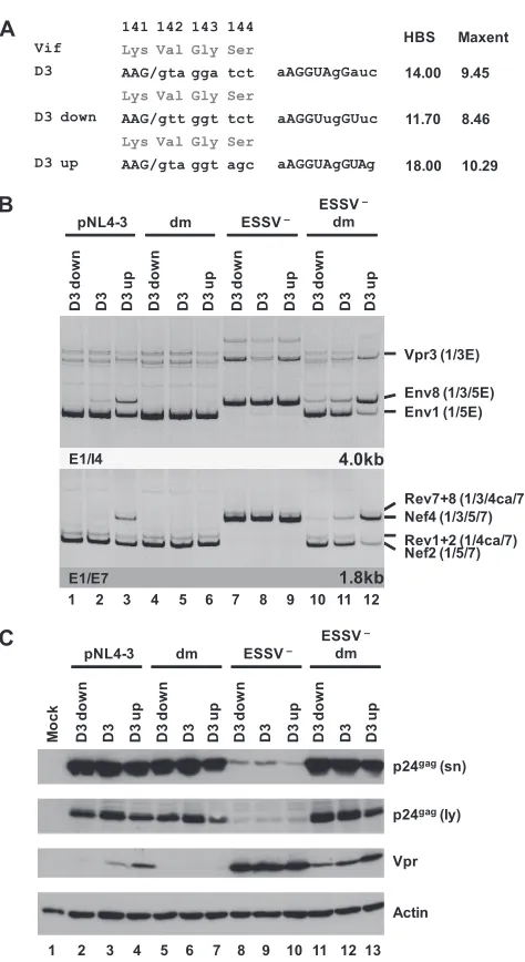
mRNA expression can be modulated by up and down
mutations of 5
=
ss D3.
The use of 3
=
ss A2 results in the formation
of
vpr
-mRNA but only when splicing at the downstream 5
=
ss D3 is
suppressed because this would remove the Vpr translational
ini-tiation codon within intron 3 from the mature transcripts.
Re-markably, ESE
vprwas shown to be critical for
vpr
mRNA
expres-sion, although it is located close to 5
=
ss D3 and separated from 3
=
ss
A2 by the repressor ESSV. It was found previously that efficient
recognition of a 5
=
ss by the U1 snRNP exerts positive feedback on
the assembly of splicing factors at the upstream 3
=
ss—most likely
via interactions across the exon (
27
–
29
). To analyze the
interde-pendence of ESE
vpr, 5
=
ss D3, and
vpr
mRNA expression,
muta-tions predicted to either decrease (D3 down) or increase (D3 up)
the intrinsic strength of the viral 5
=
ss D3 were tested in the context
of a replication-competent provirus (
Fig. 5A
). Mutations were
chosen so that the overlapping Vif ORF was not changed.
Follow-ing transient transfection of HEK 293T cells with proviral DNA,
the exon 3 abundance within the viral mRNA species was
deter-mined for each of the mutant proviruses by RT-PCR analysis (
Fig.
5B
). As expected, the extent of the complementarity between U1
snRNA and the 5
=
ss basically correlated with the amounts of exon
3 present in the viral transcripts. In the presence of ESE
vpractivity,
weakening of 5
=
ss D3 caused a decrease in the levels of exon
3-con-taining isoforms within both major viral mRNA classes, whereas
an increase in the complementarity of 5
=
ss D3 partially overcame
the general repression of exon 3 splicing by dominant negative
ESSV (
Fig. 5B
, lanes 1 to 3). This was in line with the hypothesis
that the stability of U1 snRNP binding to a 5
=
ss plays a pivotal role
in the recognition of the entire exon. However, mutant forms of
ESE
vprshowed no detectable exon 3 inclusion, regardless of their
intrinsic 5
=
ss strength (
Fig. 5B
, lanes 4 to 6), indicating a strict
requirement for functional ESE
vprto enable stable binding of the
U1 snRNP to 5
=
ss D3. Mutant ESSV was also associated with a
poor response of the
vpr3
mRNA to the distinct 5
=
ss variants in the
experiment shown (
Fig. 5B
, lanes 7 to 9) and lacked any response
in parallel experiments (data not shown). Efficient exon 3
inclu-sion was detected in each case, irrespective of up or down
muta-tions within D3 (
Fig. 5B
, e.g., Nef4, lanes 7 to 9), suggesting that in
the absence of ESSV activity, recognition of a weaker 5
=
ss can be
compensated for by stronger activation of 3
=
ss A2. Finally, when
both ESSV and ESE
vprwere mutated, the intrinsic 5
=
ss strength
again up- or downmodulated the frequency of exon 3 inclusion in
the viral mRNAs (
Fig. 5B
, e.g., Nef4, lanes 10 to 12). This
rein-TABLE 3Proteins identified by mass spectrometry
Protein IDs Gene(s) t-testPvalue
MS intensity ratio ESE/dm
No. of unique peptides
RNA-binding protein 4B Q9BQ04, E9PM61, E9PLB0, Q9BWF3-2, E7EQS3, D6R9K7, Q9BWF3-3
RBM4B, RBM4 2.83E-05 0,18 3
Flap endonuclease 1 P39748 FEN1 2.96E-07 0,32 16
Zinc finger protein 207 H0Y3M2, E1P660, O43670, O43670-2, A8MTG3
ZNF207 0.000239198 0,33 5
Serine hydroxymethyltransferase P34897, P34897-3, P34897-2, B4DLV4, H0YIZ0, G3V4W5, G3V5L0
SHMT2 0.00190648 0,31 11
RNA-binding protein 14 Q96PK6 RBM14 0.000738779 0,43 3
Mitotic checkpoint protein BUB3 O43684, O43684-2, B4DDM6 BUB3 0.00246569 0,44 7 Cleavage stimulation factor subunit 2 E7EWR4, P33240, P33240-2, B4DUD5,
E9PID8
CSTF2 3.50E-05 2,02 5
Cleavage stimulation factor subunit 3 Q12996, F5H0G6 CSTF3 1.89E-05 2,15 14 Putative DNase TATDN3 G3V151, Q17R31, Q17R31-2, E9PJE5,
E9PNH3, E9PP81, E9PRA1
TATDN3 0.00305696 2,20 11
Transformer-2 protein homolog beta P62995, E7EQD1, P62995-3, H7BXF3 TRA2B 0.0323067 2,49 8 Single-stranded DNA-binding protein,
mitochondrial
Q04837, E7EUY5, C9K0U8 SSBP1 0.0400618 2,53 4
Cleavage and polyadenylation specificity factor subunit 7
Q8N684-3, Q8N684, Q8N684-2, F5H669, F5H047, F5H6M0
CPSF7 0.000334073 3,00 11
Cleavage and polyadenylation specificity factor subunit 5
O43809, H3BND3 NUDT21 2.97E-06 3,18 14
Transformer-2 protein homolog alpha Q13595, B4DUA9, B4DQI6, Q13595-2 TRA2A 0.000404988 3,27 3 Cleavage and polyadenylation specificity
factor subunit 6
Q16630-2, Q16630, C9JGC2, F8WJN3, Q16630-3
CPSF6 0.000126486 3,68 9
Squamous cell carcinoma antigen recognized by T cells 3
Q15020, B7ZKM0 SART3 2.24E-08 4,27 4
CUGBP Elav-like family member 1 G5EA30, Q92879-4, Q92879, F8W940, Q92879-3, Q92879-2, E9PKU1, F5H0D8
CELF1 8.77E-09 4,74 9
U6 snRNA-associated Sm-like protein LSm2
Q9Y333 LSM2 1.56E-06 5,51 5
on November 7, 2019 by guest
http://jvi.asm.org/
[image:9.585.44.541.78.416.2]forces the notion that in a less favorable environment with regard
to enhancer strength, the efficiency of exon inclusion exhibits a
higher dependency on the ability of a splice site to bind the U1
snRNP on its own. Taken together, the data show that the overall
efficiency of HIV-1 exon 3 splicing is adjusted by the individual
strength of the preceding exonic splicing regulatory elements and
the intrinsic strength of 5
=
ss D3. Western blot analyses were
con-sistent with these results and revealed that Vpr expression was
under the combined control of ESSV, ESE
vpr, and 5
=
ss D3
(
Fig. 5C
). Correspondingly, increasing base pairing between the 5
=
end of U1 snRNA and 5
=
ss D3 was accompanied by a higher
abun-dance of Vpr protein within the transfected cells, whereas a
reduc-tion of the intrinsic strength showed the opposite effect on Vpr
expression (
Fig. 5C
, lanes 2 to 4 and 11 to 13). However, in the
presence of only one intact splicing regulatory element, either
ESE
vpror ESSV, exon 3 splicing efficiency was either too low or too
high to allow tuning by alterations of 5
=
ss strength (
Fig. 5C
, Vpr,
lanes 5 to 7 and 8 to 10). These results substantiate the
observa-tions that U1 snRNP binding to 5
=
ss D3 enhances the use of
up-stream 3
=
ss A2 and that splicing of exon 3 can be considered the
integrated outcome of exonic elements (ESE
vprand ESSV) and the
intrinsic strength of 5
=
ss D3.
Binding of the U1 snRNP to a nonfunctional 5
=
ss is sufficient
to augment splicing at upstream 3
=
ss A2.
The presented results
suggest that U1 snRNP binding to the 5
=
ss fulfills two functions
during pre-mRNA splicing; i.e., (i) it enhances the formation of
exon definition complexes and therefore promotes recognition of
the upstream 3
=
ss (
27
–
29
), and (ii) it commits the bound 5
=
ss to
splice site pairing with a 3
=
ss across the downstream intron into
the prespliceosome (
30
). It is hypothesized here that
vpr
mRNA
aAGGUAgGauc GUCCAUUCAUA
D3 D3 AAGGUAGGAUC
UUCCAUCCUAG
A
Vif (1/2E)
Vpr (1/3E)
Tat3 (1/3/4) Tat2 (1/2/4) Tat1 (1/4)
Env1 (1/5E) Env8 (1/3/5E) Vpr3 (1/3E)
p24gag (sn)
1 2 3 4 5 6 7 8 9 10 11 12 13 14 15 16
Nef2 (1/5/7) Rev1+2 (1/4ca/7) Nef4 (1/3/5/7)Rev7+8 (1/3/4ca/7)
B
U1 αD3 - +
ESSV + + + + - - - - + + + + - - -
-ESEvpr + -25T
>
C
-1
6
A
>
G
dm
+ -25T
>
C
-1
6
A
>
G
dm
+ -25T
>
C
-1
6
A
>
G
dm
+ -25T
>
C
-1
6
A
>
G
dm
E1/E7 E1/I4 E1/E4
1.8kb 4.0kb
FIG 4Coexpression of a modified U1 snRNP with full complementarity to 5=ss D3 induces HIV-1 exon 3 splicing andvprmRNA expression. (A) Schematic drawing of a 5=-end-modified U1 snRNA (right) perfectly matching the 5=ss D3 sequence. Mutated nucleotides are indicated by gray capital letters. Additional base pairing interactions between 5=ss D3 and the optimized 5=end of the U1 snRNA are indicated by vertical gray lines. (B) HEK 293T cells (2.5⫻105) were
transiently cotransfected with 1g of both a proviral plasmid and a U1 snRNA expression plasmid. Total RNA was isolated and subjected to RT-PCR analyses. PCR products were resolved by PAGE and stained with ethidium bromide. RT-PCR samples are shown at the top. The main viral mRNA species are indicated on the right. Viral supernatants were collected as well and analyzed for viral p24gagconcentrations by immunoblotting (bottom). E, extended exon; dm, double
mutation; sn, supernatant.
on November 7, 2019 by guest
http://jvi.asm.org/
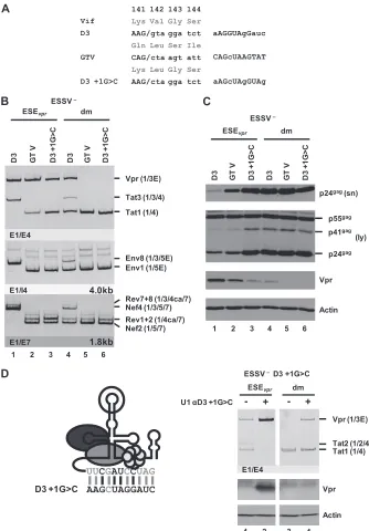
[image:10.585.135.452.62.465.2]splicing requires exon definition, so that 5
=
ss D3 is recognized by
U1 snRNA but splicing at D3 must occur with lower efficiency. To
gain a broader understanding of how
vpr
mRNA expression is
regulated by U1 snRNP binding, 5
=
ss D3 was replaced with GTV
(
17
), a U1 binding-competent but splicing-incompetent sequence
with nonetheless nearly full complementarity to U1 snRNA
(
Fig. 6A
). As a control, 5
=
ss D3 was also inactivated by a G-to-C
mutation at position
⫹
1. The resulting set of variants ranged from
a functional 5
=
ss, which supported efficient binding of the U1
snRNP, as well as splicing (D3), to sequences allowing either
effi-cient binding (GTV) or neither effieffi-cient binding nor splicing
(D3
⫹
1G
⬎
C). These variants were tested in the context of
ESSV-negative or ESSV/ESE
vprdouble-negative proviruses. Splicing
ef-ficiency at 3
=
ss A2 was determined by semiquantitative RT-PCR
analyses.
While 5
=
ss D3 was efficiently used in HEK 293T cells
trans-fected with an ESSV mutant, neither GTV nor D3
⫹
1G
⬎
C was
spliced and thus did not allow the accumulation of exon
3-con-taining mRNAs (
Fig. 6B
, Tat3, Env8, and Nef4, lanes 1 to 3). In the
presence of ESE
vpr, replacement of D3 with GTV or D3
⫹
1G
⬎
C
monotonically increased the
tat1
-to-
vpr
mRNA expression ratio
(
Fig. 6B
, Vpr and Tat1, lanes 1 to 3), which was consistent with
greater complementarity between GTV and U1 snRNA than
be-tween GTV and D3
⫹
1G
⬎
C. Even in the absence of ESE
vpr,
wild-type D3 still retained some
vpr
mRNA expression and exon 3
inclusion, although at a higher level of
tat1
mRNA expression (
Fig.
6B
, Vpr and Tat1, lane 4), while the disruption of both ESE
vprand
wild-type D3 abolished all
vpr
mRNA expression (lanes 5 and 6).
These results were in agreement with the hypothesis that U1
snRNP binding to splicing-incompetent U1 snRNA binding sites
alone suffices to augment cross-exon interactions and splicing at
3
=
ss A2.
The change in mRNA expression ratios induced by these
mu-tations was entirely consistent with the decreasing amount of Vpr
protein detected by Western blot analysis (
Fig. 6C
, Vpr).
More-over, it was observed that decreasing
vpr
mRNA splicing by both
reducing U1 snRNA complementarity at D3 and mutating ESE
vprcould rescue virus particle production of ESSV-negative provirus
(
Fig. 6C
, p24
gag[sn/ly]).
The finding that ESE
vpr-dependent binding of U1 snRNA
pro-motes the use of 3
=
ss A2 was further confirmed by the
coexpres-sion of a U1 snRNA fully complementary to splicing-inactive 5
=
ss
D3 (
⫹
1G
⬎
C) (
Fig. 6D
), which largely activated
vpr
mRNA
splic-ing (
Fig. 6D
, top), as well as Vpr protein expression (
Fig. 6D
,
bottom).
In summary, these findings recapitulate earlier studies showing
that the two functions of U1 snRNP binding to the 5
=
ss can be
dissected, supporting (i) the formation of exon definition
com-plexes and (ii) the assembly of a prespliceosome across a
down-stream intron (
27
–
29
).
DISCUSSION
Splice site recognition is commonly found to be under the
com-bined control of multiple nearby splicing regulatory elements that
can either compete or cooperate to regulate splicing activation. It
was previously shown that the HIV-1 noncoding leader exon 3
harbors a negative splicing regulatory element—termed ESSV—
within its central portion that selectively represses upstream 3
=
ss
A2 (
12
) and concomitantly inhibits exon 3 inclusion in the
differ-ent viral mRNA species (
13
). ESSV disruption results in the strong
141 142 143 144
Lys Val Gly Ser
AAG/gta gga tct
Lys Val Gly Ser
AAG/gtt ggt tct
Lys Val Gly Ser
AAG/gta ggt agc Vif
D3
D3 down
D3 up 18.00 10.29
11.70 8.46 14.00 9.45 HBS Maxent aAGGUAgGauc aAGGUugGUuc aAGGUAgGUAg
A
B
– pNL4-3 D3 d o w n D3 D3 u p dm D3 d o w n D3 D3 u pESSV –
D3 d o w n D3 D3 u p ESSV dm D3 d o w n D3 D3 u p Nef2 (1/5/7) Nef4 (1/3/5/7) Rev1+2 (1/4ca/7) Rev7+8 (1/3/4ca/7) E1/E7 1.8kb E1/I4 4.0kb Vpr3 (1/3E) Env8 (1/3/5E) Env1 (1/5E)
1 2 3 4 5 6 7 8 9 10 11 12
p24gag(ly)
p24gag(sn)
Vpr Actin pNL4-3 D3 d o w n D3 D3 u p dm D3 d o w n D3 D3 u p
ESSV –
D3 d o w n D3 D3 u p
ESSV –
dm D3 d o w n D3 D3 u p
1 2 3 4 5 6 7 8 9 10 11 12
Mo
c
k
13
C
FIG 55=ss D3 up and down mutations modulate HIV-1 exon 3 splicing and vprmRNA formation. (A) Silent mutations predicted to decrease or increase the complementarity to the 5=end of the endogenous U1 snRNA were intro-duced into viral 5=ss D3. Exonic nucleotides are denoted in uppercase letters, and intronic nucleotides are denoted in lowercase letters. Complementarity and predicted intrinsic strength by HBond score (HBS) and MaxEnt score algorithms are both shown next to the 5=ss sequence. Nucleotides complemen-tary to the U1 snRNA are in capital letters, while mismatches to the U1 snRNA are in lowercase letters. (B) HEK 293T cells (2.5⫻105) were transiently
trans-fected with 1g of each of the different infectious clones. RNA was isolated from the cells, DNase I digested, and reverse transcribed. The resultant cDNA served as the DNA template in semiquantitative PCRs using primer pairs E1/I4 and E1/E7 to specifically detect viral 4.0- and 1.8-kb viral mRNAs, respectively. Proviral mutants are shown above the panels. The main HIV-1 mRNA species are indicated at the right. (C) Protein lysates and viral supernatants were col-lected from HEK 293T cells transfected with 1g of pNL4-3 or mutant deriv-atives. Samples were loaded on 12% SDS-polyacrylamide gels and, after sepa-ration, transferred to nitrocellulose membranes. Viral proteins and␣-actin (as a loading control) were determined by probing with specific primary antibod-ies. For detection, appropriate HRP-conjugated antibodies and ECL detection reagent were applied. HBS, HBond score; MaxEnt, MaxEnt score; dm, double mutation; E, extended exon; sn, supernatant; ly, lysate.
on November 7, 2019 by guest
http://jvi.asm.org/
[image:11.585.44.281.60.494.2]accumulation of
vpr
mRNA and exon 3-containing isoforms,
which leads to viral replication incompetence. However, ESSV
does not act alone; using an
in silico
-based mutagenesis strategy,
we identified an enhancer sequence—termed ESE
vpr— upstream
of 5
=
ss D3 that was essential for exon 3 splicing in the context of
wild-type ESSV and that provided excess exon 3 splice site
activa-tion when ESSV was inactive. This noactiva-tion was strengthened by the
finding that ESSV/ESE
vprdouble-negative provirus retrieved the
141 142 143 144
Lys Val Gly Ser
AAG/gta gga tct
Gln Leu Ser Ile
CAG/cta agt att
Lys Leu Gly Ser
AAG/cta gga tct Vif
D3
GTV
D3 +1G>C
aAGGUAgGauc
CAGcUAAGTAT
aAGcUAgGUAg
A
Vpr p24gag(sn)
Actin (ly) p41gag
p24gag p55gag D3 GT
V
D3
+
1
G
>
C
D3 GT
V
D3
+
1
G
>
C
1 2 3 4 5 6
(1/3E)
B
C
E1/E4
Vpr
Tat3 (1/3/4)
Tat1 (1/4) D3 GT
V
D3
+
1
G
>
C
D3 GT
V
D3
+
1
G
>
C
E1/E7 1.8kb
E1/I4 4.0kb
Nef2 (1/5/7) Nef4 (1/3/5/7)
Rev1+2 (1/4ca/7) Rev7+8 (1/3/4ca/7) Env8 (1/3/5E) Env1 (1/5E)
D
1 2 3 4 5 6
AAGCUAGGAUC
D3 +1G>C
UUCGAUCCUAG
α
Vpr (1/3E)
Tat1 (1/4) U1 D3 +1G>C
1 2
- +
Vpr
Actin
- +
Tat2 (1/2/4)
E1/E4
3 4
ESSV –
ESEvpr dm
ESSV –
ESEvpr dm ESSV –
ESEvpr dm
D3 +1G>C
FIG 6U1 snRNP binding to a splicing-incompetent 5=ss enhancesvprmRNA expression. (A) 5=ss D3 was replaced with a splicing-incompetent sequence that perfectly matches the free 5=end of the cellular U1 snRNA except for posit