Preintegration Complex Formation during Acute Infection by
Replication-Defective Integrase Mutant Human Immunodeficiency
Virus
Xiang Li, Yasuhiro Koh, and Alan Engelman
Department of Cancer Immunology and AIDS, Dana-Farber Cancer Institute, and Department of Medicine, Harvard Medical School, Boston, Massachusetts, USA
Previous studies characterized two types of replication-defective human immunodeficiency virus type 1 (HIV-1) integrase
mu-tants: class I, which are specifically blocked at the integration step, and class II, which harbor additional virion production
and/or reverse transcription defects. Class I mutant enzymes supported little if any metal ion-dependent 3
=
-processing and DNA
strand transfer activities
in vitro
, whereas class II enzymes displayed partial or full catalytic function in studies with simplified
assay designs, suggesting that defective interaction(s) with heterologous integrase binding proteins might underlie the class II
mutant viral phenotype. To address this hypothesis, class I and II mutant enzymes were interrogated under expanded sets of
in
vitro
conditions. The majority failed to catalyze the concerted integration of two viral DNA ends into target DNA, highlighting
defective integrase function as the root cause of most class II in addition to all class I mutant virus infection defects. One mutant
protein, K264E, in contrast, could support the wild-type level of concerted integration activity. After accounting for its inherent
reverse transcription defect, HIV-1
K264Emoreover formed preintegration complexes that supported the efficient integration of
endogenous viral DNA
in vitro
and normal levels and sequences of 2-long terminal repeat-containing circle junctions during
acute infection. K264E integrase furthermore efficiently interacted
in vitro
with two heterologous binding partners, LEDGF/p75
and reverse transcriptase. Our results underscore the physiological relevance of concerted integration assays for tests of
inte-grase mutant function and suggest that the K264E mutation disrupts an interaction with an intranuclear inteinte-grase binding
part-ner that is important for HIV-1 integration.
K
ey players in retroviral integration are the viral integrase (IN)
protein, a component of virion particles, and the ends of the
linear viral DNA (vDNA) made by reverse transcription. IN
func-tions in the context of the preintegration complex (PIC), a large
nucleoprotein complex (7, 88) that can support vDNA integration
into heterologous target DNA
in vitro
(10, 42, 45). IN initially
processes the vDNA ends adjacent to conserved CA 3
=
dinucle-otides, which liberates a pGpT
OHdinucleotide from each end of
human immunodeficiency virus type 1 (HIV-1) (94). IN then uses
the processed 3
=
vDNA termini to cut opposing strands of target
DNA in a concerted fashion, which joins the vDNA ends to
chro-mosomal 5
=
phosphates (11, 41, 48). The resulting gapped
recom-bination intermediate, with unjoined vDNA 5
=
ends, is repaired
by host cell machinery to generate the integrated provirus (see
reference 33 for a recent overview of retroviral integration).
Numerous cellular factors can interact with HIV-1 IN (see
ref-erences 114 and 118 for reviews), and one in particular, lens
epithelium-derived growth factor (LEDGF)/p75, has been shown
to play an important role during HIV-1 infection (reviewed in
references 35 and 97). IN is composed of three evolutionarily
con-served domains interconnected by flexible linkers. Concon-served His
and Cys residues within the N-terminal domain (NTD) bind zinc,
whereas invariant Asp and Glu residues within the catalytic core
domain (CCD) comprise the D,D-35-E active site, which
coordi-nates Mg
2⫹ions for catalysis (reviewed in references 33 and 72).
The C-terminal domain (CTD) can engage different ligands,
in-cluding DNA (39, 120, 125), reverse transcriptase (RT) (55, 123,
130), and the SIP1/Gemin2 host factor important for reverse
tran-scription (50, 90), whereas LEDGF/p75 binding determinants are
comprised by the NTD and CCD (53, 81).
Results of biochemical experiments have indicated that a
te-tramer of HIV-1 IN catalyzes the concerted integration of two
vDNA ends (46, 51, 71, 92), findings that are consistent with
re-cent X-ray crystal structures of functional prototype foamy virus
(PFV) IN-vDNA complexes or intasomes (52, 82). PFV IN
effi-ciently integrates relatively short, oligonucleotide vDNA
sub-strates into target DNA in a concerted fashion
in vitro
(115),
whereas similar reaction conditions support aberrant integration
of single vDNA ends by HIV-1 IN (13, 53). Various parameters,
including the conditions under which IN is purified (108), length
of vDNA substrate (70), and binding ligands, such as LEDGF/p75
(53, 93), can increase the yield of concerted HIV-1 integration
products
in vitro
.
Studies that initially mutagenized the IN coding region of
pol
revealed the importance of the viral recombinase for HIV-1
rep-lication (66, 106, 109). From this start, it was evident some
muta-tions affected more than just integration. For example, a CCD
linker insertion mutant failed to release virus from transfected
cells (109). Subsequent work confirmed virus assembly/virion
Received21 September 2011Accepted10 January 2012
Published ahead of print25 January 2012
Address correspondence to Alan Engelman, [email protected]. Copyright © 2012, American Society for Microbiology. All Rights Reserved.
doi:10.1128/JVI.06386-11
on November 7, 2019 by guest
http://jvi.asm.org/
morphology defects for some HIV-1 IN mutants (2, 12, 38, 43, 60,
63, 67, 73, 77, 78, 89, 96, 100, 107, 111, 126), whereas other
mu-tants displayed defects in core uncoating (9), reverse transcription
(3, 15, 40, 67, 75–78, 80, 84, 89, 102, 104, 107, 113, 124, 126, 130),
or PIC nuclear import (3, 57, 59). In contrast, other mutations,
typified by changes of D,D-35-E active site residues, solely blocked
the integration step (2, 38, 61, 66, 67, 79, 84, 105, 111, 124). We
previously defined two phenotypic classes of replication-defective
HIV-1 IN mutant viruses to distinguish those specifically blocked
at integration (class I) from those that behave pleiotropically
(class II) (31). Due to the specific block, a hallmark of class I IN
mutant viral infection is transient accumulation of unintegrated
nuclear vDNA (2, 38, 61, 67, 79, 84, 105, 124), akin to the
pheno-type observed for wild-pheno-type (WT) HIV-1 in the presence of IN
strand transfer inhibitors (INSTIs) (54). Reverse transcription is
the most common pleiotropic defect associated with class II IN
mutant viruses (31, 32).
Central to understanding the mechanism of IN mutant virus
behavior is analysis of enzyme catalytic function, and
recombi-nant INs derived from class I and II mutant viruses have
accord-ingly been analyzed for integration activities
in vitro
. A separate IN
activity, disintegration (26), which is unlikely to be relevant
dur-ing virus infection, has nonetheless been shown to be an
impor-tant parameter because it, unlike 3
=
processing and DNA strand
transfer, can be catalyzed by the isolated CCD (14, 65). Certain
amino acid substitutions of active site D,D-35-E residues ablate
3
=
-processing, DNA strand transfer, and disintegration activities
(37, 68, 117), highlighting the requirement for divalent metal ion
coordination by the CCD for IN catalysis. In contrast, enzymes
derived from class II mutant viruses support 3
=
-processing and
DNA strand transfer activities, some even at the level of WT IN
(IN
WT) (15, 37, 40, 60, 63, 77, 78, 80, 98, 99, 102, 117). The
infec-tivity defect of IN mutant viruses can be rescued by supplying
IN
WTin
trans
as a Vpr fusion protein (47, 127), and class II IN
mutant fusion proteins, moreover, effectively
trans
-complement
class I IN mutant virus infection defects (6, 47, 77, 78, 80). Taken
together, these results highlight the catalytic competence of class II
IN mutant proteins and, by extension, that defects in auxiliary
functions, for example interaction(s) with critical protein binding
partners, might underlie the observed infection defects (6, 15, 29,
49, 59, 78, 128, 130). To address this hypothesis, 12 recombinant
IN proteins derived from class II mutant viruses were analyzed
alongside class I mutant control proteins IN
D64Nand IN
W235Ein a
variety of assays, including the most stringent test for IN function
in vitro
, the concerted integration assay. Our findings revealed that
defective IN function underlies the majority of class II mutant
virus infection defects and moreover highlight K264E as a
muta-tion that potentially disrupts an interacmuta-tion with a heterologous
factor important for integration.
MATERIALS AND METHODS
Plasmid DNAs.C-terminally His6-tagged IN proteins derived from clade B strain HIV-1NL4-3 were expressed from bacterial expression vector pKBIN6Hthr (86). Mutations were introduced by PCR usingPfuUltra DNA polymerase (Agilent Technologies, Inc., Santa Clara, CA), and se-quences of IN mutant expression vectors were verified by Sanger sequenc-ing. The plasmid pGST-RT, which encodes a fusion between glutathione S-transferase (GST) and the p66 subunit of RT (91), was a kind gift from Jeroen van Wamel and Ben Berkhout, University of Amsterdam.
Plasmids encoding full-length HIV-1NL4-3and single-round luciferase
reporter derivatives of WT and IN mutants K215A/K219A, D64N/D116N (78), D167K (102), and K264E (77) were previously described.
Protein expression and purification. Escherichia coli strain BL21(DE3) or its PC2 derivative (21) transformed with IN expression constructs was grown for 16 h at 30°C. The next day, bacteria subcultured at 1:10 in 2 liters of LB broth containing 100g/ml ampicillin were grown at 30°C until the optical density at 600 nm (OD600) reached 0.6 to 0.8, at which time IN expression was induced by the addition of 0.4 mM isopropyl--D-thiogalactopyranoside (IPTG). Cells were harvested after 4 h of induction at 28°C. The bacterial pellet was resuspended in ice-cold buffer A {25 mM Tris-HCl (pH 7.4), 1 M NaCl, 7.5 mM 3-[(3-cholamidopropyl) dimethylammonio]-2-hydroxy-1-propanesulfonate (CHAPS)]} containing 25 mM imidazole– 0.5 mM phenylmethanesulfo-nylfluoride (PMSF) and sonicated. After centrifugation for 15 min at 60,000⫻g, the supernatant was incubated with 1 ml of buffer A–25 mM imidazole– equilibrated Ni2⫹-nitrilotriacetic acid (Ni-NTA)–agarose beads (Qiagen, Valencia, CA) at 4°C for 1 h. The beads were washed twice with 20 volumes of buffer A–25 mM imidazole, followed by 30 volumes of buffer A–35 mM imidazole. IN was eluted with buffer A–200 mM imida-zole. IN-containing fractions diluted with 3 volumes of 25 mM Tris-HCl (pH 7.4)–7.5 mM CHAPS were injected into a 5-ml HiTrap heparin col-umn (GE Healthcare, Piscataway, NJ), and bound proteins were eluted with a linear gradient of 0.25 M to 1 M NaCl in 25 mM Tris-HCl (pH 7.4)–7.5 mM CHAPS using an AKTA 10 purifier. Immediately after elu-tion, 10 mM dithiothreitol (DTT) was added to each fracelu-tion, and the NaCl concentration was adjusted to 1 M. The His tag was removed from IN by using 40 U of thrombin (Sigma-Aldrich, St. Louis, MO) per mg of protein for 3 h at room temperature, which left the heterologous LVPR sequence at each C terminus. After removal of thrombin by incubation with Benzamidine beads (Novagen, Madison, WI) and ultrafiltration us-ing 9-kDa molecular weight cutoff Pierce concentrators (Thermo Fisher Scientific, Waltham, MA), IN was dialyzed overnight against buffer D (25 mM Tris-HCl [pH 7.4], 1 M NaCl, 7.5 mM CHAPS) containing 10% glycerol (wt/vol)–10 mM DTT. Protein concentration was determined by spectrophotometry, and aliquots flash-frozen in liquid N2were stored at
⫺80°C. Quantitative image analysis (FluorChem FC2; Alpha Innotech, San Leandro, CA) of Coomassie-stained gels revealed each IN preparation to be minimally 90% pure.
E. colistrain BL21 transformed with pGST-RT grown overnight at 37°C in LB–100l/ml ampicillin was diluted 1:10 the next day into 2 liters, and IPTG was added to a final concentration of 1 mM after reaching an OD600of 0.8. After 3 h at 37°C, harvested cells resuspended in 50 ml of buffer B (50 mM Tris-HCl [pH 8.0], 100 mM NaCl, 1 mM EDTA, 0.5% NP-40, 1 mM PMSF, 5 mM DTT) were sonicated at 50 W using 10-s pulses followed by 20 s of cooling over 10 min. The supernatant recovered after centrifugation for 15 min at 60,000⫻gwas incubated with 2 ml of glutathione-Sepharose 4 Fast Flow (GE Healthcare) at room temperature for 2 h, after which the beads were washed with 500 ml of phosphate-buffered saline containing 5 mM DTT. After washing with 20 ml of buffer B, protein was eluted with 20 ml of buffer E (50 mM Tris-HCl [pH 8.0], 100 mM NaCl, 1 mM EDTA, 5 mM DTT, 20 mM reduced glutathione, Complete protease inhibitor cocktail [Roche Applied Science, Indianap-olis, IN]) at room temperature. Protein concentration was determined by spectrophotometry following concentration and dialysis against buffer C (20 mM Tris-HCl [pH 7.4], 120 mM NaCl, 1 mM EDTA, 10% glycerol, 5 mM DTT, Complete protease inhibitor cocktail). Aliquots flash-frozen in liquid N2were stored at⫺80°C.
Recombinant IND64N(27), LEDGF/p75 (116), and GST fused to the C-terminal domain of LEDGF/p75 (GST-LEDGF326-530) (23) proteins expressed in bacteria were purified as previously described.
DNA substrates.Substrates that mimicked the HIV-1 U5 vDNA end were used to measure IN 3=-processing, DNA strand transfer, and disin-tegration activities. To prepare the 21-mer processing substrate, the 3= recess in annealed AE117 (5=-ACTGCTAGAGATTTTCCACAC)/AE150 (5=-GTGTGGAAAATCTCTAGCAG) was filled in with [␣-32P]TTP
Li et al.
on November 7, 2019 by guest
http://jvi.asm.org/
(3,000 Ci/mmol; PerkinElmer, Waltham, MA) using Sequenase version 2.0 T7 DNA polymerase (GE Healthcare) (41). The 30-mer substrate was similarly prepared from annealed AE143 (5=-ACTGCTAGAGATTTTCC ACACTGACTAAAA)/AE191 (5=-TTTTAGTCAGTGTGGAAAATCTCT AGCAG) DNA. To prepare the preprocessed duplex for DNA strand transfer, AE155 (5=-TTTTAGTCAGTGTGGAAAATCTCTAGCA) la-beled with [␥-32P]ATP (3,000 Ci/mmol; PerkinElmer) using T4 polynu-cleotide kinase (GE Healthcare) was annealed with AE143. The branched Y-mer disintegration substrate was prepared by annealing 5=-end-labeled AE157 (5=-GAAAGCGACCGCGCC) with equimolar amounts of AE146 (5=-GGACGCCATAGCCCCGGCGCGGTCGCTTTC), AE117 (5=-ACT GCTAGAGATTTTCCACAC), and AE156 (5=-GTGTGGAAAATCTCTA GCAGGGGCTATGGCGTCC). For each substrate, unincorporated radi-onuclide was removed by passing the labeled duplexes through Bio-Spin 6 columns (Bio-Rad, Richmond, CA) equilibrated with 10 mM Tris-HCl (pH 8.0)–20 mM NaCl– 0.1 mM EDTA.
Concerted integration of two vDNA ends into target DNA requires relatively long substrate DNA in the absence of LEDGF/p75 (53, 70). A 517-bp substrate was accordingly prepared from plasmid pU3U5 (25) following digestion with ScaI and BclI. The gel-purified DNA after 5=-end labeling was separated from unincorporated radionuclide by spin column chromatography. AE3653 (5=-CCTTTTAGTCAGTGTGGAAAATCTCT AGCA) annealed to AE3652 (5=-ACTGCTAGAGATTTTCCACACTGAC TAAAAGG) yielded the preprocessed 32-mer U5 end for LEDGF/p75-dependent integration assays (27, 53).
Recombinant IN activity assays.The 3=-processing reaction mixture (20l) contained 25 mM morpholinepropanesulfonic acid (pH 7.2), 50 mM NaCl, 10 mM MgCl2or MnCl2, 5M ZnSO4, 10 mM DTT, 0.5M IN, and 5 nM labeled DNA. Reaction mixtures were incubated at 37°C for 1 h, and reactions were stopped by addition of an equal volume of se-quencing gel sample buffer (95% formamide, 10 mM EDTA, 0.003% xy-lene cyanol, 0.003% bromophenol blue) and boiling for 2 min prior to fractionation through denaturing 20% polyacrylamide gels. Products were visualized using a Storm 820 PhosphorImager and quantified using ImageQuant version 1.2 (GE Healthcare). Radiolabeled vDNA strand transfer and disintegration reactions were conducted under the same con-ditions, except that products were fractionated through 15% sequencing gels and disintegration activity was assessed only in the presence of MnCl2. Concerted integration reaction mixtures (100l) contained 20 mM HEPES (pH 7.0), 100 mM NaCl, 10 mM MgCl2, 25M ZnCl2, 5 mM DTT, 10% polyethylene glycol 6000 (PEG 6000), 10% dimethyl sulfoxide (DMSO), 25 to 100 nM IN, 3.0 nM labeled 517-bp vDNA, and 1.5 nM pGEM-3 plasmid DNA. Reaction mixtures were incubated at 37°C for 2 h, and reactions were stopped by addition of 25 mM EDTA– 0.5% Na dode-cyl sulfate (SDS). Products deproteinized by digestion with proteinase K and precipitated with ethanol were analyzed by electrophoresis through 1.5% agarose–TAE (40 mM Tris base, 20 mM acetate, 1 mM EDTA) gels containing 0.1% SDS. Products, visualized in dried gels, were quantified using ImageQuant version 1.2.
LEDGF/p75-dependent integration reactions (36-l volume) were begun by mixing 0.55M vDNA with 0.3g pGEM-3 in 35.4 mM NaCl, 5.5 mM MgSO4, 11 mM DTT, 4.4M ZnCl2, and 22 mM HEPES-NaOH (pH 7.4). IN (2l) in dilution buffer (750 mM NaCl, 10 mM DTT, 25 mM Tris-HCl [pH 7.4]) was added, and after 15 min at room temperature LEDGF/p75 (2.0l) was added. Reactions (mixtures for which contained final IN and LEDGF/p75 concentrations of 0.8M and 0.6M, respec-tively) after 1 h at 37°C were stopped by addition of EDTA and SDS to final concentrations of 25 mM and 0.5%, respectively, and deproteinization with 30g proteinase K (Roche Applied Sciences) for 1 h at 37°C. DNAs recovered following precipitation with ethanol and separation on 1.5% agarose–TAE gels were stained with ethidium bromide (EtBr; 0.5g/ml). DNA strand transfer activity was quantified using Alpha Innotech Fluo-rChem FC2 software or ImageQuant version 1.2 for assays that included radiolabeled vDNA.
LEDGF/p75-IN binding assays.Capture of soluble IN (1g; 300 nM) by GST-LEDGF326-530(0.5g; 100 nM) or control GST (0.26g; 100 nM) prebound to glutathione-Sepharose beads (10l) was performed essen-tially as previously described (23, 102) in 100l PD buffer (25 mM Tris-HCl [pH 7.4], 150 mM NaCl, 5 mM MgCl2, 5 mM DTT, 0.1% NP-40) for 2 h at 4°C; 5g of bovine serum albumin (BSA) was included as an additional specificity control. Beads collected by settling for 20 min in the absence of centrifugation were washed with 700l PD buffer 4 times, with intermittent 20-min periods for settling. Proteins eluted with SDS gel sample buffer by boiling were fractionated through Novex bis-Tris–10% polyacrylamide gels. IN levels in gels stained with Sypro orange (Invitro-gen Corp., Carlsbad, CA) were quantified by using Alpha Innotech FluorChem FC2.
Ni-NTA beads (20l, settled volume) prewashed with PDA buffer (25 mM bis-Tris [pH 6.85], 150 mM NaCl, 2 mM MgCl2, 25 mM imidazole, 0.1% NP-40, Complete protease inhibitor cocktail) were added to reac-tion mixtures (100l) containing 5.8g LEDGF/p75 (0.95M), 0.7g IN-His6(0.2M), and 5g BSA. Following incubation at 4°C for 4 h with gentle agitation, beads washed four times with PDA buffer without cen-trifugation were mixed with 2⫻SDS gel sample buffer containing 200 mM imidazole and boiled, and resulting supernatants were fractionated through Novex bis-Tris–10% polyacrylamide gels. LEDGF/p75 levels in gels stained with Sypro orange were quantified by using Alpha Innotech FluorChem FC2.
RT-IN binding assay.IN (4.8l of a 2.9-g/ml stock in 200l buffer D) was precleared by centrifugation as follows, to reduce nonspecific ag-gregation during GST-RT binding assays. After incubation for 15 min on ice and spinning at 15,000⫻gfor 15 min at 4°C, the concentration of IN in the supernatant was determined by FluorChem FC2 imaging of Sypro orange-stained SDS-polyacrylamide gels. GST-RT pulldown of pre-cleared IN was performed as described above for GST-LEDGF326-530, ex-cept that 15g of GST-RT (1.7M) was used in place of the LEDGF fusion protein, IN was used at 3g (0.9M), and control reaction mix-tures contained 1M GST protein.
IN cross-linking.Protein cross-linking was performed as previously described (64).
Cells, viruses, and infections.HEK293T cells were maintained in Dulbecco’s modified Eagle’s medium supplemented to contain 10% fetal bovine serum, 100 IU/ml penicillin, and 100g/ml streptomycin, while SupT1 CD4⫹T cells were grown in RPMI 1640 medium modified to contain the same supplements. WT and IN mutant HIV-1NL4-3were pro-TABLE 1HIV-1 IN mutants included in this study
Change(s) Classa
Proposed
function(s) Infectivityb Reference(s)
D64N I Active site 0.06 (0.03)c 77
E69R II LEDGF binding ⬍0.01 102
V165A II LEDGF binding ⬍0.01 102, 114
R166A II LEDGF binding ⬍0.01 102
D167K II LEDGF binding 5.7 (0.1) 102
Q168A II LEDGF binding 0.8 (0.1) 102
K186Q II Multimerization 0.23 (0.28) 78
R199A II DNA binding 0.24 (0.13) 129
Q214L/Q216L II Nuclear import 0.11 (0.05) 78 K215A/K219A II Nuclear import,
DNA binding
0.02 (0.02) 78, 129
R228A II DNA binding 0.18 (0.25) 77
W235E I Multimerization 0.02 (0.01) 77
K258A II RT binding 0.09 (0.01) 77
K264E II DNA binding 0.11 (0.00) 77
aPhenotypic replication defect class. b
Percent single-round luciferase reporter virus activity relative to WT HIV-1NL4-3, with
the standard deviation shown in parentheses.
c
Value is for the double active site mutant HIV-1D64N/D116N.
on November 7, 2019 by guest
http://jvi.asm.org/
[image:3.585.298.544.77.254.2]duced by transfecting HEK293T cells with full-length molecular clones, whereas single-round derivatives carrying the luciferase reporter gene were constructed by cotransfection with a vesicular stomatitis virus gly-coprotein G (VSV-G) expression vector (78). Levels of virus production from transfected HEK293T cells were assessed in an exogenous RT assay (38, 78). Infectivities were quantified as levels of luciferase enzyme relative light units (RLU), normalized to the total protein concentration in the cell extracts (78, 85).
Viral DNA analyses.PICs extracted from SupT1 cells (4⫻107) at 7 h after acute infection with 15 ml of HEK293T cell supernatant were reacted with pTZ18U/PL target DNA, and purified DNAs were analyzed in dupli-cate by quantitative real-time PCR (Q-PCR) to determine levels of strand transfer reaction products as described previously (42, 80). In brief, back-ground values obtained from parallel Q-PCRs that omitted pTZ18U/PL-specific primers were subtracted from the samples that contained them. IN mutant activity was ascribed alongside an endpoint diluted HIV-1WT
standard curve, and specific strand transfer activity was in turn normal-ized to the level of vDNA substrate in each reaction mixture, determined by parallel Q-PCR measures of late reverse transcription (LRT) products (42, 80).
To monitor the formation of vDNA species during acute infection, SupT1 cells (4⫻106) spinoculated with 2⫻107RT-counts per minute of VSV-G-pseudotyped WT or IN mutant reporter viruses for 2 h at room temperature were lysed at multiple time points postinfection, and purified DNAs were analyzed in duplicate by Q-PCR to determine levels of LRT, 2-long terminal repeat (LTR)-containing circles, and integration prod-ucts as previously described (42, 85).
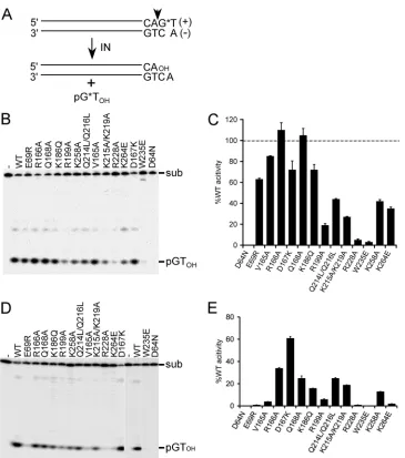
WT and IN mutant 2-LTR circle junctions (CJs) amplified by nested PCR were cloned and sequenced essentially as previously described (83). In brief, 1g of cellular DNA prepared 24 h after infection was amplified (50l) in triplicate by usingPfuUltra DNA polymerase using the primer pair AE4395 (5=-GCACCATCCAAAGGTCAGTGGATATCTG)/AE4450 FIG 13=-processing substrate design and IN activities. (A) Blunt-ended vDNA substrate, highlighting the subterminal CA 3=dinucleotide that is conserved among all retroviruses. IN processes the indicated phosphodiester bond (vertical arrowhead), releasing labeled pGTOHdinucleotide. *, positions of32P label. (B)
Polyacrylamide gel image of Mn2⫹-dependent 3=-processing reactions, highlighting the 21-bp vDNA substrate (sub) and pGT
OHcleavage product. IN was
omitted from the reaction mixture loaded in the first lane (-). (C) Mutant 3=-processing activities of the products shown in panel B, plotted as the percent INWT
function. Results are means⫾standard errors of the means (SEM) for two independent experiments. (D) The same experiment as in panel B, except reaction mixtures contained MgCl2and the vDNA substrate was 30 bp long. (E) Cumulative Mg2⫹-dependent 3=-processing activities expressed as the percent INWT
function⫾SEM for two (most mutants) to four (INW235Eand IND64N) independent experiments. Li et al.
on November 7, 2019 by guest
http://jvi.asm.org/
[image:4.585.112.477.61.474.2](5=-GCCTGGGAGCTCTCTGGCTAA). After 5 min at 95°C, reactions were cycled 35 times at 95°C (15 s), 50°C (1 min), and 72°C (30 s), fol-lowed by a final 5-min extension at 72°C. Resulting DNAs (5l) were amplified by nested PCR (50l) in triplicate using the primers AE2948 (5=-AACTAGGGAACCCACTGCTTAAG)/AE4394 (5=-GTGTGTGGTA GATCCACAGATCAAGG) under first-round reaction conditions. Pooled DNAs concentrated by precipitation with ethanol and purified by using a SNAP UV-free gel purification kit (Invitrogen Corp.) were cloned using the Zero Blunt TOPO kit (Invitrogen Corp.). Plasmid DNA quences obtained from the M13 reverse primer were parsed for LTR se-quence content and aligned by eye.
RESULTS
Experimental strategy.
HIV-1 IN proteins derived from class II
mutant viruses can efficiently
trans
-complement the infectivity
defects of class I mutant viruses and support appreciable levels of
enzyme activity
in vitro
, suggesting that some if not most class II
viral infection defects may lie in the inability of the mutant IN to
properly engage host cell cofactors (6, 15, 29, 49, 59, 78, 128, 130).
Our initial goal was to test this hypothesis by systematically
eval-uating representative class II IN mutant protein activities under a
variety of conditions that included the concerted integration of
two vDNA ends, a relatively underutilized stringent measure of
HIV-1 IN activity (51, 70).
Missense mutations throughout the IN coding region can elicit
the class II mutant viral phenotype (31, 32), and 12 representative
proteins that we and others had previously analyzed under
sim-plified integration reaction conditions were selected for study. The
mutant INs harbored amino acid substitutions in the CCD (E69R,
V165A, R166A, D167K, Q168A, K186Q, and R199A), the CTD
(R228A, K258A, and K264E), or the CCD-CTD interdomain
linker (Q214L/Q216L and K215A/K219A) (72) (Table 1). Glu69,
Val165, Arg166, Asp167, and Gln168 in particular help to form
the LEDGF/p75 binding pocket at the CCD-CCD dimer interface
(15, 22, 102), whereas Lys186 forms an intermolecular salt bridge
with NTD residue Glu11, which is important for IN
tetrameriza-tion (5, 51, 86). Arg199, Lys219, Arg228, and Lys264 contribute to
vDNA binding (64, 129), while Lys258 forms part of the CTD
binding interface with RT (123). The interdomain linker
muta-tions, which target part of a putative bipartite nuclear localization
signal (49), yield variable levels of nuclear import defects (3, 78,
95), with more robust defects observed when the mutations are
combined with additional IN changes in some (49, 59), but not all,
(95) studies. The reported single-round infectivities of the
corre-sponding mutant viruses varied from undetectable (
⬍
0.01%) to
approximately 6% of WT for HIV-1
D167K(Table 1). Two proteins
derived from class I IN mutant viruses were used as controls:
IN
D64N, harboring a conservative change of active site residue
Asp64 (27, 37), and IN
W235E, with a change of a conserved CTD
residue (67, 68). The IN proteins, expressed in
E. coli
, were
puri-fied to
ⱖ
90% homogeneity by using Ni-NTA and cation exchange
affinity chromatography methods.
3
=
processing of vDNA ends.
The 3
=
-processing substrates,
which modeled the HIV-1 U5 vDNA end, were labeled by
incor-porating radionuclide T at the plus-strand terminus, allowing
re-actions to be monitored through formation of labeled pGT
OHcleavage product in denaturing polyacrylamide gels (Fig. 1A and
B) (41, 121). IN
WTactivity was defined as the percent substrate
converted to product, and mutant activities were quantified
rela-tive to IN
WTvalues (Fig. 1C; results are summarized in Table 2).
Divalent metal ion is an essential catalytic cofactor, and because
mutant activities were typically reported using Mn
2⫹and a 21-bp
vDNA substrate (37, 40, 78, 102), these conditions were initially
used to facilitate comparisons between studies. The class II IN
mutant proteins supported a wide range of function under these
conditions, which ranged from a low of 5% of WT activity for
IN
R228Ato a high of 110% for IN
R166A(Fig. 1B and C; Table 2).
These results agree well with our own studies (78, 102, 129) as well
as other previous work (117).
The class I IN
D64Nmutant protein failed to support the
forma-tion of detectable reacforma-tion products, as expected (27, 37, 78). The
[image:5.585.41.546.79.267.2]activity of IN
W235Ewas, however, unexpectedly low (3% of IN
WT)
TABLE 2Activities of IN mutant proteinsa
Change(s) in protein
Relative activitya
3=processing DNA strand transfer
Disintegration
Mn2⫹ Mg2⫹ Mn2⫹ Mg2⫹
D64N ⫺ ⫺b ⫺b ⫺b ⫺
E69R 63 (1.0) 1.0 (0.0) 64 (1.5) 3.0 (0.5) 53 (5.5)
V165A 85 (0.5) 4.0 (0.0) 150 (7.3)c 24 (3.6) 35 (3.0)
R166A 110 (7.0) 34 (0.5) 136 (5.0) 94 (17) 56 (7.0)
D167K 72 (8.5) 61 (1.5) 144 (4.5) 119 (7.0) 81 (26)
Q168A 105 (6.5) 25 (2.0) 155 (11) 71 (14) 41 (12)
K186Q 72 (5.0) 16 (0.0) 13 (6.0) 28 (5.0) 76 (4.0)
R199A 19 (1.5) 6.0 (0.5) 4.0 (1.5) 2.0 (0.0) 329 (63)
Q214L/Q216L 44 (0.5) 25 (0.5) 27 (4.5) 3.0 (0.5) 34 (9.5)
K215A/K219A 27 (0.5) 19 (0.0) ⫺ ⫺ 57 (14)
R228A 5.0 (1.0) 1.0 (0.0) 3.0 (2.5) ⫺ 139 (25)
W235E 3.0 (0.5) ⫺b 2.0 (0.5)b ⫺b 41 (0.5)
K258A 42 (1.5) 13 (0.0) 33 (2.5) 3.0 (1.5) 152 (8.5)
K264E 35 (1.5) 2.0 (0.0) 37 (8.3)d ⫺ 49 (3.5)
aRelative to IN
WT, which in each case was set to 100%. Values are means and standard errors of the means (in parentheses) for 2 independent experiments unless otherwise
indicated.⫺, undetectable (⬍1% of INWTactivity). bBased on 4 experiments.
c
Based on 5 experiments.
dBased on 3 experiments.
on November 7, 2019 by guest
http://jvi.asm.org/
(Table 2), as this enzyme was previously reported to support WT
levels of Mn
2⫹-dependent 3
=
-processing and DNA strand transfer
activities (68). Two additional preparations of this mutant were
therefore made from a sequence-reverified bacterial expression
vector. Because all three preparations displayed the same activity
profile, we concluded that IN
W235Eis defective for 3
=
-processing
and DNA strand transfer activities (see below).
The concentration of Mg
2⫹in the human body is several
or-ders of magnitude greater than that of Mn
2⫹(74), indicating that
Mg
2⫹is the physiologically relevant IN catalytic cofactor during
HIV-1 infection. Mg
2⫹-dependent 3
=
-processing activities were
accordingly evaluated next. Likely due to the requirement for
IN-vDNA contacts distal from the reactive IN-vDNA end (44), somewhat
longer substrates, in the range of 30 to 35 bp, support robust IN
function in the presence of Mg
2⫹(36), and a 30-bp substrate was
accordingly used here. Relative IN activities were decreased across
the board under these conditions compared to prior conditions,
with greater-than-10-fold differences noted for IN
E69R, IN
V165A,
and IN
K264E(Fig. 1, compare panels D versus B and panels E versus
C; Table 2). These results are consistent with previous findings
that certain vDNA mutations preferentially affect Mg
2⫹-dependent over Mn
2⫹-dependent IN activities (44).
Single vDNA end strand transfer activities.
The DNA strand
transfer substrate was labeled at the 5
=
end of the strand that
be-comes joined to target DNA, such that the products of single
vDNA integration were detected as a ladder of bands above the
substrate in sequencing gels (Fig. 2A and B). To enable
quantifi-cation of strand transfer activity independently from 3
=
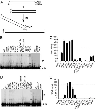
process-FIG 2DNA strand transfer assay and IN mutant activities. (A) Precleaved 30-mer vDNA substrate, indicating the position of 5=-end32P label (*). A second
oligonucleotide, which served as target acceptor DNA, is indicated by the bold lines. (B) Gel image highlighting migration positions of substrate (sub) DNA and Mn2⫹-dependent integration products (IP). (C) Mean activities⫾standard errors of the means (SEM) for three (IN
K264E), four (INW235Eand IND64N), five
(INV165A), or two (all other mutants) independent experiments under the panel B reaction conditions, plotted as the percent INWTactivity. (D) Gel image of
Mg2⫹-dependent DNA strand transfer activities. (E) Cumulative Mg2⫹-dependent activities expressed as the percent IN
WTfunction⫾SEM for two to four
(INW235Eand IND64N) independent experiments. Li et al.
on November 7, 2019 by guest
http://jvi.asm.org/
[image:6.585.111.476.66.481.2]ing, the substrate lacked the terminal GT dinucleotide that is
nor-mally removed by hydrolysis (Fig. 1A). Mn
2⫹-dependent DNA
strand transfer activities generally mimicked relative levels of 3
=
-processing activities, although two of the mutants (IN
K186Qand
IN
K215A/K219A) were at least 5-fold more defective at DNA joining
(compare Fig. 2B and 1B; Table 2). In line with the results of
3
=
-processing assays, most of the class II mutant enzymes were
defective for Mg
2⫹-dependent DNA strand transfer activity: only
IN
R166A, IN
D167K, and IN
Q168Adisplayed
⬎
50% relative activity,
with IN
K186Qand IN
V165Aharboring about 25 to 30% of IN
WTfunction (Fig. 2D and E; Table 2).
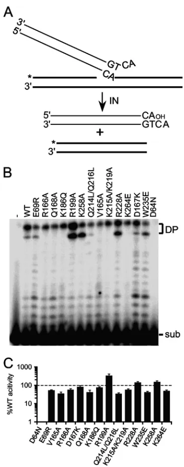
IN mutant disintegration activities.
The isolated HIV-1 CCD
is sufficient to catalyze disintegration activity (14), and NTD and
CTD mutant proteins defective for 3
=
-processing and DNA strand
transfer activities can accordingly support appreciable levels of
disintegration activity (37, 40, 68, 117). Because IN
W235Eand
IN
R228Asupported only about 2 to 5% Mn
2⫹-dependent 3
=
-processing and DNA strand transfer activities (Table 2), their
dis-integration functions were evaluated alongside the rest of the IN
proteins. The disintegration substrate mimics the product of
sin-gle U5 vDNA end integration, with label placed at the 5
=
end of the
disrupted target DNA strand (26). Nucleophilic attack by the
cor-responding 3
=
-OH group on the vDNA pA-target DNA
phospho-diester bond liberates the vDNA end and produces a joined,
labeled 30-bp strand (Fig. 3A). Consistent with our previous
re-port (37), active site mutant IN
D64Ndisplayed a very low level
(
⬍
1%) of IN
WTdisintegration activity. Class II IN mutant
activ-ities were noticeably more robust, ranging from a low of 34% for
IN
Q214L/Q216Lto a high of 329% for IN
R199A(Fig. 3B and C; Table
2). Because IN
W235Eand IN
R228Asupported about 41% and 139%
activity, respectively, we conclude that the active sites of these
enzymes are catalytically competent and that the CTD mutations
render the proteins effectively unable to catalyze vDNA 3
=
-end
processing or strand transfer activities. Functional enzyme active
sites underscore the ability of class II IN mutant proteins as well as
IN
W235Eto efficiently
trans
-complement the infection defects of
class I IN CCD mutant viruses (6, 47, 77, 78, 80).
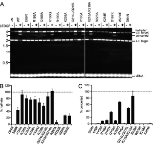
Concerted vDNA integration activities.
Unlike PFV IN (115),
HIV-1 IN is unable to efficiently integrate relatively short vDNA
substrates into target DNA in a concerted fashion in the absence of
added binding factors (13, 53). For reasons that are not entirely
clear, long vDNA substrates, in the range of several hundred base
pairs, support concerted HIV-1 IN integration activity under
cer-tain reaction conditions (70). We therefore generated a 5
=
-end-labeled 517-bp restriction enzyme fragment that harbored the
complete, blunt-ended U5 sequence at one of its ends to assess
concerted integration activity. Circular pGEM-3 target DNA
(2,867 bp) was included in the mixture to monitor the course of
the reaction: integration of a single vDNA end into this molecule
yielded a tagged nicked circle with relatively slow mobility in
aga-rose gels, whereas concerted integration of two vDNA ends after
deproteination yielded a population of approximately 3.9-kb
lin-earized pGEM-3 fragments (Fig. 4A to C). Preliminary
experi-ments with varied concentrations of IN
WTin the reaction mixture
were conducted to discern optimal levels of integration activity.
Consistent with prior reports (70), relatively minor fractions of
the vDNA substrate,
⬃
3% and 6%, were converted to the
con-certed and half-site products, respectively, under optimal
condi-tions (50 and 100 nM IN
WT) (Fig. 4B and C, lanes 1 to 4). IN
mutant activities were accordingly compared to IN
WTat protein
concentrations of 25, 50, and 100 nM. Resultant mutant half-site
and concerted integration activities were independently
quanti-fied as the percent IN
WTfunction at each protein concentration
(results are summarized in Table 3).
A subset of the mutant proteins, including IN
E69R, IN
R166A,
IN
D167K, IN
Q168A, IN
K215A/K219A, and IN
K264E, supported
rela-tively robust (
⬎
50% of IN
WT) half-site DNA strand transfer
activity (Fig. 4C, lanes 1 to 13, 29 to 31, and 35 to 40), with class
FIG 3Disintegration substrate design and IN activities. (A) Substrate illus-tration, highlighting vDNA (thin lines) and target DNA (bold lines) compo-nents as well as the position of the32P label (*). (B) Gel image showing the
migration positions of disintegration substrate (sub) and reaction products (DP). IN was omitted from the reaction mixture loaded in lane 1; the remain-ing lanes contained the indicated IN proteins. (C) Mean disintegration activ-ities⫾standard errors of the means for two independent experiments, ex-pressed as the percent INWTfunction.
on November 7, 2019 by guest
http://jvi.asm.org/
[image:7.585.328.513.62.531.2]I IN mutant control proteins IN
D64Nand IN
W235Epredictably
inactive (lanes 41 to 46) (results are summarized in Fig. 4D and
Table 3). Negligible concerted integration activities (2 to 5% of
IN
WTactivity at 50 nM and 100 nM [Fig. 4E and Table 3])
rather strikingly accompanied IN
E69R, IN
R166A, IN
Q168A, and
IN
K215A/K219A, indicating that these mutations perturb a
func-tion(s) that is central to the concerted integration of two vDNA
ends under these conditions. The remaining class II mutants
displayed half-site integration activities that ranged from a low
of 1% for IN
R228Ato 22% for IN
V165Awithout revealing
detect-able levels of concerted integration activity (Fig. 4C and Tdetect-able
3). Two of the class II mutants, IN
D167Kand IN
K264E, in
con-FIG 4Concerted integration assay design and IN mutant activities. (A) Schematic showing 5=-end-labeled blunt U5 substrate (thin lines) and circular plasmid target (bold lines) DNAs, as well as outcomes of single-end versus concerted vDNA integration. *, positions of32P label. (B) Results of preliminary titration
experiments (n⫽2) with the indicated INWTconcentrations, quantified as the percentages of substrate converted to half-site and concerted integration reaction
products. (C) Phosphorimage of agarose gel, highlighting migration positions of the 517-bp vDNA substrate and integration products. IN was omitted from the reaction mixture loaded in lane 1; the remaining reaction sets contained the indicated IN protein at 25 nM, 50 nM, or 100 nM (left to right). Positions of mass standards (in kb) are indicated to the left. (D and E) Quantitation of IN mutant half-site (D) and concerted (E) integration activities, expressed as a percentage of INWTfunction (means⫾standard errors of the means for two independent experiments).
Li et al.
on November 7, 2019 by guest
http://jvi.asm.org/
[image:8.585.111.478.60.560.2]trast, supported robust levels of concerted integration (Fig. 4C,
lanes 35 to 40, and E; Table 3).
DNA binding proteins HMGA1, HMGB1, and HIV-1
nucleo-capsid (NC) have been shown to significantly stimulate HIV-1 IN
concerted integration activity in some (16, 56) but not all (70)
studies. The lentiviral IN binding protein LEDGF/p75 (21, 24, 30,
114) has also shown variable effects on concerted integration
ac-tivity, which at low nanomolar vDNA concentrations depended
on protein concentrations (93) and/or the order by which they
were added to the reaction mixture (101). Increasing reactant
con-centrations to similar submicromolar levels of IN, LEDGF/p75,
and oligonucleotide vDNA substrate, in contrast, revealed potent
stimulation of concerted integration activity (53). We therefore
next evaluated IN mutant responses under conditions that
pro-mote efficient LEDGF/p75-dependent concerted integration
ac-tivity (53). Due to relatively high levels of vDNA substrate in these
mixtures and the reaction efficiency, substrates and products were
initially visualized following EtBr staining of agarose gels.
Integration of a single 32-bp vDNA end into pGEM-3 yields a
tagged circular product that migrates at the same position as
nicked plasmid DNA circles isolated from
E. coli
, whereas
con-certed integration in this case yielded a population of
approxi-mately 3-kb linear reaction products (53) (Fig. 5A, first three
lanes). In the absence of LEDGF/p75, IN
WTcatalyzed the
forma-tion of a low level of half-site integraforma-tion products without
detect-able concerted integration activity (Fig. 5A, lanes 1 and 2) (27, 53).
LEDGF/p75 significantly stimulated IN activity, such that the
brunt of the supercoiled pGEM-3 target DNA was consumed,
yielding palpable increases in half-site and concerted integration
reaction products (lanes 1 to 3). The effects of LEDGF/p75 on IN
mutant half-site (Fig. 5B) and concerted (Fig. 5C) integration
ac-tivities were accordingly quantified as a percentage of the IN
WTresponses. IN
D64Nand IN
W235Ewere each predictably defective for
both activities, whereas LEDGF/p75 stimulated a small amount of
IN
R228Ahalf-site activity in some but not all experiments (Table
3). Half-site integration activities of the other class II IN mutant
proteins were stimulated reasonably well by the host factor to
levels that varied from about 25% (IN
K258Aand IN
K264E) to 100%
(IN
K215A/K219A) of IN
WTlevels (Fig. 5A and B; Table 3). Akin to the
previous experiment (Fig. 4), mutant concerted integration
activ-ities varied more so than their corresponding half-site activactiv-ities.
For example, the concerted integration activities of IN
E69R,
IN
V165A, IN
R199A, IN
K258A, and IN
K264Ewere less than 10% of their
half-site values (Fig. 5; Table 3). IN
K215A/K219Aand IN
K186Q, in
contrast, catalyzed fairly similar levels of half-site and concerted
vDNA integration under these reaction conditions.
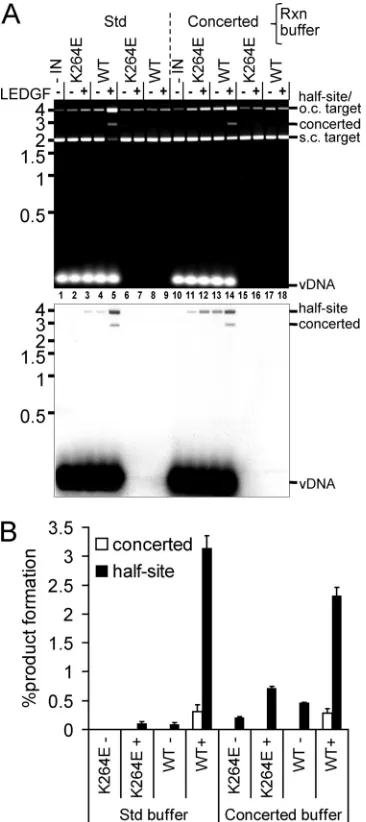
IN
K264Estood out as a protein that behaved rather differently
under different reaction conditions: it 3
=
processed and supported
the robust concerted integration of 517-bp vDNA ends, yet failed
to integrate precleaved 32-bp vDNA in a concerted fashion in the
presence of LEDGF/p75 (Fig. 4 and 5). IN
K264E-mediated 3
=
pro-cessing and integration of oligonucleotide vDNA substrate,
more-over, was largely Mn
2⫹dependent (Fig. 1 and 2; Table 2), yet its
robust response to 517-bp vDNA occurred in the presence of
Mg
2⫹(Fig. 4). To corroborate these findings, protein repurified
after resequencing the expression construct was analyzed, and this
confirmed the results. Aside from the specific divalent metal ion or
vDNA substrate, the oligonucleotide-based assays (Fig. 1 to 3)
were conducted under identical buffer conditions. In contrast, for
the concerted integration assay (Fig. 4) we used noticeably lower
levels of IN protein and included DMSO and the molecular
crowding agent PEG (70, 92, 101). To see if these parameters
con-tribute to differences in IN
K264Eactivities, 3
=
processing of the
30-mer oligonucleotide substrate was assessed under the buffer
conditions used for Fig. 4. Although this significantly suppressed
IN
WTactivity, IN
K264Enonetheless appeared equally active (data
not shown). LEDGF/p75-dependent integration activities were
also assessed under these buffer conditions. To increase the
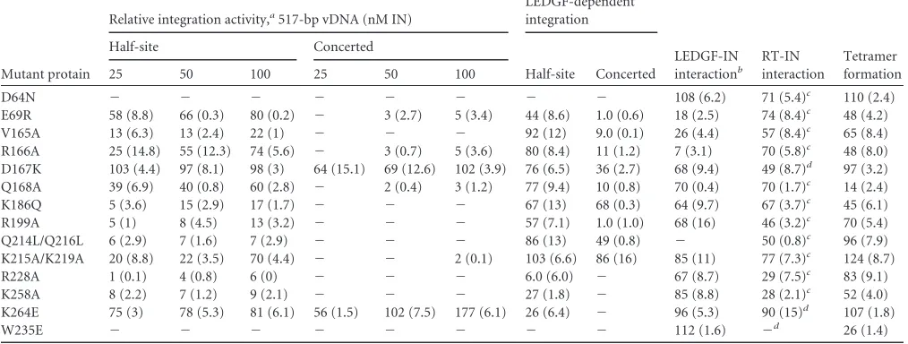
sensi-TABLE 3Relative activities of HIV-1 IN proteins
Mutant protain
Relative integration activity,a517-bp vDNA (nM IN)
LEDGF-dependent integration
LEDGF-IN interactionb
RT-IN interaction
Tetramer formation
Half-site Concerted
Half-site Concerted
25 50 100 25 50 100
D64N ⫺ ⫺ ⫺ ⫺ ⫺ ⫺ ⫺ ⫺ 108 (6.2) 71 (5.4)c 110 (2.4)
E69R 58 (8.8) 66 (0.3) 80 (0.2) ⫺ 3 (2.7) 5 (3.4) 44 (8.6) 1.0 (0.6) 18 (2.5) 74 (8.4)c 48 (4.2)
V165A 13 (6.3) 13 (2.4) 22 (1) ⫺ ⫺ ⫺ 92 (12) 9.0 (0.1) 26 (4.4) 57 (8.4)c 65 (8.4)
R166A 25 (14.8) 55 (12.3) 74 (5.6) ⫺ 3 (0.7) 5 (3.6) 80 (8.4) 11 (1.2) 7 (3.1) 70 (5.8)c 48 (8.0)
D167K 103 (4.4) 97 (8.1) 98 (3) 64 (15.1) 69 (12.6) 102 (3.9) 76 (6.5) 36 (2.7) 68 (9.4) 49 (8.7)d 97 (3.2)
Q168A 39 (6.9) 40 (0.8) 60 (2.8) ⫺ 2 (0.4) 3 (1.2) 77 (9.4) 10 (0.8) 70 (0.4) 70 (1.7)c 14 (2.4)
K186Q 5 (3.6) 15 (2.9) 17 (1.7) ⫺ ⫺ ⫺ 67 (13) 68 (0.3) 64 (9.7) 67 (3.7)c 45 (6.1)
R199A 5 (1) 8 (4.5) 13 (3.2) ⫺ ⫺ ⫺ 57 (7.1) 1.0 (1.0) 68 (16) 46 (3.2)c 70 (5.4)
Q214L/Q216L 6 (2.9) 7 (1.6) 7 (2.9) ⫺ ⫺ ⫺ 86 (13) 49 (0.8) ⫺ 50 (0.8)c 96 (7.9)
K215A/K219A 20 (8.8) 22 (3.5) 70 (4.4) ⫺ ⫺ 2 (0.1) 103 (6.6) 86 (16) 85 (11) 77 (7.3)c 124 (8.7)
R228A 1 (0.1) 4 (0.8) 6 (0) ⫺ ⫺ ⫺ 6.0 (6.0) ⫺ 67 (8.7) 29 (7.5)c 83 (9.1)
K258A 8 (2.2) 7 (1.2) 9 (2.1) ⫺ ⫺ ⫺ 27 (1.8) ⫺ 85 (8.8) 28 (2.1)c 52 (4.0)
K264E 75 (3) 78 (5.3) 81 (6.1) 56 (1.5) 102 (7.5) 177 (6.1) 26 (6.4) ⫺ 96 (5.3) 90 (15)d 107 (1.8)
W235E ⫺ ⫺ ⫺ ⫺ ⫺ ⫺ ⫺ ⫺ 112 (1.6) ⫺d 26 (1.4)
aRelative to IN
WT, which was set to 100%. Values are means, with standard errors of means in parentheses for 2 independent experiments unless otherwise indicated.⫺,
undetectable (⬍1% of INWTactivity). bResults of GST-LEDGF
326-530pulldown (Fig. 10A and B). c
Based on 3 experiments.
dBased on 4 experiments.
on November 7, 2019 by guest
http://jvi.asm.org/
[image:9.585.40.547.84.276.2]tivity of the assay, radiolabeled vDNA was incorporated at 5% of
the total substrate concentration (Fig. 6). IN
WTconverted
approx-imately 0.3% and 3% of the precleaved 32-bp substrate to
con-certed and half-site integration products, respectively, in the
pres-ence of LEDGF/p75 under both sets of reaction conditions (Fig.
6A, lanes 5 and 14; quantified in panel B). Although the conditions
that included PEG and DMSO (denoted as concerted buffer in Fig.
6) marginally stimulated IN
K264Ehalf-site integration (Fig. 6A,
bottom panel, compare lanes 11 and 12 to lanes 2 and 3),
con-certed integration activity remained undetectable. We therefore
inferred that although IN
K264Eprefers the buffer conditions used
for the experiment in Fig. 4, its robust concerted integration
ac-tivity seems particularly dependent on an aspect(s) of the
rela-tively long vDNA substrate (Fig. 4 and 6).
In vitro
activities of PICs extracted from acutely infected
cells.
Our results clarified that some class II IN mutants, in
partic-ular IN
E69R, failed to support concerted integration despite being
able to robustly integrate single vDNA ends under a variety of
reaction conditions (Tables 2 and 3). We therefore concluded that
defective IN function underscores the HIV-1
E69Rinfection defect
(102). To further address the link between purified IN activities
and HIV-1 biology, we next characterized the profiles of a subset
of mutants that supported robust concerted integration in the
context of virus infection. HIV-1
K215A/K219Aand HIV-1
K264Ewere
selected due to the relatively robust responses of these INs in one
of the utilized assays (Table 3). As HIV-1
D167Kretained about 6%
of WT infectivity (Table 1) and IN
D167Kcatalyzed concerted
vDNA integration under both sets of assay conditions, it was
in-cluded for comparison. SupT1 T cells 7 h after acute infection with
WT or IN mutant virus were lysed, and extracted PICs were
incu-bated with an excess of plasmid target DNA in the presence of
Mg
2⫹ions. Deproteinized extracts were queried by different
Q-PCR assays to ascertain levels of reverse transcription that
oc-curred prior to cell lysis and subsequent
in vitro
integration
activ-ities (42, 80). Of note, the Q-PCR PIC activity assay monitors the
formation of U5 vDNA end-target DNA junctions and therefore
does not specifically assess the concerted integration of vDNA U3
and U5 ends (42, 80).
PIC analyses rely on relatively high-multiplicity infections,
which were accomplished using viral supernatants freshly
har-vested from HEK293T cells (42). Although this approach
side-stepped normalization of input WT and IN mutant viral inocula
based on RT activity or p24 content, the experiment was internally
controlled through parallel assessment of vDNA substrate levels in
each of the cell extracts. HIV-1
D167Kand HIV-1
K264EPICs
sup-ported the formation of strand transfer reaction products,
whereas HIV-1
K215A/K219Adid not (Fig. 7A). Each virus also
re-vealed a reverse transcription defect, which under these infection
conditions varied from about 4% of WT for HIV-1
K264Eto about
35% for HIV-1
K215A/K219A(Fig. 7B). Specific PIC activities,
calcu-lated by normalizing integration activity to corresponding levels
of viral substrate DNA, revealed that HIV-1
D167Ksupported
⬃
26% of HIV-1
WTfunction, whereas HIV-1
K264Eintegration
ac-tivity exceeded that of the WT (Fig. 7C). Assessment of the
num-FIG 5LEDGF/p75-dependent integration activities. The assay design was similar to that shown in Fig. 4A, but here we used a significantly shorter (32-bp versus 517-bp) vDNA. (A) Agarose gel image revealing migration positions of vDNA substrate, supercoiled (s.c.) and open circular (o.c.) pGEM-3, as well as half-site and concerted integration reaction products. IN and/or LEDGF/p75 was omitted from reaction mixtures as indicated above the gel image. Migration positions of mass standards (in kb) are shown to the left. (B) Half-site integration activities of IN mutant proteins relative to INWT(set to 100%); means⫾standard errors
of the means for two independent experiments are shown. (C) IN mutant concerted integration activities relative to INWTfrom 2 experiments. Li et al.
on November 7, 2019 by guest
http://jvi.asm.org/
[image:10.585.136.451.64.358.2]ber of viral particles in HEK293T cell supernatants prior to SupT1
cell infection revealed an
⬃
35% release defect for HIV-1
K264Erel-ative to the WT (Fig. 7D). Correction for this difference revealed
the normalized level of HIV-1
K264Ereverse transcription to be
⬃
6% of the WT.
DNA synthesis profiles of IN mutant viruses.
The results of
the previous experiment revealed cytoplasmic HIV-1
K264EPICs,
which formed, admittedly, at significantly lower levels than
HIV-1
WT, nevertheless supported efficacious integration activity
in
vitro
. Single-round HIV-1 constructs defective for replicative
spread were utilized next to assess the fates of WT and IN mutant
DNAs during acute infection. The use of such constructs
previ-ously revealed approximate 5- to 10-fold reverse transcription
de-fects for HIV-1
D167K(102) and HIV-1
K264E(77), similar to the
responses observed in the previous experiment. Because
pseu-dotyping with the VSV-G envelope glycoprotein can significantly
stimulate levels of class II IN mutant reverse transcription relative
to the WT control (9), the heterologous envelope glycoprotein
was employed to boost the comparatively low level of HIV-1
K264EDNA synthesis. Cell extracts prepared at 2, 7, 24, and 48 h after
infection were queried by different Q-PCR assays to determine
products of LRT, 2-LTR circles, which can serve as a marker for
PIC nuclear import, and integration (Alu-PCR). HIV-1
D64N/D116Nwas used as a catalytically inactive IN mutant control.
Consistent with prior findings (9), VSV-G significantly
stimu-lated relative levels of class II HIV-1
K264Eand HIV-1
D167KIN
mu-tant reverse transcription, to
⬃
32% and 85% of HIV-1
WT,
respec-tively, at 7 h postinfection (Fig. 8A, compare with 7B), whereas
HIV-1
D64N/D116Nbehaved as the WT. The formation of HIV-1
WT2-LTR circles peaked at 24 h after infection, with the level of class
I mutant HIV-1
D64N/D116Nproduct expectedly significantly
out-weighing that of the WT (Fig. 8B) (75). HIV-1
K264Eformed circles
at a level that was indistinguishable from the WT, whereas
HIV-1
D167Kcircles formed at about 45% of the WT (Fig. 8B).
HIV-1
D167Kintegration peaked at about 6.5% of the WT level, equal to
the relative infectivity of this virus (Fig. 8C and D). Infectivity
under these conditions was therefore directly proportional to the
level of chromosomal vDNA integration. Consistently, the levels
of HIV-1
K264Eand HIV-1
D64N/D116Nintegration were below the
detection limit of the assay (Fig. 8C and D).
HIV-1
K264Eeffectively formed 2-LTR circles, indicating that
this infection was blocked after PIC nuclear entry (Fig. 8B). The
sequence of the associated 2-LTR circle junction (CJ), which
re-FIG 6LEDGF/p75-dependent INWTand INK264Eintegration activities under
different reaction buffer conditions. (A) EtBr stained image (upper panel) and phosphorimage (lower panel) for integration reactions conducted under stan-dard (Std) conditions (Fig. 5) or with the same protein, vDNA, and pGEM-3 concentrations under the Fig. 4 reaction conditions (“concerted”). Migration positions of substrates and reaction products are indicated to the right of the gel panels, whereas mass standards (in kb) are shown on the left. (B) Quanti-tation of panel A phosphorimaging results for 2 independent experiments (means⫾standard errors of the means).
FIG 7PIC formation andin vitrointegration activities. (A) PIC activities in extracts prepared from cells infected with the indicated viruses. KK/AA, K215A/K219A. (B) The same experiment as in panel A, except the Q-PCR detected the corresponding levels of vDNA substrate in the integration reac-tion mixtures. (C) PIC activities from panel A, normalized for levels of vDNA substrate (from panel B). Standard errors were calculated as M1/M2公(SEM12/
M1 2⫹SEM
2 2/M
2
2), where M
1is the average PIC activity, M2is the average
vDNA level, SEM1is the standard error of the mean of PIC activity
measure-ments, and SEM2is the SEM from vDNA measurements. (D) Relative levels of
RT activity in HEK293T cell supernatants prior to SupT1 cell infection.
on November 7, 2019 by guest
http://jvi.asm.org/
[image:11.585.300.542.63.263.2] [image:11.585.73.256.67.478.2]sults from the ligation of U3 and U5 vDNA ends by the cellular
nonhomologous DNA end-joining repair pathway (69), is
accord-ingly a metric of vDNA end integrity (62). Certain INSTIs (110) as
well as the relatively inhospitable environment of quiescent CD4
⫹T cells (119) significantly increased the frequency of 2-LTR CJ
deletions, indicative of reduced vDNA end integrity prior to
liga-tion. HIV-1
WT, HIV-1
K264E, and HIV-1
D64N/D116N2-LTR CJs
from acutely infected cells were accordingly cloned and sequenced
to assess relative end integrity of these vDNAs. Resulting
se-quences were parsed into four categories, including (i) the perfect
CJ sequence, ATCTCTAGCA
GTAC
TGGAAGGGCT (boldface
corresponds to the GT dinucleotide that is normally removed by
IN from the U5 plus-strand and U3 minus-strand termini,
respec-tively, prior to end ligation); (ii) mutation, which harbored one or
more base pair substitutions within this sequence; (iii) deletion,
with a minimum of 1 bp absent; (iv) insertion. Approximately
25% of the WT sequences were perfect CJs, with deletion and
insertion sequences accounting for about 35% each (Fig. 9; Table
4). Most insertions contained an intact copy of the polypurine
tract (PPT) or primer binding site (PBS) sequence, indicative of
aberrant primer removal by RNase H during reverse transcription
(62, 87). As expected (110), the lack of IN activity significantly
increased the frequency of perfect CJ sequences recovered from
HIV-1
D64N/D116N-infected cells, with a notable decrease in the
number of deletions (Table 4 and Fig. 9). Because the frequency
of HIV-1
K264Edeletion junctions was likewise lower than with
the WT, we inferred that the ends of nuclear HIV-1
K264EDNA
were no more prone to degradation than were the WT ends
(Fig. 9; Table 4).
Interactions with heterologous binding partners. (i) LEDGF/
p75.
The results of the work described above highlighted that
HIV-1
K264Eforms active cytoplasmic PICs and accesses cell nuclei
during acute infection. As inferred from 2-LTR CJ sequences, the
mutant vDNA ends, moreover, appeared grossly similar to the
WT, indicating that they were not prone to preferential
degrada-tion by host nucleases (119). We therefore hypothesized that the
HIV-1
K264Einfection defect might result from the inability of
IN
K264Eto interact with a nuclear protein important for vDNA
integration. A specific interaction between IN and LEDGF/p75
dictates both the frequency and distribution of lentiviral
integra-tion (35, 97), and LEDGF/p75, moreover, failed to stimulate the
concerted integration activity of IN
K264Ein vitro
(Fig. 5 and 6). The
LEDGF/p75 binding profile of IN
K264Ewas therefore compared to
the WT and other IN mutants in reciprocal pulldown assays. In
one format, soluble IN was captured by bead-bound
GST-LEDGF
326-530protein (23). As previously reported (102), some
mutations in the LEDGF/p75 binding pocket, like E69R, V165A,
and R166A, impacted the host factor interaction more
signifi-cantly than other pocket mutations, such as D167K and Q168A
(Fig. 10A and B). The Q214L/Q216L CCD-CTD interdomain
linker mutation significantly perturbed the interaction, whereas
the overlapping K215A/K219A mutation, interestingly, did not.
Consistent with prior work that indicated that the NTD and CCD
fully account for the affinity of IN for LEDGF/p75 (53, 81), each of
the CTD mutant proteins, including IN
K264E, was efficiently
re-covered with GST-LEDGF
326-530(Fig. 10A and B). We previously
reported that a reciprocal Ni-NTA pulldown assay mediated by
C-terminally His
6-tagged IN revealed some LEDGF/p75 binding
defects not evident from the GST assay (102). Accordingly
IN
D167K, IN
Q168A, and IN
K186Qeach failed to appreciably capture
LEDGF/p75 under this condition (compare Fig. 10C and D with A
and B, respectively). Because IN
K264E, by contrast, retained its
ef-ficient interaction with the host factor (Fig. 10C and D), we
con-clude that the K264E mutation does not obviously perturb the
IN-LEDGF/p75 interaction.
(ii) RT.
The IN CTD mediates a direct interaction with RT (55,
123, 130), and RT can, moreover, stimulate IN DNA strand
trans-fer activity under certain assay conditions (55). The RT-IN
inter-action is furthermore ablated by the IN mutation K258A,
suggest-ing that loss of RT bindsuggest-ing affinity might dictate class II IN mutant
reverse transcription defects (123). Binding assays were therefore
performed to assess the affinity of RT for the different IN mutant
proteins.
Preliminary experiments with IN-His
6prebound to Ni-NTA
beads failed to specifically pull down heterodimeric p66/p51 RT
protein, as RT was occasionally recovered on beads lacking IN.
Because the isolated p66 subunit bound HIV-1 IN as efficiently as
the heterodimer (55), GST-RT(p66) protein was tested next in a
GST pulldown format. Preliminary experiments again revealed
evidence of nonspecificity due to occasional recovery of IN on
GST-loaded beads, which was remedied by first preclearing the IN
proteins by centrifugation after 15 min of preincubation in
pull-down assay buffer. Consistent with the approximate 61 nM (123)
and 11 nM (112) affinity constants reported for the RT-IN and
LEDGF/p75-IN interactions, respectively, GST-LEDGF
326-530in a
parallel control sample pulled down more IN than did GST-RT
(Fig. 11A, bottom panel, lanes 2 and 3). Our assay format,
more-over, confirmed that the K258A mutation lowered the apparent
affinity of IN for RT (Fig. 11A, lane 9; results quantified in panel
FIG 8Kinetics of WT and IN mutant viral DNA synthesis and integration. (A) LRT products of the indicated VSV-G-pseudotyped viruses (N/N, D64N/D116N) were amplified from cell extracts obtained 2, 7, 24, or 48 h after infection. Values are expressed as the peak WT product formation, which occurred at 7 h. (B) 2-LTR circle Q-PCR product levels, expressed as the percent WT value, which was set to 100% at the peak (24 h postinfec-tion). (C) Integrated proviruses assessed as nested Alu-PCR. The peak WT value at 48 h postinfection was set to 100%. (D) Normalized levels of single-round viral infectivities, expressed in RLU perg of total cell pro-tein. Compiled data are from two independent experiments (means⫾ standard errors of the means).
Li et al.
on November 7, 2019 by guest
http://jvi.asm.org/
[image:12.585.42.284.64.278.2]B). The R228A mutation reduced binding to a similar extent as
K258A, whereas, interestingly, the class I mutation W235E ablated
binding (lanes 13 and 16). Because IN
K264Ewas efficiently pulled
down by GST-RT (Fig. 11; Table 3), neither the HIV-1
K264Everse transcription nor integration defect was attributable to
re-duced affinity of the mutant IN for RT.
Mutational effects on IN multimerization.
HIV-1 IN tends to
form dimers and tetramers in solution, which can be monitored
through the use of covalent cross-linking reagents (4, 24, 34, 46,
51, 64, 71). Cross-linking was accordingly utilized to assess
self-association properties of mutant proteins by quantifying the
ex-tent of tetramer formation relative to IN
WT. Four general patterns
emerged from this analysis (Fig. 12). A few of the class II mutant
proteins, IN
V165A, IN
R199A, and IN
R228A, displayed marginal
de-fects that ranged from about 15% to 35% reductions in
cross-linking efficiency. Four mutants, IN
E69R, IN
R166A, IN
K186Q, and
IN
K258A, formed tetramers about half as efficiently as IN
WT,
whereas IN
Q168Aand the class I mutant IN
W235Ewere more
defec-tive, yielding about 14% and 26% relative tetramer formation,
respectively (Fig. 10A and B; Table 3). In contrast, IN
D64N,
[image:13.585.43.543.66.447.2]FIG 9Sequences of WT and IN mutant viral 2-LTR CJs. Sequences were grouped into four classes (perfect CJ, mutation, deletion, and insertion), as defined in the text. Numbers for each class are indicated below the viral names (N/N, D64N/D116N), with total numbers of sequences obtained for each virus tabulated above the sequences (shown in parentheses). The GTAC that represents the pGT dinucleotide normally processed by IN from the U5 plus strand and U3 minus strand is indicated in bold type. Mutations are indicated in lowercase letters, and deletions are shown as dashes. Numbers to the right of sequences (in parentheses) indicate the frequency at which the mutation was determined for that virus. *, sequence detected with⬎1 virus. PBS and PPT in the insertion category represent perfect matches to the viral primer binding site (GGCGCCCGAACAGGGAC) and polypurine tract (AAAAGAAAAGGGGGG), respectively, whereas underlined bases mark partial identities to these sequences. Internal numbers in parentheses mark additional lengths of DNA insertion, which in many cases extended from the PBS and/or PPT (62).
TABLE 4Sequence frequencies among WT and IN mutant 2-LTR circle junctionsa
Sequence class
CJ frequency
WT K264E N/Nb
Perfect CJ 0.25 0.29 0.59
Mutation 0.06 0.10 0.02
Deletion 0.35 0.20 0.11
Insertion 0.35 0.40 0.28
a
Sequences are illustrated in Fig. 9.
bN/N, D64N/D116N.
on November 7, 2019 by guest
http://jvi.asm.org/
[image:13.585.40.285.632.706.2]IN
D167K, IN
Q214L/Q216L, IN
K215A/K219A, and IN
K264Eformed
te-tramers as efficiently as the WT.
DISCUSSION
In vitro
activity assays and HIV-1 IN mutant biology.
Class II IN
mutant viruses harbor a variety of replication defects that can
seemingly span the entire replication cycle, from particle assembly
and release to uncoating, reverse transcription, PIC nuclear
im-port, and integration (recently reviewed in references 8 and 32).
Class I mutant enzymes that carry alterations in IN active site
residues typically do not support detectable levels of 3
=
-processing
or DNA strand transfer activities
in vitro
, whereas numerous INs
derived from class II mutant viruses were catalytically proficient
under assay conditions that did not distinguish the cutting and
joining of single vDNA ends from the concerted integration of two
ends, as occurs during virus infection. Class II IN mutant
en-zymes, moreover, efficiently
trans
-complemented the infection
defect inherent to class I mutant viruses (6, 47, 77, 78, 80),
sug-gesting that an underlying defect of class II mutant viruses
might be the inability of the associated IN protein to properly
engage critical binding partners. Here we addressed this
over-arching hypothesis by employing a number of
in vitro
assays
that included tests for the concerted integration of two vDNA
ends into target DNA.
Assays that utilized denaturing polyacrylamide gel
electropho-resis to separate strand transfer reaction products and hence did
not distinguish half-site from concerted integration activity
re-vealed that Mn
2⫹supported more robust IN mutant activities
than Mg
2⫹(Fig. 2 and Table 2). Mg
2⫹is the likely physiologically
relevant IN cofactor, and class II mutants IN
R166A, IN
D167K, and
IN
Q168Adisplayed relatively robust Mg
2⫹-dependent DNA strand
transfer activities, whereas IN
V165Aand IN
K186Qwere about 25 to
30% as active as IN
WT. As PICs derived from HIV-1
V165A-infected
cells did not support a detectable level of
in vitro
integration
ac-tivity (80), we concluded that assays that query Mg
2⫹-dependent
single vDNA end strand transfer activity, although more relevant
than those with designs that instead utilize Mn
2⫹, do in some, but
importantly not all, cases yield results reflective of viral IN mutant
function. This conclusion was underscored by the behaviors of
IN
E69R, IN
R166A, IN
Q168A, and IN
K215A/K219Ain the concerted
inte-gration assay (Fig. 4): each enzyme supported robust levels of
half-site integration activity yet negligible levels of concerted
in-tegration activity. Three of these mutations, E69R, Q168A, and
R166A, reduced inherent IN multimerization by approximately
2-to 7-fold (Fig. 12; Table 3), suggesting that defective
tetrameriza-tion in part accounts for the inability to effectively integrate two
vDNA ends in concerted fashion. LEDGF/p75, which has been
shown to enhance lentiviral IN tetramerization (51, 86),
accord-ingly stimulated the concerted integration activities of IN
K186Q,