AND
INTERCELLULAR COMMUNICATION
IN THE ALGA STIGEOCLONIUM
A thesis presented for the Degree of Doctor of Philosophy
CONTENTS
DECLARATION
ACKNOWLEDGEMENTS
ABSTRACT
CHAPTER 1: THE PROCESS OF BIOLOGICAL PATTERN FORMATION
1. 1 1. 2
1.
3
1.
4
1. 5
1. 6
1. 7
1. 8
Introduction - Pattern and Form.
The process of biological pattern formation. The field concept in developmental biology.
1 •
3.
1 The properties of morphogenetic fields.Models of pattern formation in developmental fields.
1 •
4. 1
1. 4. 2
Prepattern theories of biological pattern format ion.
The concept of positional information. lntercellular communication during field establishment and regulation.
Gene regulation s i gna 1.
Plant Development.
the interpretation of a positional
The alga, Stigeoclonium, as a developmental system.
(v)
(Vi)
(Vi i )
CHAPTER 2 DEVELOPMENTAL MORPHOLOGY AND LIFE CYCLE OF
2. 1 2.2 2.3
2.4
ST I GEOCLON I UM
Introduction.
General Methods and Materials. Results.
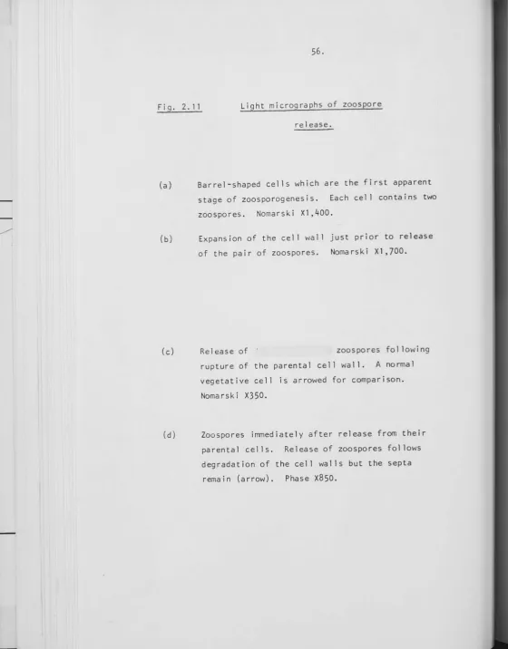
2. 3. 1 Morphology of Stigeoclonium. 2.3.2 Life cycle of Stigeoclonium. 2. 3. 3 Cell size a na 1 ys is.
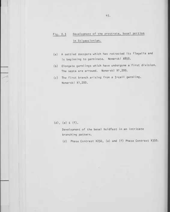
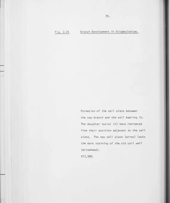
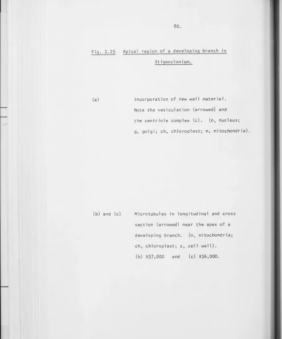
2. 3. 4 Cell Division. 2. 3. 5 Branch Development. 2. 3. 6 Norma 1 Branch Patterns.
General Discussion - Stigeoclonium as a developmental
system. 2. 4. 1
2.4.2
2. 4. 3
2.4.4
Ultrastructure of Branch Development. Cytokinesis.
Pl a smod esma ta.
Branch Patterns in Stigeoclonium.
CHAPTER 3 EXPERIMENTAL STUDIES OF BRANCHING PATTERNS IN
3. 1 3.2 3,3 3.4 3. 5 STIGEOCLONIUM Introduction.
Materials and Methods.
3, 2. 1 3, 2. 2 3.2.3
Physical treatments.
Drug treatments.
M i c ro s copy.
3, 2. 4 Studies on intercellular communication.
Results.
3. 3. 1 3.3.2 3, 3. 3
Discussion 3. 4. 1 3.4.2 3.4.3
Physical treatments.
Drug treatments.
lntercellular communication in
Stigeoclonium.
of Results.
Physical treatments.
Drug treatments.
lntercel lular communication in
S t i g eo c 1 on i um •
General Discussion on Branch Patterns in 3. 5. 1 The posit ion of branches. 3. 5. 2 The po 1 a r i ty of branches.
Stigeoclonium.
1 05 107
107 11 0
112
11 3
117 11 7 146
1 55
CHAPTER
4
A MODEL OF PATTERN FORMATION
4. 1
4.2
The universality of the process of pattern
format ion.
A model of pattern formation for Stigeoclonium.
4.
2. 1 4. 2. 24.
2. 3Gener a t i on of the s i g na l .
Transport of the signal.
The interpretation and expression
of the signal .
BIBLIOGRAPHY
210
216
217 219 221
DECLARATION
I declare that the work presented in this
thesis is my own original research and has
not been presented for a degree at any
other University.
L.B.DOOLEY
ACKNOWLEDGEMENTS
I am indebted to my supervisors Dr.H.J.Marchant and Professor B.E.S.Gunning for their guidance and support during this study. I wish to thank Professor D.J.Carr, Professor of Development Biology, Research School of Biological Sciences, Australian National University, for his continued encouragement and advice during this study.
Discussions with fellow research students, Allan Browning, Gordon Drake, Noah Pickles-Faud and Robin Scott have often aided this
research.
Financial assistance from the following sources is gratefully
acknowledged:
Commonwealth Government and the Australian National University for postgraduate scholarships.
The Australia-Japan Foundation and the International Society of
Developmental Biologists for travel grants which enabled the presentation of a paper (Dooley, L.B. and Marchant, H.J. Proceedings of the VI I Ith
International Congress of Developmental Biologists) in Tokyo at the VI I Ith International Congress of Developmental Biologists.
The Ian Potter Foundation and the European Molecular Biology Organisation for travel grants to attend the E.M.B.O. workshop on "Mathematical Models for Biologists" in Israel.
ABSTRACT
The developmental morphology and 1 ife cycle of Stigeoclonium was studied as a prelude to experimental investigations into the deter-minants of branching patterns in this alga. A general model based on the descriptive and experimental studies presented in this thesis, and placed in the broader context of plant and animal development, is discussed in the last chapter.
The filamentous green alga Stigeoclonium is among the simplest of branched organisms and grows by intercalary division without producing the complication of age gradients during pattern formation. It consists of a single file of cells. The eel ls are interconnected by plasmodesmata of approximately 30 nm in diameter.
In an unbranched erect filament of this alga al 1 cells are morphol-ogically equivalent, with the exception of the apical eel 1. The apical cell has a morphological polarity which none of the other vegetative eel ls show until they initiate branches. During branch development
the morphological equivalent of an apical eel 1 arises from an undifferent-iated vegetative eel 1 and later forms a highly differentundifferent-iated hyaline hair eel 1. Ultrastructurally branch initiation involves cytoplasmic
The branching patterns result from a dynamic equilibrium between
transverse eel l division and branch initiation. Cell division increases
the spacing between branches while branch initiation decreases the number
of cells between each branch. The spacing pattern most commonly has six
eel ls between the branches.
Experimental studies on the branching patterns in Stigeoclonium
involved a number of physical and drug treatments to investigate the
factors control] ing branch spacing and polarity.
The expression of branch polarity requires the movement of the
nucleus and associated cytoplasm to the site of polar branch initiation.
When this movement was prevented by maintaining cells at incipient
plas-molysis, apolar branches were produced from the centre of the cell at
right angles to the main filament. Altering the position of the nucleus
by centrifugation also led to a change in the site of branch initiation.
If bi nucleate eel ls are produced by caffeine treatment branches arose
adjacent to both nuclei. The movement of the nucleus was inhibited by
cytochalasin B but not colchicine treatment, suggesting that
micro-filaments may be involved in the polar migration of the nucleus during
branch initiation although no microfilaments were observed in
ultra-structural examinations.
Several physical techniques (plasmolysis, cutting, laser microbeam)
which disrupted symplastic continuity demonstrated the importance of this
pathway for transport from the apical eel ls to the base of the plant in
the potential to produce a branch and this potential is realised upon
temporary isolation from the inhibitory influence of apical eel ls.
Calcium may play a role in regulating the intercel lular transport
of a branch inhibitor. The localised deposition of cal lose near the
cross wal 1 areas during plasmolysis required free calcium, and EDAX
studies suggested a build up of calcium near the cross wal 1 areas during
plasmolysis. Prolonged treatments with the calcium ionophore A 23187
disrupted the normal polar initiation of branches and this could be due
to changes of an intracellular calcium gradient associated with the cell
cortex.
In the last chapter a model of pattern formation in Stigeoclonium
is discussed. The branching patterns in Stigeoclonium could be expressed
through a dynamic interplay between the nucleus, the cytoplasm and the
plasma-membrane which is determined by the intercel lular transport via
plasmodesmata of an inhibitor to branching. The data are consistent with
a model in which the position of branches is control led by the
intra-cellular transport of an inhibitor to branching which binds to a
CHAPTER 1
1. 1
1. 2
1. 3
1.
4
1.
5
1.
6
1.
7
1.
8
1 • 3. 1
1 •
4. 1
1. 4. 2
THE PROCESS OF BIOLOGICAL PATTERN FORMATION
Introduction - Pattern and Form.
The process of biological pattern formation.
The field concept in developmental biology.
The properties of morphogenetic fields.
Models of pattern formation in developmental fields.
Prepattern theories of biological pattern formation.
The concept of positional information.
lntercel lular communication during field establishment and regulation.
Gene regulation : the interpretation of a po s i ti o na 1 s i g na 1 .
Pl ant Development.
2 5 6
7
9 10 18 2124
25
1. 1 PATTERN AND FORM
The perception of patterns, both spatial and temporal, have been
of paramount importance in the evolution of mankind. The Palaeolithic
hunter-gatherers relied on their ability to predict the pattern of animal
and plant distribution. With the development of agriculture in the
Neolithic age, survival depended increasingly on the recognition of
seasonal weather patterns for planting and harvesting crops. Some of the
very earliest religions, such as Sun worship, almost certainly were born of
this increasing dependence on the seasonal cycles of nature. It is obvious
that 11
the need to recognise patterns arises in most human situations,
when-ever the brain receives information from any of the senses. The human
brain is a pattern recogniser par excel Jenee, providing a continuing
challenge to the scientist who can, at the most, suggest systems which
a re mere shadows of the i ncred i b 1 y powerf u 1 entity they seek to rep 1 ace. 11
(Duff, 1977).
Pattern has played an important part in artistic expression and
appreciation. Read (1949) has defined pattern as "some degree of
regularity within a 1 imited frame of reference", often perceived in a
work of art as 11the distr·ibution of line and colour in certain definite
repetitions. 11
By contrast, Read (1949) defines the form of a work of art as
11
its shape, the arrangement of its parts11
which does 11
not imply regularity,
or symmetry, or any kind of proportion.11 The distinction between pattern
defined as the spatial control of eel lular differentiation while form refers to changes in shape and the forces that generate it (Wolpert, 1971).
Much of the beauty of nature is derived from our perception of
patterns (Stevens, 1976). Four basic patterns are repeated throughout
nature: the spiral; the random meander; the explosion; and various branching patterns. Joining random points in space so that two points
connect along only one path with a central point results in variations of
these four basic patterns. (Fig.1.1). Space-fil 1 ing models show that branching patterns combine a shortness as well as a directness of path
when 1 inking points in space. The widespread occurrence of branching
patterns in such diverse systems as rivers, arteries and trees may be a
consequence of their favourable space-f i 11 i ng properties.
Pattern formation is one of the major unsolved problems in
biology. As with many problems in biology, pattern formation can be
analysed at a number of levels of organisation. At the phenomenological
level the rules governing the development of multicellular organisms can
be studied, while at the biochemical level studies on intercellular
communication and gene regulation can be undertaken. One of the most intractable problems is the identification of the elusive morphogens
which are held to control the development of patterns. However, it is likely, as several authors have maintained (Wolpert, 1971; Cooke, 1975), that an understanding of pattern formation at the phenomenological level
Fig.1.1 The Four Basic Patterns in Nature
Joining random points in space (a)
so that two points connect along
only one path with a central point
results in variations of four basic
patterns: the spiral (b); the random
meander (c); the explosion (d); and
branching patterns (e).
(From Stevens, 1976).
t.'}<o..."'f\~s o~ 1::kse.. ~ov..,r- \>os;c pa 1-tei"nS ~or 'c.\.,.e mu.\1--ic..d\'-'-\"'-"' c\..<L\l<?.\opY"\U'\t- 0
•
•
00 0
.
• • 0 0 0
0 0
• 0 0
0
0 0 0
.
.
0 0 0
• 0 0 0
0 • 0 0
•
•
0 0•
0• •
00
• 0 0
0
0 0 0 0
0 • •
•
•
.
.
0.
0
.
00
0 0
0
• • 0 0
• 0
•
0 0•
•
0
•
0 •
•
•
a•
1. 2 THE PROCESS OF BIOLOGICAL PATTERN FORMATION
Pattern formation involves dynamic spatial and temporal
inter-actions that generate and maintain a regular pattern during the development
of an organism. As with most complex biological processes, pattern
formation can be analysed at various levels of its organisation. Initially
pattern formation was studied at the multicellular level and the concept
of the developmental field was introduced to explain the regulative
features of patterns. The concept of the morphogen was proposed in an
attempt to explain the biochemical control and integration within the
organism of developmental processes. The major theories that have been proposed involve the intercellular diffusion of morphogens to form a
prepattern (Turing, 1952) or developmental gradient (Wolpert, 1971). For
example, neurosecretory chemicals which can promote formation of head and
tentacles (Schaller, 1973) and suppress formation of buds in Hydra
(Berking, 1977) have been isolated. However,the recent finding that
nerve-free hydras can undergo normal pattern-regulation (Marcum and
Campbell, 1978; Campbell, 1979) suggests that these neurosecretory factors
are either not essential for the development of Hydra, or at least not
the only means by which it can take place. Therefore there is, as yet, no unequivocal evidence that any one chemical can truly be regarded as a
morphogen in the sense required by pattern formation theories. Thus
although the phenomenological explanation of pattern formation has been successful it has proved difficult to extend this to the biochemical level.
In essence, the process of pattern formation in a developmental
field can be considered as the generation of a signal, its intercellular
In this chapter properties of developmental fields are discussed and various theories that have been proposed to explain the generation of the signal, its transport, and its interpretation are compared.
THE FIELD CONCEPT IN DEVELOPMENTAL BIOLOGY
The field concept in developmental biology arose from Driesch's discovery that spatial position is as important as developmental age in determining the fate of embryonic tissues (Driesch, 1908). He stated that the development of a part is a function of its position in relation to the whole. Child (1915, 1941) regarded the developmental field as a physio-logical gradient while Dalcq (1938) considered it as two opposite gradients with their ratios specifying position. The concept of "threshold values" of morphogenetic potential was introduced by Dalcq and Pasteels (1937, 1938) to explain the appearance of qua] itative differences within a field
initially characterized only by a quantitative variation of its morpho-genetic potential. It was held that after the threshold values had been established, development would be different either side of the threshold.
The history of the field concept in developmental biology has been surveyed by Nieuwkoop (1967). Summarizing the various concepts, he
defined the morphogenetic field as "characterized by a continuous variation of certain properties of the living biological system as a function of position and/or time, leading to an increase in the structural and functional multiplicity of the system." This definition has a dualistic character in that it not only includes the intrinsic properties of the system, but also describes its causal effect within the organism. B. l 10
secondary morphological differentiation: the end-product of a long chain
of reactions. Their detection, moreover, requires experimental
inter-vention to distinguish field effects from other strongly interwoven
developmental processes (Nieuwkoop, 1967).
Waddington (1966) regarded the developmental field as a topological
notion. "Any precise definition of a field must therefore require
reference to a multidimensional space, which would have axes on which one
could plot not only positions in time and the three dimensions of space
but also the concentrations of essential chemical compounds."
At present no precise definition of a field is possible and we
are left with the less than satisfactory definitions of Wolpert (1971)
"a field is that region in which all cells are having their positional
information specified with respect to the same set of points1' ; Robertson
and Cohen (1972): a field is 11
a collection of functionally coupled cells,
the development of which is under control of a single system"; and
Meinhardt (1978) : 11A field can be defined as an area which is controlled
by a set of d i f fu s i bl e mo r p hog ens . 11
Properties of morphogenetic (developmental) fields
Regulative Fields
Regulation refers to the capacity of developmental fields to
maintain an invariant pattern over a given size range. Two basic types
(a) Morphal lax is
Morphallactic regulation involves remodelling of the missing parts from the residue after cutting. In some planarians, and hydroids such as Tubularia, morphallaxis or rearrangement of existing cells rather than production of new cells at the cut surface results in the regeneration of the whole but diminished organism (Morgan, 1901).
(b) Epimorphosis
In epimorphosis there is growth of tissue to form a blastema in animals and callus in plants which then gives rise to the regenerated regions and produces a pattern in proportion to the whole organism. Epimorphosis has been most clearly demonstrated for amphibian 1 imb regeneration fol lowing amputation (Hay, 1966).
In Wolpert's (1969, 1971) theory of positional information duplication in epirrorphosis is explained if cells cannot have positional values more proximal than the level from which they were derived but they can have positional values corresponding to more distal regions.
2. Non-regulative or mosaic fields
squid and in general, most systems show both regulative and mosaic aspects,
but at different times. Some groups, such as insects, display mosaic ism
and regulation to varying extents. Development is probably always seen
to be regulative if the egg can be manipulated early enough (Cooke, 1975).
1
.4
MODELS OF PATTERN FORMATION IN DEVELOPMENTAL FIELDSThere are two major classes of models to explain biological
pattern formation: the prepattern theory and the positional information
theory.
In the prepattern theory a spatial singularity of the rrorphogen
is correlated with the site of cytodifferentiation. The weakness of the
theory is that it does not readily account for the regulative properties
of many developmental fields.
In the positional information theory a cell 1s position is
established by reference to a graded property between the two boundary
points of the developmental field. One attribute of positional information
theory is that it can readily explain the regulative properties of
developmental fields. One defect I ies in its dependence on a need to
fully understand the mechanism whereby a cel 1 interprets the positional
information.
Wolpert•s (1971) positional information analysis of pattern
formation is presented at the phenomenological level. By contrast, the
with biologically plausible reactions that yield a prepattern of spatial and temporal organization.
1 • 4. 1 Prepattern theories of biological pattern formation
Reaction - diffusion systems
Turing's Reaction-Diffusion Model
In a highly original paper, Turing (1952) suggested that morphogenesis could be explained by a system of interacting chemical substances cal led morphogens. He proposed that these morphogens formed a prepattern by diffusing through the tissue and reacting together auto-and cross-catalytically. This reaction-diffusion system develops a pattern because random disturbances bring about a breakdown in the inherently unstable homogeneous equilibrium.
The general form of the reaction-diffusion equations for two morphogens u and v can be written as fol lows (Fife, 1977):
2
D1 Vu + f(u,v) 1 a.
2
D2 V v + g(u,v) lb.
ut and Vt are the concentrations of the two morphogens at time t D1 and D2 are the diffusion constants
2
V is the three dimensional diffusion term
(o/
~ 2 +o/
y 2 +o/
z 2)auto-and cross-catalysis. Th~ Sou<c.e t"e.rMS ~ o..-.d.
3
f'e.o..c.r,o('\ k; A€.\:,c.S u a.Acl V.
The Gierer-Meinhardt Model of Pattern
Formation
The mode 1 of G i erer and Meinhardt ( 1972) is based on a
reaction-diffusion system of the activator-inhibitor type.
Referring to the reaction-diffusion equations la. and lb, u
would be an activator and v would be an inhibitor which interact
auto-and cross-catalytically. Increasing the amount of u further enhances
the production of u and v, whereas increasing v has the opposite effect~
Also the inhibitor diffuses more rapidly than the activator i.e. D2>D1.
The analysis of these equations by computer simulation (Gierer and
Meinhardt, 1972) has shown the appearance of spatial patterns of various
types. This model assumes a prexisting inhomogeneity in the system to
explain the generation of polarity and a shallow gradient for one of the
components. The stabi 1 ity of the pattern has not been established.
The Belousov-Zhabotinski Reaction as a model system
to study reaction-diffusion kinetics
Biologists have recently become interested in the
Belousov-Zhabotinski reaction as a model chemical system exhibiting both temporal
and spatial oscillations which are less comp] icated than biological
oscillators.
*
lh..s (es'<\~s ·," C\ ~eeJ.bctcX ·,"\\.--.;b;1:.;ov'\ "'h.<:. \... ;5. "I\E'c:eSs:c, 'j \;O ""'f'\.,._; I\c
o.i-f1lo.~ \01'.9-,o..V\.:1(2.. i "-\.-. \,; <c; 0 V\ ~ ... : .,le,\.\:
•"-
Sfc:\C; "j pQ.\-k~"s (su.-G;
The Belousov-Zhabotinski reaction consists essentially of the oxidation of malonic acid by bromate in the presence of a cerium catalyst which osci"llates between two oxidation states, cerous and eerie ions
(Atlan, 1975). The reaction oscillates regularly at intervals of about one minute producing travel I ing concentration waves which are associated with the dramatic changes of oxidation bands. The temporal oscillations
involve an autocatalytic reaction while the spatial oscillations are attributed to a reaction-diffusion process (Murray, 1976).
Such processes of auto-catalysis and reaction-diffusion were first suggested as a basis for biological morphogenesis by Turing (1952). Thus it is hoped that a greater understanding of the Belousov-Zhabotinski reaction may help to elucidate far more complex biological oscillations and ultimately provide insights into the process of biological pattern format ion.
Dissipative Structures and Development
Biological organisms are regarded as open systems in a state of dynamic equilibrium consisting of irreversible reactions which maximize free energy and minimize entropy (von Bertalanffy, 1952).
Prigogine commented in 1947 that 11classical thermodynamics is an
admirable but fragmentary theory. It is fragmentary because it can be applied only to equilibrium states in closed systems. One should there-fore attempt to find a more general theory which covers states of
Prigogine and his colleagues (see Nicolis and Prigogine, 1977)
have extended thermodynamics to irreversible processes and applied their
concept of dissipative structures to the origin, evolution and development
of 1 iving organisms.
In some systems far from equilibrium a type of order prevails
which has been termed "order through fluctuations." Although in a system
that is near to equilibrium any fluctuation away from that state will be
damped down and equilibrium rapidly restored, there also exists a class
of steady state systems, 1dissipative structures1 in which, under the
right conditions, fluctuations are amplified. These structures can then
change from a previous, ordered state to a new ordered state of lower
entropy. These systems must be steady-state, open and far from
equilibrium; the situation which prevails in biological systems. The
notion of a dissipative structure implies both a thermodynamic condition
(a critical distance from equilibrium) and a mathematical condition,
namely the bifurcation of a new solution arising beyond the instability
of the thermodynamic branch (Nicol is, 1975).
Prigogine has shown that dissipative structures can arise in
reaction-diffusion systems involving auto-catalysis such as Turing1
s
equations and the Zhabotinsky reaction (Nicolis and Prigogine,1977).
The fundamental advantage of this idea over older theories of pattern
formation (morphogenetic gradients) 1 ies in the fact that the
morphogenetic field is not postulated to be inhomogeneous from the
beginning. Rather it explains how inhomogeneity may arise.
Analysis of a reaction-diffusion system involving auto-catalysis
2
related to a parameter D/L where D is the diffusion coefficient of the
morphogen, L the length of the system (Prigogine and Lefaver, 1975). This
parameter measures the coupling between neighbouring spatial regions
and has therefore been termed the diffusive coupling. Naturally, the
2
diffusive coupling D/L depends strongly on the size of the system.
The same biochemical network could give rise to a spatial structure,
a propagating wave, or a uniform limit cycle, depending on whether it
operates in an isolated cell or in a macroscopic 11field of cells11
( N i co l i s , 1 9 7 5 ) .
It is proposed that during growth, the response of the
organism to distributions of morphogens gradually creates conditions
w h i ch des tab i 1 i z e the i n i t i a l pa t tern. At th i s po i n t of i n s tab i l i t y ,
a new pattern appears which in turn controls the developmental process
until itself destabilized by growth. Using computer simulation,
Martinez (1972) has analysed the role of eel lular division in this
succession of instabi 1 ities. It was found that the instabilities control
their own boundaries, or more exactly the relation between boundary values
and the system dimensions via cellular division. Cel 1 division alters
2
the ratio D/L which destabilizes the original pattern. This can be
summarized as fol lows:
NON-EQUILIBRIUM~~~~ INSTABILITY~~~~ DISSIPATIVE STRUCTURE
\
I
CELLULAR DIVISION
Size invariance in reaction diffusion
models of dissipative structures
Babloyantz and Hiernaux (1975) have defined the conditions
that are necessary for the non-1 inear systems (such as reaction-diffusion
models) to permit size invariance.
If the diffusion gradient of positional information is linear
as postulated by Crick (1970) and Wolpert (1971) then size invariance
implies that if a system of length~ has a concentration m of morphogen
at point rand if the size of the system is reduced to L' there must be
a point in the new system with the same concentration mas before and
such as:
r1/L1 r/L
For a reaction diffusion system size invariance requires that
the diffusion coefficient (Dm) afso cha~ges so
that:-2
D /L m
2
D1 /L' m
Recent studies of Lacall i and Harrison (1978) have shown a
regulative capacity of Turing's model when it is used to explain early
stages of pattern formation for slime moulds. Turing's model provides
a complete account of the initial establishment of the regions of cell
differentiation and their transient regulatory capacity. However,
Turing's model cannot give a good account of permanently maintained
Dissipative Structures and the development of Drosophila
A recent model of pattern formation in Drosophila (Kauffman~ .
1978) provides an elegant synthesis of many strands of thought including
gradients, thresholds, a binary combinatorial epigenetic code and
dissipative structures.
The development of the adult fly initially involves a
regional-ization of the embryo into a number of territories which will become the
various imaginal disks, and secondly, the subdivision of each of the
disks into compartments. Transdetermination of imaginal disks, whereby
the state of determination of a disk is changed, has occasionally been
observed. Homeotic~mutants can result in the conversion of one
compartment to another. It is believed that the compartment boundaries
are the thresholds between the on and off states of the wild type alleles
of so-cal led selector genes which undergo the homeotic mutations.
An analysis of transdetermination in the imaginal disks of
Drosophila led Kauffman(1969) to the conclusion that the selector genes
act 1 ike a binary switch capable of being fully on or fully off. The
states of determination of the disks are given by a combinatorial
'epigenetic code' or 'on' and 'off' states of the selector genes.
In a more recent model (Kauffman~. 1978) the switching of the
selector genes is controlled by a threshold concentration of a diffusible
morphogen. The spatial patterns in the morphogen are obtained by
considering a reaction-diffusion system as a dissipative structure so
~
H I \\ I o.,..osoec."•"e. I e_..ic\oo'Me."l\r<.l (c.1-c:sowi&c,~·,c. 'W\'-'TO."O+'S oC.C."'-"'" wl'\eY\ c.e. s w•lcn 1, 1 1 O. 1
0.S fQ.~\- 0~ OY\e. 01°'~0..1", O.C\'"e.\\J cli\~eHl\~ic.\-e jol\ thll- ~OY'M o~ Q. c..c,..,...rl~rt~
that various pattern modes are generated depending on the size of the
system and the diffusivity of the morphogen. As has been discussed
before the.pattern of spatial organization in a dissipative structure 2
depends critically on the ratio D/L with Das the diffusivity and L the
length of the system.
The early insect embryo (syncytium stage) does not grow in size,
but changes in the diffusivity of the morphogen due to eel lularization
at the blastoderm stage could initiate the symmetry breaking that is
necessary to establish new pattern modes. It is likely that as the
syncytium becomes partitioned into eel ls by eel l _membranes, resistance
to diffusion increases so that effective diffusion constants are made
smaller. If diffusion constants become smaller while the ratios of the
diffusion constants do not change, the effect is to shorten the unstable
wavelength. Mathematically, the effect is the same as if the unstable
wavelength remains constant and the spatial domain grows. As the
wavelength becomes shorter, a succession of different patterns fit on
the egg at a discrete succession of permitted wavelengths. The
sequential activation of each binary switch by the successive formation
of boundaries creates a binary combinatorial "code word11
specifying each
terminal compartment.
This model predicts the major features of Drosophila development.
The Drosophila egg is elliptical in shape and as a consequence of this the
model predicts that longitudinal compartmentation should proceed first,
followed by dorsoventral subdivision as the wavelength gradually shortens
predicts the laying down of both axes of the body with the same mechanism
and also a sequence of subdivisions which is consistent with the known
embryology of Drosophila.
The model predicts size invariance in the sequence and location of
compartmental 1 ines for moderate variations in the size of the egg. The
size invariance fol lows from the restrictions on the wavelength imposed by
the no-flux boundary conditions - the spatial gradient of concentration
at the boundaries must be flat along 1 ines perpendicular to the boundaries.
The further condition that there are only a smal 1 finite number of binary
switches 1 imits the number of chemical patterns that can be recorded and
acted upon by the egg. While the nodal compartment 1 ines are
size-invariant, they are not shape invariant.
The model also predicts the frequency of particular
trans-determinations based on the number of binary switches that must change.
The model's assumption that a homeotic mutation acts by altering the
states of a binary "selector gene" is consistent with the observed data.
1. 4. 2 The concept of positional information
Wolpert (1971) has formalized the gradient theories of development
first proposed by Dalcq (1938) and Child (1941) with the concept of
pas it i ona 1 information.
A positional value is regarded as a cellular property which is
graded from one end of a file of cells to the other between two boundary
with a map which al lows them to locate their position within the system or unit field of eel ls; a pattern is formed through the interpretation of the positional value by the particular cells. The concept of positional
information is probably best illustrated by Wolpert's French Flag model. Here the problem is to explain how a file of eel Is can form a pattern such that there is always one-third each of blue, red and white. Wolpert, (1969, 1971) proposed a simple source and sink model of a single gradient of positional information with threshold values specifying the
differentiation of the eel ls into blue, white or red. More complex models providing positional information could involve two intersecting gradients with the positional value determined by the ratio between them.
In terms of positional information, pattern formation is a two step process: first, the specification of positional information within the field, and secondly, the interpretation of this information by the cells, resulting in the expression of the pattern.
An important feature of Wolpert's theory is that the same positional information may be used to provide very different patterns, since variations of pattern mainly depend not on variant spatial
One of the most successful aspects of Wolpert1s theory is its
explanation of the regulative and size-invariant properties of
develop-mental fields. The property of size-invariance depends on the maintenance
of the boundary values of morphogen concentrations and the threshold
concentrations for differentiation over varying distances. Therefore a
smaller field will have a steeper gradient of morphogen but since the cells
have the same threshold for differentiation a smaller pattern with exactly
the same proportions wil 1 develop. In essence, the boundary values are
assumed to be fixed by a mechanism which is independent of the length of
the axis so that a regulating gradient results. Morphallactic and
epimorphic regulation in developmental fields is explained by the
con-servation of boundary values for the morphogen. In morphallaxis the
establishment of an identical boundary value for the morphogen at the cut
surface and the maintenance of a new gradient ensure size-invariance of
the pattern.
The success of \.lolpert1s model in explaining the phenomenological
aspects of development has not been extended to other levels of analysis.
The distinction between positional information and its interpretation
makes universality of developmental principles possible. Different species
could employ different codes for interpreting similar gradients of
positional information. Thus the burden for creating patterns from
monotonically changing spatial variables is then placed upon the eel lular
interpretative machinery. The positional information theory is in danger
of simply transferring the problem of pattern formation 11from one area to
another11
The phase-shift model
Goodwin and Cohen (1969) have proposed a phase-shift model for the spatial and temporal organization of developing organisms. They propose that a map of eel l position arises from wave-1 ike propagation of activity from localized cells acting as clocks or pacemakers. The gradient of positional value is established by the propagation of two waves with different velocities from a boundary pacemaker cell, the phase difference between the two waves providing a gradient in positional information.
The model is mathematically quite complex but a summary of its ii\ t.he. or igi"c..l
r"F""'
potential value is illustrated for regulating somite numbers in the /\
vertebrate embryo. A series of similar structures might form at regular intervals along the axis where maxima of the two periodic signals
coincided. A simple analogy is that of two sound waves of different frequencies which produce "beats" when their maxima coincide, at regular time intervals. Alterations in the frequency of the two signals could regulate the pattern within different tissue sizes.
The strength of the model is that it introduces the interaction between space and time in development. The temporal biochemical
organization of an individual cell is converted by functional coupling between eel ls into a spatial ordering of the temporal organization.
INTERCELLULAR COMMUNICATION DURING FIELD ESTABLISHMENT AND REGULATION
of two fundamental processes: random diffusion or a directed signal 1 ing
mechanism. Crick (1970, 1971) has discussed the imp] ications of both
these mechanisms.
A 1 inear gradient of morphogen can be established by diffusion if
a dynamic equilibrium is maintained between a source and a sink for the
morphogen. Diffusion is a random walk process and the dimensions of the
2 1
diffusion constant are LT- (L is length, T is time).
The mean distance of diffusion(;) in time t can be described by:
2 X
=
constant tThus diffusion processes are rapid over short distances but very slow over
large distances.
By contrast, a simple signal 1 ing process could be propagated at
1
constant velocity (dimensions LT ) such that:
X
constant
t
In this case the difference in time for short and long distance effects is
not as great as for a diffusional process.
Crick (1971) has divided signal 1 ing mechanisms into two broad
categories: amplitude decay and phase difference.
For the amplitude decay mechanism the amplitude of the signal could
output of each eel l was proportional to the input a eel l could use the
amplitude of the signal to give positional information.
The second type of signalling mechanism relies on the phase
difference between two signals as proposed by Goodwin and Cohen (1969).
The signals are synchronized at the source but travel with different
velocities so that a eel l can obtain positional information from the
phase difference between the two signals.
Based on considerations of the size of embryonic fields and the
time needed to set up a gradient, Crick (1970, 1971) has maintained that
diffusion can account for the morphogenetic gradients in embryonic
development. "Nature usually has such difficulty evolving elaborate
biochemical mechanisms (for example, those used in protein synthesis)
that the underlying processes are often rather simple 11
(Crick, 1970).
The plausibility of diffusional gradients for distances comparable
to embryonic fields has been established by Michalke (1977) using
monolayers of cultured cells. By culturing wild type rat liver epithelial
eel ls immediately adjacent to mutant eel ls which cannot incorporate
radio-active hypoxanthine into nucleic acids by themselves, gradients of
hypoxanthine-derived radioactivity have been observed in the mutant eel ls.
The gradient of radioactivity can be described by an exponential function
f -kD
o the form e , where D is the distance from the monolayer of wild-type
eel ls and k describes the slope of the gradient. The concentration of
radioactivity decreases by a factor of about 100 over a distance of 1 mm.
The gradient is presumed to depend on intercel lular diffusion, since this
communicative gap junctions are defective. Over certain concentration
ranges an exponential concentration gradient of diffusible ligand can be
converted to a 1 inear gradient by assuming binding of the 1 igand to a
macromolecular receptor which cannot diffuse from cell to cell (Michalke,
1977).
1.6 GENE REGULATION: THE INTERPRETATION OF A POSITIONAL SIGNAL
Development involves the sequential and regulated expression of
inherited capacities. Both the Operon model of gene regulation in bacteria
(Jacob and Monod, 1961) and the Britten-Davidson theory of gene regulation
in higher organisms (Britten and Davidson, 1969) involve control of
transcription during cell differentiation. Kauff~an(1969) has considered
the gene to be a binary switch either fully on or off and has developed
this to form complex networks of switches which could control the
development of the organism.
Pattern formation requires a mechanism for translating the
positional signal into a regulation of gene expression which results in
eel 1 differentiation at a particular position. The process of cellular
differentiation can be regarded as a dynamic interplay between the
nucleus, the cytoplasm and the plasma-membrane. It is possible that the
plasma-membrane may act as a central regulatory control point by
trans-duction of signals. A model of eel 1 differentiation involving interaction
between plasma-membrane and genome has recently been developed by Brunner
(1977). While the essential steps involved in the flow of information
from the nucleus to cell membrane are well known (transcription and
direction (membrane to nucleus) is scarce. It is believed that signal
molecules are bound to the plasma-membrane by receptor molecules and
initiate a sequence of reactions in the eel l which finally lead to the
expression of previously untranscribed parts of the genome.
The evidence for such a mechanism is well established for mitogens,
hormones, some enzyme inhibitors, and ions such as calcium (Brunner, 1977).
Some advantages of regarding the membrane as a transducer of signals to
the nucleus are that mosaic development could be explained by an unequal
and fixed distribution of receptors at the egg eel 1 surface. Also,
development which proceeds as a strict eel l 1 ineage could be a function
of sequential receptor expression. This model would require some control
of membrane receptor mobility and distribution, most probably by the
membrane-associated microtubules and microfilaments (Nicolson et al.
1977).
The crucial test of this promising theory of cell differentiation
as resulting from an interaction between the plasma-membrane and nucleus
has been suggested by Brunner (1972). The experiment would involve the
incorporation of membrane areas bearing differentiation receptors for
cell type A into intact cells of another 1 ineage (cell type B). An
attempt would then be made to transform eel 1 type B into cell type A
after stimulation of the type A differentiation receptors by species of
triggering molecule appropriate to them.
PLANT DEVELOPMENT
based on studies with animal systems. It is ironical that although Turing saw some applications of his important reaction-diffusion model
to phyl lotactic patterns in plants, very few detailed models of pattern
formation for plants have been considered until relatively recently. This
is possibly because studies on pattern formation in plants have largely been concerned with the description of histology in terms of planes of
eel 1 division and cell 1 ineages. Little experimental work has been done on the underlying critical controls of pattern formation in plants. This
is despite the fact that plants present certain features which make them very favourable for studies on pattern formation.
In a classical treatise on plant morphogenesis, Sinnott (1960)
drew attention to a number of the advantages of studies of the
develop-mental biology of plants. By their indeterminate growth, root and shoot
meristems provide regions of prolonged embryonic development as compared
with the short period of embryogenesis in animals. The fact that plants
often have multiple meristems means that experiments can be undertaken
on material of identical genetic constitution.
Except for special cases such as the egg of the marine alga Fucus (see Quatrano, 1978) very 1 ittle work has been done on pattern formation of plant embryos. This is largely because higher plant embryos are relatively inaccessible to experimental manipulation by virtue of being surrounded by layers of maternal tissue. It is possible that application of the techniques of tissue culture may alleviate the con-straints on research on,:pattern formation in plant embryos.
the leaves, flowers and fruits may suffer developmental changes in response to changes in the environment, unlike animals which respond to environmental change preferentially, by movement and modification of
behaviour. Thus plants present many excel lent opportunities for studying important interactions between environment and development.
Plant structure is not comp] icated by the interactions of diverse tissues such as the nervous system in animal development. The
develop-mental comp] ications introduced by the possession of a nervous system in even a simple animal such as Hydra (Campbel 1, 1979) have already been mentioned in section 1.2.
In general, plants possess far greater capacity for repair than animals. A single cell isolated from an adult plant may reconstitute the whole plant under appropriate conditions in cluture (Melchers, 1965). Regeneration in animals usually involves replacement of only the lost part while regeneration in plants can often involve the formation of a
whole new plant from the excised parts.
That the plant eel l is encased by a wal 1 presents certain advantages as wel 1 as some disadvantages for studies of pattern
formation. For the researcher, the disadvantage of the cel 1 wal 1 is that it prevents ready access to the eel 1 membrane during development. This is partly alleviated by the use of freeze etch techniques with the
re-generation and pattern formation.
Examples of studies on plant development which illustrate some of
the advantages of plant systems for studying pattern formation are
discussed below.
The Root Meristem
The prolonged embryonic activity of the root meristem with its
precise and predictable patterns of development has been elegantly
exploit-ed in studies on the development of the root of the water fern, Azol la
(Gunning~., 1978 a and b). The precision with which the single apical
cell divides to produce cell lineages allows the construction of a l"'\dr
predictin5 the temporal and spatial development of constituent eel ls
in the Azolla root (Gunning~., 1978a). This precise patterning was
used to advantage in studies on the anticipation of the planes of cell
division in the Azol la root by pre-prophase microtubules (Gunning~.,
1978b). However the pre-prophase band cannot itself determine cell
polarity or the plane of division (Gunning~., 1978b) and the
fundamental problem is the underlying mechanism whereby the pre-prophase
bands are positioned. It is possible that this is provided by a gradient of positional information maintained by intercellular communication.
The meristems of root and shoot have clear similarities to the
positional fields of animal embryogenesis. The primary meristematic
regions of the plant are similar in size to the embryonic fields of
animal systems
show some features characteristic of a positional field, particularly
if we think of the founder eel 1 population as a boundary region." The growth and development of the root meristem could be explained by the
interaction of two gradients which provide positional information.
Barlow (1976) suggested that one gradient might be provided by cytokinin
which would move away from its source in the quiescent centre while the
other gradient could be of an auxin which moves towards the quiescent
centre with a source in the mature cells of the root apex. The ratio of the cytokinin and auxin could determine the pathway of eel 1
differentia-tion at any posidifferentia-tion along the root meristem. Thus it should be possible to develop models of positional differentiation for the plant meristems
·which are amenable to experimental investigation.
Vascular Patterns
Vascular tissue in plants is organised in a structurally
relatively simple pattern which regenerates wel 1 after experimental
interventions such as wounding.
Jacobs (1952) clearly demonstrated IAA (Bindolyl acetic acid) to be a necessary factor in the regeneration of vascular bundles after
wounding in Coleus. Removal of a young leaf above a wound inhibited
xylem regeneration but the inhibition could be overcome by applying IAA
to the petiole of the excised leaf. Studies on the vascularisation of
callus tissue (Wetmore and Rier, 1963; Jeffs and Northcote, 1967) have shown induction of vascular ~od~\es at a particular concentration of
noci.~l~s.
These sugars all haveana:- glucosyl residue at thenon-reducing end which might imply some specificity of interaction between this residue and a binding site.
A gradient induction hypothesis which is consistent with all experimental results on the positioning of regenerating vascular cambia after the wounding and grafting of dicotyledonous sterns has been proposed by Warren Wilson and Warren Wilson (1961). The essential features of the hypothesis are a gradient of rnorphogen to the exposed surface which controls the position of regenerating vascular cambiu~ by a threshold mechanism. To account for the tendency of the regenerating cambia to unite with established cambia it is supposed that after tissues have differentiated the morphogen concentration becomes fixed in them and they induce similar levels in the immediately adjacent tissues. In a recent extension of the gradient induction hypothesis, Warren Wilson (1978) has proposed that there are two morphogens, auxin and sucrose, which
provide gradients such that the ratio between sucrose and auxin determines the position of vascular differentiation. The plausibility of the theory has been demonstrated by simulations which were found to be consistent with the results from a number of wounding and grafting experiments.
These examples of studies on pattern formation in plants
1. 8 THE ALGA STIGEOCLONIUM AS A DEVELOPMENTAL
SYSTEM
The complexity of the interactions of processes during the
development of an organism makes geometrically simple systems attractive
in the study of pattern formation. In the investigation of major problems
in developmental biology it is desirable to choose an organism which
illustrates a general problem rather than an idiosyncrasy of a particular
species. Much of the traditional work on pattern formation has been
undertaken on complex animal embryos undergoing development in
three dimensions.
Certain plant systems provide advantages due to their geometrical
simplicity and an absence of eel l movement dur i·ng pattern format ion because
of the physical constraints of the cell wall. The branching patterns they
show are among the commonest patterns in nature.
Stigeoclonium is among the simplest of branched organisms,
excluding the false branching of some procaryotic blue-green algae.
The growth of this alga by intercalary eel l division means that age
gradients are not set up in the organism. Thus, the complications of
differences in eel l age during pattern formation do not arise. In an
unbranched erect filament of Stigeoclonium al 1 cells are morphologically
equivalent with the exception of the apical cell. During the development
of a branch, the equivalent of an apical cell arises from a normal
vegetative cell. Other cells of the branch differ from those of the
main filament only in their orientation with respect to that filament.
slightly barrel-shaped. In the final stages of differentiation of a
branch, an elongate hair cell is produced at the apex. Stigeoclonium
can be considered as exhibiting a relatively simplified example of
pattern formation which involves spatial control of cellular
differen-tiation. Details of the developmental morphology and life cycle of this
alga will be discussed in the following chapter,- experimental manipulations
in Chapter 3 and a general model for pattern formation in the final
chapter.
The polarity of branching was defined in relation to the
site of branch initiation within an individual cell. "Apical"
means originating at the apical end of an individual cell,
an "apolar" branch was initiated from the centre of the cell,
while "basal II denotes a branch originating from the base of a cell.
In the majority of cases "apical" branches subsequently grew in
the direction of the apex of the multicellular filamertwhile
"basal" branches subsequently grew in the direction of the
base of the filament. "Apolar" branches usually grew at right
CHAPTER 2 DEVELOPMENTAL MORPHOLOGY AND LIFE CYCLE 0F STIGEOCLONIUM
2. 1 Introduction 34
2.2 General Methods and Materials 34
2.3
Results43
2. 3. 1
Morphology of St i g eoc 1 on i um43
2. 3. 2
Life cycle of Stigeoclonium53
2.3.3
Cell size analysis59
2.3.4
Cell division59
2. 3. 5
Branch development61
2.3.6
Normal branch patterns82
2.4
General Discussion-
Stigeoclonium 88as a developmental system.
2. 4. 1
Ultrastructure of branch deve 1 opment 882.4.2
Cytokinesis93
2. 4. 3
Pl a smod esma ta96
2. 1 INTRODUCTION
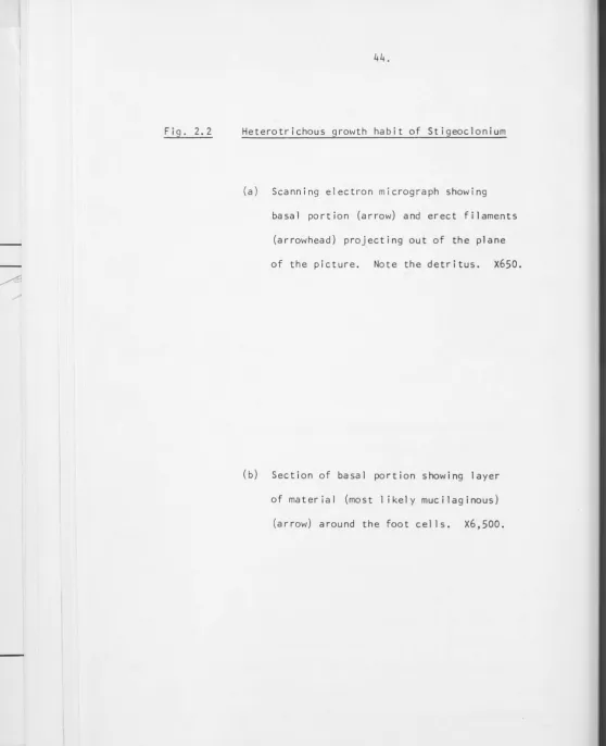
Stigeoclonium Kutzing (order, Ulotrichales; family, Chaetophoraceae) is a filamentous branched Chlorophyte having eel ls of the main axis and branches of a similar size (Cox and Bold, 1966). Stigeoclonium is a ubiquitous alga able to grow in freshwater environments throughout the world despite the fact that they vary widely in temperature and nutrient composition (Islam, 1963). It is commonly attached to rocks
in flowing streams but also grows profusely instil 1 water. Stigeoclonium (L. stigens, sharp+ L. clonium, branch) has a heterotrichous growth habit with a prostrate portion that secures the plant to the substrate and from
(f,g, «-·I)
which branched, erect filaments develop.
/\
2.2 GENERAL METHODS AND MATERIALS
2. 2. 1 Source of Stigeoclonium isolates
The culture used throughout this study was isolated from the Stepping-Stones area of Sul 1 ivan1s Creek on the campus of the Australian
National University. No attempt was made to identify the species of the plant because I consider that the criteria proposed for species
identification in the genus Stigeoclonium have not been sufficiently
( )
..,-, . \ .... I t.-V\.(O""-j'n.<)-J:..
established :ts\o..m, 1%3j Cox o.."J. ~0\J.> 1qbb. 1ne. C'4• 4 ~ ... s~d,. "
\:'~~
Slc"',L~
W4S es1:::o.\,\;s\...eJ.. ~<"OM. a. 5;"jle.. Z.oosrore. iso\o.-t.~ oc; 0.. Si"3le.. p\a,"'t. ~ro'I"\ S .... \\,va"'s C-<ee...\<. '1\.-.e. cJ\::"'<e... ...J4s"""'~"-'c.o-<"e.J
Cu'C.. \.\.Y'i-o.l-90.\ CIA.\~u.<t.. ""'~"'- l\.o Ole~..
ctlj~l
Co"-'c:O.Y"\.;>'\6..1\~£ 6j-lcr4.." ~ ~e..,cr;
"j
S-Mc.\ \ Se.j""'e.-n~S'o\'
~~1.1.f<';jM:,
y
;\ct~el'\I-S to -\<(OS'~/
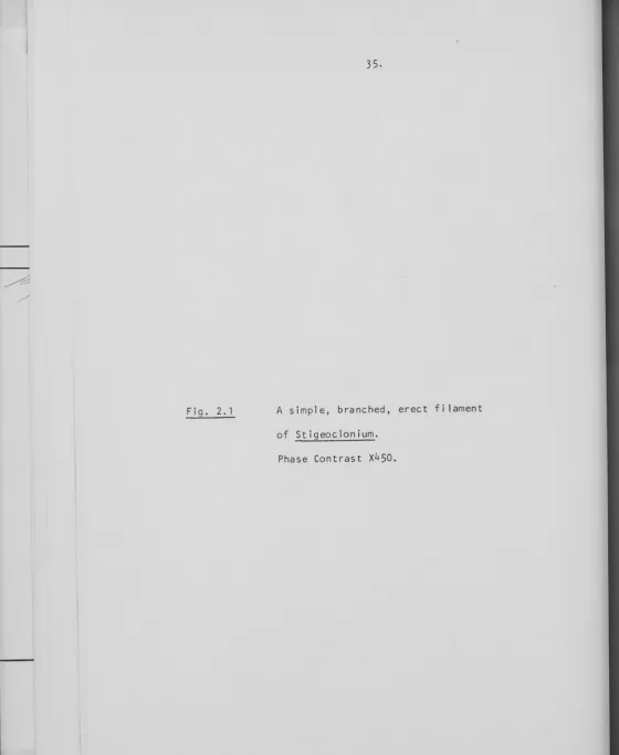
Fig. 2. 1 A simple, branched, erect filament
of Stigeoclonium.
[image:46.614.6.566.16.699.2]2.2.2
(a)
Culture Conditions
Medium
A modified Pocock's (1960) medium was used. Full details of the
medium are given in Table 2. 1. Although the medium was undefined in that
it contained soil-water, the soil came from the same source and batches
of soil-water were prepared in the same way. This medium was used because
it gave growth rates and morphologies similar to that observed in nature
and Stigeoclonium grown in it appeared better preserved ultrastructurally
than when other media were used.
Occasionally a fully-defined Bald's basal medium (Nichols and
Bold, 1965) was used.
(b) Lighting
Cultures were grown under a regime of 8 hours dark (0900 - 1700 hrs)
16 hours light (1700-0900 hrs). Illumination was by two 20-watt 11
daylight11
fluorescent lamps. Cultures were grown in 9 cm plastic Petri dishes,
placed on shelves approximately 1 foot under the 1 ights and in plastic
trays lined with silver foil, giving a light intensity of approximately
2,500 lux at the level of the petri dishes.
(c) Temperature
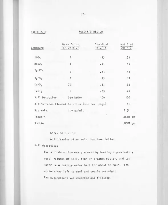
TABLE 2. la POCOCK'$ MEDIUM
Stock Solns. Standard Modified
Compound (g/100ml.) (ml./1) (ml./1)
KN03 5
.33
.33
MgS04 5
. 33
.33
KzHP04
5
.33
.33
KzC03 7
. 33
.33
CaN0 3 20
.33
.33
FeCl3
.33
.20Soi 1 Decoct ion See below 100 100
Hi 11 1
s Trace Element So 1 ut ion (see next page) 1 5
B12 soln. 1. 0 µg/m 1. 2.5
Thiamin . 0001 gm
Biotin . 0001 gm
Check pH 6.7-7.0
Add vitamins after soln. has been boiled. Soil decoction:
The soil decoction was prepared by heating approximately equal volumes of soil, rich in organic matter, and tap water in a boiling water bath for about an hour. The mixture was left to cool and settle overnight.
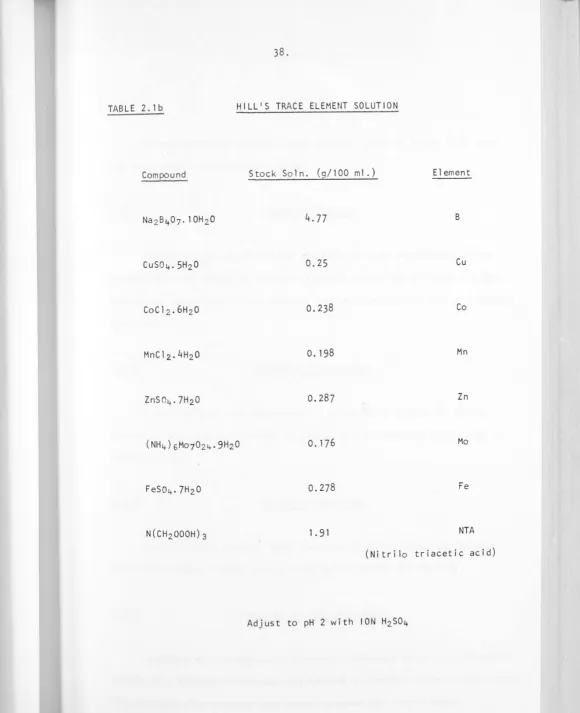
[image:49.615.12.601.17.733.2]TABLE 2. lb HILL'S TRACE ELEMENT SOLUTION
Compound Stock Soln. (g/100 ml.) Element
B
Cu
Co
Mn
Zn
Mo
Fe
NTA
(Nitrilo triacetic acid)
[image:50.615.20.601.13.726.2]Cultures
Unless otherwise stated, young actively growing plants 8-10 days old were used throughout this study.
2. 2. 4 Standard fixation
Stigeoclonium was fixed for one hour at room temperature in one percent glutaraldehyde in Pocock's Medium; washed three times in fresh medium and post-fixed in one percent osmium tetroxide in Pocock's Medium for 1 hour.
Standard dehydration
Fixed material was dehydrated in glass Petri dishes on ice by drop-wise addition of a series of graded acetone concentrations over a 2-day period.
2. 2. 6 Standard embedding
Spurr's resin (Spurr, 1969) was used for flat embedding between glass microscope slides, coated with Teflon (PTFE) (Chon~ 1q11).
Electron & Light Microscopy
Sections for transmission electron microscopy were cut using glass knives on a Reichert microtome and mounted on Formvar coated copper grids.