Recombination Causes Genome Instability
Graphical Abstract
Highlights
d
A complex between the recombination by-product and RAGs
triggers multiple DNA breaks
d
The breaks co-localize with chromosome breakpoints in
acute lymphoblastic leukemias
d
The breaks occur at many frequently mutated genes in acute
lymphoblastic leukemia
d
Cut-and-run may underpin the most common types of
lymphocyte chromosome instabilities
Authors
Christopher M. Kirkham,
James N.F. Scott, Xiaoling Wang, ...,
Peter G. Stockley, Roman Tuma,
Joan Boyes
Correspondence
j.m.boyes@leeds.ac.uk
In Brief
V(D)J recombination errors have long
been associated with chromosome
alterations in lymphoid cancers. Here,
Kirkham et al. describe an unexpected
reaction where the recombination
by-product complexes with the RAG
recombinase to trigger DNA breaks
throughout the genome. Notably, these
breaks co-localize with those found in
acute lymphoblastic leukemia.
Kirkham et al., 2019, Molecular Cell74, 1–14
Cut-and-Run: A Distinct Mechanism
by which V(D)J Recombination
Causes Genome Instability
Christopher M. Kirkham,1,3,7James N.F. Scott,1,7Xiaoling Wang,1,7Alastair L. Smith,1,4Adam P. Kupinski,1,5 Anthony M. Ford,2David R. Westhead,1Peter G. Stockley,1Roman Tuma,1,6and Joan Boyes1,8,*
1School of Molecular and Cellular Biology, Faculty of Biological Sciences, University of Leeds, Leeds LS2 9JT, UK 2Centre for Evolution and Cancer, The Institute of Cancer Research, London SM2 5NG, UK
3Present address: Charles River Laboratories, Inc., Bristol BS20 7AW, UK
4Present address: MRC Molecular Haematology Unit, Weatherall Institute of Molecular Medicine, University of Oxford, John Radcliffe
Hospital, Headington, Oxford OX3 9DS, UK
5Present address: Ipsen Bioinnovation, Milton Park, Abingdon OX14 4RY, UK
6Present address: Faculty of Science, University of South Bohemia, Ceske Budejovice, Czech Republic 7These authors contributed equally
8Lead Contact
*Correspondence:j.m.boyes@leeds.ac.uk https://doi.org/10.1016/j.molcel.2019.02.025
SUMMARY
V(D)J recombination is essential to generate antigen
receptor diversity but is also a potent cause of
genome instability. Many chromosome alterations
that result from aberrant V(D)J recombination involve
breaks at single recombination signal sequences
(RSSs). A long-standing question, however, is how
such breaks occur. Here, we show that the genomic
DNA that is excised during recombination, the
excised signal circle (ESC), forms a complex with
the recombinase proteins to efficiently catalyze
breaks at single RSSs both
in vitro
and
in vivo
.
Following cutting, the RSS is released while the
ESC-recombinase complex remains intact to
poten-tially trigger breaks at further RSSs. Consistent with
this, chromosome breaks at RSSs increase markedly
in the presence of the ESC. Notably, these breaks
co-localize with those found in acute lymphoblastic
leu-kemia patients and occur at key cancer driver genes.
We have named this reaction ‘‘cut-and-run’’ and
sug-gest that it could be a significant cause of
lympho-cyte genome instability.
INTRODUCTION
V(D)J recombination stochastically joins individual V, D, and J gene segments, to generate vast arrays of immunoglobulin and T cell receptor genes that enable vertebrates to combat almost infinite numbers of potential pathogens. Despite its obvious ad-vantages, V(D)J recombination is an inherently dangerous pro-cess because it involves breaking and rejoining DNA. Indeed, recombination errors have long been associated with chromo-some alterations (K€uppers, 2005; Roth, 2003). One of the most
common recombination errors is end donation (Roth, 2003), which occurs when a broken recombination signal sequence (RSS) is joined with a broken DNA end that was formed indepen-dently, for example, via ionizing radiation. If this brings an onco-gene under the control of the strong transcriptional elements of the antigen receptor loci, malignant transformation can result. This process is thought to underpin 30%–40% of the chromo-some translocations found in follicular and mantle cell lym-phomas (J€ager et al., 2000; Welzel et al., 2001).
More recently, whole genome sequencing studies of acute lymphoblastic leukemia (ALL) patients carrying the ETV6/ RUNX1 translocation have further implicated broken RSSs in the generation of chromosome alterations (Papaemmanuil et al., 2014). Both interstitial deletions and chromosome rear-rangements at RSSs were observed and these are thought to play an integral role in disease progression from pre- to overt leu-kemia. Notably,50% of the RAG-mediated chromosome alter-ations appear to involve only single RSSs (Papaemmanuil et al., 2014). However, a fundamental question is how V(D)J recombi-nation generates single broken RSSs for this and the end donation reaction, as stringent measures exist throughout the reaction to prevent the release of single broken ends.
Extensive measures then promote correct joining of the broken DNA ends. Initially, RAGs retain the four broken ends prior to their transfer to the classical non-homologous end joining (cNHEJ) machinery (Lee et al., 2004). Although the trans-fer mechanism is poorly understood, the acidic hinge region in the RAG2 C terminus plays a central role in shepherding the ends along the cNHEJ pathway, rather than the error-prone alternative NHEJ (aNHEJ) pathway (Coussens et al., 2013; Gigi et al., 2014). Ku70/Ku80 then bind to the signal ends, while the DNA-dependent protein kinase catalytic subunit (DNA-PKcs) and Artemis associate with the coding ends. The latter are teth-ered during processing by complexes involving DNAPK, MRN, ATM, and the XRCC4-associated protein XLF, prior to end joining by DNA ligase IV (reviewed byHelmink and Sleckman, 2012). Signal ends remain bound to RAG proteins for longer (Livak and Schatz, 1996; Ramsden and Gellert, 1995) before becoming directly ligated to generate an excised signal circle (ESC) (Figure 1A).
Beyond this extensive network of end joining proteins, yet further safeguards prevent the release of broken DNA ends: in cells deficient in cNHEJ proteins, RAG cutting results in cells be-ing directed to apoptosis via the p53-mediated pathway (re-viewed byHelmink and Sleckman, 2012). Absence of functional ATM, however, does result in increased chromosome deletions and translocations, particularly involving the TCRa/d locus (Liyanage et al., 2000). Translocations similarly result when
loss of XLF or p53 is combined with a non-functional RAG2 C ter-minus (Lescale et al., 2016). Thus, except in ATM-negative cells or engineered double mutant cells, the release of single broken RSS ends is likely to be rare. Here, we describe an unexpected mechanism that generates broken RSSs that are potential sub-strates for the observed chromosome alterations.
This mechanism involves the recombination by-product, the ESC. This retains the signal joint (SJ) where the RSSs are joined in a head-to-head configuration and can be (re)-bound by RAG proteins (Figure 1A) (Curry et al., 2007; Neiditch et al., 2002). Synaptic complex formation between the ESC and genomic RSSs or cryptic RSSs (cRSSs) can result in re-integration of the ESC as evidenced by mouse lymphocytes being littered with re-inserted ESCs (Curry et al., 2007). However, our biochemical analysis of the re-integration reaction showed that remarkably, within the synaptic complex between the SJ and an RSS, the RSS is cut much more efficiently than the head-to-head arrangement of RSSs in the SJ. We find further that following cutting, the RSS is released, generating a double strand break while the RAG-SJ complex remains intact and is free to trigger cutting at further RSSs. We have named this reaction ‘‘cut-and-run’’ and propose that the ESC triggers a double strand break (DSB) at one RSS and then ‘‘runs,’’ to potentially trigger breaks at additional RSSs. Consistent with this idea, we find that the frequency of broken RSSs increases significantly in the presence of the ESC and that these breaks
A B D
[image:3.603.97.519.101.338.2]C
Figure 1. SJ-RSS Cleavage Is Asymmetric
(A) Cartoon of deletional V(D)J recombination and the generation of an ESC.
(B) A SJ stimulates cleavage of 12- and 23-RSSs, but not vice versa. RAG cutting assays were performed using radiolabeled oligonucleotides, denoted by an asterisk above each set of lanes, carrying a 12-RSS, a 23-RSS, or a SJ sequence and separated on a native polyacrylamide gel. Unlabeled partner RSSs are present as indicated; S = SJ. Graphs represent mean of five experiments ± SD.
(C) SJ-RSS cleavage is asymmetric at a range of core RAG concentrations. As for (B) except the amount of core RAG proteins was increased over an 8-fold range, indicated by the filled arrow; a 23-RSS partner was present in all reactions.
(D) Cleavage reactions were performed as in (B) and uncut, nicked, and hairpinned (HP) DNA was separated on a denaturing gel. nt, nucleotides. Graphs represent mean of three experiments ± SD.
mirror those associated with chromosome translocations or interstitial deletions in lymphoid cancers.
RESULTS
The unique arrangement of two head-to-head RSSs in the SJ (Figure 1A) could potentially form a distinct complex with RAG proteins to influence ESC activity. To test if this is the case, RAG interactions with the SJ were examinedin vitro, and we first asked if our RAG cutting conditions obey the 12/23 rule that en-sures that efficient RAG cleavage occurs only between RSSs with different spacer lengths (reviewed bySchatz and Swanson, 2011). As expected, addition of an unlabeled partner RSS results in an10-fold increase in cutting at both 12- and 23-RSSs compared to the level of cutting in the absence of a partner RSS (Figure 1B, lanes 3 and 7).
Asymmetric Cutting within an SJ-RSS Synaptic Complex
The SJ has both a 12- and 23-RSS and therefore is expected to enhance cutting of either a 12- or 23-RSS. Consistent with this, we find that an unlabeled SJ oligonucleotide increases RAG cut-ting at both RSS types by a magnitude similar to when a single complementary RSS is added (Figure 1B, lanes 4 and 8). Remarkably, however, RAG cleavage of a SJ is low and is barely increased by addition of either a 12- or 23-RSS partner (Fig-ure 1B, lanes 10–12). These data therefore suggest that within a 12- and 23- RSS complex, each RSS enhances cutting of the other, resulting in symmetric RAG cutting. By contrast, within a SJ-RSS complex asymmetric cleavage is observed since the RSS is cleaved significantly more than the SJ.
Asymmetric SJ-RSS cutting occurs over a range of concentra-tions of partner RSS (Figure S1A). Moreover, SJ-mediated stim-ulation of RSS cutting is specific since no increase in cleavage is observed upon addition of non-RSS containing DNA (Fig-ure S1B). To test if reduced SJ cutting is due to limiting amounts of protein, RAG proteins were titrated over an 8-fold range. No substantial change in cutting at either a 12-RSS or a SJ was observed (Figure 1C). Likewise, to test if another component in the reaction is limiting, the DNA substrate was increased by 10-fold. A corresponding increase in SJ cutting was seen (Fig-ure S1C), suggesting that no individual component is limiting; instead, something intrinsic to the SJ sequence appears to pre-vent its cutting.
Previous studies showed that RSS flanking sequences influ-ence RAG cutting (Sadofsky et al., 1995). This does not explain reduced SJ cutting, however, because the flanking dinucleotide for each of the RSSs within the SJ is GT, which is cut efficiently. Therefore, to better understand this phenomenon, we next examined at which step of the RAG cutting reaction asymmetric SJ-RSS cutting occurs. RAGs first generate a nick precisely at the boundary between the heptamer and coding region; the free 30-OH group then attacks the opposite DNA strand, forming a hairpin at the coding end and a blunt double strand break at the signal end (Figure 1A) (reviewed by Gellert, 2002). To test whether SJ cutting is blocked at the nicking or hairpinning step, RAG cleavage products were electrophoresed on a dena-turing gel. Nicking and hairpinning occur efficiently at the 12- and 23-RSS when paired with either a complementary RSS or the SJ
(Figure 1D, lanes 1–4 and 5–8). Nicking of the SJ, however, is markedly lower, both in the absence of partner and upon addi-tion of a 12- or 23-RSS. Moreover, while a low level of hairpin formation is observed in the presence of a 12-RSS partner, hair-pins are almost completely absent with a 23-RSS. Together, these data suggest that there is asymmetric cleavage within a SJ-RSS complex and that SJ cutting is primarily blocked at the nicking step but hairpin formation is also prevented in a SJ/23-RSS pair.
Asymmetric Cutting Is Observed with Different SJs
In the experiments described above, the SJ (SJ1) has two consensus RSSs in a head-to-head configuration. However, RSSs can differ from the consensus, especially at non-critical bases. The most conserved bases are the first three positions in the RSS heptamer and positions 5 and 6 of the nonamer (Ramsden et al., 1994). To test if asymmetric cutting also occurs with non-consensus SJs, two common alternative heptamer se-quences, CACAATG and CACAGCC (Ramsden et al., 1994) were substituted for one of the conserved heptamers in SJ1 to generate SJ2 and SJ3. As for SJ1, these new SJs stimulate cut-ting at both a 12- and 23-RSS (Figure S2B, lanes 5, 6, 11, and 12), while the SJ itself is poorly cut, even in the presence of partner RSSs (Figure S2C). Moreover, analysis of the ESCs generated from known recombination events predicts that 72% consist of RSSs that are conserved at six or seven positions in the hep-tamer and at positions 5 and 6 in the nonamer (Table S1). This implies that most of the human ESCs generated are likely to behave similarly to those tested here. Such asymmetric SJ-RSS cutting therefore raises the possibility that RAG-ESC com-plexes could trigger breaks throughout the genome at RSSs and cRSSs, to potentially generate substrates for chromosome alterations.
Although the SJ is consistently cut much less frequently than the RSS when these elements are present intrans, when they are present incison the same DNA construct, both elements are cut (Figure S2D). A likely explanation for this is the much faster kinetics of synaptic complex formation during an intra-mo-lecular interaction that may result in cutting before RAGs are fully complexed with the SJ (below).In vivo, an SJ can be present in cisto RSSs following inversional recombination; if both the SJ and an RSS are in accessible chromatin, RAG cleavage could result in deletion of part of the antigen receptor locus. Deletional recombination to generate an ESC, however, is much more com-mon than inversional recombination, resulting in the dangers outlined above.
Asymmetric Cutting Is Caused by RAG Binding to Both RSSs in the SJ
microscopy (EM) studies (Kim et al., 2018). These studies also demonstrated that DNA nicking by RAGs requires melting of the heptamer CAC and a 180corkscrew rotation of the coding flank DNA to place the scissile phosphate in the active site (Kim et al., 2018; Ru et al., 2018). Such dynamic changes may be blocked when the coding flank is bound by a second RAG com-plex, thereby preventing cutting of the ESC.
To investigate this idea further, we compared the complexes formed on the SJ and RSS by a gel mobility shift assay. Previ-ous studies have shown that RAG proteins form two complexes with an RSS, single complex 1 (SC1) that contains two mole-cules of RAG1 and one of RAG2 and single complex 2 (SC2) that contains two molecules each of RAG1 and RAG2 (Swan-son, 2002). HMGB1 is present in all our reactions to aid cleav-age (van Gent et al., 1997) and in the presence of HMGB1, these complexes are supershifted to form HSC1 and HSC2 (Swanson, 2002). However, with the SJ, an additional, higher molecular weight complex is formed (complex C, Figure 2A, lanes 16–20). Given that the only difference between the SJ and 12- and 23-RSS oligonucleotides is the presence of an
additional RSS in the SJ, these data suggest that this slower migrating complex may result from additional RAG binding to the second RSS.
To further test if RAGs indeed bind to both RSSs of the SJ, we carried out DNase I footprinting studies by binding purified core RAG proteins to a labeled DNA fragment that carries a consensus SJ sequence. Equivalent protected regions are de-tected on both RSSs of the SJ (Figure 2B, left), and because some sites are fully protected on each RSS, this implies that both RSSs of the SJ are bound simultaneously by RAGs. Control experiments where the heptamer and nonamer sequences of each RSS are mutated (Figure 2B, middle and right) confirm that the observed protection is a result of RAG binding.
Mutating the Second RSS in the SJ Restores Symmetric Cutting
If asymmetric cutting is indeed due to simultaneous RAG binding to both RSSs of the SJ, then we would predict that mutations that abolish RAG binding to one RSS of the SJ will result in increased cutting of the SJ in the presence of an appropriate partner RSS.
[image:5.603.90.517.99.393.2]A B
Figure 2. RAGs Bind to Both RSSs in the SJ
(A) A slower migrating complex is formed with the SJ. RAG complexes were formed with labeled oligonucleotides carrying a 12-RSS (lanes 3–8), 23-RSS (lanes 10–14), or SJ (lanes 16–20) and run on a 4% polyacrylamide gel. Increasing amounts of unlabeled partner RSSs are present as indicated above the gel. When the SJ is labeled, a third complex of higher molecular weight is visible (indicated by ‘‘C’’). This is not visible in lanes containing unlabeled SJ, most likely because the paired complex has an average lifetime of400 s (Lovely et al., 2015) and would dissociate before loading on the gel. SC1/2, single complex 1/2; HSC1/2, HMGB1 single complex 1/2; PC, paired complex.
To test this idea, we used an oligonucleotide in which the nonamer of the 23-RSS within the SJ is mutated (SJ23d9). This retains a consensus 12-RSS and thus is expected to promote cutting when paired with a 23-RSS. This prediction is borne out with increased cutting of a 23-RSS but not a 12-RSS, in the presence of SJ23d9 (Figure 3A, compare lanes 4 and 8). Moreover, when an unlabeled partner 23-RSS is added to the labeled SJ23d9 oligonucleotide, a clear increase in SJ23d9 cutting is observed (Figure 3A, lane 12). Similarly, when the 12-RSS nonamer (Figure 3B) or the 12-12-RSS heptamer (Figure S3) of the SJ are mutated, increased cutting is observed when the mutated SJ is paired with a 12-RSS, resulting in almost sym-metrical cleavage within the SJ-RSS complex. These data are
therefore consistent with the idea that when RAGs bind to both sides of the SJ, this prevents SJ cleavage within an SJ-RSS complex.
Our model also predicts that if RAG binding to both RSSs of the SJs is represented by the higher molecular weight complex C seen inFigure 2A, then mutating one of the RSSs would be ex-pected to reduce the abundance of this complex. Consistent with this, we find deletion of the nonamer of the 23- or 12-RSS in the SJ reduces formation of complex C compared to the un-mutated SJ in the presence of competitor (Figures 3C and 3D). Overall, therefore, these data support the model that RAG pro-teins bind simultaneously to both sides of the SJ, leading to asymmetric cleavage.
A B
C
[image:6.603.59.539.94.490.2]D
Figure 3. Mutating One RSS within the SJ Restores Symmetric Cleavage
(A) The 23-RSS nonamer within the SJ was deleted and the oligonucleotide used in cutting reactions either as a partner for labeled 12- or 23-RSSs or as the substrate. Symmetric cutting was almost restored when SJ23d9 was paired with a 23-RSS.
(B) Deletion of the nonamer within the 12-RSS of the SJ results in more symmetric cutting with a 12-RSS. As for (A) except the oligonucleotide SJ12d9 was used. (C and D) RAG binding to mutant SJs. RAG complexes were formed with the 12-RSS, wild-type SJ, or mutant SJs and resolved on a native gel. Complex ‘‘C’’ is formed with the wild-type SJ but is reduced with SJ23d9 and SJ12d9. This is most pronounced in the presence of a partner RSS that likely outcompetes weaker RAG binding to the mutant SJ.
The SJ Triggers Cutting at a cRSS
The observed asymmetric cleavage between SJ-RSS com-plexes implies that the RAG-ESC complex could trigger DSBs at RSSsin vivo. The risks associated with this to genome stability will be determined, in part, by the locations of the ESC-directed breaks. These could occur at single RSSs in the antigen receptor loci to produce substrates for end donation reactions as well as at cRSSs, some of which lie next to oncogenes that are involved in chromosome translocations (Marculescu et al., 2002; Ragha-van et al., 2001). To test if the SJ is capable of causing breaks at these oncogene-associated cRSSs, the LMO2 cRSS was used as an example. We find that cutting is increased in the presence of either a 23-RSS or SJ partner (Figure 4A, right), suggesting that the ESC can indeed promote RAG cutting at cRSSs, a finding that is confirmed by more extensive genome-wide ana-lyses of RAG-SJ cutting (below).
The RSS Is Released following Cutting but RAGs Remain Bound to the SJ
A second factor that determines the danger posed by asym-metric cutting is the fate of the DNA following cleavage. If, following cutting, the broken RSS is released, then RAG proteins would be unable to shepherd the broken end toward the cNHEJ pathway (Lee et al., 2004), thereby enhancing the risk of translo-cations. To investigate the fate of the broken RSS, we deter-mined the stability of the RAG:RSS post-cleavage complexes. First, the release of signal ends was quantified by challenging complex stability at increasing temperatures (Figure 4B) (Cous-sens et al., 2013) and second, the release of coding ends was measured, using time course assays (Figure S4). In both cases, half of the reaction was resolved on a native polyacrylamide gel to analyze RAG-substrate complexes, and the other half was de-proteinated to analyze RAG cleavage products on cutting gels.
A
[image:7.603.101.516.92.446.2]B
Figure 4. The ESC Poses a Risk to Genome Stability
(A) The SJ triggers cutting at cRSSs. RAG cutting assays were performed using radiolabeled oligonucleotides, denoted by an asterisk, carrying a 12-RSS or the LMO2 cRSS. Unlabeled 23-RSS or SJ partners are present as indicated.
We find that the labeled signal end is released at lower temper-atures in the presence of a SJ partner (Figure 4B, lanes 7–12), compared to complexes involving either an RSS or mutant SJ partner (Figure 4B, lanes 1–6 and 13–18). Likewise, when coding ends are examined, HSC1 and HSC2 are formed with a labeled 12- or 23-RSS (Figures S4A and S4B), but upon addition of a partner, RSS cleavage is stimulated and this correlates with a substantial loss of HSC2, the active cleavage complex (Figures S4A and S4B, middle and right). By contrast, following addition of an RSS partner to a SJ, the levels of SJ complexes remain relatively unchanged (Figure S4C). These data therefore indicate that a SJ-stimulated cleavage of an RSS results in the release of the signal and coding ends, but RAG proteins remain stably associated with the SJ. This implies that the cut RSS is free to un-dergo joining with other DNA ends, while the RAG-ESC complex could potentially trigger further DNA breaks at additional RSSs, via a cut-and-run mechanism.
Asymmetry of SJ-RSS CuttingIn Vivo
To further test the cut-and-run idea, we next investigated if asymmetric RAG cutting within the SJ-RSS synaptic complex is also observedin vivo. To this end, integrase-deficient lentivi-ruses carrying a 12-RSS, a 23-RSS, or a SJ were used to transduce REH cells, a B cell line derived from an ALL patient (Rosenfeld et al., 1977) where RAG expression is at physiological levels (Bories et al., 1991). 48 h after transduction, episomal DNA, resulting from reverse transcription and homologous recombination between the LTRs of the integrase-deficient len-tiviruses, was recovered. The level of RAG cleavage of the RSS or SJ on these episomes was then measured via qPCR, us-ing a primer pair that amplifies across the RAG cleavage site so that only intact substrate is amplified. The data were normalized by amplifying a sequence unique to each substrate.
When lentiviruses carrying either a 12- or a 23-RSS were co-transduced into REH cells, so stimulation of RAG cleavage re-quires synapsis intrans, the amount of intact 12- and 23-RSS substrate was reduced to 70% and 74%, respectively, relative to samples where no partner is present (Figure 5A, left). This is consistent with the level of cutting previously published for cut-ting in transusing vectors with individual RSSs (Steen et al., 1996). When a SJ-carrying lentivirus is transduced with either a 12- or 23-RSS, in each case cutting occurs at the RSS, while negligible cleavage of the SJ is detected (Figure 5A). Therefore, in vivo, asin vitro, cleavage within a SJ-RSS complex is asym-metric. Such asymmetric cutting was also observed in NIH 3T3 cells transfected with expression vectors for RAG1 and RAG2, together with two substrate plasmids, that encode either a 12-RSS, a 23-RSS or a SJ (Figure S5A). Notably, asymmetric cutting was observed regardless of whether core RAG2 or full-length RAG2 protein was used, implying that the non-core region of RAG2, which is known to inhibit transposition and re-integration reactions (Curry et al., 2007; Elkin et al., 2003), does not influence asymmetric SJ-RSS cleavage (Figure S5B).
Ourin vitrodata suggest that asymmetric cutting in the SJ-RSS synaptic complex results from RAG binding to both SJ-RSSs in the SJ simultaneously. To test if the same is truein vivo, we used the same mutated SJ sequences as above in the REH cell/virus transduction assay, together with wild-type RSS
vec-tors. If RAG binding to the second RSS indeed blocks SJ cutting, then we predict that the mutated SJ should be cut in the pres-ence of the relevant RSS partner. As can be seen inFigure 5A, this prediction is borne out, both for the mutant SJ/12-RSS pairs (Figure 5A, middle) and also when a SJ mutated at the 23-RSS is paired with a 23-RSS (Figure 5A, right). Moreover, the same ef-fect is observed in transef-fected NIH 3T3 cells (Figure S5A). Together, these data imply that asymmetric cleavage within a SJ-RSS synaptic complex occurs via the same mechanism in vivoasin vitro.
RAGs generate blunt double strand breaks at the signal ends and to verify that the observed cutting was RAG-mediated, the recovered DNA samples were subject to ligation-mediated poly-merase chain reaction (LM-PCR). Here, a linker that recreates an ApaLI site upon ligation to a broken signal end was used to allow the presence of RAG-cleaved ends to be verified by ApaLI diges-tion. As can be seen inFigure 5B, the reduction in intact 12- and 23-RSSs inFigure 5A as well as mutated SJs is mirrored by an increase in LM-PCR product, indicating that RAG cleavage indeed appears to be involved. By contrast, only minimal cutting of the unmutated SJ is detected (Figure 5B, middle and right). Similar analyses confirmed DSBs are present at RSSs in NIH 3T3 cells whereas cutting of the SJ was undetectable (Figure S5B).
The ESC Is LigatedIn Vivo
The ESC has been reported to persistin vivowith unligated signal ends (Livak and Schatz, 1996; Ramsden and Gellert, 1995), either because these ends are ligated more slowly than coding ends or because the ESC is re-cut by RAG proteins (Neiditch et al., 2002). Because these ‘‘open’’ ESCs could potentially un-dergo re-integration, rather than cut-and-run, we were keen to assess the level of unligated signal endsin vivo. To this end, we examined the Igllocus where 60%–70% of recombination occurs between Vl1 and JCl1 (Boudinot et al., 1994), and thus the Vl1Jl1 ESC is generated in the majority of Igl-positive cells. Using LM-PCR, we find that unligated Vl1Jl1 signal ends are detectable, but only weakly (Figure 5C). To determine the proportion of signal ends that are fully ligated, we separately amplified the Vl1Jl1 ESC signal joint and the Vl1Jl1 coding joint (Figure 5D, red arrows and blue arrows, respectively) and then prepared a dilution range with known numbers of copies of each PCR product. These were used as standard curves in qPCR reactions to determine the absolute amounts of coding and signal joints in pre-B DNA samples. This showed that ESC signal joints are present at 60.4% the level of the Vl1Jl1 coding joints. Similarly, we find that 52% and 65% of the Vk16Jk5 and Vk3Jk5 ESCs, respectively, are intact. Together, these data imply that a majority of ESCs are ligated in pre-B cellsin vivo and thus have the potential to undergo cut-and-run (Figure 5D).
ESCs Cause Increased DSBsIn Vivo
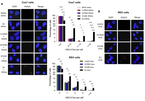
a plasmid carrying a SJ and an SV40 origin of replication to in-crease the number of SJs present. Cotransfection with RAG expression plasmids resulted in a mean of 7.3gH2AX foci per cell compared to far fewer in control cells where the RAG expres-sion vectors, or the SJ vector, were omitted or where a 12- or 23-RSS or SJ12d9 replaced the SJ (Figure 6A). This implies that the RAG-ESC complex indeed triggers genomic DSBs.
To examine whether the SJ also triggers DSBs at physiological RAG concentrations, REH cells were transduced with a lentivirus carrying a SJ, a single RSS, a mutated SJ (SJ12d9) or a control (empty) virus andgH2AX foci were measured. Although only 3–6
SJ vectors are present per cell (STAR Methods), a significant in-crease in foci was again observed with the SJ but not the single RSS or mutated SJ (Figure 6B).
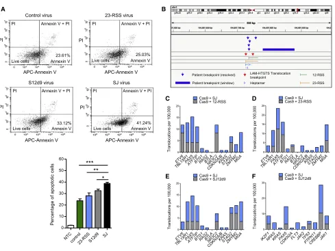
We noticed a higher level of cell death in cells transduced with the SJ-carrying lentivirus compared to those transduced with a control virus. Because unrepaired DSBs trigger apoptosis via the p53-mediated pathway, we next examined the levels of apoptosis by staining REH cells with fluorescent annexin V, which detects early apoptosis. A significant increase in staining was observed in the cells transduced with a SJ-containing virus compared to a single RSS or the control virus (Figure 7A), which
A
B
[image:9.603.59.551.97.442.2]C D
Figure 5. Cleavage of SJ-RSS Substrates Is AsymmetricIn Vivo
(A) REH cells were transduced with integrase-deficient lentiviruses carrying a 12-RSS, a 23-RSS, or a wild-type or mutant SJ sequence, as indicated. The amount of intact substrate after 48 h was measured by qPCR. Data were normalized to unique regions within each provirus, and the values given are relative to controls where lentiviruses carrying a partner sequence were not transduced. Data are represented as mean of three separate transductions ± SD. See alsoFigure S5. (B) LM-PCR was performed using the samples from (A), and the products are shown under the respective graphs. The template amplified by LM-PCR is indicated above the lanes with an asterisk. Ligation of the linker primer to a RAG cleaved end recreates an ApaLI site and digestion was used to verify RAG-mediated cutting. For the SJ mutations (and SJ), the RSS that is expected to be blunt following cleavage was amplified by LM-PCR. Amplification of each of the SJ ends is in the quantitative range and since the same primers were used to amplify the SJ-12-RSS end and SJ23d7, the increased signal with SJ23d7 indicates increased cutting, likewise for the SJ-23-RSS end and SJ12d9. SJ12d7 and SJ23d9 were cloned into the vector in the reverse orientation and gave different sized products with some primers. This also resulted in cross-reactivity of SJ23d9 with 23-RSS primers, giving an additional band (right gel, lane 13). Samples were normalized using qPCR; the bottom (PCR) shows PCRs with the normalization primers to verify the respective vectors were present.
(C) Unligated signal ends are presentin vivo. Top: cartoon of the Igllocus. Bottom: DNA from pro-B cells of PIP3 transgenic mice, where the Igllocus undergoes premature recombination in pro-B cells, was used in an LM-PCR reaction to amplify Vl1 and Jl1 signal ends. ApaLI was used to cleave the signal ends prior to LM-PCR to give a positive control for unligated signal ends. No ligase controls are shown; C indicates the no template control.
is consistent with the generation of increased DNA breaks in the presence of the ESC, as expected for the cut-and-run reaction. In the presence of SJ12d9, more apoptotic cells are present than with a single RSS, which is likely due to residual RAG bind-ing to both RSSs in SJ12d9 (Figure 3D). The levels, however, remain significantly below those with the wild-type SJ.
We next wished to test if the DSBs triggered by the RAG-ESC complex occur at RSSs, as would be predicted by the cut-and-run hypothesis, and also if the broken ends potentially form the substrates for chromosome translocations or deletions. We therefore performed linear amplification-mediated high-throughput genome-wide translocation sequencing (LAM-HTGTS) by transducing REH cells with lentiviruses designed to introduce a CRISPR/Cas9-mediated ‘‘bait’’ DSB at genomic loci and asked if there are increased translocations of ‘‘prey’’ DSBs to these sites in the presence of the SJ.
Two separate loci were chosen to introduce the Cas9-medi-ated breaks:ROSA26andIgH. We then determined the break-points that are translocated to the Cas9-mediated breaks using
the LAM-HTGTS software pipeline and the DNAGrab algorithm to predict whether RSSs are present (RSS site;https://www. itb.cnr.it/rss/analyze.html). A significant increase in transloca-tions involving broken RSSs is observed in cells transduced with the SJ-containing lentivirus compared to those transduced with Cas9-only virus (p < 0.0001; Figure S6A) or with control empty virus (IgH; p < 0.0001,ROSA; p = 0.0004) or with viruses carrying a single RSS (p = <0.0001 for both 12- and 23-RSS). Given that the 23-RSS sequence is the same in the 23-RSS and SJ lentiviruses and that these viruses differ by <300 out of 6,198 bp, the increased breaks at genomic RSSs in the presence of SJ is quite remarkable. We do, however, observe a similar level of cutting at RSSs following transduction with lentiviruses containing SJ12d9 and SJ. This might be because these two vectors differ by only 7 bp and there is some residual RAG bind-ing to both RSSs in SJ12d9 (Figure 3D). Notably, the LAM-HTGTS assay measures just the first round of cutting and, unlike the gH2AX assay, cannot detect subsequent cuts. This can potentially explain why cutting at RSSs appears similar in the γ
γ
A
[image:10.603.55.546.97.441.2]B
Figure 6. The SJ Triggers DSBIn Vivo
(A) Left:gH2AX foci in Cos7 cells transfected with RAG expression vectors and plasmids carrying the 12- or 23-RSS or SJ12d9 or SJ. One representative example of a vector alone transfection is shown. Middle, top graph: quantification ofgH2AX foci from 450 Cos7 cells per transfection condition and three independent experiments.
LAM-HTGTS assay with the SJ and SJ12d9 (Figure S6) but is very different in thegH2AX assay.
Previous whole genome sequencing studies ofETV6/RUNX1 -positive ALL patients identified 140 chromosome breakpoints that lie in close proximity to RSS-like sequences. Crucially, we find breaks that are generated in the presence of the SJ map to within a 100 bp window of 28 different ALL patient breakpoints (Table S2) (Papaemmanuil et al., 2014). The probability of this degree of overlap if the breaks were randomly located is one in 231012(p = 631013). It is also notable that three of the breaks identified with the SJ occur in 5, 5, and 6 patients, respectively, suggesting that these might be hotspots for chromosome alter-ations in ALL (Figures 7B andS7). By contrast, there is no
signif-icant increase in the degree of overlap of the breaks generated in the presence of control virus (p = 0.7), Cas9 (p = 0.07), or the 12-RSS (p = 0.6). We do, however, find four of the translocations with the 23-RSS and SJ12d9 overlap with patient breakpoints (p = 0.002), which is consistent with previous studies where sin-gle RSSs can cause some genome instability (Han et al., 1999). That said, one of the breaks identified with SJ12d9 maps pre-cisely to a breakpoint detected twice with Cas9, suggesting that it might be due to off-target Cas9 cutting.
Finally, previous whole genome sequencing and array-based genome profiling studies identified genes that are commonly mutated inETV6/RUNX1-positive ALL. These genes are often involved in B cell development and of these, 13 were found
A B
C D
[image:11.603.64.553.94.461.2]E F
Figure 7. The SJ Causes Chromosome Breaks
(A) Apoptosis in transduced REH cells as measured by annexin V and propidium iodide (PI) staining for early and late stage apoptosis, respectively. Data are represented as mean of three independent experiments ± SD; increased early stage apoptosis in the presence of the SJ compared to the other vectors is statistically significant (Student’s t test; ***p = 0.0002 [control vector], **p = 0.0064 [23-RSS], *p = 0.011 [SJ12d9]).
(B) Example of two SJ-mediated breaks that co-localize with breaks found in ALL patients. Chromosome breaks mapping close to the cRSS at 194697727 on chromosome 1 were observed in six ALL patients and in two independent LAM-HTGTS experiments. Similar co-localization was observed for 22 more patients (see alsoFigures S6,S7, andTable S2).
(C–E) Graphs showing the frequency of breaks in the 13 genes that most frequently acquire somatic mutations inETV6/RUNX1-positive ALL (Papaemmanuil et al., 2014) in the presence of the SJ compared to the 12-RSS (C), 23-RSS (D), and SJ12d9 (E), respectively.
to be enriched in RSS motifs and acquire somatic mutations most frequently (Papaemmanuil et al., 2014). Our LAM-HTGTS assay detects an increase in cleavage of nine of these genes in the presence of the SJ-containing virus that is a significant increase in the number of genes cleaved (p = 0.01–0.02), compared to the number of genes cleaved with the single RSSs or SJ12d9 virus (Figures 7C–7E;Table S3). Furthermore, using the list of genes that are commonly mutated in all types of B-ALL from the COSMIC database (https://cancer.sanger.ac. uk/cosmic) we find increased breaks in eight to ten of the eleven most commonly mutated genes in the presence of the SJ (p = 0.02–0.0002) compared to breaks with the single RSSs or SJ12d9 (Figure 7F;Table S3). These data therefore strongly imply that cut-and-run makes a significant contribution to mutation of cancer genes and to chromosome alterations in ALL.
DISCUSSION
The excision of intervening DNA from between the gene seg-ments to form an ESC by-product is an integral part of the V(D)J recombination reaction. Sealing the two signal ends was previously thought to sequester the potentially reactive DNA ends to inactivate them (Arnal and Roth, 2007). However, two re-ports published in 2007 indicated that ESCs are far from inert and can be re-bound by RAG proteins by virtue of the two RSSs and reintegrated back into the genome at RSSs or cRSSs (Curry et al., 2007; Vanura et al., 2007). While re-integration clearly oc-curs (Curry et al., 2007), we find that upon formation of a SJ-RSS synaptic complex, cleavage is asymmetric both in vitro and in vivo. Because following cleavage, the cut RSS is released but RAGs remain bound to the SJ, we propose that the RAG-ESC complex could trigger DSBs at further RSSs and cRSSs. This could continue until the ESC becomes cleaved, or until the RAG proteins are downregulated, thus generating multiple genomic DSBs in developing lymphocytes in a reaction we have named cut-and-run.
A plausible explanation for the observed asymmetric cleavage within the SJ-RSS synaptic complex is that RAG proteins bind to both RSSs of the SJ simultaneously, thus blocking effective binding of the heptamer-heptamer border and preventing effi-cient cleavage. This prediction was borne out by EMSA experi-ments that showed that a complex of higher molecular weight is formed with a SJ and by cleavage experiments with mutant SJs, which showed that deleting part of the conserved region on one side of the SJ restores symmetric cleavage by preventing simultaneous RAG binding.
We find that RAG cutting of a SJ is blocked at the nicking step although the low level of nicking seen at the SJ implies that RAG activity is not blocked entirely, but instead, the complex may have some intrinsic flexibility. Synapsis with a 12-RSS partially relieves the block because there is an increase in hairpin forma-tion. By contrast, synapsis with a 23-RSS does not allow hairpin formation. The reason for this block is unclear but it is consistent with anin vivostudy that found reintegration at Vk12-RSSs but not Jk23-RSSs (Curry et al., 2007) and may be due to the inability of a RAG/ESC/23-RSS complex to adopt the correct conforma-tion for ESC cleavage.
The low level of SJ cleavage that we observe differs from pre-vious studies where SJs were cut only3-fold less efficiently than RSSs (Neiditch et al., 2002). The difference from our data cannot be explained by the SJ sequence used because we confirmed our observation with six different SJs, including the one used previously. Instead, Neiditch et al. (2002) propose efficient SJ cleavage occurs by a nick-nick mechanism where RAGs sequentially nick at the heptamer boundary of each RSS to generate a DSB. Consistent with this, or with the failure to join signal ends until RAGs are downregulated, RAG1 and RAG2 binding maps precisely to broken signal ends detected by END-seq in thymocytes at the TCRalocus (Canela et al., 2016). By contrast, we find three different ESCs are60% uncut in primary pro- and pre-B cells (Figure 5D), although it remains to be determined whether RAGs normally associate with ligated ESCs.
Occurrence of Cut-and-RunIn Vivo
Definitive proof that cut-and-run occursin vivois inherently diffi-cult to obtain. Unlike chromosome translocations and reintegra-tion, which result in gross DNA rearrangements and insertions, cut-and-run would leave no trace other than a broken RSS. If the cleaved RSS was subsequently used in a chromosome translocation, again there would be no indication that the break-point was specifically caused by an ESC. Nevertheless, several pieces of evidence suggest that it is highly likely that cut-and-run occursin vivo.
First, extrachromosomal substrate assays indicate that cleav-age of RSSs and SJs is asymmetricin vivowith both full-length and core RAG proteins, and therefore is not anin vitroartifact. Moreover, transduction of REH cells confirmed that asymmetric cutting occurs at physiological levels of RAG proteins and at only 3–6 copies of the SJ per cell.
In addition to asymmetric cleavage, cut-and-run requires the RAG-ESC complex to synapse with genomic RSSs. Reintegra-tion of an ESC at genomic loci indicates that this is likely to be the case (Curry et al., 2007). Moreover, the increase ingH2AX foci in the presence of RAG proteins and the SJ strongly indi-cates that the RAG-ESC complex is capable of triggering DSB formation in vivo. Importantly, the LAM-HTGTS assay shows that this cutting occurs at cRSSs and that broken cRSSs are translocated to a DSB with a significantly higher frequency in the presence of the SJ than the 12- or 23-RSS alone.
to rearrange IgL or TCRaloci. Second, ESCs have been shown to persist for many weeks, even years, and upregulation of RAGs in immature B cells for receptor editing (Hillion et al., 2007) could enable RAG-ESC binding, potentially activating the cut-and-run reaction. Nonetheless, the times that RAGs and ESCs co-exist during lymphocyte development are relatively limited, which will restrict the frequency of the cut-and-run reaction.
Following RAG cleavage, the cut RSS is released and the dan-gers posed by these broken ends to genome stability depends entirely on their fate. In particular, it is unclear how asymmetric cleavage, that produces only one hairpinned and one blunt end, rather than a pair of each type of end, might affect the end-joining process. The broken RSS might be bound by the cNHEJ machinery and in the absence of a suitable partner, it may be more likely to undergo translocation. Alternatively, the DSB might be transferred to the alternative NHEJ pathway, or perhaps it is not transferred to any end-joining machinery at all. This would be consistent with studies that showed RAG proteins do not efficiently shepherd single broken ends to the cNHEJ pathway (Cui and Meek, 2007). More thorough analyses of the fate of the ends following RAG-ESC cleavage are needed to address the risks posed by cut-and-run.
Implications of Cut-and-Run
Chromosome translocations caused by aberrant recombinase activity in the production of antibodies and T cell receptors are a hallmark of many leukemias and lymphomas (K€uppers, 2005). Here, we have identified an unexpected way in which aberrant RAG activity could produce orphan DSBs that have the potential to participate in carcinogenic chromosome translocations.
One of the most common forms of translocation caused by V(D)J recombination is end donation (Roth, 2003), and it is possible that cut-and-run produces the broken RSSs that serve as substrates for this reaction. Likewise, recent studies showed a high frequency of translocations of RSS and cRSS ends to an engineered DSB (Rommel et al., 2017). Although the use of core RAG proteins in these experiments could have enhanced the release of DNA ends (Coussens et al., 2013), translocations were also observed using full-length RAG proteins. A potential mechanism by which the broken RSS and cRSS are generated, is cut-and-run.
A specific leukemia where cut-and-run could play a significant role is ALL where patients bear theETV6/RUNX1(TEL/AML1) translocation. This translocation often arises in utero, but it cannot trigger malignancy in the absence of secondary muta-tions. Instead, the ETV6/RUNX1 translocation partially stalls B cell progression at the pre-B cell stage, where RAG proteins are still expressed (Tsuzuki et al., 2004), thus providing an extended window of opportunity for secondary recombination reactions. The majority of these aberrant recombination reac-tions occur at cryptic RSSs adjacent to transcriptionally active B cell development genes, which can eventually result in full malignant transformation; 50% of the RAG-mediated chromo-some aberrations contain an RSS or cRSS at only one side of translocation (Papaemmanuil et al., 2014). Remarkably, our LAM-HTGTS data show that translocation break-points, formed
in the presence of the SJ, map to the same cRSSs as 28 ALL patient breakpoints (Papaemmanuil et al., 2014). These data strongly suggests that a plausible mechanism by which at least some single RSS or cRSS breaks are formed, is cut-and-run. Moreover, we find that SJ-mediated breaks occur in many of the genes that frequently acquire somatic mutations in ALL, strongly implying that cut-and-run is involved in the etiology of ALL. Given the longevity of the ESC, it is entirely plausible that the RAG-ESC complex also triggers breaks at other stages of lymphocyte development, leading to chromosome alterations that contribute to other lymphoid cancers.
STAR+METHODS
Detailed methods are provided in the online version of this paper and include the following:
d KEY RESOURCES TABLE
d CONTACT FOR REAGENT AND RESOURCE SHARING d EXPERIMENTAL MODEL AND SUBJECT DETAILS
B Mice B Cell lines d METHOD DETAILS
B Purification of RAG proteins from 293T cells B Purification of HMGB1
B In vitroRAG cleavage and binding assays B Calculation of ESC matches to consensus RSSs B DNase I footprint analysis of RAG-SJ binding B Preparation of GA ladder
B Signal end release assay
B Generation of 12/23-RSS and SJ lentiviruses B In vivoRAG cutting assay
B Calculation of pro-virus copies per cell
B LM-PCR of extrachromosomal substrates from NIH 3T3 cells
B LM-PCR of extra-chromosomal substrates from REH cells
B LM-PCR to detect broken Vl1-JCl1 signal ends B Quantification of ligated signal ends
B Detection ofgH2AX foci by immunofluorescence B Detection of apoptosis
B LAM-HTGTS assay
d QUANTIFICATION AND STATISTICAL ANALYSIS
d DATA AND SOFTWARE AVAILABILITY
SUPPLEMENTAL INFORMATION
Supplemental Information can be found with this article online athttps://doi. org/10.1016/j.molcel.2019.02.025.
ACKNOWLEDGMENTS
AUTHOR CONTRIBUTIONS
Conceptulization, C.M.K. and J.B.; Investigation, C.M.K., J.N.F.S., X.W., A.P.K., and J.B.; Software, A.L.S., J.N.F.S., and D.R.W.; Formal Analysis, D.R.W., A.L.S., and J.N.F.S.; Resources, A.M.F.; Writing – Original Draft, C.M.K. and J.B.; Writing – Reviewing & Editing, all authors; Funding Acquisi-tion, J.B., P.G.S., R.T., A.M.F., and D.R.W.
DECLARATIONS OF INTERESTS
The authors declare no competing interests.
Received: August 10, 2018 Revised: December 20, 2018 Accepted: February 14, 2019 Published: March 21, 2019
SUPPORTING CITATIONS
The following references appear in the Supplemental Information:Hiom and Gellert (1997); Mizushima and Nagata (1990); Tevelev and Schatz (2000).
REFERENCES
Agrawal, A., and Schatz, D.G. (1997). RAG1 and RAG2 form a stable post-cleavage synaptic complex with DNA containing signal ends in V(D)J recombi-nation. Cell89, 43–53.
Arnal, S.M., and Roth, D.B. (2007). Excised V(D)J recombination byproducts threaten genomic integrity. Trends Immunol.28, 289–292.
Bergeron, S., Anderson, D.K., and Swanson, P.C. (2006). RAG and HMGB1 Proteins: Purification and Biochemical Analysis of Recombination Signal Complexes. Methods Enzymol.408, 511–528.
Bevington, S., and Boyes, J. (2013). Transcription-coupled eviction of histones H2A/H2B governs V(D)J recombination. EMBO J.32, 1381–1392.
Bories, J.C., Cayuela, J.M., Loiseau, P., and Sigaux, F. (1991). Expression of human recombination activating genes (RAG1 and RAG2) in neoplastic lymphoid cells: correlation with cell differentiation and antigen receptor expression. Blood78, 2053–2061.
Boudinot, P., Drapier, A.M., Cazenave, P.A., and Sanchez, P. (1994). Conserved distribution of lambda subtypes from rearranged gene segments to immunoglobulin synthesis in the mouse B cell repertoire. Eur. J. Immunol. 24, 2013–2017.
Canela, A., Sridharan, S., Sciascia, N., Tubbs, A., Meltzer, P., Sleckman, B.P., and Nussenzweig, A. (2016). DNA Breaks and End Resection Measured Genome-wide by End Sequencing. Mol. Cell63, 898–911.
Coussens, M.A., Wendland, R.L., Deriano, L., Lindsay, C.R., Arnal, S.M., and Roth, D.B. (2013). RAG2’s acidic hinge restricts repair-pathway choice and promotes genomic stability. Cell Rep.4, 870–878.
Cowell, L.G., Davila, M., Ramsden, D., and Kelsoe, G. (2004). Computational tools for understanding sequence variability in recombination signals. Immunol. Rev.200, 57–69.
Cui, X., and Meek, K. (2007). Linking double-stranded DNA breaks to the recombination activating gene complex directs repair to the nonhomologous end-joining pathway. Proc. Natl. Acad. Sci. USA104, 17046–17051. Curry, J.D., Schulz, D., Guidos, C.J., Danska, J.S., Nutter, L., Nussenzweig, A., and Schlissel, M.S. (2007). Chromosomal reinsertion of broken RSS ends dur-ing T cell development. J. Exp. Med.204, 2293–2303.
Elkin, S.K., Matthews, A.G., and Oettinger, M.A. (2003). The C-terminal portion of RAG2 protects against transposition in vitro. EMBO J.22, 1931–1938. Fugmann, S.D., Villey, I.J., Ptaszek, L.M., and Schatz, D.G. (2000). Identification of two catalytic residues in RAG1 that define a single active site within the RAG1/RAG2 protein complex. Mol. Cell5, 97–107.
Gabriel, R., Lombardo, A., Arens, A., Miller, J.C., Genovese, P., Kaeppel, C., Nowrouzi, A., Bartholomae, C.C., Wang, J., Friedman, G., et al. (2011). An
un-biased genome-wide analysis of zinc-finger nuclease specificity. Nat. Biotechnol.29, 816–823.
Gellert, M. (2002). V(D)J recombination: RAG proteins, repair factors, and regulation. Annu. Rev. Biochem.71, 101–132.
Gigi, V., Lewis, S., Shestova, O., Mijuskovic, M., Deriano, L., Meng, W., Luning Prak, E.T., and Roth, D.B. (2014). RAG2 mutants alter DSB repair pathway choice in vivo and illuminate the nature of ‘alternative NHEJ’. Nucleic Acids Res.42, 6352–6364.
Grant, C.E., Bailey, T.L., and Noble, W.S. (2011). FIMO: scanning for occur-rences of a given motif. Bioinformatics27, 1017–1018.
Han, J.O., Steen, S.B., and Roth, D.B. (1999). Intermolecular V(D)J recombina-tion is prohibited specifically at the joining step. Mol. Cell3, 331–338. Helmink, B.A., and Sleckman, B.P. (2012). The response to and repair of RAG-mediated DNA double-strand breaks. Annu. Rev. Immunol.30, 175–202. Hesse, J.E., Lieber, M.R., Mizuuchi, K., and Gellert, M. (1989). V(D)J recombi-nation: a functional definition of the joining signals. Genes Dev.3, 1053–1061. Hillion, S., Youinou, P., and Jamin, C. (2007). Peripheral expression of RAG in human B lymphocytes in normal and pathological conditions is dependent on interleukin-6. Autoimmun. Rev.6, 415–420.
Hiom, K., and Gellert, M. (1997). A stable RAG1-RAG2-DNA complex that is active in V(D)J cleavage. Cell88, 65–72.
Hu, J., Meyers, R.M., Dong, J., Panchakshari, R.A., Alt, F.W., and Frock, R.L. (2016). Detecting DNA double-stranded breaks in mammalian genomes by linear amplification-mediated high-throughput genome-wide translocation sequencing. Nat. Protoc.11, 853–871.
J€ager, U., Bo¨csko¨r, S., Le, T., Mitterbauer, G., Bolz, I., Chott, A., Kneba, M., Mannhalter, C., and Nadel, B. (2000). Follicular lymphomas’ BCL-2/IgH junc-tions contain templated nucleotide inserjunc-tions: novel insights into the mecha-nism of t(14;18) translocation. Blood95, 3520–3529.
Kim, D.R., Dai, Y., Mundy, C.L., Yang, W., and Oettinger, M.A. (1999). Mutations of acidic residues in RAG1 define the active site of the V(D)J recom-binase. Genes Dev.13, 3070–3080.
Kim, M.-S., Chuenchor, W., Chen, X., Cui, Y., Zhang, X., Zhou, Z.H., Gellert, M., and Yang, W. (2018). Cracking the DNA Code for V(D)J Recombination. Mol. Cell70, 358–370.e4.
K€uppers, R. (2005). Mechanisms of B-cell lymphoma pathogenesis. Nat. Rev. Cancer5, 251–262.
Landree, M.A., Wibbenmeyer, J.A., and Roth, D.B. (1999). Mutational analysis of RAG1 and RAG2 identifies three catalytic amino acids in RAG1 critical for both cleavage steps of V(D)J recombination. Genes Dev.13, 3059–3069. Lee, G.S., Neiditch, M.B., Salus, S.S., and Roth, D.B. (2004). RAG proteins shepherd double-strand breaks to a specific pathway, suppressing error-prone repair, but RAG nicking initiates homologous recombination. Cell117, 171–184.
Lefranc, M.P., Clement, O., Kaas, Q., Duprat, E., Chastellan, P., Coelho, I., Combres, K., Ginestoux, C., Giudicelli, V., Chaume, D., and Lefranc, G. (2005). IMGT-Choreography for immunogenetics and immunoinformatics. In Silico Biol. (Gedrukt)5, 45–60.
Lescale, C., Abramowski, V., Bedora-Faure, M., Murigneux, V., Vera, G., Roth, D.B., Revy, P., de Villartay, J.-P., and Deriano, L. (2016). RAG2 and XLF/ Cernunnos interplay reveals a novel role for the RAG complex in DNA repair. Nat. Commun.7, 10529.
Livak, F., and Schatz, D.G. (1996). T-cell receptor alpha locus V(D)J recombi-nation by-products are abundant in thymocytes and mature T cells. Mol. Cell. Biol.16, 609–618.
Liyanage, M., Weaver, Z., Barlow, C., Coleman, A., Pankratz, D.G., Anderson, S., Wynshaw-Boris, A., and Ried, T. (2000). Abnormal rearrangement within the alpha/delta T-cell receptor locus in lymphomas from Atm-deficient mice. Blood96, 1940–1946.
Marculescu, R., Le, T., Simon, P., Jaeger, U., and Nadel, B. (2002). V(D)J-mediated translocations in lymphoid neoplasms: a functional assessment of genomic instability by cryptic sites. J. Exp. Med.195, 85–98.
Mizushima, S., and Nagata, S. (1990). pEF-BOS, a powerful mammalian expression vector. Nucleic Acids Res.18, 5322.
Nagawa, F., Kodama, M., Nishihara, T., Ishiguro, K., and Sakano, H. (2002). Footprint analysis of recombination signal sequences in the 12/23 synaptic complex of V(D)J recombination. Mol. Cell. Biol.22, 7217–7225.
Nagawa, F., Hirose, S., Nishizumi, H., Nishihara, T., and Sakano, H. (2004). Joining mutants of RAG1 and RAG2 that demonstrate impaired interactions with the coding-end DNA. J. Biol. Chem.279, 38360–38368.
Neiditch, M.B., Lee, G.S., Huye, L.E., Brandt, V.L., and Roth, D.B. (2002). The V(D)J recombinase efficiently cleaves and transposes signal joints. Mol. Cell9, 871–878.
Papaemmanuil, E., Rapado, I., Li, Y., Potter, N.E., Wedge, D.C., Tubio, J., Alexandrov, L.B., Van Loo, P., Cooke, S.L., Marshall, J., et al. (2014). RAG-mediated recombination is the predominant driver of oncogenic rearrange-ment in ETV6-RUNX1 acute lymphoblastic leukemia. Nat. Genet.46, 116–125. Raghavan, S.C., Kirsch, I.R., and Lieber, M.R. (2001). Analysis of the V(D)J recombination efficiency at lymphoid chromosomal translocation breakpoints. J. Biol. Chem.276, 29126–29133.
Ramsden, D.A., and Gellert, M. (1995). Formation and resolution of double-strand break intermediates in V(D)J rearrangement. Genes Dev.9, 2409–2420. Ramsden, D.A., Baetz, K., and Wu, G.E. (1994). Conservation of sequence in recombination signal sequence spacers. Nucleic Acids Res.22, 1785–1796. Rogakou, E.P., Pilch, D.R., Orr, A.H., Ivanova, V.S., and Bonner, W.M. (1998). DNA double-stranded breaks induce histone H2AX phosphorylation on serine 139. J. Biol. Chem.273, 5858–5868.
Rommel, P.C., Oliveira, T.Y., Nussenzweig, M.C., and Robbiani, D.F. (2017). RAG1/2 induces genomic insertions by mobilizing DNA into RAG1/2-indepen-dent breaks. J. Exp. Med.214, 814–831.
Rosenfeld, C., Goutner, A., Venuat, A.M., Choquet, C., Pico, J.L., Dore, J.F., Liabeuf, A., Durandy, A., Desgrange, C., and De The, G. (1977). An effect hu-man leukaemic cell line: Reh. Eur. J. Cancer13, 377–379.
Roth, D.B. (2003). Restraining the V(D)J recombinase. Nat. Rev. Immunol.3, 656–666.
Roth, D.B., Zhu, C., and Gellert, M. (1993). Characterization of broken DNA molecules associated with V(D)J recombination. Proc. Natl. Acad. Sci. USA 90, 10788–10792.
Ru, H., Chambers, M.G., Fu, T.-M., Tong, A.B., Liao, M., and Wu, H. (2015). Molecular Mechanism of V(D)J Recombination from Synaptic RAG1-RAG2 Complex Structures. Cell163, 1138–1152.
Ru, H., Mi, W., Zhang, P., Alt, F.W., Schatz, D.G., Liao, M., and Wu, H. (2018). DNA melting initiates the RAG catalytic pathway. Nat. Struct. Mol. Biol.25, 732–742.
Sadofsky, M.J., Hesse, J.E., McBlane, J.F., and Gellert, M. (1993). Expression and V(D)J recombination activity of mutated RAG-1 proteins. Nucleic Acids Res.21, 5644–5650.
Sadofsky, M.J., Hesse, J.E., van Gent, D.C., and Gellert, M. (1995). RAG-1 mu-tations that affect the target specificity of V(D)j recombination: a possible direct role of RAG-1 in site recognition. Genes Dev.9, 2193–2199.
Schatz, D.G., and Swanson, P.C. (2011). V(D)J recombination: mechanisms of initiation. Annu. Rev. Genet.45, 167–202.
Steen, S.B., Gomelsky, L., and Roth, D.B. (1996). The 12/23 rule is enforced at the cleavage step of V(D)J recombination in vivo. Genes Cells1, 543–553. Swanson, P.C. (2002). A RAG-1/RAG-2 tetramer supports 12/23-regulated synapsis, cleavage, and transposition of V(D)J recombination signals. Mol. Cell. Biol.22, 7790–7801.
Tevelev, A., and Schatz, D.G. (2000). Intermolecular V(D)J recombination. J. Biol. Chem.275, 8341–8348.
Tsuzuki, S., Seto, M., Greaves, M., and Enver, T. (2004). Modeling first-hit functions of the t(12;21) TEL-AML1 translocation in mice. Proc. Natl. Acad. Sci. USA101, 8443–8448.
van Gent, D.C., Hiom, K., Paull, T.T., and Gellert, M. (1997). Stimulation of V(D)J cleavage by high mobility group proteins. EMBO J.16, 2665–2670. Vanura, K., Montpellier, B., Le, T., Spicuglia, S., Navarro, J.M., Cabaud, O., Roulland, S., Vachez, E., Prinz, I., Ferrier, P., et al. (2007). In vivo reinsertion of excised episomes by the V(D)J recombinase: a potential threat to genomic stability. PLoS Biol.5, e43.
Welzel, N., Le, T., Marculescu, R., Mitterbauer, G., Chott, A., Pott, C., Kneba, M., Du, M.Q., Kusec, R., Drach, J., et al. (2001). Templated nucleotide addition and immunoglobulin JH-gene utilization in t(11;14) junctions: implications for the mechanism of translocation and the origin of mantle cell lymphoma. Cancer Res.61, 1629–1636.
STAR
+
METHODS
KEY RESOURCES TABLE
REAGENT or RESOURCE SOURCE IDENTIFIER Antibodies
Anti-phospho-histone H2A.X (Ser139) Cell Signaling Technology Cat#2577;RRID:AB_2118010 AlexaFluor488 anti-rabbit IgG (H+L) F(ab’)2Fragment Cell Signaling Technologies Cat#4412;RRID:AB_1904025
AlexaFluor568 goat anti-rabbit IgG Life Technologies Cat#A11011;RRID:AB_143157 APC-conjugated Annexin V Thermo Fisher Scientific Cat#17-8007-72; RRID:AB_2575165 Bacterial and Virus Strains
DH5acompetent cells Thermo Fisher Scientific Cat#18265017 BL21(DE3) competent cells NEB Cat#C25271 Endura competent cells Lucigen Cat#60240 Chemicals, Peptides, and Recombinant Proteins
Protease inhibitors Roche Cat#11697498001 Amylose resin NEB Cat#E80225 Ni-NTA agarose resin QIAGEN Cat#30230 HiTrap Q XL column GE Healthcare Cat#17-5158-01 Proteinase K Roche Cat#03115879001
NheI NEB Cat#R3131
ApaI NEB Cat#R0114
EcoRV NEB Cat#R0195
ApaLI NEB Cat#R0507
Klenow DNA polymerase NEB Cat#M0212 T4 DNA ligase NEB Cat#M0202 Vent (exo) DNA polymerase NEB Cat#M0254 Taq DNA polymerase NEB Cat#M0267 DNase I Worthington Cat#DPRF
a-32P dCTP Perkin Elmer Cat#NEG013H
g-32P ATP Perkin Elmer Cat#NEG002A
Poly-L-lysine Sigma Cat#P4704 Vectashield plus DAPI Vector laboratories Cat#H-1200 Polyethylenimine Sigma Cat#408727 Polybrene Merck Cat#TR-1003-G dsGreen Gel staining solution Lumiprobe Cat#A10010 Critical Commercial Assays
Annexin V Apoptosis detection kit - APC Thermo Fisher Scientific Cat#88-8007-72 SensiFAST SYBR No-Rox Bioline Cat#BIO-72005 QuantiFluor dsDNA System Promega Cat#E2671 MiSeq Reagent kit v2 (500 cycle) Illumina Cat#MS-102-2003 PhiX Control v3 kit Illumina Cat#FC-110-3001 Deposited Data
FASTQ raw sequence files from LAM-HTGTS This paper Uploaded to NCBI SRA; PRJNA483469 Experimental Models: Cell Lines
REH - human ALL cell line carrying theETV6/RUNX1
(TEL-AML) fusion gene
Greaves lab, Institute of Cancer Research, London;Rosenfeld et al., 1977
N/A NIH 3T3 - mouse embryo fibroblast cell line Bonifer lab, University of Birmingham N/A
Continued
REAGENT or RESOURCE SOURCE IDENTIFIER Cos 7 - African green monkey kidney fibroblast cell line Harris lab, University of Leeds N/A HEK293T - human embryonic kidney cell line Harris lab, University of Leeds N/A Experimental Models: Organisms/Strains
CBA/C57BL/6J mice University of Leeds Facility N/A
l5-IRF4 transgenic mice Bevington and Boyes, 2013 PIP3 mice Oligonucleotides
Primers used in RAG cutting assays are given inTable S4
This paper;Table S4 N/A Primers used in PCR assays are given inTable S5 This paper;Table S5 N/A Adaptors and common primers for HTGTS Hu et al., 2016 N/A Recombinant DNA
Plasmid: pETM11-HMGB1 A gift from Professor Marco Bianchi N/A Plasmid: pEF-McR1 This paper;Table S6 N/A Plasmid: pEF-McR2 This paper;Table S6 N/A Plasmid: pJH290 Steen et al., 1996; Han et al., 1999 N/A Plasmid: pJH29012SJ This paper;Table S6 N/A Plasmid: pJH29023SJ This paper;Table S6 N/A Plasmid: pJd1+ This paper;Table S6 N/A Plasmid: pSJ+ This paper;Table S6 N/A Plasmid: p23+ This paper;Table S6 N/A Plasmid: p12+ This paper;Table S6 N/A Plasmid: pSJ12d7+ This paper;Table S6 N/A Plasmid: pSJ23d7+ This paper;Table S6 N/A Plasmid: pSJ12d9+ This paper;Table S6 N/A Plasmid: pSJ23d9+ This paper;Table S6 N/A Plasmid: pSJ12d9CAC+ This paper;Table S6 N/A Plasmid: pSJ23d9CAC+ This paper;Table S6 N/A Plasmid: pSJ12d923d9CAC+ This paper;Table S6 N/A Plasmid: pJH548 Sadofsky et al., 1993 N/A Plasmid: pEFflRAG2 This paper;Table S6 N/A Plasmid: pEFcRAG2 This paper;Table S6 N/A Plasmid: pWPI Addgene Cat#12254 Plasmid: pWPI-12RSS This paper;Table S6 N/A Plasmid: pWPI-23RSS This paper;Table S6 N/A Plasmid: pWPI-SJ This paper;Table S6 N/A Plasmid: pWPI-mutant SJ This paper;Table S6 N/A Plasmid: LentiCRISPR-v2 Addgene Cat#52961 Plasmid: pCMVR8.74 Addgene Cat#22036 Plasmid: pMD2.G Addgene Cat#12259 Software and Algorithms
HTGTS pipeline Hu et al., 2016 http://robinmeyers.github.io/transloc_ pipeline/
GraphPad Prism N/A https://www.graphpad.com/ IMGTrepertoire (The international ImMunoGeneTics
information system)
Lefranc et al., 2005 http://www.imgt.org Recombination Signal Sequences Site Cowell et al., 2004 https://www.itb.cnr.it/rss/ FIMO Grant et al., 2011 http://meme-suite.org/ COSMIC COSMIC mutation data (genomic
screens) Release v85
CONTACT FOR REAGENT AND RESOURCE SHARING
Further information and requests for resources and reagents should be directed to and will be fulfilled by the corresponding author, Joan Boyes (j.m.boyes@leeds.ac.uk).
EXPERIMENTAL MODEL AND SUBJECT DETAILS
Mice
PIP3 mice have been described previously (Bevington and Boyes, 2013) and express the IRF4 cDNA under the control of the pro-B cell specific lambda promoter and LCR. Non-transgenic CBA/C57BL/6J were obtained from the University of Leeds animal facility. Animals were sacrificed at 5-7 weeks, bone marrow was removed from femurs and used for the isolation of pro- or pre-B cells by flow cytometry. A single mouse was used to isolate DNA from pro-B or pre-B cells per independent experiment; equivalent numbers of male and female animals were used overall. All animal procedures were performed under Home Office license PPL 70/7697, following review by the University of Leeds ethics committee. They were housed in a full barrier facility, with no more than six animals per cage, where all mice are free of common pathogens, including murine norovirus,PasteurellaandHelicobacter.
Cell lines
REH cells were established from the peripheral blood of a 15-year old female with acute lymphoblastic leukemia (Rosenfeld et al., 1977). This cell line carries t(12:21) that generates theETV6/RUNX1(TEL-AML) fusion gene. Cells were maintained in RPMI1640 with 10% fetal calf serum at 37C in 5% CO2and were grown at a density between 53105and 23106(https://www.dsmz.de/ catalogues/details/culture/ACC-22.html).
NIH 3T3 (male mouse embryo fibroblast) cells, Cos 7 (male African green monkey kidney fibroblast) cells and HEK293T (female human embryonic kidney) cells were maintained in DMEM with 10% fetal calf serum, 3.8 mM L-glutamine, 50 U/ml penicillin and 0.05 mg/ml streptomycin at 37C in 5% CO2.These adherent cell lines were passaged every 2-3 days to ensure that they remained sub-confluent. They have not been authenticated.
METHOD DETAILS
Purification of RAG proteins from 293T cells
MBP-tagged core RAG proteins were purified from HEK293T cells according toBergeron et al. (2006). HEK293T cells were seeded at 13106cells per 10 cm plate. The following day, the medium was changed three hours prior to transfection; the cells were then trans-fected with 5mg each of the plasmids pEF-McR1 and pEF-McR2 (Table S6) using PEI. Cells were harvested after 48 hours, washed twice in PBS and frozen until use. All purification steps were performed at 4C. Briefly, cell pellets (typically from 30 dishes) were thawed on ice and resuspended in a total of 10 mL of lysis buffer (10 mM NaPi[pH 7.2], 0.5 M NaCl, 1 mM DTT, 0.25% TWEEN-20, 1x protease inhibitors with EDTA (Roche), 1 mM PMSF, 1 mM benzamidine). Cells were lysed by using a Dounce homogenizer for 20 strokes with a tight-fitting pestle, then centrifuged for 40 minutes at 11,000 xg(30,000 rpm), 4C, in a SW55 Ti rotor (Beckman). The supernatant was loaded onto a 1 mL amylose column equilibrated with buffer A (10 mM NaPi[pH 7.2], 0.5 M NaCl, 1 mM DTT, 0.25% TWEEN-20). The column was washed with 10 column volumes (CV) of buffer A, followed by 10 CV of buffer B (buffer A without TWEEN-20) and protein was eluted with 10 CV buffer C (Buffer A without TWEEN-20, with 10 mM maltose). Fractions were analyzed by SDS-PAGE and RAG-containing fractions were pooled and dialysed twice for three hours against dialysis buffer (25 mM Tris-HCl [pH 8.0], 150 mM KCl, 2 mM DTT, 10% glycerol). Dialysed protein was aliquoted, snap-frozen in a dry ice/ethanol bath and stored at80C.
Continued
REAGENT or RESOURCE SOURCE IDENTIFIER
The R Project for Statistical Computing N/A https://www.r-project.org/
Analysis of translocations to RSSs This paper Deposited on GitHub:https://github. com/boyeslab/cutandrun/
Analysis of chromosome distribution of translocations
This paper Deposited on GitHub:https://github. com/boyeslab/cutandrun/
Analysis of overlaps between target genes and translocations
This paper Deposited on GitHub:https://github. com/boyeslab/cutandrun/
Analysis of the intersection between ALL patient breakpoints and translocations