A Single Native Ganglioside
GM1-Binding Site Is Sufficient for
Cholera Toxin to Bind to Cells and
Complete the Intoxication Pathway
The Harvard community has made this
article openly available.
Please share
how
this access benefits you. Your story matters
Citation
Jobling, Michael G., ZhiJie Yang, Wendy R. Kam, Wayne I. Lencer,
and Randall K. Holmes. 2012. “A Single Native Ganglioside
GM1-Binding Site Is Sufficient for Cholera Toxin To Bind to Cells and
Complete the Intoxication Pathway.” Edited by R. John Collier. mBio
3 (6). https://doi.org/10.1128/mbio.00401-12.
Citable link
http://nrs.harvard.edu/urn-3:HUL.InstRepos:41483391
Terms of Use
This article was downloaded from Harvard University’s DASH
repository, and is made available under the terms and conditions
applicable to Other Posted Material, as set forth at
http://
A Single Native Ganglioside GM
1-Binding Site Is Sufficient for
Cholera Toxin To Bind to Cells and Complete the Intoxication
Pathway
Michael G. Jobling,aZhiJie Yang,a* Wendy R. Kam,bWayne I. Lencer,b,cand Randall K. Holmesa
Department of Microbiology, University of Colorado School of Medicine, Aurora, Colorado, USAa; GI Cell Biology, Department of Pediatrics, Children’s Hospital, Boston,
Massachusetts, USAb; and Harvard Medical School, Harvard Digestive Diseases Center, Boston, Massachusetts, USAc
* Present address: Zhijie Yang, Veterans Affairs Medical Center at San Francisco (VAMC) and University of California at San Francisco (UCSF), San Francisco, California, USA
ABSTRACT Cholera toxin (CT) fromVibrio choleraeis responsible for the majority of the symptoms of the diarrheal disease chol-era. CT is a heterohexameric protein complex with a 240-residue A subunit and a pentameric B subunit of identical 103-residue B polypeptides. The A subunit is proteolytically cleaved within a disulfide-linked loop to generate the A1 and A2 fragments. The B subunit of wild-type (wt) CT binds 5 cell surface ganglioside GM1(GM1) molecules, and the toxin-GM1complex traffics from
the plasma membrane (PM) retrograde through endosomes and the Golgi apparatus to the endoplasmic reticulum (ER). From the ER, the enzymatic A1 fragment retrotranslocates to the cytosol to cause disease. Clustering of GM1by multivalent toxin
binding can structurally remodel cell membranes in ways that may assist toxin uptake and retrograde trafficking. We have re-cently found, however, that CT may traffic from the PM to the ER by exploiting an endogenous glycosphingolipid pathway (A. A. Wolf et al., Infect. Immun. 76:1476 –1484, 2008, and D. J. F. Chinnapen et al., Dev. Cell 23:573–586, 2012), suggesting that multi-valent binding to GM1is dispensable. Here we formally tested this idea by creating homogenous chimeric holotoxins with
de-fined numbers of native GM1binding sites from zero (nonbinding) to five (wild type). We found that a single GM1binding site is
sufficient for activity of the holotoxin. Therefore, remodeling of cell membranes by mechanisms that involve multivalent bind-ing of toxin to GM1receptors is not essential for toxicity of CT.
IMPORTANCEThrough multivalent binding to its lipid receptor, cholera toxin (CT) can remodel cell membranes in ways that may assist host cell invasion. We recently found that CT variants which bind no more than 2 receptor molecules do exhibit toxicity, suggesting that CT may be able to enter cells by coopting an endogenous lipid sorting pathway without clustering receptors. We tested this idea directly by using purified variants of CT with zero to five functional receptor-binding sites (BS). One BS enabled CT to intoxicate cells, supporting the conclusion that CT can enter cells by coopting an endogenous lipid-sorting pathway. Al-though multivalent receptor binding is not essential, it does increase CT toxicity. These findings suggest that achieving higher receptor binding avidity or affecting membrane dynamics by lipid clustering and membrane remodeling may be driving forces for evolution of AB5subunit toxins that can bind multivalently to cell membrane lipid receptors.
Received27 September 2012Accepted5 October 2012Published30 October 2012
CitationJobling MG, Yang Z, Kam WR, Lencer WI, Holmes RK. 2012. A single native ganglioside GM1-binding site is sufficient for cholera toxin to bind to cells and complete the intoxication pathway. mBio 3(6):e00401-12. doi:10.1128/mBio.00401-12
EditorR. John Collier, Harvard Medical School
Copyright© 2012 Jobling et al. This is an open-access article distributed under the terms of the Creative Commons Attribution-Noncommercial-Share Alike 3.0 Unported License, which permits unrestricted noncommercial use, distribution, and reproduction in any medium, provided the original author and source are credited.
Address correspondence to Randall K. Holmes, randall.holmes@ucdenver.edu. M.G.J. and Z.Y. contributed equally to this article
C
holera toxin (CT) produced byVibrio cholerae, the major fac-tor responsible for the severe watery diarrhea characteristic of the disease cholera, acts by intoxicating the intestinal epithelial cells (1). CT is an AB5toxin with a single enzymatic A subunit(CTA) and five identical B polypeptides that form a pentameric B subunit (CTB). The toxin must first bind to host cells to begin a complex process whereby it gains access to its target protein in the cytoplasm. The B subunit has five identical sites that can bind to ganglioside GM1(GM1) receptors in the plasma membrane (PM).
The receptor-toxin complex formed at the PM is endocytosed and transported to the endoplasmic reticulum (ER) (2, 3). Retrograde transport of CT to the ER is not mediated efficiently by other gangliosides and nonceramide lipids. The A subunit is not re-quired for this trafficking pathway (4). The carboxy-terminal (A2)
domain of the A subunit interacts noncovalently with the B pen-tamer to stabilize the heterohexameric AB5structure of the
holo-toxin. The A subunit has a disulfide-linked loop between Cys-187 and Cys-199, and the enzymatically active A1 fragment (CT-A1) is generated during synthesis of CT byV. cholerae protease(s) or subsequently in the gut or during interaction with the target cell (by host proteases) by cleavage after Arg-192. Once CT is in the ER, the disulfide bond in CTA is reduced, and in a PDI-dependent process, CT-A1 dissociates from the rest of the toxin (5, 6). CT-A1 then retrotranslocates to the cytoplasm by a process that is depen-dent on the ER-associated degradation pathway (7–9). CT-A1 may masquerade as a misfolded protein, since it is inherently un-stable at physiological temperatures and stabilizing it can prevent it from exiting the ER (10). In the cytosol, CT-A1 escapes
on October 5, 2019 by guest
http://mbio.asm.org/
dation by the proteasome due to the paucity of lysine residues in its structure. It then forms an allosterically activated complex by binding to an ADP ribosylation factor (ARF), and it ADP ribosy-lates the alpha subunit of the stimulatory G protein, leading to constitutive activation of adenylate cyclase. In the human intesti-nal cell line T84, an increased concentration of cyclic AMP elicits a Cl– secretory response which can be measured
electrophysi-ologically in real time as a change in short circuit current,Isc(11).
A key step in the intoxication process is the transport of the toxin from the cell surface to the ER. How GM1confers the
spec-ificity for lipid trafficking has not yet been determined. Pentam-eric binding of GM1by the B subunit of CT may itself induce
membrane curvature and induce invagination to begin the entry process, as is seen with simian virus 40 (SV40) (12) and Shiga toxin (13–15). Interestingly, invasion by SV40 occurs only with GM1that has native long-chain acyl groups. Clustering of GM1
may enable the toxin to associate with lipid rafts that serve as platforms for trafficking of CT through the retrograde pathway or parts of it. Lipid rafts are viewed as highly dynamic microdomains that may self-assemble in membranes from sphingomyelin, cho-lesterol, glycolipids, and proteins that favor a lipid-ordered mi-croenvironment (reviewed in references 16 to 18). Some functions attributed to lipid rafts may require interactions with protein components or scaffolds to stabilize them, extend their lifetimes, or facilitate their coalescence into larger physiologically significant structures. By binding to and cross-linking five GM1molecules,
CTB might serve as such a protein scaffold and promote the func-tion of lipid rafts in toxin trafficking. Conversion of PM sphingo-myelin to ceramide or acute depletion of membrane cholesterol both prevent endocytosis of CT (19, 20), consistent with a role for lipid rafts in CT trafficking. A requirement for the lipid raft-associated proteins flotillin 1 and flotillin 2 in a zebra fish model of intoxication by cholera toxin (21) also supports the key role of lipid rafts in trafficking of CT.
Our previous study (22) showed that a mixture of CT holotox-ins producedin vivoand having chimeric B pentamers with from zero to two wild-type (wt) GM1binding sites (BS) was still capable
of intoxicating host cells. Because genetic methods, not chemical modifications, were used to prepare these toxin variants, the structures of their GM1BS were fully defined and were known to
be either the same as wt sites or inactive. Thus, having at most 2 wt GM1 BS was sufficient for CT to intoxicate host cells, albeit at
reduced efficiency. Left unanswered, however, was the question of whether a holotoxin with only one binding site for the GM1
recep-tor can function. A toxin molecule able to bind only a single GM1
molecule would be completely unable to cluster GM1molecules or
scaffold them into microdomains and would therefore be unable to induce membrane curvature (2, 23). We designed the experi-ments reported here to produce defined holotoxin variants that have from zero to five native GM1BS in their B pentamers, and we
compared their abilities to bind to GM1and intoxicate T84 cells.
We found that holotoxins able to interact with only a single GM1
molecule can nevertheless still complete the intoxication process, demonstrating directly that binding a single GM1molecule
per-mits the toxin to enter the host cell, complete the trafficking pro-cess, and deliver the toxic CT-A1 fragment to the cytoplasm. However, we also found that eliminating even a single binding site from wt holotoxin produced a detectable attenuation in toxicity.
RESULTS
Production and purification of cholera toxin variants with 0, 1, 2, 3, 4, or 5 native GM1binding sites.To produce CT holotoxins
with defined combinations of wt and mutant CTB subunits, we made three compatible plasmid constructs encoding either tagged wt CTB, CTB-G33D, or wt CTA and expressed them in the same
Escherichia colistrain to produce a holotoxin pool with B pentam-ers consisting of tagged wt CTB and/or CTB-G33D, from which to purify all six possible holotoxin variants. The tag is a 34-amino-acid peptide (denoted GSH6) which is genetically appended after the Met103 codon of native ctxB and encodes glycosylation (bold)
and sulfation sites (underlined) (24),
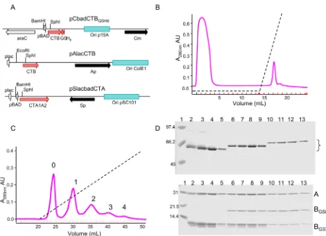
SSSGGGGSSH-PNNTSNNTSSAEDYEYPS, followed by six His residues. Each plasmid has a different replication origin, antibiotic selection, and combination of promoters (Fig. 1A; Table 1). ThectxAgene, en-coding CTA, is expressed from dual pLac and pBAD promoters. Holotoxins and free B pentamers were purified from whole-cell lysates by metal-ion affinity chromatography on Talon resin, and free B pentamers were removed by passing the eluate pool over cation exchange resin, resulting in binding of the free CTB pen-tamers to the resin and recovery of holotoxin in the flowthrough (Fig. 1B). Holotoxins with different numbers of BS were then separated by anion-exchange chromatography (Fig. 1C). By vary-ing the amount of each inducer, we sought to alter the ratio of wt and mutant B subunits in the holotoxin pool. However, for prac-tical purposes, we found that a single condition of relatively low levels of arabinose (0.0005% to express GSH6-tagged wt-CTB) and high levels of isopropyl--d-thiogalactopyranoside (IPTG) (400M to express native-size CTB-G33D) gave acceptable yields of holotoxin with homopentameric and singly or doubly tagged heteropentameric B subunits (with zero, one, and two wt BS, re-spectively) and detectable but lower yields of the triple, quadruple, and homopentameric tagged B subunits as described in detail in the next paragraph. This production strain we designated AMBT (forA, mutant CTB, wt CTB-tagged). Simply by swapping the wt and G33D alleles in the respective CTB-encoding vectors and keeping the same inducer ratios, we were able to express the single or double tagged heteropentameric and homopentameric wt CTB subunits with three, four, and five wt BS, respectively, from the production strain designated ABMT (A, wt CTB, mutant CTB-tagged). Using these strategies, from two production strains, we produced all six variant holotoxins, which had from zero to five wt BS and a maximum of two tagged subunits. A third production strain, designated ABBT (A, wt CTB, wt CTB-tagged), in which both the GSH6-tagged and untagged B polypeptides were wt CTB, was used under the same expression conditions to produce con-trol holotoxins with five native BS and zero, one, or two tagged CTB subunits.
Figure 1 shows the purification process for the AMBT strain (mutant CTB-G33D, wt CTB tagged). Since CTB pentamers nat-urally bind to metal affinity resins (25), cholera toxin variants can be purified to near-homogeneity by a single passage over Talon resin. The imidazole eluate pool from the Talon resin contained a mixture of holotoxin and some free CTB pentamers. These free pentamers were removed by cation exchange chromatography, where at pH 8.0 in 50 mM Tris buffer, holotoxin passed through the column while free pentamers bound and could be eluted sub-sequently with a salt gradient (Fig. 1B). The heterogenous mixture of holotoxins was further separated into its components by
anion-Jobling et al.
on October 5, 2019 by guest
http://mbio.asm.org/
exchange chromatography. The GSH6 tag not only changed the size of the B monomer but also changed its predicted pI from 8.24 for native CTB to 6.21 for CTB-GSH6. The respective predicted pIs for the CTB-G33D variant changed from 7.31 for the native length to 5.91 for the GSH6-tagged variant. All holotoxins bound to the anion exchange matrix, and at least five peaks could be discerned following elution with a salt gradient (Fig. 1C). Samples
from individual fractions were analyzed by SDS-PAGE either without (Fig. 1D, upper) or after (Fig. 1D, lower) boiling in sam-ple loading buffer. The unboiled samsam-ples in lanes 4 to 13 showed a single band corresponding to assembled holotoxin (AB5), with a
[image:4.594.53.523.63.404.2]small amount of contaminating protein of slower mobility in lanes 2 and 3. The major fractions from each peak are homoge-nous and are consistent with each peak containing a single species FIG 1 Plasmid constructs and purification of variant holotoxins from strain AMBT. (A) Schematic representation of plasmid constructs showing the following: the promoters (arrowheads online) pBAD, arabinose inducible, and pLac, IPTG inducible; replication origins (shaded boxes); antibiotic resistance determinants (solid arrows); and other genes (toxin subunits, shaded arrows, and AraC regulatory protein, open arrow). (B) Cation exchange (HS20) chromatographic separation of holotoxin and CTB pentamers from Talon eluates of extracts from strain AMBT. Holotoxins elute in the flowthrough, whereas CTB pentamers bind and are eluted from the column with a salt gradient (dashed line, 0 to 1 M NaCl). Inside ticks on thexaxis mark fractions collected. (C) Anion exchange (HQ20) chromatographic separation of individual holotoxin variants (labels 0 to 4 on peaks correspond to the number of tagged CTB polypeptides in the holotoxin variant) eluted with a salt gradient (dashed line, 0 to 1 M NaCl). (D) Coomassie-stained SDS-PAGE analysis of peaks 0 (lanes 2 to 5), 1 (lanes 6 to 9) and 2 (lanes 10 to 13); size standards (in kDa) are in lane 1. Upper panel, unboiled samples separated on a 10% gel; bracket identifies holotoxins; lower panel, boiled samples on a 15% gel; “A” indicates CTA polypeptide, “BGSH6” indicates GS-H6-tagged wt CTB polypeptide; and “BG33D” indicates CTB-G33D mutant polypeptide.
TABLE 1Plasmids used in this study
Plasmid name [alternate designation] Toxin subunit, promoter(s), selection, origin Source or reference
pSlacbadCTA [0726S2(07)] CTA, pLac and pAraBAD, Spr, OripSC1010 This study
pAlacCTB [pLMP1] CTB, pLac, Ampr, Ori
ColE1 38
pAlacCTBG33D[pLMP148] CTB-G33D, pLac, Ampr, OriColE1 42
pCbadCTBGSH6 [0114C1(08)] CTB-GSH6, pAraBAD, Cmr, Orip15a This study
pCbadCTBG33DGSH6 [0114C2(08)] CTB-G33D-GSH6, pAraBAD, Cmr, Orip15a This study
pCTB-GS [pYF4] CTB-GS, pLac, Ampr, Ori
ColE1 24
0417C1(06) CTB-Linker-His6, pAraBAD, Cmr, Orip15a This study
1126C2(07) CTB-G33D-Linker-His6, pAraBAD, Cmr, Orip15a This study
on October 5, 2019 by guest
http://mbio.asm.org/
[image:4.594.41.545.628.723.2]of variant holotoxin, The assembled holotoxins in the unboiled samples bind less SDS than if they were fully denatured and there-fore do not migrate in direct proportion to their molecular weights. The relative molecular weight (Mr) for each holotoxin
variant is increased by the presence of one or more tagged CTB subunits. Each of these holotoxins completely disassembles into its component polypeptides after boiling (Fig. 1D, lower panel; CTA, 29 kDa; CTB-GSH6-tagged monomer, 14.4 kDa; and native CTB-G33D monomer, 11.5 kDa). The holotoxin in peak 0 (Fig. 1C) has homopentameric CTB-G33D subunits and no func-tional GM1BS, while holotoxins in peaks 1 and 2 have a mixture of
wt CTB-GSH6 and CTB-G33D pentamers with 1 and 2 GM1BS,
respectively. The fractions loaded in lanes 2 to 5 contain CTA plus 5 CTB-G33D (peak 0), and those in lanes 6 to 9 contain CTA plus 1 wt CTB-GSH6 and 4 CTB-G33D (peak 1), and those in lanes 10 to 13 contain CTA plus 2 wt CTB-GSH6 and 3 CTB-G33D (peak 2). Only these first three peaks were well enough separated to be purified efficiently from the extract of production strain AMBT. The fractions corresponding to each peak were pooled, dialyzed, and rerun over the same anion-exchange column to achieve a very high degree of purity. To make holotoxins with 3 and 4 native GM1BS, an expression and purification run was done using strain
[image:5.594.302.540.66.251.2]ABMT, which produced three peaks similar to those shown in Fig. 1C, containing holotoxins with zero, one, or two GSH6-tagged CTB-G33D subunits and five, four, or three native GM1
BS, respectively. Finally, as noted above, strain ABBT was used for expression and purification of wt control holotoxins, all with 5 GM1BS and zero, one, or two GSH6-tagged CTB subunits.
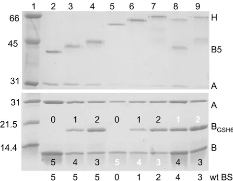
Analysis of the eight final highly purified holotoxin prepara-tions by SDS-PAGE and Coomassie blue staining is shown in Fig. 2. The upper panel shows a gel run with unboiled samples, and the lower panel shows a gel run with samples boiled in sample buffer to dissociate the components. Without boiling, holotoxins with untagged or tagged wt CTB pentamers (Fig. 2, upper panel, lanes 2 to 4) dissociated into free CTA and stable pentameric B subunits that ran near the 45-kDa marker (lane 1). Interestingly, the presence of one or more CTB-G33D polypeptides increased stability of the heterohexameric AB5holotoxins. Consequently,
unboiled holotoxins with three or more CTB-G33D polypeptides (Fig. 2, upper panel, lanes 5 to 7) remained fully associated and ran near the 66-kDa marker (lane 1), whereas unboiled holotoxins with one or two CTB-G33D subunits exhibited partial dissocia-tion into B pentamer and free CTA (Fig. 2, upper panel, lanes 8 and 9). The high stability of the heterohexameric AB5holotoxins
containing five, four, or three CTB-G33D polypeptides is also ev-ident in the upper panel of Fig. 1D. As expected, increasing the number of tagged CTB polypeptides resulted in moderate de-creases in observed mobilities of the pentamer bands (Fig. 2, up-per panel, compare lanes 2, 3, and 4) and the holotoxin bands (Fig. 2, upper panel, compare lanes 5, 6, and 7), and increasing the number of CTB-G33D polypeptides resulted in slight increases in mobilities of the holotoxin bands (Fig. 2, upper panel, compare lanes 6 to 8 and lanes 7 to 9). Upon boiling, all holotoxins disso-ciated and resolved into their individual polypeptide components (Fig. 2, lower panel). All holotoxins have one CTA subunit. Ho-lotoxins in lanes 2 and 5 are predicted to have five native CTB monomers; holotoxins in lanes 3, 6, and 8 have are predicted to have one tagged CTB and 4 untagged CTB monomers; and holo-toxins in lanes 4, 7, and 9 are predicted to have two tagged and three untagged CTB monomers.
Stoichiometry of the individual polypeptides in each purified holotoxin variant was confirmed experimentally by densitometric scanning of the gel in the lower panel of Fig. 2. To adjust for differences in loading of the purified holotoxins (ranging from 0.6g in lane 6 to 2.9g in lane 2), the observed density for each band was expressed as a fraction of the total density for all bands in the same lane. The observed fractional densities were then com-pared with the expected values based on the predicted molecular mass of each polypeptide (CTA, 27.2 kDa; wt CTB, 11.6 kDa; and tagged CTB, 15.3 kDa) and the assumption that binding of Coo-massie blue is proportional to the mass of each peptide. The results (see Table S1 in the supplemental material) were generally within 15% of expected values, but all observed values for tagged B units were higher than expected, suggesting that the tagged sub-units bound proportionately more Coomassie blue stain than na-tive CTA or CTB. Corrected for loading differences, the expected stoichiometric ratios for the tagged B subunit bands in lanes 3 versus 4, 6 versus 7, and 8 versus 9 were 1:2, and the corresponding observed ratios were 1:2.1, 1:1.7, and 1:1.9, respectively.
Ganglioside GM1-binding activities of cholera toxin
vari-ants.Relative binding activity of each holotoxin preparation with from zero to five binding sites (and with zero, one, or two tagged B subunits) to ganglioside GM1receptor was measured by ELISA
with a fixed amount of toxin (5 ng, 60 fmol) added to individual wells coated previously with serial dilutions of GM1, and bound
toxin was detected with polyclonal rabbit anti-CTB and horserad-FIG 2 Coomassie-stained SDS-PAGE analysis of all eight purified holotoxin variants. Upper panel, unboiled samples separated on a 10% gel; lower panel, boiled samples separated on a 15% gel. Bio-Rad low range size standards (in kDa) are in lane 1. Five-BS holotoxins with 0, 1, or 2 tagged B subunits (lanes 2, 3, and 4, respectively), a 0-BS holotoxin (5 G33D B subunits, lane 5), or 1- to 4-BS holotoxins (lanes 6, 7, 9, and 8, respectively) are shown. “H” indicates a holotoxin heterohexamer (AB5), “B5” indicates a CTB pentamer, “A” indi-cates a CTA polypeptide, “BGSH6” indicates a GSH6-tagged CTB monomer,
“B” indicates a CTB monomer, and “wt BS” indicates the number of native GM1BS in each holotoxin variant. Numbers on bands in the lower gel show the
numbers of the respective monomer in the pentameric B subunit of each holotoxin (the middle band contains GSH6-tagged CTB monomers, and the lower band contains CTB monomers; black numbers designate functional GM1-binding sites per CTB pentamer, and white numbers designate
G33D-containing, non-GM1-binding sites per CTB pentamer). Amounts of toxin
loaded per lane varied from 0.6g for lane 6 up to 2.9g for lane 2 (2.9, 1.1, 1.15, 0.8, 0.6, 0.85, 2.55 and 1.95g for lanes 2 to 9, respectively).
Jobling et al.
on October 5, 2019 by guest
http://mbio.asm.org/
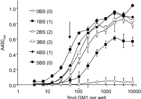
ish peroxidase (HRP)-conjugated secondary antibody. Figure 3 shows that at high GM1density, toxins with more than one native
GM1 BS bound almost as well as native cholera toxin. The
single-BS holotoxin showed a lower plateau signal at high GM1
density than holotoxins with more than one BS, which we inter-pret to be due to a less favorable equilibrium between binding and release for the holotoxin with one BS for GM1. With more than
one BS, a toxin molecule is expected to exhibit faster initial bind-ing to immobilized GM1, multivalent binding as the density of
immobilized GM1increases, and slower dissociation from GM1,
resulting in increased avidity of binding. In wells coated with 75 nM GM1(50l, 3.75 pmol), toxins with 2 or more native GM1
BS bound at more than 90% of the wt level, toxin with a single BS bound significantly less at 60% of the wt level, and toxin with no BS gave a minimal signal (4% or less of the wt level). At lower GM1
densities, there were significant differences between all variants, and in wells coated with 1.2 nM GM1(50l, 60 fmol), toxins with
four or three BS bound at 54 and 49% of wt levels, respectively, and toxins with 2, 1, or no BS bound at 25, 12, and 1% of wt levels, respectively. The amount of GM1required to coat each well and
give 50% of the maximal signal was calculated to be 65, 100, 150, 240, and 850 fmol per well for wt holotoxin and for holotoxin variants with 4, 3, 2, or 1 wt BS, respectively.
Activities of toxin variants on mouse Y1 cells and real-time electrophysiological measurements of variant toxin activities on polarized human colonic T84 cells.Biological activities of the eight holotoxin preparations (the untagged and singly and doubly tagged native holotoxin controls and the variant holotoxins with zero to four wt BS and one or two tags) were initially tested in an overnight assay on mouse Y1 adrenal cell monolayers, on which cholera toxin causes an easily scorable morphological change (rounding of intoxicated cells). In this assay, approximately 2 ng of unnicked native cholera toxin caused rounding of 75 to 100% of the cells (48 U/100 ng). The G33D holotoxin showed some round-ing at only the highest concentration tested (33 ng, extrapolatround-ing
to less than 1 U/100 ng). All holotoxin variants with one or more BS showed rounding of Y1 cells at between 24 and 48 U/100 ng, showing that a single GM1binding site is sufficient to intoxicate
mouse Y1 cells at near-wt levels. Failure to detect significantly less toxicity after overnight exposure of mouse Y1 adrenal cells to ho-lotoxin variants with from 1 to 4 GM1binding sites suggests that
any differences in delivery of the wt CT-A1 fragment from the cell surface to the cytosol by these holotoxin variants were not rate limiting for development of the morphological manifestations of intoxication.
To assess the biological consequences of decreased numbers of BS in a more quantitative manner, we determined the real-time electrophysiological effects of the holotoxin variants on polarized human intestinal cells (T84 cell line) by measuring the short cir-cuit current required to eliminate the potential difference in-duced by the cAMP-dependent Cl–secretory response resulting
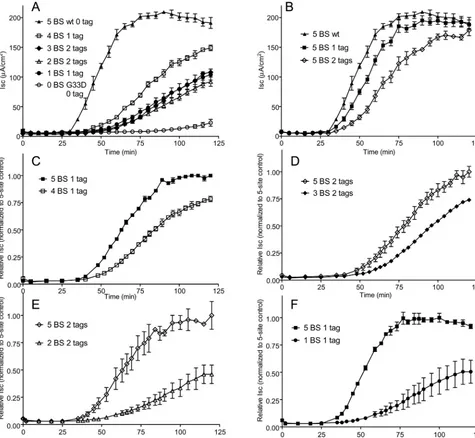
from CT-A1-mediated ADP ribosylation of Gs␣and constitutive activation of adenylate cyclase. Figure 4A to 4F shows the results of these experiments. Panel A shows that loss of even a single GM1BS
attenuated the activity of cholera toxin to some degree, an effect which increased as the number of native GM1BS was lost. The
time of onset of intoxication was also delayed as the number of native GM1BS decreased. Nevertheless, toxin with a single native
GM1 BS had clearly detectable activity. As expected, the G33D
holotoxin with no native GM1BS had almost no activity over the
time frame of the experiment: 1.5% of wt toxin signal over base-line at 90 min and less than 9% of wt signal at 120 min. Since these variants have differing numbers of C-terminal GSH6 tags (zero, one, or two), we also examined the effect that the number of tags had on the activity of wt holotoxin with five BS (Fig. 4B). The presence of the tags modestly affected both the time of onset of intoxication, a measure of toxin-GM1trafficking from
the PM to the ER of host cells, and the rate of increase ofIsc. The
effect was greater for doubly tagged holotoxin, although it eventually showed a similar maximalIsc. To control for these
effects of the B-subunit tags, toxins with different numbers of native GM1 BS but the same number of tags were compared.
For these analyses, data were normalized by setting the maxi-mal signal for the five native GM1BS toxins in each comparison
to 1.00 (Fig. 4C to F). For the toxin variants with one tag on the B subunit, the holotoxin with four native GM1BS exhibited a
slight (25%) attenuation of toxicity relative to holotoxin with five native GM1BS, seen as a decrease in maximalIscbut with
no delay in onset of intoxication (Fig. 4C). A similar result (30% decrease of maximalIsc) was seen for comparisons of the
holotoxin variants with three or five native GM1BS and two
CTB-GSH6-tagged subunits (Fig. 4D). When the number of native GM1BS was reduced to two or one in doubly or singly wt
B-subunit-tagged holotoxin variants, the peakIscdecreased by
more than 50% and also the onset of intoxication was delayed (Fig. 4E and 4F), suggesting defects in entry of CT into the cell or transport to the ER (or both).
As an initial step toward investigating whether CT variants with five and one native GM1BS trafficked from the cell surface to
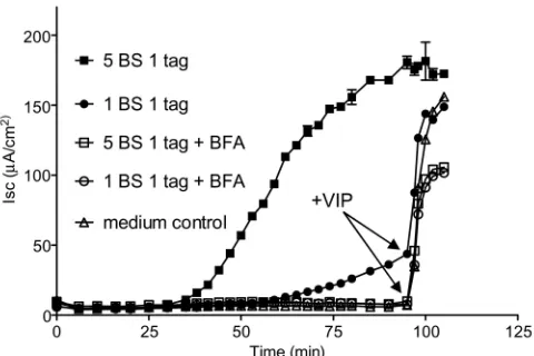
the ER by the same pathway, we examined the effects of brefeldin A (BFA) on their toxicity for T84 cell monolayers. We found that BFA completely inhibited theIsc(Fig. 5) induced by both single
and five native GM1BS toxin variants. BFA- treated cells, however,
still responded to addition of the cAMP agonist vasoactive intes-tinal polypeptide (VIP) at 90 min, showing that the toxin-treated FIG 3 GM1ELISA of variant holotoxins. Toxins with from zero (0BS) to five
(5BS) BS bound to ELISA plates coated with serial dilutions of GM1; the graph
shows mean signal⫾SD for triplicate wells. Wells were coated with serial 2-fold dilutions of GM1starting at 7.5 pmol/well (50l of 150 nM), and 5 ng
of holotoxin was added to each well (approximately 60 fmol). The downward arrow marks the point where toxin and GM1are equimolar (59 fmol/well),
assuming all GM1molecules are bound. Numbers of tagged B subunits in each
holotoxin are indicated in parentheses.
on October 5, 2019 by guest
http://mbio.asm.org/
[image:6.594.43.283.68.237.2]cells retained viability and were competent for cAMP-dependent Cl–secretion (I
sc). Thus, a toxin variant with a single native GM1
BS, like native CT holotoxin, traffics through a BFA-sensitive pathway to exert its toxic effects.
DISCUSSION
Previous studies have shown that holotoxins with reduced num-bers of GM1BS are still able to intoxicate host cells, albeit with
attenuated activity. The initial studies (26, 27) used chemical modification and denaturation-renaturation to generate mixed populations of holotoxins predicted to have one or two (nonna-tive but ac(nonna-tive) GM1BS. In our previous study (22), we used
ge-netic methods to create populations of chimeric holotoxins with
only one or two completely native GM1BS and showed that these
chimeras were still capable of intoxicating host cells but with at-tenuated activity.
More recently, we studied how a membrane lipid might specify trafficking of toxin in the retrograde pathway by using fluorophore-labeled GM1and imaging toxin trafficking in live
cells (28). We found that a subset of GM1species, those with
unsaturated ceramide domains, sorted efficiently from the PM to the trans-Golgi network and the ER. Cross-linking by toxin bind-ing was dispensable for such GM1trafficking, but membrane
cho-lesterol and the lipid raft-associated proteins actin and flotillin were required. Our results implicated an endogenous protein-dependent mechanism of lipid sorting that is protein-dependent on cer-FIG 4 Real-time electrophysiological comparisons of each holotoxin variant against wild-type or control-tagged toxins. (A) Comparison of wt holotoxin versus holotoxins with 4, 3, 2, 1, or no native GM1BS. Isc, short circuit current inA/cm2. (B) Effect of adding one or two GSH6-tagged CTB subunits to wt holotoxin
with 5 native GM1BS. (C to F) Normalized data for tag-controlled comparisons of 5-BS holotoxin versus 4-BS (C), 3-BS (D), 2-BS (E), or 1-BS (F) holotoxin
variants. Maximum signal for the tagged wt holotoxin in panels C through F was adjusted to 1.00, and all data points in the panel were normalized to that value. Data points show the mean response⫾SE;n⫽2 to 4 for A;n⫽3 to 4 for B;n⫽2 for C to F; each study was reproduced at least once.
Jobling et al.
on October 5, 2019 by guest
http://mbio.asm.org/
[image:7.594.56.531.70.508.2]amide structure and could explain how the toxin gains access to the ER of host cells to induce disease (28).
This work shows that multivalent binding to GM1is indeed
dispensable for CT toxicity. To do this, we prepared and purified homogenous preparations of holotoxins with defined numbers of GM1BS. Monomeric binding of CT to single molecules of GM1
permits the holotoxin to intoxicate the host cell. Our results con-firm and expand our studies on trafficking of the single GM1
lip-ids. Conversely, loss of even a single GM1BS resulted in a
measur-able diminishment of toxicity in the T84 line of polarized human intestinal cells. This result is also consistent with findings of our studies of trafficking of the non-cross-linked GM1 molecules,
where we found that GM1 cross-linking by toxin binding
en-hanced entry of CT into the ER. Thus, although multivalent bind-ing to GM1by CT is fundamentally dispensable, it does have a
significant effect on retrograde trafficking from the cell surface to the ER and on toxicity.
One way that multivalent binding could affect CT function would be by enhancing the binding avidity for cell membranes containing GM1. In this and in our previous studies of the CT
binding site mutants, we observe a loss of avidity for binding GM1
when the GM1binding pockets are mutated. In the current
stud-ies, the loss of apparent avidity for the one-binding-site species is 10-fold, but the toxin still binds to GM1applied to the well at
nanomolar concentrations, suggesting this may not fully explain the strong loss of toxin function.
It is possible that the attenuated toxicity seen with holotoxins with reduced numbers of native GM1BS could be enhanced by the
presence of one or more CTB-G33D polypeptides that confer in-creased stability to the holotoxins (and thus might decrease the delivery of the CT-A1 subunit to the cytosol). We raise this idea because we observed that insertion of even a single CTB-G33D monomer into the pentameric B subunit rendered the holotoxin partially resistant to dissociation by SDS, and increasing the num-ber of CTB-G33D monomers resulted in holotoxins that were completely resistant to dissociation by SDS. Conversely, there is noa priorireason to assume that the presence of one or more
CTB-G33D polypeptides would affect chaperone-mediated re-lease of CT-A1 from the nicked and reduced holotoxin in the ER, which appears to facilitate CT-A1 retrotranslocation from the ER to the cytosol (29). Further experiments will be required to test these possibilities.
Another way that multivalent binding to GM1might affect CT
function would be through reorganization of membrane structure and function induced by scaffolding GM1into ceramide-based
nanodomains, as suggested by studiesin vitroandin vivo(14, 15, 30–33). Significantly, studies of the closely related AB5subunit
Shiga toxin show that multivalent binding to the glycosphingo-lipid GB3 spontaneously induces high-curvature membrane tu-bules by coupling the toxin-lipid complex to membrane shape (31). Multivalent binding of CT to GM1in model membranes also
induces spontaneous membrane curvature, implying a similar coupling of the toxin-glycosphingolipid complex formation to membrane shape (32, 34, 35). This could allow partitioning of the CT-GM1complex into highly curved sorting tubules of the sorting
endosome, which is required for retrograde transport, and explain why the toxins with more than 1 GM1binding site are more potent
in intoxication. It is also possible that multivalent binding to GM1
may be needed to activate intracellular signaling pathways that enhance uptake and trafficking, as for Shiga toxin (36).
Effects either on binding avidity or on membrane structure and trafficking dynamics could underlie how the CTB subunit, and the other AB5toxins, evolved as pentameric structures. This
and our other recent studies, however, show that the most funda-mental function exploited by CT is to coopt an endogenous gly-cosphingolipid sorting pathway from the PM to the ER that is essential for toxin entry into the cytosol and the induction of dis-ease.
MATERIALS AND METHODS
Bacterial strains and construction of expression clones.All chimeric toxins in this study were produced by expression from recombinant plas-mids inEscherichia coliBW27784 (araFGHpCP18-araE[37]). This strain does not metabolize arabinose and constitutively expresses the arabinose transporter, and therefore a uniform degree of induction occurs in all cells of a culture at any arabinose concentration. To produce the expression strains, genes encoding native-length CTB or carboxy-terminally ex-tended CTB tagged with a glycosylation-sulfation signal (24) followed by a hexahistidine peptide were cloned on compatible plasmids with differ-ent selectable markers and inducible promoters. These encoded resistance to ampicillin (Ap) with the IPTG-induciblelacUV5promoter or resis-tance to chloramphenicol (Cm) and the arabinose-induciblearaBAD
promoter, along with the regulatoraraC. Variants of these CTB clones were also made with G33D substitutions that eliminate GM1binding. A
separate plasmid (pSlacbadCTA) selectable with spectinomycin (Sp) and compatible (with a pSC101 origin) with bothctxBplasmids was used to independently express thectxAgene under control of both thelacand
araBADpromoters. Each expression strain thus contained three plasmids,
encoding a native CTB subunit (wt or G33D variant, Ap, andlac pro-moter), a GSH6-tagged CTB subunit (G33D or wt, Cm, andara pro-moter), and the wt CTA subunit (Sp andaraandlacpromoters). Details of construction, including full DNA sequences, are available on request. Plasmids created or used in this study are listed in Table 1. Plasmids pAlacCTB and pAlacCTBG33Dencode Ap resistance and the wt and G33D
native-length variants of CTB, respectively, under control of thelac pro-moter and have previously been published as pLMP1 and pLMP148, re-spectively (38). Carboxy-terminally linker-hexahistidine-tagged variants of CTB and CTB-G33D were made in a compatible Cmr
arabinose-inducible vector, pAR3 (39), using the hexa-His tag from pT7sh6 (40) and a glycine-rich repeat from the M13 phage PIII protein, encoding XX FIG 5 Electrophysiological comparison of the effect of brefeldin A on
single-tagged holotoxins with 5 or 1 native GM1BS. Assays were conducted with
(open symbols) or without (solid symbols) brefeldin A (10M) present. At 90 min, 10 nM vasoactive intestinal polypeptide (VIP) was added to all assays except the 5-BS treatment to show that monolayers remained viable and were responsive to VIP stimulation. Data points show mean response⫾SE for duplicate monolayers.
on October 5, 2019 by guest
http://mbio.asm.org/
[image:8.594.44.284.66.226.2](EX)4XDPRVPSS (where X is GGGS) inserted between residues 102 and 103 of CTB. Initial experiments to make wt and CTB-G33D mixed pen-tamers were done using these plasmids, but we saw significant proteolysis of the polyglycine linker-tagged variants, and subsequent experiments were done with variants that had the linker replaced with the GSH6 tag— pCbadCTBGSH6 and pCbadCTBG33DGSH6.
Toxin expression and purification.A 400-ml LB culture at 30°C was inoculated with a 1/8 volume of an overnight culture with appropriate antibiotic selection (Sp and Ap at 100g/ml and Cm at 25g/ml) and grown to anA600of 1.2, when it was induced with 400M IPTG and 0.0005% l-arabinose with incubation continued overnight. Cells were col-lected by centrifugation, resuspended in 20 ml phosphate-buffered saline (PBS), and lysed by mixing for 20 min at room temperature with 0.5 mg/ml lysozyme and Elugent detergent (EMD Biosciences, Inc., La Jolla, CA) to 2%. Viscosity was reduced by sonication four times (10 s each) on ice, and the lysate was cleared by centrifugation at 15,000 rpm for 20 min in an SS34 rotor. Toxin was purified from the supernatants by Talon chromatography as detailed by the manufacturer (Clontech Labo-ratories, Inc., Mountain View, CA). Imidazole eluates were dialyzed against 50 mM Tris-HCl (pH 8.0) (buffer A). All chromatographic sepa-rations were conducted on an Akta purifier (GE Healthcare Biosciences, Pittsburgh, PA) in 4.6-mm by 100-mm Poros perfusion chromatography columns packed with Poros HS 20 cation exchange medium or Poros HQ 20 anion exchange medium. For cation exchange, the column was equil-ibrated with 5 column volumes (CV) buffer A and washed with 5 CV buffer A after sample loading. Bound material was eluted with 10 CV of a linear gradient of 0 to 100% buffer B (50 mM Tris-HCl, pH 8.0, 1M NaCl). For anion exchange, the column was equilibrated with 5 CV buffer A, washed with 5 CV of buffer A after sample loading, and was eluted with a 40-CV (initial separation) or 10-CV (second purification) linear gradient of 0 to 100% buffer B. Anion exchange fractions were pooled and concen-trated using Microcon Ultracel YM-10 filter devices (EMD Millipore, Bil-lerica, MA).
Ganglioside GM1 ELISA. Ninety-six-well microtiter plates were coated overnight with 50l of 2-fold serial dilutions of ganglioside GM1 (Supelco, Sigma-Aldrich, St. Louis, MO) in PBS, starting at 150 nM, and then blocked with 10% horse serum in PBS. All steps were performed at 37°C for 1 h, after which samples were aspirated off and wells were washed three times with PBS plus 0.05% Tween 20. Each holotoxin was assayed in triplicate by incubating 100l of 50-ng/ml holotoxin per well (approxi-mately 60 fmol), followed by 1/5,000 rabbit anti-cholera toxin B subunit (B10) and then 1/2,000 HRP-conjugated goat anti-rabbit serum (Thermo Fisher Scientific Inc., Rockford, IL), followed byo-phenylenediamine (OPD) substrate (Sigma-Aldrich), and the reaction was stopped with 3 M HCl. Absorbance was read at 450 nm.
Y1 assay.Mouse Y1 adrenal cells (ATCC CCL-79) were grown at 37°C in a humidified 5% CO2atmosphere in RPMI medium with 5% fetal bovine serum with 1⫻penicillin-streptomycin (Life Technologies), and 96-well plates were seeded with 104 cells per well.
One-hundred-microliter volumes of 2-fold serial dilutions of toxins were added to semi-confluent monolayers and incubated overnight, followed by scoring for toxin-induced rounding. One unit of toxin was defined as the amount contained in the last dilution giving 75 to 100% rounding of cells.
T84 cells and electrophysiology.Measurements of short-circuit cur-rent (Isc) on monolayers (0.33-cm2inserts) of the polarized human
intes-tinal cell line T84 were performed as previously described (41).
SUPPLEMENTAL MATERIAL
[image:9.594.299.547.64.730.2]Supplemental material for this article may be found athttp://mbio.asm.org /lookup/suppl/doi:10.1128/mBio.00401-12/-/DCSupplemental.
Table S1, DOC file, 0.1 MB.
ACKNOWLEDGMENTS
This work was supported by NIH grant R01 AI31940 to R. K. Holmes and by NIH grants DK48106, DK090603, and DK53056 and Harvard Diges-tive Disease Center grant DK34854 to W. I. Lencer.
REFERENCES
1.Holmgren J, Sánchez J.2011. Cholera toxin—A foe & a friend. Indian J. Med. Res.133:153–163.
2.Ewers H, Helenius A.2011. Lipid-mediated endocytosis. Cold Spring Harb. Perspect. Biol. 3:a004721.http://dx.doi.org/10.1101/cshperspect .a004721.
3.Wernick NL, Chinnapen DJ, Cho JA, Lencer WI.2010. Cholera toxin: an intracellular journey into the cytosol by way of the endoplasmic reticu-lum. Toxins (Basel)2:310 –325.
4.Lencer WI, Moe S, Rufo PA, Madara JL.1995. Transcytosis of cholera toxin subunits across model human intestinal epithelia. Proc. Natl. Acad. Sci. U. S. A.92:10094 –10098.
5.Taylor M, Banerjee T, Ray S, Tatulian SA, Teter K. 2011. Protein-disulfide isomerase displaces the cholera toxin A1 subunit from the holo-toxin without unfolding the A1 subunit. J. Biol. Chem.286:22090 –22100. 6.Moore P, Bernardi KM, Tsai B.2010. The Ero1␣-PDI redox cycle regu-lates retro-translocation of cholera toxin. Mol. Biol. Cell21:1305–1313. 7.Teter K, Jobling MG, Holmes RK.2003. A class of mutant CHO cells
resistant to cholera toxin rapidly degrades the catalytic polypeptide of cholera toxin and exhibits increased endoplasmic reticulum-associated degradation. Traffic4:232–242.
8.Rodighiero C, Tsai B, Rapoport TA, Lencer WI.2002. Role of ubiquiti-nation in retro-translocation of cholera toxin and escape of cytosolic deg-radation. EMBO Rep.3:1222–1227.
9.Schmitz A, Herrgen H, Winkeler A, Herzog V.2000. Cholera toxin is exported from microsomes by the Sec61p complex. J. Cell Biol.148: 1203–1212.
10. Massey S, et al.2009. Stabilization of the tertiary structure of the cholera toxin A1 subunit inhibits toxin dislocation and cellular intoxication. J. Mol. Biol.393:1083–1096.
11. Lencer WI, et al.1997. Proteolytic activation of cholera toxin and
Esch-erichia colilabile toxin by entry into host epithelial cells. Signal
transduc-tion by a protease-resistant toxin variant. J. Biol. Chem.272:15562–15568. 12. Ewers H, et al.2010. GM1 structure determines SV40-induced
mem-brane invagination and infection. Nat. Cell Biol.12:11–18.
13. Johannes L, Römer W.2010. Shiga toxins—from cell biology to biomed-ical applications. Nat. Rev. Microbiol8:105–116.
14. Johannes L, Mayor S.2010. Induced domain formation in endocytic invagination, lipid sorting, and scission. Cell142:507–510.
15. Safouane M, et al.2010. Lipid consorting mediated by Shiga toxin in-duced tubulation. Traffic11:1519 –1529.
16. Cao X, Surma MA, Simons K.2012. Polarized sorting and trafficking in epithelial cells. Cell Res.22:793– 805.
17. Reeves VL, Thomas CM, Smart EJ.2012. Lipid rafts, caveolae and GPI-linked proteins, p 3–13.InJasmin JF, Frank PG, Lisanti MP (ed), Caveo-lins and caveolae, roles in signaling and disease mechanisms, vol729. Springer, New York, NY.
18. Simons K, Sampaio JL.2011. Membrane organization and lipid rafts. Cold Spring Harb. Perspect. Biol.3:a004697.http://dx.doi.org/10.1101 /cshperspect.a004697.
19. Saslowsky DE, Lencer WI.2008. Conversion of apical plasma membrane sphingomyelin to ceramide attenuates the intoxication of host cells by cholera toxin. Cell Microbiol.10:67– 80.
20. Wolf AA, Fujinaga Y, Lencer WI. 2002. Uncoupling of the cholera toxin-GM1ganglioside receptor complex from endocytosis, retrograde
Golgi trafficking, and downstream signal transduction by depletion of membrane cholesterol. J. Biol. Chem.277:16249 –16256.
21. Saslowsky DE, et al.2010. Intoxication of zebra fish and mammalian cells by cholera toxin depends on the flotillin/reggie proteins but not Derlin-1 or -2. J. Clin. Invest.120:4399 – 4409.
22. Wolf AA, et al.2008. Attenuated endocytosis and toxicity of a mutant cholera toxin with decreased ability to cluster ganglioside GM1molecules.
Infect. Immun.76:1476 –1484.
23. Sorre B, et al.2009. Curvature-driven lipid sorting needs proximity to a demixing point and is aided by proteins. Proc. Natl. Acad. Sci. USA106: 5622–5626.
24. Fujinaga Y, et al.2003. Gangliosides that associate with lipid rafts mediate transport of cholera and related toxins from the plasma membrane to endoplasmic reticulum. Mol. Biol. Cell14:4783– 4793.
25. Dertzbaugh MT, Cox LM.1998. The affinity of cholera toxin for Ni2⫹
ion. Protein Eng.11:577–581. Jobling et al.
on October 5, 2019 by guest
http://mbio.asm.org/
26. De Wolf WS, Dierick WS.1994. Regeneration of active receptor recog-nition domains on the B subunit of cholera toxin by formation of hybrids from chemically inactivated derivatives. Biochim. Biophys. Acta1223: 285–295.
27. De Wolf WS, Dams E, Dierick WS.1994. Interaction of a cholera toxin derivative containing a reduced number of receptor binding sites with intact cells in culture. Biochim. Biophys. Acta1223:296 –305.
28. Chinnapen DJ, et al.2012. Lipid sorting by ceramide structure from plasma membrane to ER for the cholera toxin receptor ganglioside GM1. Dev. Cell23:573–586.
29. Tsai B, Rodighiero C, Lencer WI, Rapoport TA.2001. Protein disulfide isomerase acts as a redox-dependent chaperone to unfold cholera toxin. Cell104:937–948.
30. Römer W, et al.2010. Actin dynamics drive membrane reorganization and scission in clathrin-independent endocytosis. Cell140:540 –553. 31. Römer W, et al.2007. Shiga toxin induces tubular membrane
invagina-tions for its uptake into cells. Nature450:670 – 675.
32. Tian A, Capraro BR, Esposito C, Baumgart T.2009. Bending stiffness depends on curvature of ternary lipid mixture tubular membranes. Bio-phys. J.97:1636 –1646.
33. Panasiewicz M, Domek H, Hoser G, Kawalec M, Pacuszka T. 2003. Structure of the ceramide moiety of GM1 ganglioside determines its oc-currence in different detergent-resistant membrane domains in HL-60 cells. Biochemistry42:6608 – 6619.
34. Sorre B, et al.2009. Curvature-driven lipid sorting needs proximity to a
demixing point and is aided by proteins. Proc. Natl. Acad. Sci. U. S. A. 106:5622–5626.
35. Heinrich M, Tian A, Esposito C, Baumgart T.2010. Dynamic sorting of lipids and proteins in membrane tubes with a moving phase boundary. Proc. Natl. Acad. Sci. U. S. A.107:7208 –7213.
36. Torgersen ML, Skretting G, van Deurs B, Sandvig K.2001. Internaliza-tion of cholera toxin by different endocytic mechanisms. J. Cell Sci.114: 3737–3747.
37. Khlebnikov A, Datsenko KA, Skaug T, Wanner BL, Keasling JD.2001. Homogeneous expression of the PBADpromoter inEscherichia coliby
con-stitutive expression of the low-affinity high-capacity AraE transporter. Microbiology147:3241–3247.
38. Jobling MG, Palmer LM, Erbe JL, Holmes RK.1997. Construction and characterization of versatile cloning vectors for efficient delivery of native foreign proteins to the periplasm ofEscherichia coli. Plasmid 38:158 –173.
39. Pérez-Pérez J, Gutiérrez J.1995. An arabinose-inducible expression vec-tor, pAR3, compatible with ColE1-derived plasmids. Gene158:141–142. 40. Tripet B, et al.2004. Structural characterization of the SARS-coronavirus
spike S fusion protein core. J. Biol. Chem.279:20836 –20849.
41. Lencer WI, Delp C, Neutra MR, Madara JL.1992. Mechanism of cholera toxin action on a polarized human intestinal epithelial cell line: role of vesicular traffic. J. Cell Biol.117:1197–1209.
42. Merritt EA, et al.1995. Surprising leads for a cholera toxin receptor-binding antagonist: crystallographic studies of CTB mutants. Structure 3:561–570.
