R E S E A R C H A R T I C L E
Open Access
ER
b
1 represses basal-like breast cancer epithelial
to mesenchymal transition by destabilizing EGFR
Christoforos Thomas
1*, Gayani Rajapaksa
1†, Fotis Nikolos
1†, Ruixin Hao
1, Anne Katchy
1, Catherine W McCollum
1,
Maria Bondesson
1, Phil Quinlan
2, Alastair Thompson
2, Savitri Krishnamurthy
3, Francisco J Esteva
4and
Jan-Åke Gustafsson
1Abstract
Introduction:Epithelial to mesenchymal transition (EMT) is associated with the basal-like breast cancer phenotypes. Sixty percent of basal-like cancers have been shown to express wild-type estrogen receptor beta (ERb1). However, it is still unclear whether the ERbexpression is related to EMT, invasion and metastasis in breast cancer. In the present study, we examined whether ERb1 through regulating EMT can influence invasion and metastasis in basal-like cancers.
Methods:Basal-like breast cancer cells (MDA-MB-231 and Hs578T), in which ERb1 was either overexpressed or down-regulated were analyzed for their ability to migrate and invade (wound-healing assay, matrigel-coated Transwell assay) as well as for the expression of EMT markers and components of the EGFR pathway
(immunoblotting, RT-PCR). Co-immunoprecipitation and ubiquitylation assays were employed to examine whether ERb1 alters epidermal growth factor receptor (EGFR) protein degradation and the interaction between EGFR and the ubiquitin ligase c-Cbl. The metastatic potential of the ERb1-expressing MDA-MB-231 cells was evaluated in vivo
in a zebrafish xenotransplantation model and the correlation between ERb1 and E-cadherin expression was examined in 208 clinical breast cancer specimens by immunohistochemistry.
Results:Here we show that ERb1 inhibits EMT and invasion in basal-like breast cancer cells when they grow either
in vitroor in vivoin zebrafish. The inhibition of EMT correlates with an ERb1-mediated up-regulation of miR-200a/b/ 429 and the subsequent repression of ZEB1 and SIP1, which results in increased expression of E-cadherin. The positive correlation of ERb1 and E-cadherin expression was additionally observed in breast tumor samples. Down-regulation of the basal marker EGFR through stabilization of the ubiquitin ligase c-Cbl complexes and subsequent ubiquitylation and degradation of the activated receptor is involved in the ERb1-mediated repression of EMT and induction of EGFR signaling abolished the ability of ERb1 to sustain the epithelial phenotype.
Conclusions:Taken together, the results of our study strengthen the association of ERb1 with the regulation of EMT and propose the receptor as a potential crucial marker in predicting metastasis in breast cancer.
Introduction
In the last decade, genomic studies have identified five breast cancer intrinsic subtypes (Luminal A, Luminal B, HER2 (overexpressing theERBB2), basal-like and clau-din-low) [1,2]. In a recent study, an integrated analysis of copy number and gene expression split the intrinsic
subtypes revealing novel subgroups with distinct clinical outcome, including a high-risk ERa-positive subgroup and a subset of ERa-positive and ERa-negative cases with a favorable outcome. According to this analysis, the majority of the basal-like tumors formed a high-genomic instability subgroup with relatively good long-term outcomes (after five years) [3]. Basal-like pheno-types represent tumors that express markers that are characteristic of the myoepithelium of the normal mam-mary gland, such as epidermal growth factor receptor (EGFR), p63 and the basal cytokeratins CK14, CK5/6
* Correspondence: chthomas@uh.edu †Contributed equally
1Department of Biology and Biochemistry, Center for Nuclear Receptors and
Cell Signaling, University of Houston, 3605 Cullen Blvd., Houston, TX 77204, USA
Full list of author information is available at the end of the article
and CK17 [1,4]. They show partial overlap with the tri-ple-negative breast cancers that are characterized by a lack of HER2 gene amplification and estrogen and pro-gesterone receptor expression. Approximately 75% of triple-negative breast cancers are classified as basal-like tumors on the basis of their overall gene-expression pro-file. The basal-like phenotype represents a more homoge-neous group of cancers than the group of cancers defined by triple negativity [5]. Basal-like tumors are often resis-tant to chemotherapy and develop disresis-tant metastases in characteristic tissues, such as lung and brain [6]. Recent studies have suggested a correlation between the basal phenotypes and epithelial to mesenchymal transition (EMT) [7].
EMT has been reported to promote invasion during the progression of breast carcinomas and it is considered as an essential early step in tumor metastasis [8,9]. EMT is characterized by loss of cellular adhesion, which is mediated by down-regulation of adhesion molecules, such as CD44 and E-cadherin [10,11]. The expression of E-cadherin is regulated by a number of transcriptional repressors, which include SNAIL, SLUG, SIP-1 (ZEB-2), δEF1 (ZEB-1) and TWIST [12-15]. The family of micro-RNAs 200 (miR-200a, miR-200b, miR-200c, miR-141 and miR-429) and the miR-205A regulate the expression of the transcriptional repressors of E-cadherin ZEB-1 and ZEB-2 and, consequently, the levels of E-cadherin in breast cancer cells and tissues. A decrease in the expres-sion of these microRNAs has been observed in cells that have undergone EMT and in mesenchymal regions of metaplastic breast cancer lacking E-cadherin expression [16]. Up-regulation of components of the EGFR signaling pathway, such as ERK2, has also been reported to influ-ence the levels of E-cadherin by regulating the transcrip-tional repressors ZEB-1 and ZEB-2 [17,18].
The potential role of estrogen receptors in regulating EMT and aggressive behavior in breast cancer has recently been under investigation [19]. Although a decline of ERa levels is detected in invasive breast cancers, a few studies have shown regulation of cell migration and invasion by ERa[20,21]. Recent studies have also associated the ERb isoforms ERb1, ERb2 and ERb5 with the regulation of cell migration and invasion in prostate cancer [22,23]. Down-regulation of the fully functional ERbisoform ERb1 (also known as wild-type ERb) promoted EMT in prostate cancer cells and this correlated with the loss of ERb1 in high Glea-son grade invasive prostate carcinoma [22]. Interestingly, patients with triple-negative breast cancer that were treated with adjuvant tamoxifen have been shown to have signifi-cantly better survival when the tumors were positive for ERb1 [24]. In addition, clinical findings showed an inverse correlation between ERb1 positivity and expression of EGFR, a crucial component in basal-like cancers that drives
proliferation and EMT [25]. Given that down-regulation of ERb1 has been observed in invasive breast cancers, in this study we hypothesized that ERb1 functions to maintain an epithelial phenotype in breast cancer and examined whether ERb1 reduces the invasiveness of basal cancer cells by repressing EMT [26].
Materials and methods
Cells, reagents and transfections
The breast cancer cell lines (MDA-MB-231, Hs578T and MCF-7) and the lung cancer cell line (H1299) were obtained from the ATCC. In 17b-estradiol (E2) experi-ments, cells were maintained in phenol red-free media con-taining two or five percent dextran-coated charcoal (DCC)-treated fetal calf serum (FCS). Transforming growth factor
b (TGF-b) and EGF experiments were performed in serum-free or 0.5% FCS media with recombinant human TGF-b1 (5 ng/ml; R & D Systems, Minneapolis, MN USA) for one to three days or EGF (10 ng/ml; Sigma) for 24 h. MDA-MB-231 and Hs578T cells were infected with lenti-viruses containing the plenti6/V5 empty vector or the recombinant pLenti6/V5-D-FLAG-ERb1 and pLenti6/V5-D-FLAG-ERaplasmids as described previously [27]. Cells were transfected twice with ERb-specific siRNAs (Invitro-gen, Carlsbad, CA USA), target sequences 1# 5’
-TTAGC-GACGTCTGTCGCGTCTTCAC-3’; 2# 5’-TTACGAC
Migration and invasion assays
In the wound-healing assay, cells were allowed to form monolayers at 24-well plates. The monolayer was scratched with a pipette tip to form the wound. Twelve hours later, images of the wound were taken using a 10× objective in an OLYMPUS IX51 microscope equipped with an OLYMPUS camera (OLYMPUS, Center Valley, PA USA) and cells in the wound area in five independent fields were counted.
In the invasion assay, cells were seeded in matrigel-coated 6.5 mm Transwell champers (8 μm pore size; BD Biosciences, San Jose, CA USA). Six hours later, the cells that had been translocated to lower compartments of the wells and attached to the lower surface of the filter were fixed in methanol and stained with crystal-violet. The stained cells were counted in five indepen-dent fields in each Transwell.
Immunofluorescence and microscopy
Cells were plated onto 18 mm2coverslips, fixed in 3% par-aformaldehyde (PFA) and 2% sucrose for 15 minutes at room temperature (RT), permeabilized in 20 mM Tris HCI pH 7.5, 75 mM NaCl, 300 mM sucrose, 3 mM MgCl2 and 0.5% Triton-X-100 for 15 minutes at RT and blocked with 5% goat serum in phosphate-buffered saline (PBS) for 1 h at RT. Slides were stained with an E-cadherin antibody (BD Biosciences) at 4°C overnight, washed, incubated with secondary antibody and images were collected on an OLYMPUS BX51 microscope equipped with an OLYM-PUS XM10 camera (OLYMOLYM-PUS, Center Valley, PA USA).
RNA extraction and real-time PCR
Total RNA was isolated using TriZol reagent (Invitrogen) and reverse-transcribed to cDNA using a SuperScript™ II reverse transcriptase kit (Invitrogen). Real-Time PCR was performed using the SyBr green PCR kit (Applied Biosystems, Grand Island, NY USA). EGFR mRNA levels were additionally analyzed using TaqMan mRNA assay according to the manufacturer’s instructions (Applied Biosystems). All quantitative data were normalized to GAPDH and actin-b. For microRNAs, real-time PCR was performed as above using TaqMan microRNA assays (Applied Biosystems). All microRNA data are expressed relative to a U6 small nuclear (sn) RNA TaqMan PCR performed on the same sample. The sequences of the pri-mers used for qPCR are listed in the Additional file 1, Table S1.
Immunoblotting and immunoprecipitation
Cells were lysed in RIPA buffer, including protease and phosphatase inhibitors as previously described [28]. For separation of cytoplasmic and nuclear fractions, cells were suspended in a cold buffer containing 10 mM Hepes pH 7.0, 10 mM KCI, 0.1 mM EDTA, 1 mM DTT
and 0.5 mM PMSF. After 15 minutes’incubation on ice, the homogenate was mixed with 10% NP-40 and centri-fuged for 30 sec. The nuclear pellet was resuspended in a cold buffer containing 10 mM Hepes-KOH pH 7.9, 400 mM NaCI, 0.1 mM EDTA, 5% glycerol, 1 mM DTT and 0.5 mM PMSF and the nuclear extract was isolated by centrifugation. The blots were performed as previously described [28]. Primary antibodies used in immunoblot-ting include: ERa, E-cadherin, N-cadherin, cadherin-11, vimentin, ZEB-1, SIP1, Lamin A/C and Tubulin (Santa Cruz Biotechnology, Santa Cruz, CA USA), actin-b (Sigma St. Louis, MO USA), EGFR (Santa Cruz Biotech-nology, Cell Signaling, Danvers, MA USA), ERb1 (14C8; GeneTex, Irvine, CA USA), which detects an N-terminal epitope and recognizes the ERbisoforms derived from alternative splicing of the last exon, including ERb1 and an in-house antibody that detects an epitope in ligand binding domain of ERb1 (amino acids 320 to 527)) [29], SNAIL (Abcam, Cambridge, MA USA), p-ERK1/2 and total ERK1/2 (Cell Signaling), c-Cbl (BD Biosciences). Recombinant ERb1 (Invitrogen) was loaded in SDS-PAGE gels and used as a positive control. For ubiquityla-tion analysis, cells were lysed in RIPA buffer containing protease inhibitor cocktail (Roche, Branchburg, NJ USA). The lysates were briefly sonicated and cleared by centri-fugation at 4°C. Supernatants were incubated with anti-EGFR antibody overnight at 4°C and A/G agarose beads for 2 h at 4°C. The immunocomplexes were washed three times, boiled in 2× sample buffer and immunoblotted with anti-ubiquitin antibody (Santa Cruz Biotechnology). For the EGFR-c-Cbl co-immunoprecipitations, cells were lysed in a buffer containing 50 mM Hepes pH 7.4, 150 mM NaCI, 1 mM EDTA, 1 mM EGTA, 1% Nonidet P-40, 1% glycerol including protease and phosphatase inhibitors. Lysates were incubated on ice for 30 minutes without soni-cation, cleared by centrifugation and the cleared lysates were subjected to immunoprecipitation as described.
Zebrafish tumor model and generation of fluorescent cells
Novato, CA USA), were used for the microinjection. Injected embryos were kept at 32°C and were examined every day for tumor invasion using a fluorescent micro-scope (OLYMPUS IX51) equipped with an OLYMPUS XM10 camera. Information for the zebrafish lines is included in the Additional file 2, Supplementary materi-als and methods.
Patient information
A tissue microarray consisting of 240 breast cancer sam-ples was constructed by the Tayside Tissue Bank. Access to tumor samples was approved by the Tayside Regional Ethics Committee with written informed consent from contributing patients. Clinical history and tumor charac-teristics were available for 238 cases. The clinicopathologi-cal characteristics of these patients are summarized in the Additional file 3, Table S2. The majority of the patients received adjuvant endocrine therapy or combined endo-crine therapy and chemotherapy, with or without radio-therapy. Among these patients, 74.7% were ERa-positive, 53.7% were PR-positive and 14.5% were HER2-positive. Histologically, 192 invasive ductal carcinomas (80.6%), 14 invasive lobular carcinomas (5.8%), 5 tubular carcinomas (2.1%), 5 mucinous carcinomas (2.1%) and 22 other histo-logical or mixed types (9.2%) were included.
Antibody validation and immunohistochemistry
The anti-ERb1 antibody (clone PPG5/10, Dako, Carpin-teria, CA USA), which is specific for the C-terminal amino acid sequences of ERb1, was used for immunohistochem-istry (IHC). This antibody was validated by immunocyto-chemistry. Briefly, H1299 human lung cancer cells were stably transfected with the pIRES empty vector (Clontech) or the recombinant pIRES-ERb1 or pIRES-ERb2 plasmids. Control, ERb1 and ERb2-expressing cells were fixed with 10% formalin. The cell suspension was centrifuged and the cell pellet was folded in sharkskin filter paper using four overlapping edges and placed within the base of a tissue cassette. The cassette was placed in a specimen bucket with 10% formalin. The formalin-fixed cell material was embedded in paraffin, cut at 5μm intervals and used for H&E staining and IHC.
For immunohistochemistry, formalin-fixed, paraffin-embedded sections were de-paraffinized with xylene and rehydrated through a graded alcohol series. For antigen retrieval, the slides were immersed in 10 mM sodium citrate buffer (pH 6.0) and maintained at a sub-boiling temperature for six minutes. The endogenous peroxidase activity was blocked by incubation in 0.3% hydrogen perox-ide solution for 20 minutes. The slperox-ides were first incubated with 1% bovine serum albumin (BSA) to block non-specific staining and then with the primary antibody overnight at 4°C in a humidified chamber. The sections were then pro-cessed according to the Dako DAB detection kit.
The results of the immunohistochemistry were assessed by a pathologist (SK) in a blinded fashion. Each specimen was assigned a score according to the intensity of the nuclear staining (for ERb1) and cytoplasmic and mem-brane staining (for E-cadherin) (no staining = 0, weak staining = 1, moderate staining = 2, strong staining = 3) and the extent of stained cells (0% = 0, 1 to 24% = 1, 25 to 49% = 2, 50 to 74% = 3, 75 to 100% = 4). The final immu-noreactive score was determined by multiplying the inten-sity score with the extent of the score of stained cells, ranging from 0 (the minimum score) to 12 (the maximum score). We defined ERb1 expression as low (score 0 to 4), medium (score 5 to 8) and high (score 9 to 12). For E-cad-herin, we defined a 0 score as negative and a 1 to 12 as positive.
Statistical analysis
The correlation between expression of ERb1 and E-cad-herin, respectively, was determined using Pearson’s corre-lation test. All statistical tests were two-sided andP-values less than or equal to 0.05 were considered as statistically significant. The statistical analyses were performed using SPSS 20.0 software (SPSS, IBM, Armonk, NY USA).
Results
ERb1 is required for the epithelial breast cancer phenotype
Basal-like phenotypes are high-grade (grade III), ERa -negative invasive breast tumors that express EMT markers and show cadherin switching as a consequence of tumor de-differentiation [7]. Previous studies have shown a decline of ERb1 expression from ductal carcinomain situ (DCIS) to invasive cancer and an association of the recep-tor with the repression of mesenchymal characteristics in invasive prostate cancer [22,30,31]. We hypothesized that ERb1 regulates EMT in breast cancer and that low ERb1 expression in a proportion of basal-like cancers is asso-ciated with mesenchymal characteristics and poor clinical outcome. To test this hypothesis, we stably expressed ERb1 in the invasive triple-negative breast cancer MDA-MB-231 and Hs578T cells and compared the expression levels achieved in these cells with the endogenous expres-sion of ERs in MCF-7 cells (Additional file 4, Figure S1). According to recent studies, MDA-MB-231 and Hs578T cells most resemble the claudin-low breast cancer subtype; however, as basal-like tumors, they display low expression of the luminal and HER2 gene clusters and express low amounts of ERb1 [32]. Induction of ERb1 expression pro-moted morphological changes in these cells characterized by the loss of the “fibroblastoid-like” phenotype and the acquisition of an epithelial-like compact morphology (Figure 1A, B, upper panel). Furthermore, a more spindle-shaped morphology was observed when endogenous ERb1
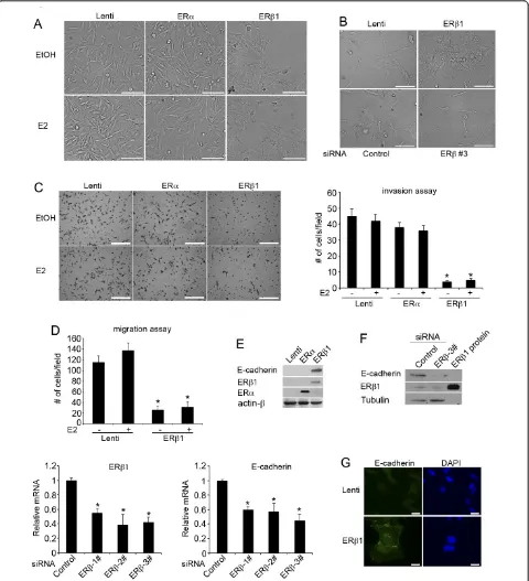
Figure 1ERb1 inhibits invasion and migration in breast cancer cells by regulating EMT.(A)Control (Lenti), ERa- and ERb1-expressing MDA-MB-231 cells following incubation with EtOH or 17b-estradiol (E2) for 24 h (scale bars, 50μm).(B)Control (Lenti) and ERb1-expressing Hs578T cells (upper panel) and Hs578T cells that were transiently transfected with a siRNA targeting luciferase (Control) or a specific ERbsiRNA (siRNA 3#) (lower panel) were photographed (scale bars, 100μM).(C)Control (Lenti), ERa- and ERb1-expressing MDA-MB-231 cells were incubated with EtOH or E2 and assessed for invasion by using matrigel-coated Transwell chambers. The cells that were translocated to the lower surface of the filter were shown (left panel) (scale bars, 500μm). The graph shows the mean (cell number per field) of three separate
(Figure 1B, lower panel). Induction of ERb1 expression altered the morphology of the MDA-MB-231 and Hs578T cells in the absence of ligand. The morphology of the ERb1-expressing MDA-MB-231 cells following treatment with 17b-estradiol (E2) was similar to that of the untreated cells (Figure 1A). Consistent with the changes in the mor-phology, induction of ERb1 expression in MDA-MB-231 cells repressed invasion and migration (Figure 1C, D), functions characteristic of EMT [33]. Although induction of ERb1 and ERaexpression resulted in a similar activa-tion of an ERE-luciferase reporter, ERafailed to promote epithelial morphology and reduce the invasiveness of MDA-MB-231 cells (Figure 1A, C; Additional file 4, Figure S1; Additional file 5, Supplementary figure legends). Similar to the impact on the cellular morphology and invasiveness, only ERb1 inhibited cadherin switching as shown by the up-regulation of epithelial E-cadherin in both MDA-MB-231 and Hs578T cells and down-regula-tion of the mesenchymal cadherin-11 in MDA-MB-231 and N-cadherin in Hs578T cells (Figures 1E and 2A, B; Additional file 6 Figure S2A). The positive correlation between ERb1 and E-cadherin expression was confirmed by the decrease of E-cadherin mRNA and protein levels
when ERb1 was knocked down in MDA-MB-231 cells
(Figure 1F). In line with the results from the immuno-blotting analysis, immunofluorescence showed higher expression of E-cadherin in the cell surface of the ERb 1-expressing cells compared to the control cells (Figure 1G). This suggests that ERb1 up-regulates the functional form of E-cadherin that promotes cell-cell adhesion. No altera-tion in the levels of the mesenchymal marker vimentin was detected in ERb1-expressing MDA-MB-231 cells sug-gesting that ERb1 induces cell-cell adhesion in these cells by primarily regulating the expression of cadherin (Figure 2B; Additional file 6 Figure S2B).
miR-200 and ZEB1/2 are involved in ERb1-mediated regulation of E-cadherin
A number of transcription factors (SNAIL, SLUG, TWIST, SIP-1 and ZEB-1) have been shown to promote EMTin
vitroby acting as transcriptional repressors of E-cadherin [34,35]. Nuclear translocation of SNAIL has been shown to repress E-cadherin expression in ERb1 knockdown prostate cancer cells [22]. Based on these data, we exam-ined whether SNAIL inhibition is involved in the ERb 1-mediated induction of E-cadherin expression that we observed in breast cancer cells. Surprisingly, induction of ERb1 expression in MDA-MB-231 cells neither altered the expression nor the intracellular localization of SNAIL as assessed by immunoblotting using cytoplasmic and nuclear extracts from control and ERb1-expressing cells as well as immunofluorescence microscopy using a SNAIL Ab (Additional file 7, Figure S3A, B). Instead, up-regulation of ERb1 in MDA-MB-231 and Hs578T cells repressed the
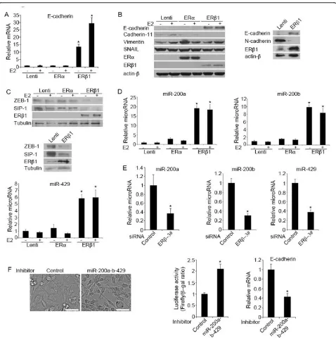
expression of the transcriptional repressors of E-cadherin ZEB-1 and SIP-1 (Figure 2C). Given that recent studies have reported that the microRNA-200 family and miR-205 regulate EMT by targeting ZEB-1 and SIP-1, we examined whether the expression of members of the microRNA-200 family and miR-205 were up-regulated prior to repression of ZEB-1 and SIP-1 expression in ERb1-expressing cells [16]. Using quantitative real-time PCR we found that the cluster of miR-200b-200a-429 was up-regulated by more than 7-fold in the ERb1-expressing MDA-MB-231 and Hs578T cells (3.5-fold increase only for miR-429 in Hs578T cells) (Figure 2D; Additional file 8, Figure S4A). In addition, reduction of endogenous ERb1 expression in MDA-MB-231 and Hs578T cells by ERbsiRNA led to a decrease in the expression of miR-200a, miR-200b and miR-429 (Figure 2E; Additional file 8, Figure S4B). In contrast to the cluster of miR-200b-200a-429, the cluster miR-200c-141 and the miR-205 were unchanged in ERb 1-expressing MDA-MB-231 cells (Additional file 9, Figure S5). We also examined how important is the up-regulation of miR-200a-b-429 for the ERb1-mediated repression of EMT. We transfected the ERb1-expressing MDA-MB-231 cells with inhibitors of miR-200a, miR-200b and miR-429 and assessed the level of functional knockdown of miR200a-b-429 by a reporter assay, in which the comple-mentary sequence of miR200a-b-429 was introduced in the 3’UTR of a luciferase reporter gene. Transfection of the cells with miR200a-b-429 inhibitors resulted in a more than two-fold increase in luciferase activity compared with the negative control inhibitor suggesting that a greater than 50% inhibition of the miR200a-b-429 function had been achieved by the miR200a-b-429 inhibitors (Figure 2F). Inhi-bition of miR200a-b-429 partially reversed the ERb 1-mediated epithelial phenotype and caused a 50% reduction in the expression of E-cadherin (Figure 2F). These data strengthen the role of ERb1 in regulating EMT and suggest a mechanism through which the receptor may regulate E-cadherin expression.
ERb1 inhibits EMT by repressing EGFR signaling
Figure 2ERb1 induces the expression of E-cadherin by up-regulating members of the microRNA 200 family and repressing the expression of ZEB-1 and SIP-1.(A)E-cadherin mRNA levels in control (Lenti), ERa- and ERb1-expressing MDA-MB-231 cells following
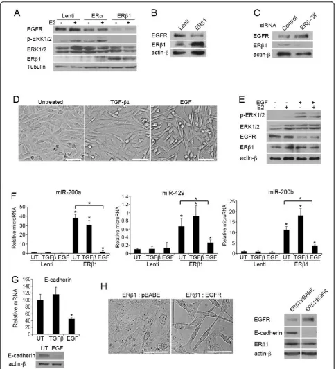
reduction in the EGFR protein levels in MDA-MB-231 and Hs578T cells and decreased the phosphorylation of ERK1/2 as assessed by immunoblotting using an ERK1/2 phospho-specific antibody (Figure 3A, B). Furthermore, reduction of endogenous ERb1 expression in MDA-MB-231 cells led to up-regulation of EGFR (Figure 3C). Analy-sis of EGFR mRNA by qPCR showed the same levels in control and ERb1-expressing cells as well as in cells where ERb1 had been knocked down, suggesting that ERb1 does not regulate the transcription ofEGFRgene (Additional file 6, Figure S2C). To test whether the ERb1-EGFR inter-action is a critical regulator of EMT in basal-like breast cancer cells, we treated the ERb1-expressing cells with EGF or the EMT inducer TGF-b1 for 24 h. For the same purpose, we stably transfected the ERb1-expressing MDA-MB-231 cells with an empty vector or a plasmid that encodes wild-type EGFR. As expected, treatment of the cells with EGF restored the phosphorylation of ERK1/2, decreased the cell-cell contact observed in the ERb 1-expressing cells and abolished the ERb1-mediated up-regulation of miR-200a-200b-429 and the increased levels of E-cadherin (Figure 3D-F, 3G). In contrast, treatment of the cells with TGF-b1, for the same time period as for EGF, failed to reverse the ERb1-induced phenotype in MDA-MB-231 cells (Figure 3D, F, G). As in the case of EGF treatment, EGFR overexpression induced a more fibroblastoid morphology in ERb1-expressing cells, which was accompanied by down-regulation of E-cadherin (Figure 3H).
ERb1 induces degradation of EGFR by enhancing the EGFR-c-Cbl interaction
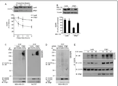
Given that ERb1 altered only the protein but not the mRNA levels of EGFR, we set out to investigate whether ERb1 regulates EGFR at a post-transcriptional level. Spe-cifically, we hypothesized that ERb1 induces degradation of the EGFR protein. EGFR degradation occurs through a process that includes ubiquitylation of the receptor, accelerated endocytosis and degradation by proteasomal and lysosomal hydrolases [39]. In chase experiments, expression of ERb1 reduced the half-life of EGFR sug-gesting that EGFR protein turnover was enhanced by ERb1 (Figure 4A). Treatment of the cells with the protea-some inhibitor MG-132 inhibited the ERb1-dependent reduction in EGFR protein abundance (Figure 4B) con-firming that EGFR down-regulation in ERb1-expressing cells was due to increased degradation. Given that ubi-quitylation is an important step in the degradation of EGFR, we carried out ubiquitylation assays to test whether ERb1 induces ubiquitylation of EGFR. Interest-ingly, the levels of the ubiquitylated EGFR were dramati-cally increased in ERb1-expressing MDA-MB-231 and Hs578T cells (Figure 4C). Furthermore, the ubiquitylated EGFR was decreased when ERb1 was knocked down in
MDA-MB-231 cells (Figure 4D). Given that ubiquityla-tion of the activated EGFR is mediated by members of the Cbl family of RING domain E3 ubiquitin ligases, including the c-Cbl [40], we examined whether ERb1 promotes ubiquitylation of EGFR by inducing its associa-tion with c-Cbl. In control MDA-MB-231 cells, immuno-precipitation of EGFR under nondenaturing conditions showed a rapid but transient recruitment of c-Cbl to EGFR with a barely detectable c-Cbl-EGFR association at 45 minutes following EGF induction. ERb1-expressing MDA-MB-231 cells showed enhanced and more sus-tained c-Cbl-EGFR association with high amounts of c-Cbl recruited to EGFR even at 45 minutes following EGF induction (Figure 4E). These results strengthen our hypothesis that ERb1 down-regulates EGFR by inducing its degradation.
ERb1 inhibits invasion of breast cancer cellsin vivo
To study the role of ERb1 in regulating early events of the metastatic cascade, we used a zebrafish tumor model in which theTg(flk1:EGFP)/casper zebrafish embryos were implanted with the highly metastatic human MDA-MB-231 cells. TheTg(flk1:EGFP)/casper embryos lack pigmentation and express green fluorescent protein (GFP) in the vascular system for direct visualization of vascular development [41]. Both control (Lenti) and ERb1-expressing MDA-MB-231 cells were stably transfected with either DsRed or AmCyan fluorescent proteins. A mixture of either control-DsRed and ERb1-AmCyan cells or control-AmCyan and ERb1-DsRed cells were injected into the perivitelline cavity at 48 hours post-fertilization (hpf), at which time the immune system of the fish is not yet developed. The zebrafish were first imaged 3 h after implantation (Figure 5A, B, upper panels). Invasion and dissemination of DsRed and AmCyan cells were monitored daily in zebrafish. At five days post-injec-tion (dpi), both DsRed and AmCyan MDA-MB-231 control cells had significantly disseminated away from the primary injection site, including the head and the tail regions, whereas ERb1-expressing MD-MB-231 cells labeled with either DsRed or AmCyan remained at the primary site (Figure 5A-D). Out of 45 embryos that were injected with both control and ERb1-expressing cells, 27 embryos had disseminated control cells, and only 2 embryos had dissemi-nated control and ERb1-expressing cells. However, in these two zebrafish, the ratio of control:ERb1 disseminated cells was more than 8:1 (Figure 5E; Additional file 11, Figure S7). Our results show that the difference in metastatic potential between the control and the ERb1-expressing cells is due to their different capacity to invade and disseminate.
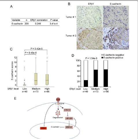
ERb1 and E-cadherin levels are positively correlated in breast cancers
ERb1 and E-cadherin protein levels in breast tumor samples. We utilized a tissue microarray of 240 primary untreated and unselected breast cancers. Clinical history and tumor characteristics (tumor type, age, size, grade, lymph node status, ERa, PR and HER2 status) that were available for 238 cases are summarized in Additional file 3, Table S2. ERb1 and E-cadherin protein levels were determined by IHC using an ERb1 specific antibody (PPG5/10; DAKO). The specificity of the ERb1 antibody was confirmed by immunocytochemistry (Additional file 12 Figure S8A, B). A total of 32 samples were excluded from the analysis due to the absence of tumor or
presence of benign tumor in the core. We carried out Pearson’s correlation analysis with the information of ERb1 and E-cadherin expression from the 208 cancers. Pearson’s correlation analysis showed a strong positive correlation between ERb1 and E-cadherin expressions (P= 3.41e-4) (Figure 6A).
[image:10.595.58.540.87.438.2]To better understand the correlation between ERb1 and E-cadherin, we divided the breast cancer samples into three groups based on ERb1 levels defined by their expres-sion scores and studied the difference of E-cadherin expression among the three groups. The expression of E-cadherin in each group was represented by its median
Figure 4ERb1 induces ubiquitylation and degradation of EGFR.(A)Control (Lenti) and ERb1-expressing MDA-MB-231 cells were incubated in the presence of 100μM cycloheximide and 10 ng/ml EGF for the indicated times and analyzed for EGFR expression by immunoblotting. Treatment with EGF induces phosphorylation of EGFR and this accounts for the retarded electrophoretic mobility of EGFR at times 0.5 to 2. Lower panel: the graph represents the quantification of EGFR protein abundance from three independent experiments with SEM andP-value (*)
score (Figure 6C) or positive percentage (Figure 6D). As shown in Figure 6C, the median scores of E-cadherin in tumors expressing low levels of ERb1 were significantly smaller than in those with higher ERb1 levels. Similarly, the positive percentage analysis for E-cadherin showed a positive correlation with ERb1 levels (Figure 6D). These results are consistent with our findings that ERb1 up-regu-lates E-cadherin in breast cancer cell lines.
Discussion
Although basal-like breast cancers in general are associated with relatively poor prognosis, they are heterogeneous, including diverse subgroups in terms of chemotherapy
[image:11.595.57.540.91.467.2]response and risk of developing distant metastases [2,6,7]. Interestingly, ERb1 positivity has recently been associated with better survival in triple-negative cancers that were treated with tamoxifen and inversely correlated with the expression of EGFR, an important marker in the immuno-histochemical identification of basal-like cancers [24,25,36]. One process that has been attributed to primary tumor metastasis is EMT. Here we examined whether ERb1 through regulating EMT can influence invasion and metas-tasis in basal-like cancers. ERb1 repressed the mesenchy-mal spindle-shaped morphology of the MDA-MB-231 and Hs578T cells and enhanced cell-cell contact. ERb1 altered the morphology of these cells in the absence of ligand.
This is in agreement with our previous data showing increased transcriptional activity following expression of ERb1 in MDA-MB-231 cells in the absence of ERb ago-nists. The increased transcriptional activity in the absence of ligand was correlated with the phosphorylation of ERb1 at ser-87 [28]. As a result of the changes in the morphol-ogy, ERb1 inhibited migration and reduced the invasive-ness of MDA-MB-231 cells. When control and ERb 1-expressing cells were injected into zebrafish embryos, only the control cells disseminated to distant sites suggesting that ERb1 functions as a crucial anti-invasive factor. Given that expression of EMT markers and cadherin switching have been reported to correlate with the basal-like pheno-types inin vitromodel systems and in specimens from patients [7], we examined whether ERb1 inhibits invasion and migration by regulating EMT in cells with basal char-acteristics. ERb1 was found to induce the expression of E-cadherin by inhibiting its transcriptional repressors ZEB1/2 and up-regulating the miR-200a, miR-200b and miR-429, which correlate with the epithelial breast cancer phenotype (Figure 6E).
ERK2 has recently been shown to affect the ZEB1/2 regulatory pathway of E-cadherin expression in human mammary cells [17,18]. ERK1/2 are activated by diverse pathways including that initiated by EGFR [38]. Overex-pression of EGFR promotes migration and invasion of basal cells and its expression correlates with poor survi-val in basal-like cancers [36,37]. Since ERb1 was found to inhibit EMT by down-regulating the ZEB1/2 pathway in basal-like cells, we tested whether repression of EGFR and ERK1/2 signaling are involved in ERb1-mediated up-regulation of E-cadherin and the subsequent inhibi-tion of cell migrainhibi-tion and invasion. Indeed, ERb1 induced a decrease in EGFR protein levels without alter-ing the transcription of the EGFR gene followed by down-regulation of the phosphorylated ERK1/2 forms. Induction of EGFR signaling in ERb1-expressing cells through up-regulation of EGFR or treatment of the cells with EGF reversed the ERb1-dependent epithelial phe-notype, suggesting that EGFR is a critical factor in the ERb1-mediated regulation of EMT.
Given that inhibition of transcription was not involved in ERb1-mediated down-regulation of EGFR, we exam-ined whether ERb1 promotes degradation of the tyrosine kinase receptor. EGFR degradation is a complex process that involves ubiquitylation of the activated receptor by the E3 enzyme Cbl and subsequent proteolysis by pro-teosomal and lysosomal hydrolases [39]. ERb1 was found to induce ubiquitylation and degradation of EGFR by enhancing the EGFR-c-Cbl association. Ubiquityla-tion is an important process of a negative regulatory cir-cuit that terminates EGFR signaling by targeting the receptor for degradation [42]. Our data show for first time that ERb1, by inducing these negative feedback
pathways, is likely to exert a role of EGFR inhibitor and tumor suppressor function.
Interestingly, it has recently been shown that ERb decreases the expression of insulin-like growth factor II-mRNA binding protein 3 (IMP-3) by repressing EGFR transcription in MDA-MB-231 cells [43]. In our study, the transcription of EGFR was not altered when ERb1 was expressed or knocked down in MDA-MB-231 and Hs578T basal-like cells. Instead, as mentioned above, ERb1 promotes degradation of EGFR by inducing its ubiquitylation in both MDA-MB-231 and Hs578T cells.
By examining 208 clinical breast cancer specimens, we found that the expression of ERb1 was significantly asso-ciated with the expression of E-cadherin. This correlation has not previously been reported. However, since the dis-covery of ERb, it has been shown that the association of ERbto other clinicopathological indicators is likely to be divergent in different breast cancer cohorts analyzed by IHC using different ERbantibodies. The tumor cohort examined in our study included a different number of HER2-positive (14.5%) and probably triple-negative breast cancers compared with the cohorts utilized in some of the recent studies that examined large number of samples with well-validated antibodies (HER2-positive, Honma
et al. 2008: 5.25% and Novelliet al. 2008: 31.9%) [24,25, 44,45]. Such differences in the characteristics of the clini-cal cancer samples as well as differences in the specificity of the ERbantibodies used in these studies, may explain why the correlation between ERb1 and E-cadherin expres-sion has not been previously observed. This positive ERb1-E-cadherin association is consistent with the ERb 1-mediated up-regulation of E-cadherin observed in breast cancer cells. It is possible that there are some limitations in the relevance of these results since the level of ERb 1-expression achieved in our cells may not reflect the levels of expression seen in clinical samples. Despite these limita-tions, taken together, our results propose a role for ERb1 in up-regulating E-cadherin in breast cancer cells. This suggests that the low ERb1 levels may be the primary cause of low E-cadherin expression and induction of EMT in some breast cancers. Since EMT correlates with a group of basal-like breast cancers that often develop metastases in distant sites [7], ERb1 may play a crucial role in repressing invasive behavior and inhibiting metas-tasis in this subset of breast cancers. Our data show that ERb1 impedes EMT and influences invasion by down-regulating EGFR, which is expressed in basal-like cancers. These results strengthen the possibility that ERb1 can help to identify patients with basal-like cancer with lower risk to develop metastasis.
Conclusions
with EMT. Our findings indicate that ERb1 inhibits EMT and reduces the invasiveness of basal-like breast cancer cells by up-regulating the epithelial marker E-cadherin. ERb1 induces the expression of E-cadherin by down-regulating EGFR, an oncogenic factor that is expressed in basal-like cancers. ERb1 was found to ter-minate EGFR signaling by targeting the receptor for degradation. Our data support the notion that ERb1 can serve as a clinical marker to identify patients with basal-like cancer that have lower risk to develop metastasis.
Additional material
Additional file 1: Table 1S. Oligonucleotides used in qPCR. The table lists the sequences of the oligonucleotides used in qPCR.
Additional file 2: Supplementary materials and methods. The file contains supplementary information for the zebrafish lines used in xenotransplantation study.
Additional file 3: Table S2. Clinicopathological characteristics of 238 breast cancer patients. The table contains the clinicopathological characteristics of 238 breast cancer patients.
Additional file 4: Figure S1. Functional analysis of ERaand ERb1 in MDA-MB-231 cells. The figure shows the functionality of ERaand ERb1 in MDA-MB-231 cells.
Additional file 5: Supplementary figure legends. The file contains the figure legends for the supplementary figures S1-S8.
Additional file 6: Figure S2. Regulation of EMT markers by ERb1. Description: The figure shows how ERb1 regulates some of the EMT markers.
Additional file 7: Figure S. ERb1 does not alter the intracellular localization of SNAIL. The figure shows how ERb1 affects the intracellular localization of SNAIL.
Additional file 8: Figure S4. ERb1 regulates the expression of miR-200a, miR-200b and miR-429. The figure shows the regulation of miR-200a, miR-200b and miR-429 by ERb1 in Hs578T cells.
Additional file 9: Figure S5. Regulation of miR-200c, miR-141 and miR-205 by ERb1. The figure shows the regulation of miR-200c, miR-141 and miR-205 by ERb1.
Additional file 10: Figure S6. Differences in the expression of EGFR between the ERa-positive(MCF-7) and the triple-negative (MDA-MB-231 and Hs578T) cells. The figure shows the different expression levels of EGFR in MCF-7, MDA-MB-231 and Hs578T breast cancer cells.
Additional file 11: Figure S7. Dissemination patterns of ERb 1-expressing cells in zebrafish. The figure shows the dissemination patterns of ERb1-expressing cells in zebrafish.
Additional file 12: Figure S8. Validation of the anti-ERb1 antibody by immunocytochemistry. The figure shows the specificity of the anti-ERb1 antibody used in immunohistochemistry.
Abbreviations
BSA: bovine serum albumin; CK: cytokeratin; DCC: dextran-coated charcoal; DCIS: ductal carcinomain situ; dpi: days post-injection; EGFR: epidermal growth factor receptor; EMT: epithelial to mesenchymal transition; ERα: estrogen receptorα; ERβ: estrogen receptorβ; ERKs: extracellular-signal-regulated kinases; FCS: fetal calf serum; GFP: green fluorescent protein; hpf: hours post-fertilization; IACUC: Institutional Animal Care and Use Committee; IHC: immunohistochemistry; IMP-3: insulin-like growth factor II mRNA binding protein 3; PBS: phosphate-buffered saline; RT: room temperature; RT-PCR: reverse transcription polymerase chain reaction; TGF-β: transforming growth factorβ
Acknowledgements
We thank Igor Bado, Anders Strom, Cecilia Williams, Ivan Nalvarte, Luisa A. Helguero and Ryan Butler for technical help, and Sang-Hyuk Chung and Chiara Gabbi for help with statistical analysis. This paper was supported by grants from The Emerging Technology Fund of Texas, CPRIT, The Welch Foundation (E-0004) and The Swedish Cancer Society.
Author details
1Department of Biology and Biochemistry, Center for Nuclear Receptors and
Cell Signaling, University of Houston, 3605 Cullen Blvd., Houston, TX 77204, USA.2Department of Surgery and Molecular Oncology, University of Dundee,
DD1 9SY Dundee, UK.3Department of Pathology, The University of Texas MD Anderson Cancer Center, 1515 Holcombe Blvd., Houston, TX 77030, USA.
4Department of Breast Medical Oncology, The University of Texas MD
Anderson Cancer Center, 1515 Holcombe Blvd., Houston, TX 77030, USA.
Authors’contributions
CT conceived, designed and supervised the study, performed or participated in all experiments, their analysis and interpretation, and wrote the manuscript. JAG supervised research and edited the manuscript. GR participated in thein vitroexperiments with the cell lines and in the immunohistochemical staining of the tissue sections. FN participated in most of the cell-based studies. RH and CWM performed the experiments with the transplantation of human cells in zebrafish. MB supervised the zebrafish xenotransplantation assay and participated in editing the manuscript. AK assisted in the experiments with the microRNAs. PQ assisted in the clinical data analysis. SK participated in the design of the clinical study and evaluated the immunohistochemical staining. FJE and AT participated in the design of the clinical study and supervised the analysis of the clinical breast cancer specimens. All authors read and approved the final manuscript.
Competing interests
The authors declare that they have no competing interests.
Received: 1 July 2012 Revised: 19 September 2012 Accepted: 12 November 2012 Published: 16 November 2012
References
1. Perou CM, Sorlie T, Eisen MB, van de Rijn M, Jeffrey SS, Rees CA, Pollack JR, Ross DT, Johnsen H, Akslen LA, Fluge O, Pergamenschikov A, Williams C, Zhu SX, Lonning PE, Borresen-Dale AL, Brown PO, Botstein D:Molecular portraits of human breast tumours.Nature2000,406:747-752. 2. Prat A, Perou CM:Deconstructing the molecular portraits of breast
cancer.Mol Oncol2011,5:5-23.
3. Curtis C, Shah SP, Chin SF, Turashvili G, Rueda OM, Dunning MJ, Speed D, Lynch AG, Samarajiwa S, Yuan Y, Graf S, Ha G, Haffari G, Bashashati A, Russell R, McKinney S, Langerod A, Green A, Provenzano E, Wishart G, Pinder S, Watson P, Markowetz F, Murphy L, Ellis I, Purushotham A, Borresen-Dale AL, Brenton JD, Tavare S, Caldas C,et al:The genomic and transcriptomic architecture of 2,000 breast tumours reveals novel subgroups.Nature2012,486:346-352.
4. Sorlie T, Tibshirani R, Parker J, Hastie T, Marron JS, Nobel A, Deng S, Johnsen H, Pesich R, Geisler S, Demeter J, Perou CM, Lonning PE, Brown PO, Borresen-Dale AL, Botstein D:Repeated observation of breast tumor subtypes in independent gene expression data sets.Proc Natl
Acad Sci USA2003,100:8418-8423.
5. Rakha E, Ellis I, Reis-Filho J:Are triple-negative and basal-like breast cancer synonymous?Clin Cancer Res2008,14:618-619, author reply 618. 6. Kreike B, van Kouwenhove M, Horlings H, Weigelt B, Peterse H, Bartelink H,
van de Vijver MJ:Gene expression profiling and histopathological characterization of triple-negative/basal-like breast carcinomas.Breast
Cancer Res2007,9:R65.
7. Sarrio D, Rodriguez-Pinilla SM, Hardisson D, Cano A, Moreno-Bueno G, Palacios J:Epithelial-mesenchymal transition in breast cancer relates to the basal-like phenotype.Cancer Res2008,68:989-997.
8. Yori JL, Seachrist DD, Johnson E, Lozada KL, Abdul-Karim FW, Chodosh LA, Schiemann WP, Keri RA:Kruppel-like factor 4 inhibits tumorigenic progression and metastasis in a mouse model of breast cancer.
9. Thiery JP:Epithelial-mesenchymal transitions in tumour progression.Nat
Rev Cancer2002,2:442-454.
10. Hazan RB, Qiao R, Keren R, Badano I, Suyama K:Cadherin switch in tumor progression.Ann N Y Acad Sci2004,1014:155-163.
11. Maeda M, Johnson KR, Wheelock MJ:Cadherin switching: essential for behavioral but not morphological changes during an epithelium-to-mesenchyme transition.J Cell Sci2005,118:873-887.
12. Bolos V, Peinado H, Perez-Moreno MA, Fraga MF, Esteller M, Cano A:The transcription factor Slug represses E-cadherin expression and induces epithelial to mesenchymal transitions: a comparison with Snail and E47 repressors.J Cell Sci2003,116:499-511.
13. Comijn J, Berx G, Vermassen P, Verschueren K, van Grunsven L, Bruyneel E, Mareel M, Huylebroeck D, van Roy F:The two-handed E box binding zinc finger protein SIP1 downregulates E-cadherin and induces invasion.Mol Cell2001,7:1267-1278.
14. Eger A, Aigner K, Sonderegger S, Dampier B, Oehler S, Schreiber M, Berx G, Cano A, Beug H, Foisner R:DeltaEF1 is a transcriptional repressor of E-cadherin and regulates epithelial plasticity in breast cancer cells.
Oncogene2005,24:2375-2385.
15. Yang J, Mani SA, Donaher JL, Ramaswamy S, Itzykson RA, Come C, Savagner P, Gitelman I, Richardson A, Weinberg RA:Twist, a master regulator of morphogenesis, plays an essential role in tumor metastasis. Cell2004,117:927-939.
16. Gregory PA, Bert AG, Paterson EL, Barry SC, Tsykin A, Farshid G, Vadas MA, Khew-Goodall Y, Goodall GJ:The miR-200 family and miR-205 regulate epithelial to mesenchymal transition by targeting ZEB1 and SIP1.Nat Cell Biol2008,10:593-601.
17. Shin S, Blenis J:ERK2/Fra1/ZEB pathway induces epithelial-to-mesenchymal transition.Cell Cycle2010,9:2483-2484.
18. Shin S, Dimitri CA, Yoon SO, Dowdle W, Blenis J:ERK2 but not ERK1 induces epithelial-to-mesenchymal transformation via DEF motif-dependent signaling events.Mol Cell2010,38:114-127.
19. Thomas C, Gustafsson JA:The different roles of ER subtypes in cancer biology and therapy.Nat Rev Cancer2011,11:597-608.
20. Wang X, Belguise K, Kersual N, Kirsch KH, Mineva ND, Galtier F, Chalbos D, Sonenshein GE:Oestrogen signalling inhibits invasive phenotype by repressing RelB and its target BCL2.Nat Cell Biol2007,9:470-478. 21. Ye Y, Xiao Y, Wang W, Yearsley K, Gao JX, Shetuni B, Barsky SH:ERalpha
signaling through slug regulates E-cadherin and EMT.Oncogene 29:1451-1462.
22. Mak P, Leav I, Pursell B, Bae D, Yang X, Taglienti CA, Gouvin LM, Sharma VM, Mercurio AM:ERbeta impedes prostate cancer EMT by destabilizing HIF-1alpha and inhibiting VEGF-mediated snail nuclear localization: implications for Gleason grading.Cancer Cell17:319-332. 23. Leung YK, Lam HM, Wu S, Song D, Levin L, Cheng L, Wu CL, Ho SM:
Estrogen receptor beta2 and beta5 are associated with poor prognosis in prostate cancer, and promote cancer cell migration and invasion.
Endocr Relat Cancer17:675-689.
24. Honma N, Horii R, Iwase T, Saji S, Younes M, Takubo K, Matsuura M, Ito Y, Akiyama F, Sakamoto G:Clinical importance of estrogen receptor-beta evaluation in breast cancer patients treated with adjuvant tamoxifen therapy.J Clin Oncol2008,26:3727-3734.
25. Marotti JD, Collins LC, Hu R, Tamimi RM:Estrogen receptor-beta expression in invasive breast cancer in relation to molecular phenotype: results from the Nurses’Health Study.Mod Pathol2010,23:197-204. 26. Leygue ER, Watson PH, Murphy LC:Estrogen receptor variants in normal
human mammary tissue.J Natl Cancer Inst1996,88:284-290. 27. Hartman J, Edvardsson K, Lindberg K, Zhao CY, Williams C, Strom A,
Gustafsson JA:Tumor repressive functions of estrogen receptor beta in SW480 colon cancer cells.Cancer Res2009,69:6100-6106.
28. Thomas CG, Strom A, Lindberg K, Gustafsson JA:Estrogen receptor beta decreases survival of p53-defective cancer cells after DNA damage by impairing G(2)/M checkpoint signaling.Breast Cancer Res Treat2011,
127:417-427.
29. Saji S, Jensen EV, Nilsson S, Rylander T, Warner M, Gustafsson JA:Estrogen receptors alpha and beta in the rodent mammary gland.Proc Natl Acad
Sci USA2000,97:337-342.
30. Skliris GP, Munot K, Bell SM, Carder PJ, Lane S, Horgan K, Lansdown MR, Parkes AT, Hanby AM, Markham AF, Speirs V:Reduced expression of oestrogen receptor beta in invasive breast cancer and its re-expression
using DNA methyl transferase inhibitors in a cell line model.J Pathol
2003,201:213-220.
31. Shaaban AM, O’Neill PA, Davies MP, Sibson R, West CR, Smith PH, Foster CS:
Declining estrogen receptor-beta expression defines malignant progression of human breast neoplasia.Am J Surg Pathol2003,
27:1502-1512.
32. Prat A, Parker JS, Karginova O, Fan C, Livasy C, Herschkowitz JI, He X, Perou CM:Phenotypic and molecular characterization of the claudin-low intrinsic subtype of breast cancer.Breast Cancer Res2010,12:R68. 33. Yang J, Weinberg RA:Epithelial-mesenchymal transition: at the
crossroads of development and tumor metastasis.Dev Cell2008,
14:818-829.
34. Peinado H, Portillo F, Cano A:Transcriptional regulation of cadherins during development and carcinogenesis.Int J Dev Biol2004,48:365-375. 35. Peinado H, Olmeda D, Cano A:Snail, Zeb and bHLH factors in tumour
progression: an alliance against the epithelial phenotype?Nat Rev Cancer
2007,7:415-428.
36. Nielsen TO, Hsu FD, Jensen K, Cheang M, Karaca G, Hu Z, Hernandez-Boussard T, Livasy C, Cowan D, Dressler L, Akslen LA, Ragaz J, Gown AM, Gilks CB, van de Rijn M, Perou CM:Immunohistochemical and clinical characterization of the basal-like subtype of invasive breast carcinoma.
Clin Cancer Res2004,10:5367-5374.
37. Hirsch DS, Shen Y, Wu WJ:Growth and motility inhibition of breast cancer cells by epidermal growth factor receptor degradation is correlated with inactivation of Cdc42.Cancer Res2006,66:3523-3530. 38. Ramos JW:The regulation of extracellular signal-regulated kinase (ERK)
in mammalian cells.Int J Biochem Cell Biol2008,40:2707-2719. 39. Levkowitz G, Waterman H, Ettenberg SA, Katz M, Tsygankov AY, Alroy I,
Lavi S, Iwai K, Reiss Y, Ciechanover A, Lipkowitz S, Yarden Y:Ubiquitin ligase activity and tyrosine phosphorylation underlie suppression of growth factor signaling by c-Cbl/Sli-1.Mol Cell1999,4:1029-1040. 40. Pennock S, Wang Z:A tale of two Cbls: interplay of c-Cbl and Cbl-b in
epidermal growth factor receptor downregulation.Mol Cell Biol2008,
28:3020-3037.
41. Lee SL, Rouhi P, Dahl Jensen L, Zhang D, Ji H, Hauptmann G, Ingham P, Cao Y:Hypoxia-induced pathological angiogenesis mediates tumor cell dissemination, invasion, and metastasis in a zebrafish tumor model.Proc
Natl Acad Sci USA2009,106:19485-19490.
42. Frosi Y, Anastasi S, Ballaro C, Varsano G, Castellani L, Maspero E, Polo S, Alema S, Segatto O:A two-tiered mechanism of EGFR inhibition by RALT/ MIG6 via kinase suppression and receptor degradation.J Cell Biol2010,
189:557-571.
43. Samanta S, Sharma VM, Khan A, Mercurio AM:Regulation of IMP3 by EGFR signaling and repression by ERbeta: implications for triple-negative breast cancer.Oncogene2012,31:4689-4697.
44. Novelli F, Milella M, Melucci E, Di Benedetto A, Sperduti I, Perrone-Donnorso R, Perracchio L, Venturo I, Nistico C, Fabi A, Buglioni S, Natali PG, Mottolese M:A divergent role for estrogen receptor-beta in node-positive and node-negative breast cancer classified according to molecular subtypes: an observational prospective study.Breast Cancer Res2008,10:R74.
45. Shaaban AM, Green AR, Karthik S, Alizadeh Y, Hughes TA, Harkins L, Ellis IO, Robertson JF, Paish EC, Saunders PT, Groome NP, Speirs V:Nuclear and cytoplasmic expression of ERbeta1, ERbeta2, and ERbeta5 identifies distinct prognostic outcome for breast cancer patients.Clin Cancer Res
2008,14:5228-5235.
doi:10.1186/bcr3358
Cite this article as:Thomaset al.:ERb1 represses basal-like breast cancer epithelial to mesenchymal transition by destabilizing EGFR.