R E S E A R C H A R T I C L E
Open Access
Elevated DDX21 regulates c-Jun activity and rRNA
processing in human breast cancers
Yandong Zhang
1,2, Kathleen C Baysac
1,2, Lian-Fai Yee
1,2, Anthony J Saporita
1,2and Jason D Weber
1,2*Abstract
Introduction:The DDX21 RNA helicase has been shown to be a nucleolar and nuclear protein involved in
ribosome RNA processing and AP-1 transcription. DDX21 is highly expressed in colon cancer, lymphomas, and some breast cancers, but little is known about how DDX21 might promote tumorigenesis.
Methods:Immunohistochemistry was performed on a breast cancer tissue array of 187 patients. In order to study the subcellular localization of DDX21 in both tumor tissue and tumor cell lines, indirect immunofluorescence was applied. The effect of DDX21 knockdown was measured by cellular apoptosis, rRNA processing assays, soft agar growth and mouse xenograft imaging. AP-1 transcriptional activity was analyzed with a luciferase reporter and bioluminescence imaging, as well as qRT-PCR analysis of downstream target, cyclin D1, to determine the mechanism of action for DDX21 in breast tumorigenesis.
Results:Herein, we show that DDX21 is highly expressed in breast cancer tissues and established cell lines. A
significant number of mammary tumor tissues and established breast cancer cell lines exhibit nuclear but not nucleolar localization of DDX21. The protein expression level of DDX21 correlates with cell proliferation rate and is markedly induced by EGF signaling. Mechanistically, DDX21 is required for the phosphorylation of c-Jun on Ser73 and DDX21 deficiency markedly reduces the transcriptional activity of AP-1. Additionally, DDX21 promotes rRNA processing in multiple breast cancer cell lines. Tumor cells expressing high levels of endogenous DDX21 undergo apoptosis after acute DDX21 knockdown, resulting in significant reduction of tumorigenicityin vitroandin vivo.
Conclusions:Our findings indicate that DDX21 expression in breast cancer cells can promote AP-1 activity and rRNA processing, and thus, promote tumorigenesis by two independent mechanisms. DDX21 could serve as a marker for a subset of breast cancer patients with higher proliferation potential and may be used as a therapeutic target for a subset of breast cancer patients.
Introduction
Breast cancer is the second-most common cancer diag-nosed in women in the United States. Multiple factors have been found to contribute to breast cancer develop-ment. Proliferation of breast cancer cells requires signals from growth factors such as estrogen and epidermal growth factor (EGF). These factors activate one of the most important transcription factors in the nucleus, acti-vating protein-1 (AP-1), which governs the transcription of key molecules involved in cell cycle progression and
survival, as well as oncogene-induced transformation and cancer cell invasion [1-4]. Previous studies have shown that inhibition of AP-1 activity in breast cancer cells in-duces cell cycle arrest and cell death as well as reduced cell invasionin vitroandin vivo[5-8].
The AP-1 protein family primarily consists of homodi-mers of Jun family members (including v-jun, c-jun, jun B, Jun D) and heterodimers of the Jun family with Fos family components (v-fos, c-fos, fosB, Fra-1, Fra2), or Jun family with activating transcription factors (ATF2, ATF3, B-ATF) [2,4]. Jun-Jun and Jun-Fos dimers preferentially bind to the phorbol ester tumor promoter response element (TRE) while Jun-ATF dimers prefer to bind to c-AMP-responsive element (CRE) [9]. Jun/AP-1 can influence the transcription of a plethora of proteins involved in prolifer-ation, apoptosis, invasion and tumorigenesis [3]. By itself, * Correspondence:[email protected]
1
ICCE Institute, Washington University School of Medicine, 660 South Euclid Avenue Campus 8069, St. Louis, MO 63110, USA
2
Department of Internal Medicine, Division of Molecular Oncology, Siteman Cancer Center, Washington University School of Medicine, 660 South Euclid Avenue Campus 8069, St. Louis, MO 63110, USA
© 2014 Zhang et al.; licensee BioMed Central Ltd. This is an Open Access article distributed under the terms of the Creative Commons Attribution License (http://creativecommons.org/licenses/by/2.0), which permits unrestricted use, distribution, and reproduction in any medium, provided the original work is properly credited. The Creative Commons Public Domain Dedication waiver (http://creativecommons.org/publicdomain/zero/1.0/) applies to the data made available in this article, unless otherwise stated.
c-Jun can transform fibroblast cells and has been classified as a proto-oncogene, albeit a weak one [10,11]. A strong synergistic effect occurs between AP-1 and RasV12 alleles in cell transformation and tumorigenesis [10,12]. An up-stream mitogen-activated protein (MAP) kinase pathway that activates Jun N-terminal kinase (JNK) can activate c-Jun. JNK phosphorylates c-Jun on Ser63 and Ser73 [13,14], although phosphorylation on Ser73 of c-Jun plays a more critical role than Ser63 in its activation [15] .
The DDX21 DEAD box RNA helicase has been recog-nized as an important nucleolar protein involved in ribo-some RNA processing as previous groups have found that depletion of DDX21 results in significant reduction of 18S and 28S rRNA levels in numerous cell types [16-18] and DDX21 has been found to associate with 45S and 32S rRNA species [18]. DDX21 mRNA expression has been correlated with disease-free survival in breast cancer pa-tients [19] and accumulation of DDX21 has been observed in colon cancers and lymphomas [20,21]. DDX21 has also been shown to interact with c-Jun and has been implicated in c-Jun-mediated cellular differentiation [22]. Knockdown of c-Jun causes a diffusion of exclusively nucleolar DDX21 to partially nuclear localization [18].
In this report, we found that DDX21 is highly expressed in breast cancer tissues compared to normal breast tissue and its expression is pivotal to maintain enhanced breast cancer cell proliferation and growth. Surprisingly, a signifi-cant number of breast cancer tissues and breast cancer cell lines show nuclear localization of DDX21 protein. In cells expressing high levels of c-Jun, such as MDA-MB-231 cells, DDX21 associated with c-Jun, was required for c-Jun phosphorylation, and was essential for endogenous AP-1 activity. Moreover, DDX21 helicase activity was re-quired to enhance the oncogenic activity of RasV12, sug-gesting that DDX21 activities might provide requisite functions during cellular transformation. Our results dem-onstrate that DDX21 is an important growth and prolifer-ation modifier that regulates oncogene-induced mammary tumorigenesis, and implicate its potential therapeutic value in breast cancers.
Material and methods Cell culture
MCF-7, 231, SKBR3, 361, MDA-MB-468, CAMA-1, and BT549 breast cancer cells were cul-tured in Dulbecco’s modified Eagle’s medium (DMEM) supplemented with 10% fetal bovine serum (FBS) and peni-cillin-streptomycin. HCC70, HCC712 (obtained from Dr. Matthew Ellis, Washington University), HCC1428, HCC 1806, ZR751, and T47D breast cancer cells were cultured in complete RPMI media supplemented with 10% FBS and penicillin-streptomycin. All cells were maintained at 37°C in 5% CO2. All cell lines were purchased from American Type Culture Collection (ATCC) unless otherwise noted.
Antibodies
Antibodies were obtained from Bethyl Laboratories, Montgomery, TX, USA: anti-glyceraldehyde 3-phosphate dehydrogenase (GAPDH), anti-DDX21; Cell Signaling, Danvers, MA, USA: anti-p53, anti-cyclin D1, anti-c-jun, anti-phospho-c-jun (S73), anti-phospho-c-jun (S63); and Santa Cruz Biotechnology, Dallas, TX, USA: anti-epithelial growth factor receptor (EGFR), anti-tubulin, anti-Ras.
Immunoprecipitation and western blots
Cells were lysed in buffer containing 1% NP-40, 50 mM Tris, pH 7.5, 150 mM NaCl, and supplemented with protease and phosphatase inhibitor cocktails (Sigma-Aldrich, St Louis, MO, USA). After incubation on ice for 10 minutes, cell lysates were sonicated to ensure complete disruption. Lysates were then centrifuged for 10 minutes at 13,000 rpm and supernatants were subjected to protein quantification assay. For western blots, 50μg of cell lysate was loaded on a pre-cast mini-gel (Bio-Rad, Hercules, CA, USA), followed by transferring to polyvinylidene fluoride (PVDF) membrane (Merck Millipore, Billerica, MA, USA). For immunoprecipitation, cell lysates were diluted to 1 mg/ml with lysis buffer; 500μg of total cell lysate was incubated with 2μg of indicated antibody for 2 hours at 4° C, followed by addition of protein A/G Sepharose beads and further incubated for 1 hour at 4°C. After centrifuga-tion at 1,500 rpm for 2 minutes, beads were washed three times with cell lysis buffer before analysis.
Plasmids
Full-length coding sequence for DDX21 was cloned from early passage human BJ fibroblast total cDNA. Primers (for-ward primer: TAGTACTCGAGCCACCA TGCCGGGAA AACTCCGTAGT; reverse primer: CATGTGGATCCT GAC TTGTCATCGTCATCCTTGTAATCTTGACCAAA TGCTTTACTGAA) were used to clone the open reading frame (ORF) of DDX21 into pLVX lentiviral vector at Hin-dIII/ BamHI sites. Quick-change site-directed mutagenesis kit (Stratagene, La Jolla, CA, USA) was used to carry out the mutation K236R. Primers (forward primer: 5′
-CGGA-CAGGAACTGGGAGGACATT CTCCTTTGCC-3′;
re-verse primer: 5′-GGCAAAGGAGAATGTCCTCCCAG T
TCCTGTCCG-3′) were designed using free software from Stratagene. Five different pLKO.1 plasmids containing short hairpin RNA (shRNA) to target each of DDX21 were obtained from the Genome Institute at Washington Uni-versity. A pLKO.1 scrambled shRNA control plasmid was purchased from Addgene, Cambridge, MA, USA.
Quantitative RT-PCR
Figure 1(See legend on next page.)
Zhanget al. Breast Cancer Research2014,16:449 Page 3 of 18
all designed by Primer Express 2.0 software and purchased from Integrated DNA Technologies, Coralville, IA, USA. The following primers were used in quantitative reverse-transcriptase-polymerase chain reaction (qRT-PCR): for
cyclin D1, forward primer: 5′-AAGCTGTGCATCTA
CACCGA-3′; reverse primer: 5′-CTTGAGCTTGTTCAC
CAGGA-3′. For GAPDH, forward primer: 5′-CCCACTC
CTCCACCTTTGAC-3′; reverse primer: 5′
-CATACCAG-GAAATGAGCTTGACAA-3′. For all qPCR analysis, SYBR
green mix (Bio-Rad) was utilized.
[methyl-3H]-methionine labeling of rRNA
Equal numbers of cells were subjected to starvation in methionine-free media containing 10% dialyzed FBS for
30 minutes. Cells were then treated with 50 μCi/mL
[methyl-3H]-methionine and chased in complete media containing an excess of unlabeled methionine (10 μM) for the indicated times. Samples were lysed in RNASolv reagent (Omega Bio-Tek, Norcross, GA, USA) and extracted RNA was separated on agarose-formaldehyde gels and transferred to a Hybond XL membrane (GE Healthcare, Little Chalfont, UK). The membrane was cross-linked and sprayed with En3Hance (Perkin-Elmer, Waltham, MA, USA) prior to autoradiography.
Immunofluorescence and microscopy
Cells were fixed with 10% formalin/10% methanol. Cells were then incubated with rabbit anti-DDX21 (Bethyl La-boratories) at a 1:200 dilution. Goat anti-rabbit rhoda-mine was applied to facilitate the visualization of DDX21 protein. To mark cell nucleoli, mouse anti-nucleophosmin (NPM) or anti-upstream binding factor (UBF) was used at a dilution of 1:100, followed by incu-bation with goat anti-mouse fluorescein isothiocyanate (FITC).
Apoptosis analysis
Apoptosis assays were performed with Vybrant apoptosis kit #2 (Molecular Probes, Eugene, OR, USA) according to the manufacturer’s protocol.
Soft agar assay
1.0 × 10⁴ cells were mixed in 4.0 mL 0.3% agar/DMEM/ 10% FBS as the top agar and plated into 60 mm plates with 4.0 mL 0.6% agar/DMEM/10% FBS as the base agar.
Plates were incubated at 37°C, checked every three days, and fed with 2.0 ml 0.3% agar/ DMEM/10% FBS every week. Colonies were photographed and counted two to three weeks later.
Cell cycle analysis
Cells were trypsinized and washed with phosphate-buffered saline (PBS). Cells were then resuspended in PBS and 100% ethanol was added drop-wise to obtain a final ethanol concentration of 75%. Cells were centrifuged at 2,000 rpm at 4°C for 2 minutes. Cells were then washed with PBS and resuspended in PI working solution (PBS containing 1% FBS, 250 μg/ml of RNase A, 30μg/ml of propidium iodide). Cells were filtered through a 35 μm strainer cap (Becton Dickinson, Franklin Lakes, NJ, USA) before being subjected to fluorescence-activated cell sort-ing (FACS) analysis.
Immunohistochemistry
Human tumor tissue microarrays were purchased from CYBRDI (CC08-11-007). This tissue microarray contains self-paired tumor and adjacent normal tissues from 50 cases of stage I to stage III invasive ductal carcinoma. A rabbit polyclonal anti-human DDX21 antibody (Bethyl La-boratories) was used at 1:25. Tissue slides were deparaffi-nized in xylene and rehydrated in a series of graded alcohols and the antigen was retrieved in 0.01 M sodium citrate buffer (pH 6.0) using a steamer. The sections were then treated with 1% hydrogen peroxide in methanol for 30 minutes to exhaust endogenous peroxidase activity. After a 1 h preincubation in 10% normal FBS to prevent nonspecific staining, the samples were incubated with pri-mary antibody at room temperature for 2 hours. Standard protocol was then followed based on DAKO (Glostrup, Denmark) Envision kit using polymer to amplify signals. A separate breast tissue microarray was purchased from US Biomax, Rockville, MD, USA (BR1503). This tissue micro-array contains duplicate cores for three cases of normal tissues, three fibroadenoma, two cystosarcoma phyllodes, seven intraductal carcinomas and sixty invasive ductal car-cinomas. Tissue slides were prepared as above. Rabbit anti-DDX21 was applied at 1:25 dilution onto the tissue slide and incubated at room temperature for 2 hours with humidity. After PBS washes, secondary goat anti-rabbit Alexa Fluor 594 was used at a dilution of 1:200 for half an
(See figure on previous page.)
Figure 2(See legend on next page.)
Zhanget al. Breast Cancer Research2014,16:449 Page 5 of 18
hour at room temperature. After PBS washes, slow-fade 4',6-diamidino-2-phenylindole (DAPI) with mounting media was applied and each tissue slide was checked under fluores-cence microscope and images were taken. Both immunore-active intensity and percentage of stained cells in different areas of the same slide were analyzed according to criteria described previously [23]. DDX21 expression was designated as 0 = no staining; 1 = weak; 2 = moderate; and 3 = strong. Additional points were scored as one, two, or three when the percentage of positive cells was less than 25%, 25% to 50%, or greater than 50%, respectively.
Bioluminescence imaging
Nonobese diabetic/severe combined immunodeficient (NOD/SCID) female mice were purchased from Charles River Laboratories International, Inc., Wilmington, MA, USA and received standard institutional care. They were five weeks old at the time of surgery. MDA-MB-231 cells stably expressing luciferase reporter (a kind gift from Dr. Marc Diamond, Washington University in St. Louis) were infected with lentiviruses encoding shScrambled RNA or shRNA-DDX21. Two days postinfection, cells were trypsinized, washed twice in PBS, counted, and kept on ice prior to injection. Mice were anesthetized and an inverted Y incision was made between the fourth and fifth sets of nipples to expose the fourth mammary fat pads bilaterally. 5 × 10⁴MDA-MB-231 cells were im-planted into the fourth mammary glands. The final
vol-ume was 50 μl per mammary gland. The incision was
sutured and mice were monitored for tumor growth at the indicated time points via bioluminescence imaging. For bioluminescence imaging of live animals, previously described mice were injected intraperitoneally with 150 μg/g D-luciferin (Biosynth, Naperville, IL, USA) in PBS, anesthetized with 2.5% isoflurane, and imaged with a charge-coupled device (CCD) camera-based biolumin-escence imaging system (IVIS 100; Caliper, Hopkinton, MA, USA; exposure time 10 to 300 seconds, binning 8, field of view 12, f/stop 1, open filter). Signal was dis-played as photons/sec/cm2/sr. Regions of interest (ROI)
were defined manually around the abdomen, throat, and whole body using Living Image and IgorPro Software (Version 2.50) (Wavemetrics, Portland, OR, USA).
Mouse xenograft model
NUDE female mice were purchased from Charles River Laboratories International, Inc. and received standard in-stitutional care. They were five weeks old at the time of surgery. For NUDE mice injection, alternate reading frame (Arf)−/−mouse embryonic fibroblasts (MEFs) were in-fected as indicated. Fibroblasts were trypsinized and re-suspended in PBS at a concentration of 1 × 10⁷ cells/ml. Five-week-old NUDE mice were injected subcutaneously with 1 × 10⁶cells along their flank, with sample sizes of five mice per condition. Four weeks postinfection, tumors were dissected, photographed and weighed.
Animal approval
The Animal Studies Committee of Washington Univer-sity has approved the use of animals in conjunction with this study through Animal Welfare Assurance # A-3381-01 and Approval # 2A-3381-0130226 granted on November 18, 2013. All mouse experiments followed the guidelines of the USDA Animal Welfare Act and the Public Health Service Policy on the use of animals in research.
Results
DDX21 is highly expressed in breast carcinoma
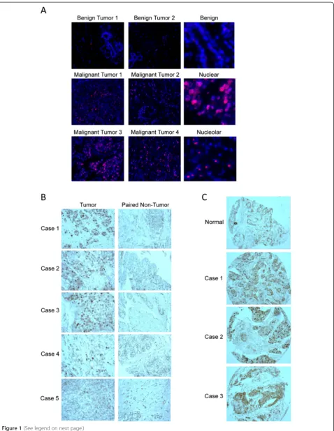
We performed immunohistochemistry staining for DDX21 with several sets of human breast cancer tissue samples to determine whether DDX21 overexpression occurs in breast cancer. First, we performed immunofluorescence staining for DDX21 with a breast tumor tissue array including 67 cases of various stages and subtypes of human breast can-cer. Our results indicated that DDX21 was highly expressed in 25% of the 67 cases of breast tumor tissues, while normal breast tissue and benign tumors all exhibited very weak staining for DDX21. Representative images of these tumor tissues are shown in Figure 1A. Estrogen receptor (ER), progesterone receptor (PR), human epidermal growth
(See figure on previous page.)
Figure 3(See legend on next page.)
Zhanget al. Breast Cancer Research2014,16:449 Page 7 of 18
factor receptor 2 (HER2) status and DDX21 scores are listed in Additional file 1: Table S1. DDX21 staining was primarily observed in nuclei or in nucleolar structures (Figure 1A). Strong DDX21 staining correlated with a high rate of proliferation as detected by Ki67 on these tumor tissues (Additional file 1: Table S1). However, not all of Ki67-positive breast cancer tissues displayed strong DDX21 staining, suggesting that DDX21 is not simply a marker of proliferation. ER and PR status showed no cor-relation with DDX21 expression levels, while DDX21 appeared to be highly expressed in a majority of HER2-positive breast cancers (Additional file 1: Table S1). In addition, we also performed a second tissue array with 50 pairs of nontumor and tumor tissues from unique breast cancer patients (grade I to grade III). Nearly 20% of breast tumor tissues exhibited stronger immunostaining of DDX21 than the paired normal breast tissue in grade I to grade III breast carcinomas (Figure 1B). Similarly, immu-nostaining of a third breast cancer tissue array showed that DDX21 was highly expressed in 16 out of 70 cases (Figure 1C). Additionally, The Cancer Genome Atlas (TCGA) datasets from recently sequenced human basal breast cancer samples showed overexpression of DDX21 mRNA in 14/81 (17%) samples (cBioPortal). Taken to-gether, DDX21 is highly expressed in a significant portion of breast cancer tissues where it localizes to either nuclei or nucleoli.
Nuclear and nucleolar DDX21 is highly expressed in established breast cancer cell lines
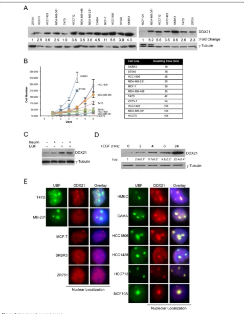
We also analyzed DDX21 protein levels in a panel of established breast cancer cell lines. All of these cancer cells displayed detectable but varying DDX21 protein ex-pression levels (Figure 2A). DDX21 protein levels were significantly higher in cancer cell lines (compared to non-transformed MCF10A cells) that proliferated faster, such as HCC1806, SKBR3, MDA-MB-231 (Figure 2A and B). Conversely, slower proliferating cell lines such as ZR751, HCC70 and HCC1428, expressed much lower DDX21 levels (Figure 2A and B). However, tubulin levels may not serve as the most accurate internal protein control, thus allowing for some differences in our observed expression patterns. In general, and although not absolute, we show
that there is a correlation between higher DDX21 levels and overall cell proliferation rate.
In an effort to more accurately assess the connection between proliferation rates and DDX21 expression, we altered the proliferation rate of MCF10A cells through the addition of defined mitogens. MCF10A cells are a nontu-morigenic cell line derived from a human mammary gland and its proliferation is dependent on growth factors such as insulin and epidermal growth factor (EGF). Addition of in-sulin to deprived MCF10A, which only slightly stimulated cell proliferation, resulted in a minimal increase in DDX21 protein expression (Figure 2C). However, addition of EGF, which induced a near maximal proliferation rate, resulted in a significant increase in DDX21 protein expression that was not further affected by insulin (Figure 2C). A time-course analysis following EGF stimulation showed a rela-tively rapid induction of DDX21 protein expression as early as 2 hours poststimulation (Figure 2D). Thus, DDX21 ap-pears to be a mitogen-induced protein whose expression is indicative of proliferation status in vitro. This appears to correlate with a report showing that DDX21 can be regu-lated by activated MEK signaling [24].
We next performed immunofluorescence analysis to de-termine the cellular localization of endogenous DDX21 in the same panel of established breast cancer cell lines. Con-sistent with the immunofluorescence results of primary breast cancer tissues shown in Figure 1A, we found that DDX21 protein was both nuclear and nucleolar. Specifically, DDX21 localized to the nucleoplasm of T47D, MDAMB231, MCF-7, SKBR3 and ZR751 cell lines (Figure 2E), while it lo-calized to nucleoli in CAMA, HCC1806, HCC1428, HC C712 and MDA-MB-468 as well as primary human mam-mary epithelial cells (HMECs) (Figure 2E).
DDX21 is essential for breast cancer cell proliferation and survival
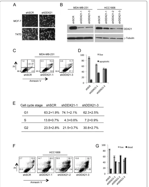
Given the elevated protein expression of DDX21 in highly proliferative breast cancer cell lines, we sought to deter-mine whether DDX21 was an essential protein for cell proliferation and survival. We utilized multiple shRNAs tar-geting endogenous human DDX21 to effectively lower DDX21 protein expression. Following successful knock-down of DDX21, we observed a consistent two-to-threefold
(See figure on previous page.)
Figure 4(See legend on next page.)
Zhanget al. Breast Cancer Research2014,16:449 Page 9 of 18
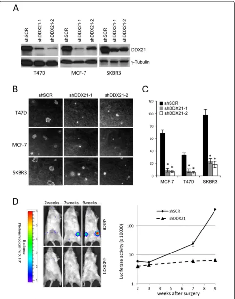
increase in cell death. Live cell imaging of MCF-7 and T47D cells revealed an increase in apoptotic cells following DDX21 knockdown as evidenced by smaller and more re-fractive cells (Figure 3A). Reduction of DDX21 with mul-tiple shRNAs in MDAMB231 and HCC1806 cells was confirmed by western blot analysis (Figure 3B). Annexin V staining was used to mark apoptotic cells and results showed a clear increase in apoptotic MDA-MB-231 cells, where DDX21 was primarily nuclear, after DDX21 knock-down (Figure 3C and 3D). Cell cycle analysis revealed a slight, but reproducible, reduction in S phase that coincided with either an increase in G1 or G2/M phases (Figure 3E). Consistent with these results, knockdown of DDX21 in HCC1806 cells, where DDX21 localized primarily to nucle-oli, resulted in dramatic increases in cell death. However, Annexin V and PI staining of HCC1806 cells indicated that DDX21 knockdown in HCC1806 cells resulted in necrotic cell death rather than apoptosis (Figure 3F and 3G). Taken together, these results show that elevated DDX21 expres-sion is required in order to maintain the viability and cell cycle progression of highly proliferative breast cancer cells.
DDX21 is required for the tumorigenicity of breast cancer cellsin vitroandin vivo
To assess the long-term effects of DDX21 deficiency on proliferation and tumorigenesis, we utilized multiple shRNAs targeting endogenous human DDX21 to effect-ively knock down DDX21 protein expression in three highly proliferative breast cancer cell lines (Figure 4A). DDX21 knockdown had a dramatic impact on anchorage-independent cell growth with lower levels of DDX21 resulting in impaired growth in semi-solid soft agar (Figure 4B and C) and indicating that DDX21 might be re-quired for mammary cell tumorigenicity. In addition, we also performed xenograft implants to assessin vivo tumori-genesis. Severe combined immunodeficiency (SCID) mice were injected with equal numbers of MDA-MD-231 cells carrying a luciferase reporter and either shScrambled or shDDX21 constructs. At nine weeks post-surgery, the five-shScrambled mice developed steadily increasing signals that were significantly higher than the five-shDDX21 mice (Figure 4D). These results indicate a clear requirement for DDX21 protein expression during breast cancer tumorigen-esisin vitroandin vivo. Our results are also consistent with
previous findings that MCF-7 cells (high DDX21) grow significantly faster in vivo than ZR-75-1 cells (low DDX21) [25].
DDX21 modulates AP-1 activity
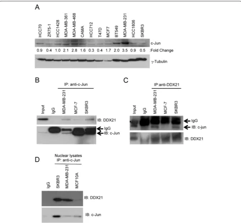
To begin to understand the molecular function of DDX21, we turned to one of its only known interacting partners, c-Jun [22]. We first examined whether DDX21 interacted with c-Jun in our panel of established breast cancer cell lines. We found that c-Jun was highly expressed in MDA-MB-231 and to a lesser extent in BT549 cells while the remaining lines had very low levels of c-Jun protein expression (Figure 5A). Protein extracts from MDA-MB-231, MCF-7 and SKBR3 cells were sub-jected to co-immunoprecipitation assays using anti-c-Jun or anti-DDX21 antibodies. We determined that indeed DDX21 associated with c-Jun in each of the cells in recip-rocal immunoprecipitations (Figure 5B and C). Addition-ally, these interactions were enriched when nuclei were isolated prior to performing the immunoprecipitations (Figure 5D), arguing that the interaction of c-Jun and DDX21 in these cells is occurring in the nucleoplasm.
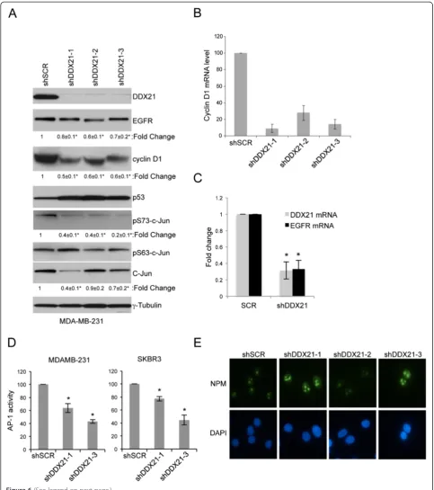
As c-Jun is a critical component of the AP-1 transcrip-tion factor, we hypothesized that DDX21 might some-how modulate AP-1 activity in breast cancer cells. To test this hypothesis, we knocked down DDX21 expres-sion in MDA-MB-231 cells and assayed c-Jun activation using antibodies that recognized the phosphorylation of c-Jun on two critical serine residues, Ser63 and Ser73 [12]. DDX21-depleted cells displayed significantly re-duced levels of phosphorylated c-Jun on Ser73, the pri-mary site for c-Jun activation, while maintaining consistent total c-Jun levels (Figure 6A).
Importantly, AP-1 activity governs the transcriptional in-duction of cyclin D1 to promote the enhanced proliferation of established breast cancer cells [1]. Consistent with our connection between DDX21 and c-Jun phosphorylation status, knockdown of DDX21 resulted in a significant de-crease in cyclin D1 protein expression (Figure 6A). Simi-larly, expression of EGFR, another target of AP-1 [4], was also reduced at the protein level by up to 30 to 40% in all three DDX21 knockdown samples (Figure 6A) as well as at the mRNA level (Figure 6C). As expected, cyclin D1 mRNA levels were substantially reduced in DDX21 knockdown
[image:10.595.61.538.90.103.2](See figure on previous page.)
cells (Figure 6B), underscoring the transcriptional relation-ship between DDX21 and known AP-1 targets. To more directly assess the endogenous AP-1 transcriptional activity in DDX21-depleted cells, a firefly luciferase reporter was utilized. We first infected MDA-MB-231 cells with lentivi-ruses encoding an AP-1-firefly luciferase reporter, and then infected these transduced cells with a second lentivirus en-coding the shScramble or shRNA-DDX21. Cells were then transiently transfected with a plasmid encoding a Renilla
[image:11.595.59.537.88.530.2]luciferase reporter to control for transfection efficiency. After normalization toRenilla luciferase activity, we mea-sured a 40 to 50% reduction in AP-1 activity in DDX21 knockdown cells (Figure 6D). Correspondingly, we ob-served a reduction in AP-1-directed luciferase activity in SKBR3 cells following DDX21 knockdown (Figure 6D). Taken together, our results demonstrate that DDX21, through its interaction with c-Jun, is required for AP-1 ac-tivity and its ability to transcriptionally induce cyclin D1
Figure 5c-Jun interacts with DDX21 in breast cancer cell lines. (A)Panel of breast cancer cell lines was screened by western blot analysis with anti-c-Jun antibody. Fiftyμg of whole cell lysates were subjected to the analysis and tubulin is used as a loading control. Quantitation of the signal was made by Image J software and normalized by tubulin levels.(B)500μg of protein lysates were subjected to immunoprecipitation with anti-c-Jun antibody with mouse IgG as a negative control, 25μg of SKBR3 cell lysate was used as input control. Immune complexes were blotted with anti-DDX21 and anti-c-Jun.(C)Reciprocal immunoprecipitation was also performed with the above-mentioned cell lysates. Western blot analysis was performed to confirm the association between c-Jun and DDX21 in breast cancer cell lines.(D)c-Jun immunoprecipitation was performed on purified nuclear lysates from the indicated cell lines. Western blot analysis was performed to confirm the association between c-Jun and DDX21.
Zhanget al. Breast Cancer Research2014,16:449 Page 11 of 18
and EGFR. To rule out the possibility that these findings occurred because of a loss in nucleolar integrity, we stained nucleoli from MDA-MB-231 cells with antibodies recogniz-ing nucleophosmin (NPM). We observed no change in nu-cleolar staining of NPM when comparing shSCR and shDDX21 cells (Figure 6E), indicating an intact nucleolus in all cells.
Nucleolar DDX21 is required for rRNA processing in breast cancer cells
DDX21 was originally identified as a protein that might be involved in rRNA processing based solely on its nucleolar localization. To determine whether DDX21 is a critical component of rRNA processing in our breast cancer sys-tem where DDX21 is primarily nucleolar, we first per-formed an rRNA pulse-chase assay with HCC1806 cells in which DDX21 localizes to the nucleolus. As shown by Figure 7A, DDX21 knockdown caused the accumulation of the rRNA precursor 47S and reduced the total levels of mature 28S and 18S rRNAs, indicating a requirement for DDX21 in the initial processing of the 47S transcript. To determine whether other rRNA processing steps were af-fected by DDX21 reduction, we followed rRNA processing for 50 minutes. DDX21 knockdown primarily inhibited rRNA processing at the initial 47S processing step and did not result in the accumulation of other downstream inter-mediates that were not processed properly (Figure 7B), again underscoring the role of DDX21 in the first step 47S rRNA processing.
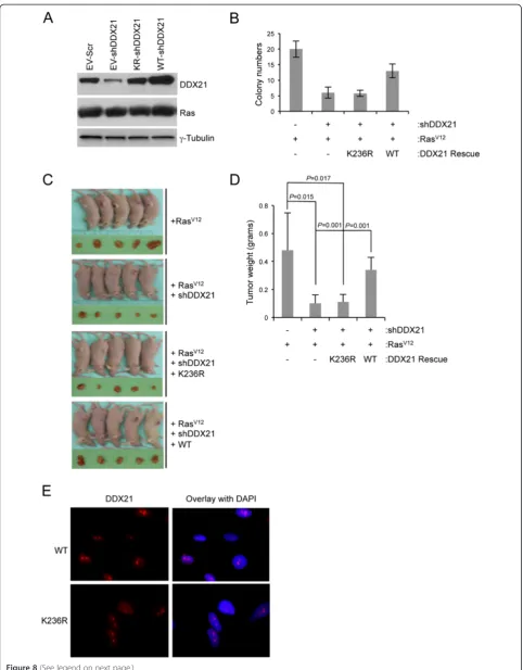
DDX21 RNA helicase activity is required for efficient RasV12cell transformation
Rasis a frequent target for mutation in human cancers. Constitutive activation of Ras through activating muta-tions, such as RasV12, leads to oncogenic transformation of immortalized mouse fibroblasts [26]. C-Jun has been
shown to synergize with oncogenic RasV12 to induce
tumor formation [12,27]. To determine whether DDX21 enhances RasV12’s transforming activity, immortalized (Arf-null) MEFs were consecutively infected with lenti-viruses encoding wild-type DDX21 and retrolenti-viruses
encoding oncogenic RasV12, and then shRNA for mouse
endogenous DDX21. Conversely, Arf-deficient MEFs
were infected with lentiviruses encoding a helicase-defective DDX21 mutant at lysine 236. Lysine 236 re-sides in the ATP-binding motif of DDX21 and mutation of lysine 236 to arginine (K236R) disrupts its ATP-binding activity and severely attenuates its helicase ac-tivity. Protein expression levels of DDX21 and Ras are shown in Figure 8A. The tumorigenicity of RasV12 was then analyzed with various levels of DDX21 by soft agar assay. Reduction in endogenous DDX21 resulted in a significant attenuation in soft agar colony formation and wild-type DDX21 overexpression rescued the ability of RasV12-transformed Arf-null MEFs to grow in soft
agar following endogenous DDX21 knockdown
(Figure 8B). However, the helicase dead mutant K236R DDX21 failed to restore the transforming activity of RasV12(Figure 8B), implying that DDX21 helicase activ-ity was required for effective RasV12transformation. To determine whether this phenotype could be observed in vivo, the above-mentioned cells were injected into the flanks of NUDE mice and the resulting tumors were excised after four weeks. Consistent with our in vitro results, only wild-type DDX21 was able to rescue the tumorigenicity of RasV12 (Figure 8C and D), indicating that DDX21 and its RNA helicase activity is pivotal to maintain RasV12 transforming activity in vivo. Im-portantly, nucleolar localization of DDX21 per se was not sufficient for its function in promoting cellular transformation as the K236R helicase dead mutant lo-calized to nucleoli (Figure 8E) but failed to rescue transformation.
Discussion
We report here that DDX21 is overexpressed in a signifi-cant number of human breast cancers (up to 25%). Its over-expression preferentially occurs in highly proliferative breast cancer tissues and cells. Apart from its predominant nucleolar localization in many breast cancer cells, we also found that DDX21 localized to the nucleoplasm in a signifi-cant number of breast cancer tissues as well as established
(See figure on previous page.)
Figure 6DDX21 modulates AP-1 activity in MDA-MB-231 cells. (A)MDA-MB-231 cells were either infected with shSCR or shRNA-DDX21 lentiviruses. Whole cell lysates were subjected to western blot analysis with the indicated antibodies with tubulin as an internal control. Standard error of the mean is indicated with fold-change.*,P<0.05.(B)RT-PCR analysis was performed for cyclin D1 mRNA levels after normalization with GAPDH mRNA levels. Error bars were taken from three independent experiments.*,P<0.001.(C)RT-PCR analysis was performed for EGFR mRNA levels after normalization with GAPDH mRNA levels. Error bars were taken from three independent experiments.*,P<0.005.(D)MDA-MB-231 and SKBR3 cells were infected with AP-1 reporter lentiviruses to detect endogenous AP-1 activity. After verification of AP-1 luciferase activity, these cells were infected with either shSCR or shRNA-DDX21. Two days postinfection, equal numbers of cells were transfected with pGL-Renilla-luciferase plasmids. Equal numbers of cells were then analyzed for both firefly andRenillaluciferase activity. Data presented is firefly luciferase activity after normalization withRenillaluciferase and then further normalized to shSCR control.*,P<0.001 (n =3).(E)MDA-MB-231 cells infected with shSCR or shRNA-DDX21 lentiviruses were stained with antibodies recognizing NPM and visualized by indirect immunofluorescence. DAPI stain was used to mark nuclei. Images are representative of over 100 cells for each condition. DAPI, 4',6-diamidino-2-phenylindole; EGFR, epithelial growth factor receptor; GAPDH, glyceraldehyde 3-phosphate dehydrogenase; NPM, nucleophosmin; RT-PCR, reverse transcriptase-polymerase chain reaction.
Zhanget al. Breast Cancer Research2014,16:449 Page 13 of 18
cancer cell lines. Levels of c-Jun did not appear to regulate the nuclear localization of DDX21, since in MDA-MB-231 cells that had the highest c-Jun levels, DDX21 predomin-antly localized to the nucleus. This suggests that other regulatory mechanisms might influence the subcellular localization of DDX21. This localization is significant because in these cells we found an association between DDX21 and the c-Jun transcription factor. Originally thought to play a role in rRNA processing due to its nucle-olar localization [28], nucleoplasmic localization of DDX21 implies functions outside of rRNA processing. This was re-cently demonstrated when DDX21 was found to form a complex with DDX1 and DHX36 to sense double-stranded RNA in the cytoplasm of dendrite cells [28]. Our results point to its important function in the nucleus: to promote the activation of c-Jun in epithelial cells. DDX21 sets the ac-tivation state of nuclear AP-1 complexes and facilitates its transcriptional activity toward key players in cell prolifera-tion such as cyclin D1. Particularly, in MDA-MB-231 cells where endogenous c-Jun levels are high, DDX21 expression appears to be critical for maintaining AP-1 transcriptional activity. Short hairpins targeting endogenous DDX21 re-sulted in lowered DDX21 and concomitant decreases in c-Jun Ser73 phosphorylation, a phospho-residue required for AP-1 activation. While it is uncertain how DDX21 might enhance c-Jun phosphorylation through its interaction with c-Jun, lower DDX21 protein expression clearly led to a sig-nificant reduction in classic AP-1 transcriptional targets such as cyclin D1 and EGFR.
[image:14.595.56.292.86.705.2]Large-scale genome sequencing of human cancers has revealed that cancer cells possess large numbers of gen-etic and epigengen-etic alterations [29-31]. While many of these alterations are likely to be random due to the gen-omic instability of tumor cells, only a few are regarded as true oncogenes that drive the cancer phenotype [32]. However, increased dependence of cancer cells on nor-mal cellular functions also plays essential roles for main-taining cancer cell phenotype. These genes are often called non-oncogenes in reference to their requirement for cellular transformation while not possessing trans-forming activities alone [33,34]. Here, we have shown that DDX21 indeed could fall into this category of genes whose activities are required but not sufficient for
Figure 8(See legend on next page.)
Zhanget al. Breast Cancer Research2014,16:449 Page 15 of 18
cellular transformation. DDX21 expression is high in many primary human breast tumors [19] (and herein) and established breast cancer cell lines. Knockdown of DDX21 resulted in dramatic increases in cell death in numerous breast cancer cell lines. DDX21 expression was required to maintain breast cancer survival and proliferation in an in vivo mouse mammary gland. Im-mortal fibroblasts also required DDX21 expression to be efficiently transformed by oncogenic RasV12 alleles. To-gether, these results point to an essential role for DDX21 in tumor cell survival, proliferation, and transformation through two independent activities: rRNA processing and c-Jun activation. However, DDX21 alone is not suffi-cient to transform immortal cells, underscoring its po-tential role as a non-oncogene. Our results strengthen the concept that has been posited in recent years: that cancer cells rely on non-oncogenes in order to maintain and even enhance their tumorigenic phenotype.
The family of DEAD/DEAH box RNA helicases has recently emerged as a large family (54 members) of multifunctional proteins involved in various steps of RNA and DNA metabolism [35-38]. It is becoming ap-parent that many of these helicases perform functions that are either fundamental in many basic cellular pro-cesses such as rRNA synthesis or are more specific in promoting cancer cell growth and proliferation. Even though their detailed molecular mechanisms remain largely undiscovered, our report on DDX21 function un-derscores the potential importance and diversity of DEAD/DEAH helicases in promoting cancer and cer-tainly warrants a broader evaluation of the activities of this protein family.
Conclusions
In this study, we found DDX21 overexpressed in highly proliferative primary breast carcinomas. DDX21 was found to localize to the nucleus and nucleolus in a num-ber of breast cancer tissues as well as in numerous established breast cancer cell lines. In multiple breast cancer cell lines, DDX21 protein knockdown triggered cell cycle arrest as well as massive cell death and caused a significant reduction in anchorage-independent cell
growthin vitroas well asin vivo. We found that nuclear DDX21 interacted with the activating protein (AP-1) component, c-Jun. DDX21 was required for c-Jun Ser73 phosphorylation and activation of AP-1 transcriptional activity, while nucleolar DDX21 promoted cell growth by enhancing rRNA processing. Furthermore, DDX21 RNA helicase activity was required for effective RasV12 transformation. These data highlight the importance of DDX21 in breast cancer cell proliferation and transform-ation through its promotion of AP-1 activity, rRNA pro-cessing and its cooperation with known oncogenes.
Additional file
Additional file 1:DDX21 expression scores and patient information.
Abbreviations
ARF:alternate reading frame; DAPI: 4',6-diamidino-2-phenylindole; DMEM: Dulbecco’s modified Eagle’s medium; EGF(R): epithelial growth factor (receptor); ER: estrogen receptor; FACS: fluorescence-activated cell sorting; FBS: fetal bovine serum; FITC: fluorescein isothiocyanate;
GAPDH: glyceraldehyde 3-phosphate dehydrogenase; Her-2: human epidermal growth factor receptor 2; HMEC: human mammary epithelial cell; MEF: mouse embryonic fibroblast; NPM: nucleophosmin; ORF: open reading frame; PBS: phosphate-buffered saline; PR: progesterone receptor; PVDF: polyvinylidene fluoride; qRT-PCR: quantitative reverse transcriptase-polymerase chain reaction; SCID: severe combined
immunodeficiency; shRNA: short hairpin RNA; UBF: upstream binding factor.
Competing interests
The authors declare that they have no competing interests.
Authors’contributions
YZ designed the study, performed the experiments, wrote the manuscript, approved the final version, and agreed to be accountable for all aspects of the work. KCB performed EGFR experiments and nucleolar integrity assays, edited the manuscript, approved the final version, and agreed to be accountable for all aspects of the work. LY helped in the growth curves for most of the cell lines, interpreted data for the work, edited the manuscript, approved the final version, and agreed to be accountable for all aspects of the work. AJS helped to perform NUDE mice injections, interpreted data for the work, contributed to the draft of the manuscript, approved the final version, and agreed to be accountable for all aspects of the work. JDW supervised and designed the study, revised the manuscript, approved the final version, and agreed to be accountable for all aspects of the work. All authors have read and approved the final version of this manuscript.
Authors’information
YZ is currently an Assistant Professor in South University of Science and Technology of China. AJS is currently a staff scientist in Millipore (St. Louis). (See figure on previous page.)
Acknowledgements
The authors thank the members of the Weber and Helen Piwnica-Worms laboratories for their technical input and suggestions, in particular Shirong Cai, Lynn Collins, and Julie Prior. The authors thank the RNAi Consortium, Children’s Discovery Institute, and The Genome Institute at Washington University for providing lentiviral knockdown constructs. The results shown here for DDX21 mRNA expression in basal breast cancers are based upon data generated by the TCGA Research Network: http://cancergenome.nih.gov/.
Grant support
This work was supported by a Siteman Cancer Center Frontier Award (JD Weber), US NIH 1R01CA120436 (JD Weber), and a Department of Defense Era of Hope Scholar Award BC075004 (JD Weber).
Received: 13 November 2013 Accepted: 19 September 2014
References
1. Shen Q, Uray IP, Li Y, Krisko TI, Strecker TE, Kim HT, Brown PH:The AP-1 transcription factor regulates breast cancer cell growth via cyclins and E2F factors.Oncogene2008,27:366–377.
2. Angel P, Karin M:The role of Jun, Fos and the AP-1 complex in cell-proliferation and transformation.Biochim Biophys Acta1991,1072:129–157. 3. Eferl R, Wagner EF:AP-1: a double-edged sword in tumorigenesis.Nat Rev
Cancer2003,3:859–868.
4. Vogt PK, Bos TJ:jun: oncogene and transcription factor.Adv Cancer Res
1990,55:1–35.
5. Liu Y, Ludes-Meyers J, Zhang Y, Munoz-Medellin D, Kim HT, Lu C, Ge G, Schiff R, Hilsenbeck SG, Osborne CK, Brown PH:Inhibition of AP-1 transcription factor causes blockade of multiple signal transduction pathways and inhibits breast cancer growth.Oncogene2002,21:7680–7689.
6. Liu Y, Lu C, Shen Q, Munoz-Medellin D, Kim H, Brown PH:AP-1 blockade in breast cancer cells causes cell cycle arrest by suppressing G1 cyclin expression and reducing cyclin-dependent kinase activity.Oncogene
2004,23:8238–8246.
7. Lu A, Zhang F, Gupta A, Liu J:Blockade of AP1 transactivation abrogates the abnormal expression of breast cancer-specific gene 1 in breast cancer cells.J Biol Chem2002,277:31364–31372.
8. Ludes-Meyers JH, Liu Y, Munoz-Medellin D, Hilsenbeck SG, Brown PH:AP-1 blockade inhibits the growth of normal and malignant breast cells. Oncogene2001,20:2771–2780.
9. Hai T, Curran T:Cross-family dimerization of transcription factors Fos/Jun and ATF/CREB alters DNA binding specificity.Proc Natl Acad Sci U S A
1991,88:3720–3724.
10. Schutte J, Minna JD, Birrer MJ:Deregulated expression of human c-jun transforms primary rat embryo cells in cooperation with an activated c-Ha-ras gene and transforms rat-1a cells as a single gene.Proc Natl Acad Sci U S A1989,86:2257–2261.
11. Bos TJ, Monteclaro FS, Mitsunobu F, Ball AR Jr, Chang CH, Nishimura T, Vogt PK:Efficient transformation of chicken embryo fibroblasts by c-Jun requires structural modification in coding and noncoding sequences. Genes Dev1990,4:1677–1687.
12. Smeal T, Binetruy B, Mercola DA, Birrer M, Karin M:Oncogenic and transcriptional cooperation with Ha-Ras requires phosphorylation of c-Jun on serines 63 and 73.Nature1991,354:494–496.
13. Kallunki T, Deng T, Hibi M, Karin M:c-Jun can recruit JNK to phosphorylate dimerization partners via specific docking interactions.Cell1996,87:929–939. 14. Kallunki T, Su B, Tsigelny I, Sluss HK, Derijard B, Moore G, Davis R, Karin M:
JNK2 contains a specificity-determining region responsible for efficient c-Jun binding and phosphorylation.Genes Dev1994,8:2996–3007. 15. Smeal T, Hibi M, Karin M:Altering the specificity of signal transduction
cascades: positive regulation of c-Jun transcriptional activity by protein kinase A.Embo J1994,13:6006–6010.
16. Henning D, So RB, Jin R, Lau LF, Valdez BC:Silencing of RNA helicase II/ Gualpha inhibits mammalian ribosomal RNA production.J Biol Chem
2003,278:52307–52314.
17. Yang H, Zhou J, Ochs RL, Henning D, Jin R, Valdez BC:Down-regulation of RNA helicase II/Gu results in the depletion of 18 and 28 S rRNAs in Xenopus oocyte.J Biol Chem2003,278:38847–38859.
18. Holmstrom TH, Mialon A, Kallio M, Nymalm Y, Mannermaa L, Holm T, Johansson H, Black E, Gillespie D, Salminen TA, Langel U, Valdez BC,
Westermarck J:c-Jun supports ribosomal RNA processing and nucleolar localization of RNA helicase DDX21.J Biol Chem2008,283:7046–7053. 19. Cimino D, Fuso L, Sfiligoi C, Biglia N, Ponzone R, Maggiorotto F, Russo G,
Cicatiello L, Weisz A, Taverna D, Sismondi P, De Bortoli M:Identification of new genes associated with breast cancer progression by gene expression analysis of predefined sets of neoplastic tissues.Int J Cancer
2008,123:1327–1338.
20. Bonzheim I, Irmler M, Klier-Richter M, Steinhilber J, Anastasov N, Schafer S, Adam P, Beckers J, Raffeld M, Fend F, Quintanilla-Martinez L:Identification of C/EBPbeta target genes in ALK + anaplastic large cell lymphoma (ALCL) by gene expression profiling and chromatin immunoprecipitation. PLoS One2013,8:e64544.
21. Jung Y, Lee S, Choi HS, Kim SN, Lee E, Shin Y, Seo J, Kim B, Jung Y, Kim WK, Chun HK, Lee WY, Kim J:Clinical validation of colorectal cancer biomarkers identified from bioinformatics analysis of public expression data.Clin Cancer Res2011,17:700–709.
22. Westermarck J, Weiss C, Saffrich R, Kast J, Musti AM, Wessely M, Ansorge W, Seraphin B, Wilm M, Valdez BC, Bohmann D:The DEXD/H-box RNA helicase RHII/Gu is a co-factor for c-Jun-activated transcription.Embo J
2002,21:451–460.
23. Loupakis F, Pollina L, Stasi I, Ruzzo A, Scartozzi M, Santini D, Masi G, Graziano F, Cremolini C, Rulli E, Canestrari E, Funel N, Schiavon G, Petrini I, Magnani M, Tonini G, Campani D, Floriani I, Cascinu S, Falcone A:PTEN expression and KRAS mutations on primary tumors and metastases in the prediction of benefit from cetuximab plus irinotecan for patients with metastatic colorectal cancer.J Clin Oncol2009,27:2622–2629. 24. Ambrosini G, Pratilas CA, Qin LX, Tadi M, Surriga O, Carvajal RD, Schwartz
GK:Identification of unique MEK-dependent genes in GNAQ mutant uveal melanoma involved in cell growth, tumor cell invasion, and MEK resistance.Clin Cancer Res2012,18:3552–3561.
25. Hoffmann J, Bohlmann R, Heinrich N, Hofmeister H, Kroll J, Kunzer H, Lichtner RB, Nishino Y, Parczyk K, Sauer G, Gieschen H, Ulbrich HF, Schneider MR:Characterization of new estrogen receptor destabilizing compounds: effects on estrogen-sensitive and tamoxifen-resistant breast cancer. J Natl Cancer Inst2004,96:210–218.
26. Bos JL:ras oncogenes in human cancer: a review.Cancer Res1989,
49:4682–4689.
27. Smeal T, Binetruy B, Mercola D, Grover-Bardwick A, Heidecker G, Rapp UR, Karin M:Oncoprotein-mediated signalling cascade stimulates c-Jun activity by phosphorylation of serines 63 and 73.Mol Cell Biol1992,
12:3507–3513.
28. Zhang Z, Kim T, Bao M, Facchinetti V, Jung SY, Ghaffari AA, Qin J, Cheng G, Liu YJ:DDX1, DDX21, and DHX36 helicases form a complex with the adaptor molecule TRIF to sense dsRNA in dendritic cells.Immunity2011,
34:866–878.
29. Cancer Genome Atlas Research Network:Comprehensive genomic characterization defines human glioblastoma genes and core pathways. Nature2008,455:1061–1068.
30. Ding L, Getz G, Wheeler DA, Mardis ER, McLellan MD, Cibulskis K, Sougnez C, Greulich H, Muzny DM, Morgan MB, Fulton L, Fulton RS, Zhang Q, Wendl MC, Lawrence MS, Larson DE, Chen K, Dooling DJ, Sabo A, Hawes AC, Shen H, Jhangiani SN, Lewis LR, Hall O, Zhu Y, Mathew T, Ren Y, Yao J, Scherer SE, Clerc K,et al:Somatic mutations affect key pathways in lung
adenocarcinoma.Nature2008,455:1069–1075.
31. Wood LD, Parsons DW, Jones S, Lin J, Sjoblom T, Leary RJ, Shen D, Boca SM, Barber T, Ptak J, Silliman N, Szabo S, Dezso Z, Ustyanksky V, Nikolskaya T, Nikolsky Y, Karchin R, Wilson PA, Kaminker JS, Zhang Z, Croshaw R, Willis J, Dawson D, Shipitsin M, Willson JK, Sukumar S, Polyak K, Park BH, Pethiyagoda CL, Pant PV,et al:The genomic landscapes of human breast and colorectal cancers.Science2007,318:1108–1113.
32. Hahn WC, Weinberg RA:Modelling the molecular circuitry of cancer. Nat Rev Cancer2002,2:331–341.
33. Luo J, Solimini NL, Elledge SJ:Principles of cancer therapy: oncogene and non-oncogene addiction.Cell2009,136:823–837.
34. Solimini NL, Luo J, Elledge SJ:Non-oncogene addiction and the stress phenotype of cancer cells.Cell2007,130:986–988.
35. Lai MC, Chang WC, Shieh SY, Tarn WY:DDX3 regulates cell growth through translational control of cyclin E1.Mol Cell Biol2010,
30:5444–5453.
36. Parsyan A, Shahbazian D, Martineau Y, Petroulakis E, Alain T, Larsson O, Mathonnet G, Tettweiler G, Hellen CU, Pestova TV, Svitkin YV, Sonenberg N:
Zhanget al. Breast Cancer Research2014,16:449 Page 17 of 18
The helicase protein DHX29 promotes translation initiation, cell proliferation, and tumorigenesis.Proc Natl Acad Sci U S A2009,
106:22217–22222.
37. Saporita AJ, Chang HC, Winkeler CL, Apicelli AJ, Kladney RD, Wang J, Townsend RR, Michel LS, Weber JD:RNA helicase DDX5 is a p53-independent target of ARF that participates in ribosome biogenesis. Cancer Res2011,71:6708–6717.
38. Wortham NC, Ahamed E, Nicol SM, Thomas RS, Periyasamy M, Jiang J, Ochocka AM, Shousha S, Huson L, Bray SE, Coombes RC, Ali S, Fuller-Pace FV:The DEAD-box protein p72 regulates ERalpha-/oestrogen-dependent transcription and cell growth, and is associated with improved survival in ERalpha-positive breast cancer.Oncogene2009,28:4053–4064.
doi:10.1186/s13058-014-0449-z
Cite this article as:Zhanget al.:Elevated DDX21 regulates c-Jun activity and rRNA processing in human breast cancers.Breast Cancer Research
201416:449.
Submit your next manuscript to BioMed Central and take full advantage of:
• Convenient online submission
• Thorough peer review
• No space constraints or color figure charges
• Immediate publication on acceptance
• Inclusion in PubMed, CAS, Scopus and Google Scholar
• Research which is freely available for redistribution

![Figure 7 Nucleolar DDX21 promotes rRNA processing in breastcancer cells. (A) HCC1806 cells were infected with shSCR orshRNA-DDX21, two days postinfection, cells were pulsed with[3H-methyl] methionine and chased for 1 hour](https://thumb-us.123doks.com/thumbv2/123dok_us/8274420.281607/14.595.56.292.86.705/figure-nucleolar-promotes-processing-breastcancer-infected-postinfection-methionine.webp)