R E S E A R C H A R T I C L E
Open Access
miR-638 mediated regulation of
BRCA1
affects
DNA repair and sensitivity to UV and cisplatin in
triple-negative breast cancer
Xiaohui Tan
1, Jin Peng
1, Yebo Fu
1, Shejuan An
1, Katayoon Rezaei
2, Sana Tabbara
2, Christine B Teal
3, Yan-gao Man
4,
Rachel F Brem
5and Sidney W Fu
1,6*Abstract
Introduction:Triple-negative breast cancer (TNBC) represents 15 to 20% of all types of breast cancer; however, it accounts for a large number of metastatic cases and deaths, and there is still no effective treatment. The deregulation of microRNAs (miRNAs) in breast cancer has been widely reported. We previously identified that miR-638 was one of the most deregulated miRNAs in breast cancer progression. Bioinformatics analysis revealed that miR-638 directly targetsBRCA1. The aim of this study was to investigate the role of miR-638 in breast cancer prognosis and treatment. Methods:Formalin-fixed, paraffin-embedded (FFPE) breast cancer samples were microdissected into normal epithelial and invasive ductal carcinoma (IDC) cells, and total RNA was isolated. Several breast cancer cell lines were used for the functional analysis. miR-638 target genes were identified by TARGETSCAN-VERT 6.2 and miRanda. The expression of miR-638 and its target genes was analyzed by real-time qRT-PCR and Western blotting. Dual-luciferase reporter assay was employed to confirm the specificity of miR-638 target genes. The biological function of miR-638 was analyzed by MTT chemosensitivity, matrigel invasion and host cell reactivation assays.
Results:The expression of miR-638 was decreased in IDC tissue samples compared to their adjacent normal controls. The decreased miR-638 expression was more prevalent in non-TNBC compared with TNBC cases. miR-638 expression was significantly downregulated in breast cancer cell lines compared to the immortalized MCF-10A epithelial cells. BRCA1was predicted as one of the direct targets of miR-638, which was subsequently confirmed by dual-luciferase reporter assay. Forced expression of miR-638 resulted in a significantly reduced proliferation rate as well as decreased invasive ability in TNBC cells. Furthermore, miR-638 overexpression increased sensitivity to DNA-damaging agents, ultraviolet (UV) and cisplatin, but not to 5-fluorouracil (5-FU) and epirubicin exposure in TNBC cells. Host cell reactivation assays showed that miR-638 reduced DNA repair capability in post UV/cisplatin-exposed TNBC cells. The reduced proliferation, invasive ability, and DNA repair capabilities are associated with downregulatedBRCA1expression. Conclusions:Our findings suggest that miR-638 plays an important role in TNBC progression viaBRCA1deregulation. Therefore, miR-638 might serve as a potential prognostic biomarker and therapeutic target for breast cancer.
* Correspondence:sfu@gwu.edu
1
Department of Medicine (Division of Genomic Medicine), The George Washington University School of Medicine and Health Sciences, 2300 Eye St. NW, Ross Hall 402C, Washington, DC 20037, USA
6Department of Microbiology, Immunology and Tropical Medicine, The
George Washington University School of Medicine and Health Sciences, 2300 Eye St. NW, Ross Hall 402C, Washington, DC 20037, USA
Full list of author information is available at the end of the article
Introduction
Breast cancer is the leading cause of cancer-related deaths in women [1]. Clinically, this heterogeneous disease is categorized into four major molecular subtypes: Luminal A, Luminal B, HER2 type and triple-negative/basal-like. Triple-negative breast cancer (TNBC) constitutes approxi-mately 15 to 20% of all breast cancer cases, with the worst outcome of all subtypes [2]. Systemic treatment for Luminal A and B is based on inhibitors of ERsignaling, whereas patients with tumors overexpressingHER2 recep-tor can be treated with HER2-targeting agents. For pa-tients with TNBC, however, there is no targeted therapy available, and chemotherapy has limited duration of effect in later stages of the disease [3].
The precise causes of breast cancer are still unclear. Epigenetic and genetic alterations have long been thought of as two related mechanisms in both the initial develop-ment and in breast cancer progression [4]. Breast cancer susceptibility gene 1 (BRCA1) is the most well-known gene linked to breast cancer risk [5,6].BRCA1plays mul-tiple roles in DNA damage response pathways including DNA double-strand break repair, DNA base-excision re-pair (BER) [7] and nucleotide-excision rere-pair (NER) [8]. Deficiency inBRCA1expression tends to exhibit defective DNA repair, which is a critical mechanism of tumorigen-esis [9]. Brca1-deficient murine mammary epithelial cells are more sensitive to anticancer treatment, such as cis-platin [10].
The crosstalk between the genome and the epigenome offers new possibilities for diagnosis and therapy [11]. Epi-genetics has been extended to microRNAs (miRNAs). Ma-ture miRNAs are single-stranded RNA molecules of about 18 to 24 nucleotides, which are endogenously stable and evolutionarily conserved molecules regulating target gene expression [12]. miRNA signatures are associated with clinicobiological features of breast cancer [13,14]. The advantage of miRNA approaches is based on its ability to concurrently target multiple effectors of pathways. Due to their stability and size, miRNAs can be readily extracted from formalin-fixed, paraffin-embedded (FFPE) samples, or circulating blood as stable markers for cancer detection [15]. miRNA-based anticancer therapies have recently been explored, either alone or in combination with current targeted therapies [16].
Breast cancer is a genetically and phenotypically com-plex disease [17]. The classic linear multi-step model of breast cancer progression has been observed based on his-tomorphological and epidemiological data. The earliest neoplastic stage of progression is atypical ductal hyperpla-sia (ADH), in which molecular alterations occur in breast epithelium of a normal terminal duct lobular unit. Subse-quent molecular alterations occur in ADH, resulting in ductal carcinomain situ(DCIS), another early neoplastic stage, upon which additional events occur, resulting in
invasive ductal carcinoma (IDC) [18]. In our previous work, we identified deregulated miRNAs in the progres-sion of breast cancer development using FFPE samples from breast cancer tissue. We found that 21, miR-200b/c, miR-141, and miR-183 were consistently upregu-lated in ADH, DCIS and IDC compared to normal, while miR-638 was uniquely downregulated in ADH and DCIS [19]. Differentially expressed miR-638 has been detected in the majority of tumors [20-25]. More interestingly, up-regulation of miR-638 could be a biomarker in response to DNA damage [26]. In the present study, we aim to understand the molecular mechanisms of miR-638 de-regulation in breast cancer by investigating its effects on proliferation, invasion, DNA repair and sensitivity to anti-cancer drugs/UV light in breast anti-cancer, with a particular focus on TNBC.
Materials and methods
FFPE breast cancer samples and microdissection
The tissue blocks were retrieved from the tissue repository of the Armed Forces Institute of Pathology (AFIP) with its IRB (Institutional Review Board) approval. This study was approved by the IRB of the George Washington Univer-sity. All specimens are anonymized and not coded; there-fore they cannot be linked back to the individual subject identities in any way. No consent was needed for this study. The FFPE blocks were subject to microdissection into IDC and normal components as described previously [19].
Breast cancer cell lines and cell culture
The human breast cancer cell lines, MDA-MB-231, Hs578T, MCF-7 and T47D were purchased from the American Type Culture Collection (ATCC), and cul-tured in Dulbecco’s modified Eagle’s medium (DMEM) (Lonza Group Ltd, Basel, Switzerland) supplemented with 10% fetal bovine serum (FBS) and 1% penicillin and streptomycin antibiotics. Immortalized MCF-10A cells were cultured in mammary epithelial cell growth medium (MEGM) (CC-3150, Lonza) containing 100 ng/ ml of cholera toxin to make a complete growth culture medium. All cell lines were grown in a 37°C humidified incubator with 5% CO2.
Total RNA extraction
K at 55°C overnight and then treated with DNase I. After washing, total RNA, including the small miRNA fraction, was reconstituted in distilled water. Quantity and quality of the total RNA samples were assayed by the NanoDrop1000 Spectrophotometer (Thermo Fisher Scientific, Waltham, MA, USA).
Quantitative real-time reverse transcription-PCR (qRT-PCR) assay
The Taqman MiRNA Reverse Transcript Kit (Applied Bio-systems, Foster City, CA, USA), which features a stem-loop RT primer specifically hybridizing with a miRNA was used. The reverse transcription was performed using the MultiScribe Reverse Transcriptase. Specifically, 10 ng of the total RNA was used to start the RT step following the manufacturer’s protocol. The RT reactions were carried out at 16°C for 30 minutes, 42°C for 30 minutes, 85°C for 5 minutes and then held at 4°C. To verify miRNA expres-sion, a final volume of 20 μl for each PCR reaction mix-ture consisting of 10μl TaqMan Universal Master Mix II with no UNG (Applied Biosystems), 1μl of 20 x Taqman miR-638 PCR primer (Ambion), 2 μl of 1:1 diluted RT products and 7 μl nuclease-free water. qPCR was per-formed using the ABI 7300 Real-Time PCR System (Ap-plied Biosystems). The conditions for qPCR were 95°C for 10 minutes, followed by 40 cycles of 95°C for 15 seconds and 60°C for 60 seconds. The mean quantity values of the miRNA expression were normalized by U6 snRNA. Pri-mer sequences are available upon request.
miRNA target analysis
The potential target genes of miR-638 were analyzed using the TARGETSCAN-VERT 6.2 [27] and miRanda, which help identify targets based on comparative sequence analysis, seed match complementation and Z-score for assigned untranslated regions (UTRs) and coding sequence (CDS) region. A group of selected target genes were further analyzed.
Dual luciferase reporter assay
The 3′-untranslated region (3′-UTR) of the BRCA1 -wild-type (W) or -mutant (M) was cloned to the firefly luciferase-expressing vector, pEZX-MT05 (Genecopoeia, Rockville, MD, USA). The BRCA1-W and -M CDS of miR-638 binding site was constructed in pGL3 plasmid (a gift from Dr. Wen Chen from Sun Yat-sen University, China). For the luciferase assay, cells (7 × 10⁵ cells per well in a 24-well plate) were co-transfected with the 3′ UTR or CDS of BRCA1 reporter vector and miR-638 mimics or the control vector pEZX-MT05 negative-control (mock) using FuGENE Transfection Reagent (Promega, Madison, WI, USA). Luciferase activities were determined with the Dual-Luciferase Reporter System
(Genecopoeia). Each sample was measured in triplicate using the Glomax Luminometer (Promega).
Protein extraction and Western blot analysis
Proteins were extracted from cell lines using RIPA Buffer (Thermo Fisher Scientific) according to the manufactur-er's protocol. Proteins were separated by SDS-PAGE using a 4 to 15% Mini-PROTEAN TGX™ Precast Gel (Bio-Rad Laboratories, Hercules, CA, USA) and transferred over-night at 30 V in a 4°C cold room. The membrane was blocked prior to the addition of the primary antibody with 5% milk in Tris-buffered saline (TBS) with 0.05% Tween 20. The membrane was incubated overnight with either BRCA1 rabbit polyclonal antibody (9010S, Cell Signaling Technology, Danvers, MA, USA) at a dilution of 1:1000 in TBS buffer with 0.05% Tween and 5% milk, or GAPDH (MA5-15738) mouse monoclonal antibody (Sigma-Al-drich, St Louis, MO, USA) at a dilution of 1:2,000 in TBS buffer with 0.05% Tween. The membrane was washed three times with TBS/0.05% Tween and incubated with anti-rabbit immunoglobulin G (IgG) conjugated to horse-radish peroxidase (7074S, Cell Signaling) forBRCA1, anti-mouse IgG (7076S, Cell Signaling) for GAPDH at a 1:2,000 dilution in TBS/0.05% Tween and 5% milk. The Super Signal WestFemo Maximum Sensitivity Substrate (Thermo Fisher Scientific) was used according to the manufacturer's protocol to visualize protein expression and the band intensities were quantified by the ImageJ software.
Transfection of miR-638 mimic and miR-638 inhibitor in human breast cancer cell lines
Using transient transfection, 2.4 × 10⁵ cells of each cell line were seeded in a 6-well plate, cultured in DMEM medium supplemented with 10% FBS in a 37°C humidi-fied incubator with 5% CO2. After overnight incubation, cells reached 30% to 50% confluence and were transi-ently transfected with miR-638 mimic, miR-638 inhibitor or mock (Ambion) by Lipofectamine RNAiMAX reagent (Life Technologies) using the Opti-MEM medium (Life Technologies). Cells were subjected to further analysis after 24 h transfection.
Matrigel invasion assay
serum-free DMEM medium was added to the upper and lower chambers and allowed to rehydrate for 2 h in a 37°C cell culture incubator. After 2 h rehydration, the medium was removed from the upper and lower chambers, 750μl of DMEM with 10% FBS and 0.1% bovine serum albumin (BSA) was added to the pre-wetted lower chambers. Then 2.5 × 10⁴ cells for MDA-MB-231, 3 × 10⁴ for Hs578T, 5 × 10⁴for MCF-7, 8 × 10⁴for T47D, transfected by either miR-638 or mock for 24 h were seeded onto the top chamber of pre-wetted inserts, cultured in 500μl serum-free DMEM with 0.1% BSA in the top chamber. Cells were incubated in a matrigel chamber in a 37°C humidified in-cubator with 5% CO2for 24 h for MDA-MB-231 and 48 h for Hs578T, MCF-7 and T47D. Next, the noninvasive cells were removed from the upper surface of the membrane by scrubbing with a cotton swab and the invasive cells present on the bottom of the membrane were fixed, stained with the Diff-Quick staining solution and counted (five microscope fields under the 10X lens). Experiments were done in duplicate for each cell line twice. Cell counts were performed on five non-overlapping random fields for each chamber, and four chambers were counted for each experimental point. The percentage of invasive cells was normalized to the corresponding control.
UVC/chemosensitivity and MTT assays
The miR-638 mimic or mock transfected cells were washed with phosphate-buffered saline (PBS) 100 μl of MTT working solution (5 mg/ml stock MTT diluted in the Opti-MEM media to 0.5 mg/ml working solution) was added to each well and incubated at 37°C with 5% CO2for 3 h. The MTT solution was carefully removed and 100μl DMSO was added to each well and incubated in a 37°C humidified incubator with 5% CO2for 30 min. Color development was measured using a spectropho-tometer at 570 nm on a plate reader (Bio-Tek Instru-ments, Winooski, VT, USA) and quantified following the manufacturer’s protocol (Promega). For the MTT che-mosensitivity assay, cells were treated with UVC (10 J/ m2) and various concentrations of cisplatin (0.5 to 8μg/ mL), 5-fluorouracil (5-FU, 5 to 400 μg/mL), or epirubi-cin (0.025 to 1.6 μg/mL). After 48 h, MTT solution was added and absorbance was measured.
Plasmid treatment with UV light and anticancer drugs and host cell reactivation assay
pCMVLuc reporter gene plasmid (a kind gift from Dr. Kenneth H. Kraemer, National Cancer Institute, NIH) was dissolved in 10 mm Tris-HCl, 1 mm EDTA, pH 8 (TE buffer) to a final concentration of 100 μg /ml and poured in a petri dish to form a one-dimensional 2 mm thick layer. The petri dish was placed on ice and irradi-ated by 1,000 J/m2of UV light. For the drug treatment, 1 μl aliquots of a stock solution of 1 μg/μl cisplatin,
10μg/μl 5-FU and 0.01μg/μl epirubicin (Sigma-Aldrich) in TE were added to 10 μg plasmid DNA dissolved in 200μl TE buffer and the samples were incubated at 37°C for the 6 hours. At the end of the incubation period, 1 M NaCl was added to a final concentration of 0.2 M NaCl. The plasmid DNA was precipitated with 2 volumes of ethanol, washed with 70% ethanol before dissolving in TE buffer.
DNA repair capability of cells was assessed using the host cell reactivation (HCR) assay with the pCMVLuc reporter gene plasmid treated by UV or anticancer drugs [29]. Briefly, 4 μl (200 ng) of CsCl-purified pCMVLuc damaged or non-damaged were co-transfected with 50 nM miR-638 mimic into cells using Lipofectamine 2000 (Invitrogen). To estimate the DNA repair capacity after miR-638 knockdown, we co-transfected pCMVLuc with 50 mol of miR-638 inhibitor. Relative luciferase activities are presented as a percentage of activities obtained with treated versus untreated control plasmids.
Statistical analysis
Data was expressed as mean ± standard error (SE). Permu-tation test was performed for MTT assay between control and miR-638 mimic transfected groups. The Student’s t test (two-tailed) was applied to the matrigel assays between control and miR-638 transfected groups.P value less than 0.05 or 0.01 was considered statistically sig-nificant and presented with one and two asterisks respectively.
Results
Decreased expression of miR-638 in breast cancer
In our previous work, we found that miR-638 expression was decreased in both ADH and IDC stages in breast cancer [19]. To further evaluate the relationship between miR-638 expression and progression of breast cancer, we examined miR-638 expression in 30 breast cancer sam-ples after microdissecting into normal and IDC compo-nents (Figure 1A) by qRT-PCR. Downregulated miR-638 expression was detected in 18 of 30 (60%) cases, includ-ing 15 of 20 (75%) non-TNBC and 3 of 10 (30%) TNBC cases compared with their adjacent normal samples (Figure 1B). Thus, deregulation of miR-638 is more prevalent in non-TNBC compared to TNBC cases.
miR-638 target gene identification
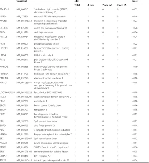
TargetScan and miRanda were used to identify target genes for miR-638 (miRbase.org website). We obtained a list of target genes with the information pertaining to the binding sites for miR-638, includingBRCA1(Table 1). To identify the common target genes for miR-638, we nar-rowed down the functional pathway enrichment analysis using two different algorithms. Since BRCA1is a multi-functional tumor suppressor protein and plays multiple roles in DNA damage response pathways, we focused on BRCA1as a target gene for this study.
miR-638 exerted diverse effects onBRCA1expression depending upon the subtypes of breast cancer cell lines
To validate the computational predictions and the bio-logical effect of miR-638 targetingBRCA1, we carried out in vitro luciferase reporter assays. miR-638 has been
[image:5.595.60.537.89.452.2]reported to inhibitBRCA1expression by targetingBRCA1 in CDS but not in the 3’UTR [30]. We performed lucifer-ase reporter assay with the pGL3 plasmid containing miR-638-binding site in BRCA1 CDS region (Figure 2A). We found that the luciferase activities vary in different breast cancer cell lines after successful transfection of miR-638 mimics (Figure 2B). Luciferase activities were significantly decreased in miR-638-transfected TNBC cell lines MDA-MB-231 and Hs578T, but not in estrogen receptor (ER)-positive cell lines T47D and immortalized MCF-10A cells. Inversely, the luciferase activities were significantly in-creased in miR-638-transfected MCF-7 cells. There was no significant difference in luciferase activities between controls and mutant miR-638 transfectants (Figure 2B). Inconsistent with the luciferase assay results, significant downregulation of BRCA1 was observed in TNBC cell lines MDA-MB-231 and Hs578T, but upregulation of
BRCA1 was shown in ER-positive cell lines, MCF-7 and T47D (Figure 2C). These results demonstrate that miR-638 may specifically regulate BRCA1 in TNBC, not inER+ cells, which suggests that the function of miR-638 might be blocked or antagonized by hormonal receptor expression.
Overexpression of miR-638 inhibited proliferation in TNBC cell lines
[image:6.595.57.540.113.639.2]Since the expression of miR-638 was significantly lower in both breast cancer cell lines and tissues compared to nor-mal cells and tissues, we next focused on the functional ef-fects of miR-638 on breast cancer cells. To address this, Table 1 A representative list of target genes for miR-638
Target gene Representative transcript
Gene name Conserved
sites
Total context + score
Total 8-mer 7mer-m8 7mer-1A
STARD10 NM_006645 StAR-related lipid transfer (START) domain containing 10
1 1 0 0 −0.53
NPAS4 NM_178864 neuronal PAS domain protein 4 1 1 0 0 −0.44
MKLN1 NM_001145354 muskelin 1, intracellular mediator containing kelch motifs
1 0 0 1 −0.27
CCDC92 NM_025140 coiled-coil domain containing 92 1 0 0 1 −0.26
SHPK NM_013276 sedoheptulokinase 1 0 0 1 −0.26
RIMKLB NM_020734 ribosomal modification protein rimK-like family member B
1 0 1 0 −0.24
PGK1 NM_000291 phosphoglycerate kinase 1 1 0 0 1 −0.22
HP1BP3 NM_016287 heterochromatin protein 1, binding protein 3
1 0 0 1 −0.21
LMO4 NM_006769 LIM domain only 4 1 0 0 1 −0.21
PAK2 NM_002577 p21 protein (Cdc42/Rac)-activated kinase 2
1 0 0 1 −0.2
MARCKS NM_002356 myristoylated alanine-rich protein kinase C substrate
1 0 0 1 −0.19
FRMPD4 NM_014728 FERM and PDZ domain containing 4 1 0 0 1 −0.18
EMILIN3 NM_052846 elastin microfibril interfacer 3 1 0 0 1 −0.18
MYCL1 NM_001033081 v-myc myelocytomatosis viral oncogene homolog 1, lung carcinoma derived (avian)
1 0 0 1 −0.18
LOC100507050 NM_001195528 hypothetical LOC100507050 1 0 0 1 −0.18
ISOC2 NM_001136201 isochorismatase domain containing 2 1 0 0 1 −0.18
EDN3 NM_207032 endothelin 3 1 0 0 1 −0.18
BRCA1 NM_007294 breast cancer 1, early onset 1 0 0 1 −0.16
TSPAN1 NM_005727 tetraspanin 1 1 0 0 1 −0.16
BUB3 NM_004725 budding uninhibited by
benzimidazoles 3 homolog (yeast)
1 0 0 1 −0.15
SP8 NM_182700 Sp8 transcription factor 1 0 0 1 −0.15
ZNF24 NM_006965 zinc finger protein 24 1 0 0 1 −0.14
KDSR NM_002035 3-ketodihydrosphingosine reductase 1 0 0 1 −0.14
KPNA6 NM_012316 karyopherin alpha 6 (importin alpha 7) 1 0 0 1 −0.13
SP7 NM_001173467 Sp7 transcription factor 1 0 0 1 −0.12
NOVA1 NM_002515 neuro-oncological ventral antigen 1 1 0 0 1 −0.11
SENP1 NM_014554 SUMO1/sentrin specific peptidase 1 1 0 0 1 −0.06
SRSF1 NM_001078166 serine/arginine-rich splicing factor 1 1 0 0 1 −0.05
EPHA7 NM_004440 EPH receptor A7 1 0 0 1 −0.04
miR-638 mimic, miR-638 inhibitor or mock control was transfected into breast cancer cells. The cellular prolifera-tion rate of breast cancer cell lines was determined by MTT assay. As expected, overexpression of miR-638 inhibited proliferation in TNBC cell lines, MDA-MB-231 and Hs578T, while increased cell growth in ER-positive MCF-7 cells, and had almost no effect in T47D cells com-pared to the mock-transfected control cells (Figure 3A). Conversely, transfection of miR-638 inhibitor resulted in an increased cell growth in MDA-MB-231 and Hs578T cells and a decreased cell growth in MCF-7 and no signifi-cant change in T47D cells. Surprisingly, decreased cell proliferation was also observed in miR-638-transfected MCF-10A cells, which are defined as‘normal’breast epi-thelial cells. However, there was no significant difference when miR-638 inhibitor was transfected into MCF-10A cells. These results demonstrate that miR-638 can inhibit cell proliferation in TNBC but not in ER-positive breast cancer cells, suggesting that overexpression of miR-638 may be a promising therapeutic option for TNBC. The downregulation of miR-638 could be a prognostic indica-tor for the aggressiveness.
Overexpression of miR-638 affected invasion ability in breast cancer cell lines
To determine if overexpression of miR-638 affects the inva-sive ability of breast cancer cell lines, cell invasion assay was performed using the BD matrigel. We found that miR-638-overexpressing TNBC cell lines exhibited significant inhibition of invasion ability (60% in MDA-MB-231 and 70% in Hs578T) compared to the control. For ER-positive cell lines, miR-638-overexpressing MCF-7 cell line showed no invasion changes (P= 0.90) while T47D decreased its invasion activity, compared to the control (P= 0.47) (Figure 3B and C). This data suggests that miR-638 plays an important role in cell invasion, specifically in TNBC.
Overexpression of miR-638 sensitizes the TNBC cell lines to DNA-damaging agents
Overexpression of BRCA1in MCF7 cell line has been re-ported to result in an increased resistance to cisplatin [31]. Based on our evidence, we reasoned that overexpression of
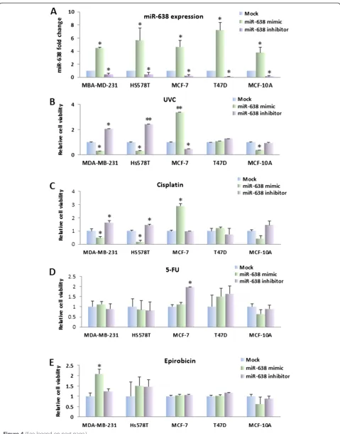
miR-638 could sensitize TNBC cells to DNA-damaging agents by means of their ability to reduceBRCA1 expres-sion and curb the activation of BRCA1in TNBC cells. To evaluate this hypothesis, we treated breast cancer cells transfected with miR-638 mimic or inhibitor (Figure 4A) with DNA-damaging agents (UVC, cisplatin and 5-FU) and non-DNA-damaging agents (epirubicin). UVC/chemosensi-tivity was determined by the MTT assay 48 hours after treatment. As shown in Figure 4B and C, miR-638 expres-sion significantly increased sensitivity to UV and cisplatin in TNBC cell lines MDA-MB-231 and Hs578T, but re-duced sensitivity in MCF-7 cells as compared with mock. Interestingly, a similar effect of cisplatin was observed in MCF-10A cells. The effect of miR-638 on sensitivity of UV and cisplatin was not observed in ER-positive cell line, T47D. Conversely, knockdown of miR-638 using miR-638 inhibitor increased cell viability in MDA-MB-231 and Hs578T cells after treatment with UV and cisplatin. It is notable that miR-638 introduction did not change the sen-sitivity to 5-FU and epirubicin in breast cancer cell lines (Figure 4D and E). These results suggested that the inhib-ition of miR-638 increased sensitivity to partial of DNA-damaging agents in TNBC but not in non-TNBC cells.
miR-638 overexpression significantly reduced post-UV/ drugs host cell reactivation activity in TNBC cells
miR-638 is involved in DNA repair pathway regulation dur-ing carcinogen exposure [26].BRCA1 appears to promote cell survival after DNA damage by participating in repair pathways [32]. We then asked whether the deregulation of miR-638 increases the sensitivity of the TNBC cell lines to cisplatin and UV via the DNA repair pathway. We mea-sured luciferase activity by co-transfecting miR-638 mimic or inhibitor, along with pCMU-Luc vector [29], which was pre-treated by UVC, DNA-damaging and non-DNA-damaging agents respectively into the breast cancer cell lines. For UV and cisplatin exposure, we found that TNBC cell lines MDA-MB-231 and Hs578T exhibited significantly reduced luciferase activity, while MCF-7 cell lines showed opposite effect. There were no changes in luciferase activity for T47D cells (Figure 5A and B). Conversely, contrary effect on DNA repair capability was observed in MDA-MB-231,
(See figure on previous page.)
Figure 2miR-638-mediated regulation ofBRCA1in breast cancer cell lines. (A)The top part shows the predicted location of the miR-638 binding site of the 3’-UTR ofBRCA1. The bottom part indicates the binding site in the CDS ofBRCA1(BRCA1CDS) as well as the mutantBRCA1
Hs578T and MCF-7 cells after co-transfection of miR-638 inhibitor and the pCMU-Luc vector pre-treated with UVC and cisplatin. Our data indicates that miR-638 im-pairs the DNA repair capability by regulatingBRCA1 ex-pression in TNBC cell lines.
[image:9.595.60.538.88.554.2]For 5-FU exposure, reduced DNA repair capability was only observed in Hs578T cells but not in MDA-MB-231, MCF-7 and T47D cells when co-transfected with miR-638 mimic and pCMU-Luc vector pre-treated with 5-FU. No significant DNA repair capability changes were
Figure 3miR-638 modulates proliferation in breast cancer cells and inhibits invasive ability in TNBC cells. (A)Effects of miR-638 on cell proliferation were determined by MTT assay. The proliferation rate decreased after transfection of miR-638 mimic (gray bars) in TNBC cell lines MDA-MB-231 and Hs578T as well as in MCF-10A, and increased in MCF-7 cells, and had no effect in T47D cells, compared to the mock control (white bars). Transfection of miR-638 inhibitor (dark bars) promoted proliferation in TNBC cell lines, MDA-MB-231 and Hs578T, but inhibited proliferation in MCF-7 cells compared to the mock. There were no obvious changes in T47D cells in either mimic or inhibitor transfection, while MCF10A exhibited similar pattern as TNBC cells. Values represent the mean ± SE for three independent experiments. (*P<0.05).(B)Transwell assays with matrigel were performed for the invasion activity of breast cancer cells
transfected with either miR-638 mimic or the mock control. Overexpression of miR-638 reduces cell invasion in TNBC cell lines, MDA-MB-231 and Hs578T, but not in ER-positive cells, MCF-7 and T47D.(C)Invasion ability of the cells was displayed as a percentage of the absolute cell numbers. Results are displayed as mean data ± SE. (*P<0.05 and**P<0.001). Five fields of unit area on each membrane or whole membrane were counted for cell numbers, and the
observed in cell lines when co-transfected with miR-638 mimic, and pCMU-Luc vector pre-treated with epirubi-cin. On the other hand, DNA repair capability did not change when co-transfected miR-638 inhibitor and pCMU-Luc pre-treated with either 5-FU or epirubicin. Our results demonstrate that the miR-638 exerts a dis-tinct effect on DNA repair toward anticancer drugs in TNBC and ER-positive breast cancer cells.
Discussion
The objective of this study was to decipher the role of miR-638 in breast cancer tumorigenesis and treatment. We revealed a dynamic miR-638 expression during the process of breast cancer progression. In addition, we demonstrated that different expression patterns of miR-638 are correlated with its functions in different types of breast cancer cells. Moreover, we found that the role of miR-638 on tumorigenesis, radiation and drug sensitivity was mediated by targeting BRCA1 through DNA repair pathway. Our results suggest that aberrant expression of miR-638 contributes to human breast cancer progres-sion, invasion and sensitivity of radiation and chemo-therapy, particularly in TNBC.
miR-638 expression changes during development of breast cancer
miRNA may exhibit different expression levels between normal and cancer cells [33]. For example, miR-142-3p and miR-9 were upregulated in squamous cell carcinoma in comparison to normal bronchial tissues. The alterations of miRNA expression were probably correlated with the pathways maintaining the malignant phenotypes or clin-ical outcomes [26]. miR-638 has been characterized to be downregulated in majority of tumors, but upregulated in hepatocellular liver cancer. Our previous data indicated that miR-638 was downregulated in ADH and IDC during the breast cancer developing stages compared to normal breast tissues [19]. In this study, we analyzed miR-638 expression in a new cohort of 30 breast cancer FFPE samples. We showed that downregulation of miR-638 pre-sented in the majority of the IDCs compared to their adja-cent normal tissues. These results support the notion that dysfunction of miR-638 is essential in maintaining the ma-lignant phenotype of cancer. A larger cohort study will help understand the exact role of miR-638 in breast can-cer development and progression. In addition, we assessed
the expression of miR-638 in breast cancer cell lines. The expression of miR-638 was low in breast cancer cell lines compared with the MCF-10A cells (Figure 1C). Based on these data, we hypothesize that deregulation of miR-638 is involved in the process of breast cancer progression.
miR-638 is a double-faced gene expression regulator
It is currently believed that miRNAs elicit their effect by silencing the expression of target genes [34]. However, miRNAs may also function to positively regulate gene expression [35,36]. miR-638 has been reported to inhibit BRCA1 expression in different cancer cell lines by tar-geting BRCA1 in CDS, but not in 3’ UTR [30]. In our study, overexpression of miR-638 suppressedBRCA1 ex-pression in TNBC cells, MDA-MB-231 and Hs578T, but not in ER-positive cell lines. These data support the no-tion that miR-638 exerts diverse effects depending upon breast cancer types. Since TNBC lacks expression ofER, PR and HER2, we believe that hormones might be in-volved in miR-638-mediated BRCA1 regulation, which requires further studies. Nicolosoet al. observed an op-posite BRCA1 regulation of miR-638 in MCF-7 cells [30]. A possible explanation might be that miR-638 ex-hibits distinctBRCA1regulation in cell populations with different phases of cell cycle. Our data suggests that miR-638 elicits differential efficacy by silencing or indu-cing the expression ofBRCA1 in different breast cancer cell lines. These findings demonstrate that miR-638 ex-hibits a dual function in response to environmental stimuli depending upon the state of cell malignancy. It might be a useful biomarker for surveillance of chemical exposure in breast cancer treatment.
miR-638 plays an important role in cell proliferation and invasion
Previous studies have demonstrated that miR-638 regu-lates cell growth and smooth muscle cell proliferation and migration [37], and negatively regulates BRCA1 expres-sion. In esophageal squamous cell carcinoma, miR-638 promotes cell proliferationin vitro[23]. We analyzed the functional consequences after overexpressing miR-638 in breast cancer cell lines. Our data indicated that miR-638 has different functions on cell behaviors in different types of breast cancer cells. miR-638 inhibited cell proliferation in TNBC cells, while promoted cell proliferation in ER-positive cells. Interestingly, miR-638 exerts similar effect
[image:11.595.61.544.89.103.2](See figure on previous page.)
on inhibiting proliferation in MCF-10A, which is defined as ‘normal’ breast epithelial cells. Although MCF-10A is non-tumorigenic, it is a triple-negative human breast cell line [38]. Our data supports that miR-638 inhibits cell proliferation in TNBC.
Importantly, miR-638 also alters cell invasion ability. The invasion was decreased in TNBC cell lines after miR-638 overexpression, but increased in MCF-7 cells. We hypothesize that miR-638 functions as a tumor-suppressor or oncomir in different types of breast cancer cells or stages during breast cancer progression. How-ever, miR-638 also suppresses the invasion in T47D cells although no significant downregulation of BRCA1 was observed. The possibility might be that miR-638 regu-lates invasion in T47D via other pathways instead of BRCA1-related DNA repair pathway.
miR-638 is associated with radiation and chemotherapy sensitivity of breast cancer cell lines via DNA repair pathway
miRNAs has been implicated in response to DNA damage and repair. Some miRNAs are involved in DNA damage re-sponse and/or DNA repair, which would affect cellular sen-sitivity to DNA-damaging agents [39]. In order to explore whether miR-638 expression could be used as a biomarker for predicting tumor response to chemotherapy and radio-therapy for breast cancer, we found that overexpression of miR-638 impaired DNA repair in breast cancer cell lines, which suggests that miR-638 might correspond to the cel-lular stress upon radiation and DNA-damaging agents.
Conclusions
We previously found that miR-638 was one of the most deregulated miRNAs in breast cancer progression. In present work, we demonstrated that miR-638 directly regu-latesBRCA1expression in breast cancer, implying a critical role of miR-638 in the course of breast carcinogenesis and biological behaviors. miR-638 exerts distinct effects on cell proliferation and invasion in different types of breast can-cer. In addition, miR-638 enhanced radiation and chemo-therapy sensitivity in TNBC cells by regulating BRCA1 expression via DNA repair pathways. Taken together, miR-638 may serve as a potential prognostic biomarker and therapeutic target for breast cancer.
Abbreviations
5-FU:5-fluorouracil; ADH: atypical ductal hyperplasia;BRCA1: breast cancer susceptibility gene 1; BSA: bovine serum albumin; CDS: coding sequence; DCIS: ductal carcinomain situ; DMEM: Dulbecco’s modified Eagle’s medium; ER: estrogen receptor; FBS: fetal bovine serum; IDC: invasive ductal carcinoma; IgG: immunoglobulin G; FFPE: formalin-fixed, paraffin-embedded; HCR: host cell reactivation; MEGM: mammary epithelial cell growth medium; miRNAs: microRNAs; PBS: phosphate-buffered saline; TBS: Tris-buffered saline; TNBC: triple-negative breast cancer; UTR: untranslated region.
Competing interests
The authors declare that they have no competing interests.
Authors’contributions
XT contributed to the conception and design of the project, development of methodology, acquisition of data, analysis and interpretation of data, bioinformatics and statistical analyses, and writing of the manuscript. JP performed qRT-PCR and matrigel invasion assays, and helped write the manuscript. YF performed RNA isolation from FFPE samples and data analysis. SA performed cell culture, plasmids preparation and data analysis. KR, ST, CBT and YM identified pathological cases with clinical information and performed microdissection. RFB participated in the design of the project and the manuscript preparation. SWF designed and interpreted data, and wrote and revised the manuscript. All authors read and approved the final version of this manuscript.
Acknowledgements
We would like to thank Dr. Kenneth H. Kraemer (NCI/NIH) for providing us with the pCMVLuc reporter gene plasmid and Dr. Wen Chen (Faculty of Preventive Medicine, School of Public Health, Sun Yat-sen University, China) for the pGL3 plasmid. We thank Woojin Lee, the W.T. Gill, Jr., Summer Research Fellow at GW School of Medicine and Health Science for qRT-PCR technical assistance. This research was supported by the Dr. Cyrus and Myrtle Katzen Cancer Research Center Grant at The George Washington University (to SWF and RFB), the National Cancer Institute Grant (1R21 CA159103-01) (to SWF), the Hao Foundation grant (to SWF) and the Elaine H. Snyder Cancer Research Award (to SWF).
Author details
1Department of Medicine (Division of Genomic Medicine), The George
Washington University School of Medicine and Health Sciences, 2300 Eye St. NW, Ross Hall 402C, Washington, DC 20037, USA.2Department of Pathology, The George Washington University School of Medicine and Health Sciences, 2150 Pennsylvania Avenue NW, Washington, DC 20037, USA.3Department of Surgery, The George Washington University School of Medicine and Health Sciences, 2150 Pennsylvania Avenue NW, Washington, DC 20037, USA. 4
Research Lab and International Collaboration, Bon Secours Cancer Institute, Bon Secours Health System, 5801 Bremo Road, Richmond, VA 23226, USA. 5
Department of Radiology, The George Washington University School of Medicine and Health Sciences, 2300 M St., NW, Room 822, Washington, DC 20037, USA.6Department of Microbiology, Immunology and Tropical Medicine, The George Washington University School of Medicine and Health Sciences, 2300 Eye St. NW, Ross Hall 402C, Washington, DC 20037, USA.
Received: 19 March 2014 Accepted: 28 August 2014
[image:13.595.58.535.90.102.2](See figure on previous page.)
References
1. Jemal A, Bray F, Center MM, Ferlay J, Ward E, Forman D:Global cancer statistics.CA Cancer J Clin2011,61:69–90.
2. Boyle P:Triple-negative breast cancer: epidemiological considerations and recommendations.Ann Oncol2012,23:vi7–vi12.
3. Crown J, O'Shaughnessy J, Gullo G:Emerging targeted therapies in triple-negative breast cancer.Ann Oncol2012,23:vi56–vi65.
4. Sharma S, Kelly TK, Jones PA:Epigenetics in cancer.Carcinogenesis2010,
31:27–36.
5. Turner N, Tutt A, Ashworth A:Hallmarks of 'BRCAness' in sporadic cancers. Nat Rev Cancer2004,4:814–819.
6. Mueller CR, Roskelley CD:Regulation of BRCA1 expression and its relationship to sporadic breast cancer.Breast Cancer Res2003,5:45–52. 7. Alli E, Sharma VB, Sunderesakumar P, Ford JM:Defective repair of oxidative
dna damage in triple-negative breast cancer confers sensitivity to inhibition of poly(ADP-ribose) polymerase.Cancer Res2009,69:3589–3596. 8. Hartman AR, Ford JM:BRCA1 induces DNA damage recognition factors
and enhances nucleotide excision repair.Nat Genet2002,32:180–184. 9. Deng CX, Wang RH:Roles of BRCA1 in DNA damage repair: a link
between development and cancer.Hum Mol Genet2003,12:R113–R123. 10. Alli E, Sharma VB, Hartman AR, Lin PS, McPherson L, Ford JM:Enhanced
sensitivity to cisplatin and gemcitabine in Brca1-deficient murine mammary epithelial cells.BMC Pharmacol2011,11:7.
11. You JS, Jones PA:Cancer genetics and epigenetics: two sides of the same coin?Cancer Cell2012,22:9–20.
12. Slezak-Prochazka I, Durmus S, Kroesen BJ, van den Berg A:MicroRNAs, macrocontrol: regulation of miRNA processing.RNA2010,16:1087–1095. 13. Iorio MV, Ferracin M, Liu CG, Veronese A, Spizzo R, Sabbioni S, Magri E,
Pedriali M, Fabbri M, Campiglio M, Ménard S, Palazzo JP, Rosenberg A, Musiani P, Volinia S, Nenci I, Calin GA, Querzoli P, Negrini M, Croce CM:
MicroRNA gene expression deregulation in human breast cancer. Cancer Res2005,65:7065–7070.
14. Fu SW, Chen L, Man YG:miRNA biomarkers in breast cancer detection and management.J Cancer2011,2:116–122.
15. Berger FRM:Micro-RNAs as potential new molecular biomarkers in oncology: have they reached relevance for the clinical imaging sciences? Theranostics2013,30:943–952.
16. Costa PM, PdLM:MicroRNAs as molecular targets for cancer therapy: on the modulation of microRNA expression.Pharmaceuticals (Basel)2013,
6:1195–1220.
17. Riaz M, van Jaarsveld MT, Hollestelle A, der Smissen WJ P-v, Heine AA, Boersma AW, Liu J, Helmijr J, Ozturk B, Smid M, Wiemer EA, Foekens JA, Martens JW:miRNA expression profiling of 51 human breast cancer cell lines reveals subtype and driver mutation-specific miRNAs.Breast Cancer Res2013,15:R33.
18. Moulis S, Sgroi DC:Re-evaluating early breast neoplasia.Breast Cancer Res
2008,10:302.
19. Chen L, Li Y, Fu Y, Peng J, Mo MH, Stamatakos M, Teal CB, Brem RF, Stojadinovic A, Grinkemeyer M, McCaffrey TA, Man YG, Fu SW:Role of deregulated microRNAs in breast cancer progression using FFPE tissue. PLoS One2013,8:e54213.
20. Kahlert C, Klupp F, Brand K, Lasitschka F, Diederichs S, Kirchberg J, Rahbari N, Dutta S, Bork U, Fritzmann J, Reissfelder C, Koch M, Weitz J:Invasion front-specific expression and prognostic significance of microRNA in colorectal liver metastases.Cancer Sci2011,102:1799–1807.
21. Lin Y, Zeng Y, Zhang F, Xue L, Huang Z, Li W, Guo M:Characterization of microRNA expression profiles and the discovery of novel microRNAs involved in cancer during human embryonic development.PLoS One
2013,8:e69230.
22. Wang JL, Hu Y, Kong X, Wang ZH, Chen HY, Xu J, Fang JY:Candidate microRNA biomarkers in human gastric cancer: a systematic review and validation study.PLoS One2013,8:e73683.
23. Zhang X, Wei J, Zhou L, Zhou C, Shi J, Yuan Q, Yang M, Lin D:A functional BRCA1 coding sequence genetic variant contributes to risk of esophageal squamous cell carcinoma.Carcinogenesis2013,34:2309–2313. 24. Sand M, Skrygan M, Sand D, Georgas D, Hahn SA, Gambichler T, Altmeyer P,
Bechara FG:Expression of microRNAs in basal cell carcinoma.Br J Dermatol2012,167:847–855.
25. Zhu DX, Zhu W, Fang C, Fan L, Zou ZJ, Wang YH, Liu P, Hong M, Miao KR, Liu P, Xu W, Li JY:miR-181a/b significantly enhances drug sensitivity in
chronic lymphocytic leukemia cells via targeting multiple anti-apoptosis genes.Carcinogenesis2012,33:1294–1301.
26. Li D, Wang Q, Liu C, Duan H, Zeng X, Zhang B, Li X, Zhao J, Tang S, Li Z, Xing X, Yang P, Chen L, Zeng J, Zhu X, Zhang S, Zhang Z, Ma L, He Z, Wang E, Xiao Y, Zheng Y, Chen W:Aberrant expression of miR-638 contributes to benzo(a)pyrene-induced human cell transformation.Toxicol Sci2013,
125:382–391.
27. TargetScan: prediction of microRNA targets.[http://www.targetscan.org/] 28. Fu Y, Lian Y, Kim KS, Zhang L, Hindle AK, Brody F, Siegel RS, McCaffrey TA,
Fu SW:BP1 homeoprotein enhances metastatic potential in ER-negative breast cancer.J Cancer2010,1:54–62.
29. Tan X, Anzick SL, Khan SG, Ueda T, Stone G, Digiovanna JJ, Tamura D, Wattendorf D, Busch D, Brewer CC, Zalewski C, Butman JA, Griffith AJ, Meltzer PS, Kraemer KH:Chimeric negative regulation of p14ARF and TBX1 by a t(9;22) translocation associated with melanoma, deafness, and DNA repair deficiency.Hum Mutat2013,34:1250–1259.
30. Nicoloso MS, Sun H, Spizzo R, Kim H, Wickramasinghe P, Shimizu M, Wojcik SE, Ferdin J, Kunej T, Xiao L, Manoukian S, Secreto G, Ravagnani F, Wang X, Radice P, Croce CM, Davuluri RV, Calin GA:Single nucleotide
polymorphisms inside microRNA target sites influence tumor susceptibility.Cancer Res2010,70:2789–2798.
31. Husain A, He G, Venkatraman ES, Spriggs DR:BRCA1 up-regulation is associated with repair-mediated resistance to
cis-diamminedichloroplatinum(II).Cancer Res1998,58:1120–1123. 32. Kennedy RD, Quinn JE, Mullan PB, Johnston PG, Harkin DP:The role of
BRCA1 in the cellular response to chemotherapy.J Natl Cancer Inst2004,
96:1659–1668.
33. Mascaux C, Laes JF, Anthoine G, Haller A, Ninane V, Burny A, Sculier JP:
Evolution of microRNA expression during human bronchial squamous carcinogenesis.Eur Respir J2009,33:352–359.
34. He L, Hannon GJ:MicroRNAs: small RNAs with a big role in gene regulation.Nat Rev Genet2004,5:522–531.
35. Place RF, Li LC, Pookot D, Noonan EJ, Dahiya R:MicroRNA-373 induces expression of genes with complementary promoter sequences. Proc Natl Acad Sci USA2008,105:1608–1613.
36. Janowski BA, Younger ST, Hardy DB, Ram R, Huffman KE, Corey DR:
Activating gene expression in mammalian cells with promoter-targeted duplex RNAs.Nat Chem Biol2007,3:166–173.
37. Li P, Liu Y, Yi B, Wang G, You X, Zhao X, Summer R, Qin Y, Sun J: MicroRNA-638 is highly expressed in human vascular smooth muscle cells and inhibits PDGF-BB-induced cell proliferation and migration through targeting orphan nuclear receptor NOR1.Cardiovasc Res2013,99:185–193. 38. Subik K, Lee JF, Baxter L, Strzepek T, Costello D, Crowley P, Xing L, Hung
MC, Bonfiglio T, Hicks DG, Tang P:The expression patterns of ER, PR, HER2, CK5/6, EGFR, Ki-67 and AR by immunohistochemical analysis in breast cancer cell lines.Breast Cancer (Auckl)2010,4:35–41.
39. Wang Y, Taniguchi T:MicroRNAs and DNA damage response: implications for cancer therapy.Cell Cycle2013,12:32–42.
doi:10.1186/s13058-014-0435-5
Cite this article as:Tanet al.:miR-638 mediated regulation ofBRCA1 affects DNA repair and sensitivity to UV and cisplatin in triple-negative breast cancer.Breast Cancer Research201416:435.
Submit your next manuscript to BioMed Central and take full advantage of:
• Convenient online submission
• Thorough peer review
• No space constraints or color figure charges
• Immediate publication on acceptance
• Inclusion in PubMed, CAS, Scopus and Google Scholar
• Research which is freely available for redistribution