RATIONAL DESIGN AND
MECHANISTIC STUDIES OF
A PHOSPHOHYDROLASE
FROM
ENTEROBACTER
AEROGENES
A thesis submitted for the degree of Doctor of Philosophy of The Australian National University
AND
THE AUSTRALIAN NATIONAL UNIVERSITYby
Sylvia Hsu-Chen Yip
This thesis presents original research that I have performed under the supervision of Professor David Ollis at the Research School of Chemistry. I have made an effort to acknowledge the contributions of individuals to this thesis as listed below as well as in the relevant parts of the thesis:
■ Plasmid DNA of gpdQ in pCY76 was obtained from Dr Colin Jackson and Dr Hye-Kyung Kim.
■ E. coli DHlOß cells carrying the putative ugp operon cloned in the plasmid pBlueScript were prepared by Dr Sean Yu-McLoughlin. ■ Primers for PCR sequencing, namely 4pCY76 and M13 were
originally obtained from Dr Jian-Wei Liu who also designed the latter. 4pCY76 was designed by Dr Bradley Stevenson.
■ Plasmid DNA of opdA in pETMCSI was obtained courtesy of Dr Jee-Loon Foo.
■ Magnetic circular dichroism studies on wild-type GpdQ, mutants N80D and N80A were performed by Dr Kieran Hadler during his PhD candidature at the University of Queensland.
■ Dr Paul Carr and Kelly Nguyen were involved in the X-ray diffraction studies, data collection and analysis of wild-type GpdQ crystal structure 3D03.
■ Dr Jee-Loon Foo performed the soaking experiments for all the GpdQ crystals made in this study.
■ Crystallisation screens of GpdQ mutant F21K were set up by Miss Tracy Murray.
First and foremost, I would like to extend my heartfelt gratitude to my supervisor Professor David Ollis, whose support, guidance and help from the initial to final stages of my candidature have helped me to develop a solid understanding and passion for protein research. I am also greatly indebted to Dr Jian-Wei Liu for his technical help in the laboratory; Dr Paul Carr for sharing his time and expertise in protein crystallography and structural analysis and for putting up with my lack of knowledge in these areas. A very huge ‘thank you’ goes to Drs Sean Yu-McLoughlin and Colin Jackson, my ‘predecessors’ in GpdQ research, whose previous work made the enzyme such an interesting subject to work on.
I would like to thank all the past and present members of the Ollis and Otting research groups - Jee-Loon (my saviour each time I messed up my experiments), Chris, Morgan, Hye-Kyung, Amalia, Tracy, Sapna, Joe, Bradley, Ruhu, Karin, Kiyoshi, Hiromasa, Xuncheng, Tiffany, Laura, Xinying and Kelly. It has been sheer joy working with you all these 3.5 years.
Special thanks to Associate Professors Gerhard Schenk and Lawrence Gahan of the University of Queensland, their students Kieran, Eleanor and Fernanda who collaborate with the Ollis group on GpdQ research. I am also grateful to the Australian National University and CSIRO for financially supporting my studies.
GpdQ is a phosphohydrolase from Enterobacter aerogenes whose rare ability to hydrolyse simple diesters of phosphoric acid like dimethyl phosphate and diethyl phosphate first caught the attention and interest of researchers in the 1970s. When GpdQ was characterised further in more recent times, it was found to have an exceptionally broad substrate repertoire. The enzyme has primarily phosphodiesterase activity - capable of hydrolysing a wide range of phosphodiesters including glycerophosphodiesters that are believed to be its natural substrates. Additionally, GpdQ has low phosphomonoesterase, phosphotriesterase and phosphonate monoesterase activities.
In this study, the promiscuity of GpdQ was exploited and we set out to improve the enzyme’s activity on one of the non-physiological phosphodiesters, bis (p-nitrophenyl) phosphate. Directed evolution was the main strategy used to achieve this goal. After the first four rounds of evolution, the hexameric quaternary structure of the catalytically improved GpdQ mutants was found to have broken down and high activity was associated with the dimer. Rational design was then employed to identify mutations that would promote dissociation and form a stable GpdQ dimer. These rationally-designed mutants were included for gene shuffling from the fifth round of directed evolution onwards. At the end of this study, eight full rounds of evolution were completed. Three GpdQ dimers displaying ~500-fold &cat/Km improvement were successfully engineered.
Acknowledgements... iii
Abstract...v
Table of contents... vii
List of figures... xii
List of tables...xv
List of abbreviations and acronyms used in this dissertation...xvii
Abbreviations for amino acids...xxi
List of publications... xxii
1 INTRODUCTION AND LITERATURE REVIEW...1
1.1 Enzymes...3
1.1.1 Enzyme active sites...5
1.1.2 Metalloenzymes...8
1.2 Organophosphate compounds... 9
1.2.1 The chemistry of organophosphate compounds... 9
1.2.2 Organophosphate compounds and their uses...10
1.2.3 The toxicity of organophosphate triesters...12
1.3 GpdQ - A novel glycerphosphodiesterase from Enterobacter
aerogenes... 13
1.3.1 Growth of
Escherichia coli
co-expressing OpdA and GpdQ
using paraoxon as the phosphorus source... 13
1.3.2 Characteristics and properties of GpdQ... 15
1.4 The structure of GpdQ...18
1.4.1 Overall quaternary structure... 18
1.4.2 Structural domains in a GpdQ monomer... 22
1.4.3 The conserved catalytic domain, active site and binuclear
metal center...23
1.4.4 The dimerisation domain...26
1.4.5 The cap domain... 28
1.5 Rv0805 - A 3’, 5’-cyclic nucleotide phosphodiesterase from
mycobacterium tuberculosis...32
1.6 Protein engineering strategies...34
1.6.1 Protein engineering... 34
1.6.2 Rational design...36
1.6.3 Directed evolution...37
1.6.4 A semi-rational approach to enzyme engineering - combining
rational design and directed evolution... 39
1.7 Overview of thesis... 40
2 EXPERIMENTAL PROCEDURES... 55
2.1 Introduction... 57
2.2 Strains and vectors...57
2.3.2 Site-directed mutagenesis polymerase chain reaction (SDM-PCR)
...62
2.3.3 Agarose gel electrophoresis and gel purification...66
2.3.4
Dpn\
digestion of template DNA... 67
2.3.5 Transformation... 68
2.3.6 Single-colony mini-cultures and plasmid isolation...69
2.3.7 DNA verification...69
2.3.8 DNA quantitaion, PCR sequencing and ethanol precipation of
DNA... 70
2.3.9 Site-saturation mutagenesis...72
2.4 Directed evolution...72
2.4.1 Genetic library creation...74
2.4.1.1. A Preparation of gene insert...74
2.4.1.1 .A. 1 Error-prone polymerase chain reaction (epPCR).... 74
2.4.1.1. A.2 Purification of PCR products... 77
2.4.1.1. A.3 Digestion with restriction enzymes...78
2.4.1.1 .B Preparation of cloning vector... 80
2.4.1.1 .B. 1 Preparation of
Nde\
and
EcoRl
cloning sites... 80
2.4.1.1 .B.2 Dephosphorylation of vector DNA... 80
2.4.1.2 Gel purification of vector and insert DNA...81
2.4.1.3 DNA quantitation...82
2.4.1.4 Ligation... 83
2.4.1.5 Quantitative and qualitative analyses of libraries...84
2.4.1.5. A Qualitative analysis...84
2.4.1.5. B Quantitative analysis...86
2.4.1.6 Creating future libraries (second generation libraries and
onward) using staggered extension process (StEP)...86
2.4.2 Developing methods to screen libraries for improved variants .... 90
2.4.2.1
In vivo
agar plate-based screenings with
phosphotriester and phosphodiester compounds...90
2.4.2.2
In vitro
random screening with bis (p-nitrophenyl) phosphate..
...92
2.5 Protein expression and purification... 94
2.5.1 Protein expression...94
2.5.2 Protein purification...96
2.5.2.1 Protocol A - McLoughlin
et al.
(modified)...97
2.5.2.2 Protocol B - Ghanem
et al.
(modified)... 98
2.5.2.3 Protocol C - Ghanem’s method, ‘reversed’...99
2.6 Kinetic analysis of GpdQ proteins...101
2.7 Determination of protein molecular weight... 104
2.8 Protein crystallography... 106
MECHANISTIC STUDIES OF GpdQ... 113
3.1 Introduction... 115
3.2 A summary of mutagenesis studies of GpdQ active site residues....118
3.3 Mutational analysis of ß-metal ligands... 120
3.3.1 N80D... 120
3.3.2 H156Q, H195Q, H195N and H195A... 121
3.3.2.1 H156Q...121
3.3.2.2 HI95 - H195Q, H195N and H195A... 121
3.3.3 Magnetic circular dichroism (MCD) studies on N80 mutants.... 122
3.3.4 N80A and H156A - Biphasic kinetics and negative cooperativity...
...122
3.3.4.1 Obtaining apparent values of kinetic parameters (Km and Vmax)
using the Eadie-Hofstee curve... 122
3.3.4.2 Assessing negative cooperativity in N80A and H156A using
the Hill equation...124
3.4 Mutational analysis of a-metal ligands... 128
3.4.1 D8 - D8N and D8A... 128
3.4.2 H 10-H 10A...128
3.5 Mutational analysis of the bridging ligand, D50...129
3.6 Mutagenesis of H81, the conserved histidine...130
3.6.1 Introduction and background... 130
3.6.2 Results...133
3.6.3 Discussion...133
3.6.4 Conclusions on H81... 135
3.7 Conclusions...135
4 DIRECTED EVOLUTION OF GpdQ - SCREENING METHODS
AND PART ONE...143
4.1 Introduction...145
4.2 In vivo agar plate-based screening of GpdQ mutant libraries...145
4.2.1 McLoughlin’s growth assay method... 146
4.2.2 Cho’s top agar method...149
4.3 In vitro random screening of GpdQ mutant libraries...151
4.4 Directed evolution of GpdQ (Rounds 1-4)... 152
4.4.1 High affinity evolution...152
4.4.2 High activity evolution...155
4.5 Dissociation of oligomeric structure in mutant 3-25...159
4.5.1 Initial detection... 159
4.5.2 Determination of protein molecular weight in mutant 3-25... 161
4.5.3 Discovery of two forms of activities in mutant 3-25... 161
4.6.4 H217R... 166
4.7 Conclusions on directed evolution of GpdQ (Part One)...168
5 OPTIMISATION OF GpdQ OLIGOMERIC STRUCTURE WITH
RATIONAL DESIGN FOR BIS (P-NITROPHENYL) PHOSPHATE
ACTIVITY...175
5.1 Introduction... 177
5.2 Factors affecting the dissociation equilibrium... 178
5.3 C269 mutants...181
5.3.1 C269S and C269A...181
5.3.2 3-25C269A and 2-35-C269A...183
5.3.3 Hysteretic kinetics of C269 mutants... 184
5.4 N53 mutants... 191
5.4.1 N53D and N53A...193
5.4.2 Comparison of GpdQ N53 mutants with Rv0805 D66A... 194
5.5 Truncation at the cap domain of GpdQ... 195
5.5.1 Y257stop... 196
5.5.2 C54G-Y221 H-Y257stop...197
5.5.3 Conclusions on the cap domain of GpdQ... 199
5.6 Truncation at the dimerisation domain of GpdQ...200
5.7 F21... 201
5.7.1 F21K...201
5.7.2 F21K-C54G-Y221 H-Y257stop... 204
5.7.3 The preliminary crystallisation of F21K...204
5.8 W261R...205
5.9 Conclusions...206
6 DIRECTED EVOLUTION OF GpdQ (PART TWO)... 209
6.1 Introduction...211
6.2 Directed evolution of GpdQ (Rounds 5-8)...213
6.2.1 Screening results...213
6.2.2 Expression, purification and characterisation of eighth round
mutants...217
6.3 Structure-function studies of mutations R12L, N103D and S127A .. 220
6.3.1 R12L...220
6.3.2 N103D...222
6.3.3 S127A...223
6.4 Conclusions on directed evolution of GpdQ (Part Two)...224
7 FINAL CONCLUSIONS AND FUTURE DIRECTIONS...227
7.1 Mutational anlysis of active site residues and mechanistic studies of
GpdQ... 229
C - Nucleotide sequence of vector pCY76... 244
D - Sequences of primers used in this research project...246
E - Proteolytic activation of GpdQ... 248
F - Factors influencing the negative cooperativity of GpdQ mutant N80A
...251
F.l N80A phosphodiesterase activity with bis (p-nitrophenyl)
phosphate...251
F.2 N80A phosphomonoesterase activity p-nitrophenyl phosphate...252
F.3 Intra/inter-molecular negative cooperativity in N80A... 254
1
.2
:1.3:
1.4:
1.5:
1.6:1.7:
1.8:1.9:
1.10:1.1 1:
1.12:
1.13:
1.14
1.15
1.16:
1.17:
1.18:
1.19:
1.20: 1.2 1:1.22:
1.23:
1.24:
2.1:2.2:
2.3:
2.4:
2.5:
2.6:
2.7:
2
.8
:2.9:
2.10: 2.11:Chemical structures of the common classes of O Ps...
Examples of phosphotriesters...
The phosphodiester bonds in DNA...
Examples of phosphodiesters...
The proposed catabolic pathway for the breakdown of methyl
paraoxon MPO)...
Comparison of the 5.6-kb DNA fragment isolated from
E.
aerogenes
and the
E. coli ugp
operon...
Glycerophosphodiesters and their hydrolysis...
The GpdQ dimer...
The GpdQ hexamer
The GpdQ monomer...
The active sites of a/ß sandwich dimetallo-phosphohydrolases ....
The active site of GpdQ...
The dimerisation domain of GpdQ...
Electrostatic potential mapped to the molecular surface of the
GpdQ monomer...
The GpdQ dimer and the intertwining dimerisation domains...
The domain swapping mechanism by the cap domain and the
inter-chain disulfide bond...
Inter-chain interactions by the cap domain of GpdQ...
The GpdQ hexamer with the cap domains highlighted...
The cap domain in a GDPD...
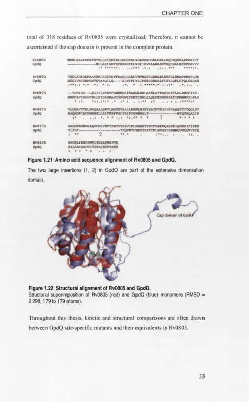
Amino acid sequence alignment of Rv0805 and GpdQ...
Structural alignment of Rv0805 and GpdQ...
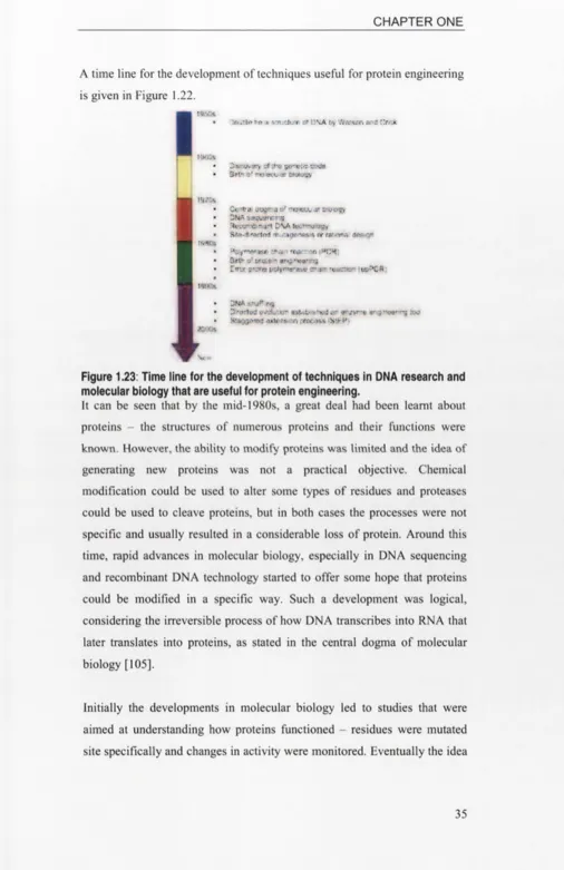
Time line for the developments in molecular biology...
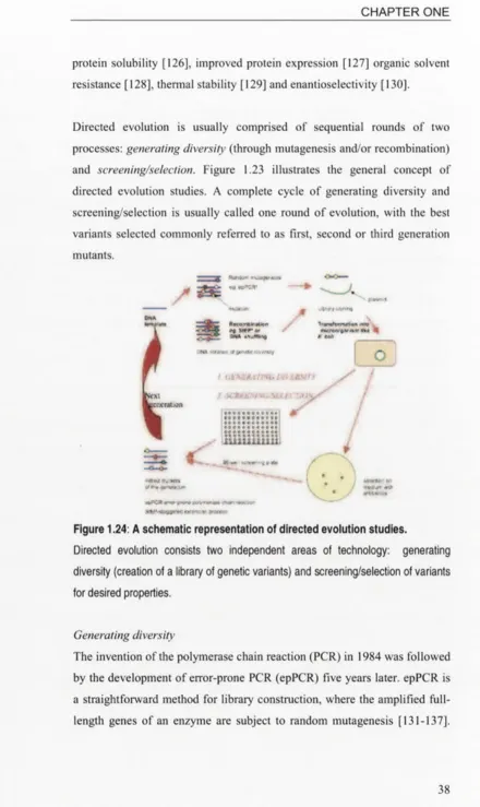
A schematic representation of directed evolution studies...
The physical map of pCY76 vector...
The process of making a site-directed mutant...
DNA sequence alignment of the W261R mutant and
wild-type GpdQ...
Primer dimerisation...
Partially complementary W261R forward (W261R-f)and
reverse (W261R-r) primers...
SDM-PCR product visualisation with agarose gel electrophoresis..
Agarose gel presentation of pDNA samples digested with
Ndel
and
EcoRl
...
The process of creating a library of
gpdQ
mutants...
An agarose gel showing epPCR amplification products...
DNA UV spectrum measurement graphs...
A 1.0% agarose gel showing
gpdQ
insert and cut pCY76 plasmid
just before ligation...
StEP-D against wild-type GpdQ, showing mutations G259R
and C269A... 89
2.15: An example of a secondary screening during the sixth round of
directed evolution...
94
2.16: A comparison of GpdQ proteins expressed in TBA and
LBA media... 96
2.17: Drop assay of fractions eluted from Q Sepharose column... 100
2.18: A profile of the soluble fraction of crude lysate eluted in Q
Sepharose column... 100
2.19: A profile of wild-type GpdQ eluted in Superdex 200
column... 100
2.20: Purified GpdQ visualised in a 17.5% SDS-PAGE gel... 101
2.21: Hydrolysis of bpNPP and /?NPP... 102
2.22: Elution profile of molecular weight markers from Superdex 200
size exclusion column... 105
2.23: Calibration curve obtained with molecular weight markers eluted
from Superdex 200 size exclusion column... 105
3.1: The active site of GpdQ and active site mutants made in
this study... 117
3.2: Michaelis-Menten plots of wild-type GpdQ, N80A and Hl 56A... 123
3.3: Eadie-Hofstee plots of wild-type GpdQ, N80A and H l56A... 124
3.4: Hill plots of wild-type GpdQ N80A and H156A...
127
3.5: SDS-PAGE gel showing D50A after lysis using the French
press... 129
3.6: The extensive hydrogen-bonding network of H81... 131
4.1: Size exclusion chromatography of GpdQ mutant 3-5... 160
4.2: Oligomeric dissociation of GpdQ mutant 3-25... 160
4.3: The inter-molecular disulfide bond in GpdQ... 162
4.4: Locations of the cap domains at the interface between the dimeric
units of GpdQ... 163
4.5: Structure-function studies of G259R... 164
4.6: The Y221-Y250 interaction in GpdQ... 165
4.7: Structure-function studies of H217R... 167
5.1: An excerpt of Superdex 200 migration profile of GpdQ triple
mutant enzyme 3-25... 179
5.2: The effects of enzyme concentration on oligomeric dissociation in
GpdQ... 180
5.3: The effects of metal ions on oligomeric dissociation in GpdQ... 180
5.4: Gel filtration analysis of C269A... 181
5.5: Hysteretic kinetics of GpdQ mutant C269S... 183
5.11: Structural alignment of the active sites of Rv0805 and GpdQ... 191
5.12: The active site of bacteriophage
X
Ser/Thr protein phosphatase
and the hydrogen network of H76 and D52... 195
5.13: SDS-PAGE gel showing the expressed Y257stop compared to
N53A... 196
5.14: Dissociation of GpdQ into dimers and monomers... 198
5.15: The insolubility of GpdQ mutant Y204stop... 200
5.16: Drop assay of GpdQ mutant Y204stop... 201
5.17: The inter-chain hydrophobic patch in GpdQ... 202
5.18: A detailed view of the inter-chain hydrophobic patch in GpdQ...
202
5.19: The F2IK GpdQ dimer... 203
5.20: GpdQ F2IK crystals... 205
6.1: The hydrophobic pocket constituted by residues C54-E59 and the
two salt link interactions formed by R12... 221
6.2: An excerpt of the Superdex 200 elution profile of R12L... 221
6.3: N103 - a surface residue in GpdQ... 222
6.4: A close-up diagram of N103 and K84 on a GpdQ monomer... 223
6.5: Location of S127 at the periphery of the GpdQ active site... 224
7.1: A figure showing how the cap domain of chain B overhangs the
active site of chain A... 230
E. l: SDS-PAGE visualisation of purified wild-type and D168A GpdQ
enzymes... 249
F. l: The Michaelis-Menten curves of N80A assayed with bpNPP in
different conditions... 252
F.2: A sample of kinetic progress curve of N80A hydrolysis of 20 mM
bpNPP... 253
libraries generated in this study with different concentrations of
Mn2+... 75
2.2: Comparing the mean average specific activities of 24 mutants from
each of Libraries A and B... 75
2.3: Comparing the relative average activities of 24 mutants from each of
Libraries StEP-C and StEP-D... 89
3.1: List of active site mutants made, characterised and discussed in
this study... 115
3.2: Kinetic parameters of GpdQ active site mutants... 119
3.3: Apparent values of
k
c&t, Km and
kcJ K m
of N80A and H156A... 125
3.4: Values of the Hill constant
(n)
of wild-type GpdQ, N80A and Hl 56A
as derived from the plots in Figure 3.4 and their corresponding
correlation coefficient (R2) values... 127
3.5: Kinetic findings from mutagenesis studies of the conserved
histidine
residues
in
various
structurally
related 132
dimetallophosphatases...
3.6: Results of kinetic assays of H81 mutants at pH 9.0... 133
3.7: Results of kinetic assays of H81 mutants at pH 6.0... 133
4.1: Kinetic parameters of GpdQ-catalysed hydrolysis of various
organophosphate compounds used in this project, as published by
Ghanem
et al
... 146
4.2: Results of the growth assays of
E. coli
strains phosphate-free MOPS
minimal medium each supplemented with a different source of
inorganic phosphate... 147
4.3: The 20 fittest mutants showing highest activities when assayed with
0.25 mM b/?NPP during the third round of directed evolution... 153
4.4: Kinetic parameters of the best variants acquired after the first three
rounds of high affinity evolution of GpdQ with substrate b/?NPP... 154
4.5: Kinetic parameters of single site mutants C54G, H217R, Y221H
and G259R... 154
4.6: The genotypes of the 16 mutants selected from the first round of high
activity evolution... 157
4.7: List of best mutants from second to fourth rounds of high
activity evolution... 158
4.8: Exploration of point mutations derived from the third round of high
catalytic evolution of GpdQ... 159
4.9: Characterisation of 3-25 hexamer and dimer with b/?NPP... 161
5.1: Characterisation of C269S and C269A... 180
5.2: Assay results of hexamers and dimers of mutants 3-25-C269S
of C54G-Y221 H-Y257stop... 196
5.7: Kinetic properties of F2IK relative to the wild type GpdQ... 201
5.8: Kinetic properties
of the dimeric and
monomeric forms of
quadruple mutant F21K-C54G-Y221H-Y257stop... 202
5.9: Kinetic results of W261R... 204
6.1: Directed evolution of GpdQ (Part Two)... 214
6.2: An elucidation of the genotypes and activity improvement (relative to
the wild-type) of the 14 best mutants from the 8th round of
directed evolution... 215
6.3: Kinetic parameters of the five characterised eighth round mutants... 218
6.4: Kinetic results of single site mutants R12L, N103D and S127A
compared to the wild type... 219
E. 1 Kinetic results of D168A... 251
F. l A summary of the kinetic parameters and the Hill constant values of
N80A assayed in various conditions... 253
F.2 /?NPP activities of wild-type GpdQ, N80D, N80A with Mn2+or C02+.. 255
F.3 Values of the Hill coefficient (n) and correlation coefficient (R2) for
A b brevations and acronym s
Full nam es
3-25 G pdQ m utant w ith m utations C 54G , Y 221H and G 259R
°C D egree C elsius
Ä A ngstrom
A B C A T P -binding cassette A chE A cetylcholinesterase A D P A denosine diphosphate A M P A denosine m onophosphate APS A m m onium persulfate A TP A denosine triphosphate A m S A m m onium sulfate
b N ucleotide base
bp N ucleotide base pair
bpN PP bis (/?tfra-nitrophenyl) phosphate
B is-T ris propane l,3-bis(tris(hydroxym ethyl)m ethylam ino)propane BSA B ovine serum album in
C aC l2 C alcium chloride
cA M P C yclic adenosine m onophosphate C d2+ C adm ium (II) ion
C IP C a lf intestinal alkaline phosphatase
cm centim eter
C o2+ C obalt (ll) ion C oC l2 C obalt chloride
CUSO4 C upric sulfate
Da D alton
dA TP D eoxyadenosine triphosphate dC T P D eoxycytidine triphosphate D EP D iethyl phosphate
D M P D im ethyl phosphate D M SO D im ethylsulfoxide D N A D eoxyribonucleic acid
dN TP D eoxyribonucleotide triphosphate
E. a ero g e n es E n te ro b a c te r a ero g e n es E. co li ' E sc h e ric h ia co li
EB E lution buffer
ED T A E thylenediam inetetraacetic acid epPC R E rror-prone polym erase chain reaction EPR E lectron param agnetic resonance E tB r Ethidium brom ide
EtO H E thanol
GDPD GlpQ GpdQ gpdQ GPD GPE HC1 H3PO4 HEPES k kDa K2HPO4 K2SO4 KH2PO4 ^cat Km L 1 LB LBA M m P
M. tuberculosis
MCD MES Mg2+ MgCl2 MgSÜ4 min MJ0936 Mn2+ MnCl2 MOPS MPO mQH20 n nm NaCl NaOAc NaOH Na2HP04 NH4CI Glycerophosphodiester phosphodiesterase Periplasmic GDPD from E. coli
Phosphohydrolase from Enterobacter aerogenes
Gene expressing the phosphohydrolase from E. aerogenes
Glycerophosphodiester
s??-glycerolphosphoethanolamine Hydrochloric acid
Phosphoric acid
4-2-hydroxyethyl-l -piperazineethanesulfonic acid 103 or kilo
Kilodalton
Dipotassium hydrogen orthophosphate Potassium sulfate
Potassium dihydrogen orthophosphate Catalytic constant
Michaelis constant
Leucine or liter, depending on context Liter
Luria-Bertani medium
Luria-Bertani-ampicillin medium
Methionine or molar, depending on context 10’3 or mili
1 O'6 or micro
Mycobacterium tuberculosis
Magnetic circular dichroism
2- (N-morpholino)ethanesulfonic acid Magnesium (II) ion
Magnesium chloride Magnesium sulfate Minute
Phosphodiesterase from Methanococcus jannaschii
Manganese (II) ion Manganese chloride
3- (N-morpholino)propanesulfonic acid Methyl paraoxon or paraoxon-methyl
Reagent grade water purified and deionised using the Milli-Q system
1 O'9 or nano nanometer Sodium chloride Sodium acetate Sodium hydroxide
OPAA
OpdA
opdA
OPH ORF
P. furiosus
PCR PDE pDNA Pi Pi pho phoA phoB pNPP PP-1 PTE RE RNA rpm Rv0805 s SB SDM SDS-PAGE SSM StEP
T. maritima
TB TBA TBE TEMED TIM Tm Tricine Tris U
Organophosphorus acid anhydrase (OPAA) from
Altermonas sp.
Phosphotriesterase from Agrobacterium radiobacter
Gene expressing the phosphotriesterase from A. radiobacter
Phosphotriesterase from Pseudomonas diminuta
Open reading frame
Pyrococcus furiosus
Polymerase chain reaction Phosphodiesterase
Plasmid DNA Inorganic phosphate Isoelectric point
Bacterial regulon that expresses genes responsible for the transport and assimilation of inorganic phosphate Gene from pho regulon that expresses alkaline phosphatase
Positive regulatory gene in pho regulons /rara-nitrophenyl phosphate
Protein phosphatase 1 Phosphotriesterase
Restriction endonuclease or enzyme Ribonucleic acid
Revolutions per minute
3’,5’-cyclic nucleotide phosphodiesterase from
Mycobacterium tuberculosis
Second
Sodium hydroxide-boric acid buffer Site-directed mutagenesis
Sodium dodecyl sulfate polyacrylamide gel electrophoresis
Site-saturation mutagenesis Staggered extension process
Thermotoga maritima
Terrific broth medium
Terrific broth-ampicillin medium Tris-boric acid-EDTA buffer
N,N,N’,N’,-tetramethylethylenediamine Common (a/ß)g tertiary fold in
triosephosphateisomerases and other proteins Melting temperature
U V U ltra v io le t
V V a lin e o r v o lt, d e p e n d in g o n c o n te x t
v /v V o lu m e p e r v o lu m e ra tio
vs V e rs u s
w /v W e ig h t p e r v o lu m e ra tio
Y E N B Y e a s t e x tr a c t a n d n u trie n t b ro th m e d iu m
Z n 2+ Z in c (II) io n
letter codes:
A Alanine
C Cysteine
D Aspartic acid/aspartate
E Glutamic acid/glutamate
F Phenylalanine
G Glycine
H Histidine
I Isoleucine
K Lysine
L Leucine
M Methionine
N Asparagine
P Proline
Q Glutamine
R Arginine
S Serine
T Threonine
V Valine
W Tryptophan
Schenk, G., and Ollis, D. L. (2008) Malonate-bound structure of the glycerophosphodiesterase from Enterobacter aerogenes (GpdQ) and characterization of the native Fe2+ metal-ion preference. Acta Crystallographica Section F-Structural Biology and Crystallization
Communications 64, 681-685.
2. Hadler, K. S., Tanifum, E. A., Yip, S. H. C., Mitic, N., Guddat, L. W., Jackson, C. J., Gahan, L. R., Nguyen, K., Carr, P. D., Ollis, D. L., Hengge, A. C., Larrabee, J. A., and Schenk, G. (2008) Substrate- promoted formation of a catalytically competent binuclear center and regulation of reactivity in a glycerophosphodiesterase from Enterobacter aerogenes. Journal of the American Chemical Society 130, 14129-14138.
3. Hadler, K. S., Mitic, N., Yip. S. H. C., Gahan, L. R., Ollis, D. L. Schenk, G. and Larrabee, J. A. (2009) Electronic structure analysis of the binuclear metal center in the bioremediator glycerophosphodiesterase (GpdQ) from Enterobacter aerogenes.
/
CHAPTER ONE: INTRODUCTION AND LITERATURE REVIEW
This dissertation is about directed evolution, rational design and mechanistic studies of a phosphohydrolase enzyme from Enterobacter aerogenes. The enzyme in question is GpdQ, which has very high levels of activity with phosphodiesters and low levels of activity with other types of phosphate esters.
This chapter presents introductions and overviews of:
1.1 Properties and limitations of enzymes: the long-term aims of this work.
1.2 Organophosphate compounds that are substrates of GpdQ.
1.3 An introduction to GpdQ - its origin, isolation and cloning, catalytic properties.
1.4 The structure of GpdQ.
1.5 Rv0805, a 3 \5 ’-cyclic nucleotide phosphodiesterase from
Mycobacterium tuberculosis that has the closest sequence and
structural similarity to GpdQ.
1.6 Protein engineering strategies used in this study, namely directed evolution and rational design. This section also explains how rational design complements directed evolution in engineering enzymes.
1.7 The overview of this thesis.
1.1 ENZYMES
of chemists and applications for enzymes are increasingly being found in industrial and academic settings. However, their properties impose practical limits upon their use. There are a few obvious examples. For instance, enzymes are not stable over wide temperature ranges. Most enzymes from enteric bacteria work at the optimum temperature of 37°C and suffer from denaturation at high temperatures. More importantly, not all reactions of industrial importance are found in nature and an appropriate enzyme to catalyse a reaction may not exist naturally. If we could design or evolve enzymes for particular applications then these molecules would provide an efficient and clean way to carry out chemical conversions. This thesis is concerned with the process by which the substrate specificity of an enzyme can be altered. The field of directed evolution is still rapidly developing and much work needs to be done in understanding how to evolve enzymes and understanding the molecular consequences of this evolution. For reasons outlined later, I chose to work with an enzyme that had a very broad specificity and set out to increase its activity towards a relatively poor substrate. Specifically I was interested in enhancing the ability of GpdQ to degrade non-physiological substrates - simple phosphate esters. This study was intended to be the fist step in evolving GpdQ to degrade a variety of phosphate esters. It was also intended to provide some understanding how the enzymes could possibly be redesigned to catalyse new hydrolysis reactions.
enhance activity? These are the types of questions that we would like to address with the results from the directed evolution studies.
The structure of a protein is traditionally described on four levels - primary (the amino acid sequence of a protein), the secondary structure (a-helices and ß-sheets), tertiary structure (subunit or chain structure - the arrangement of secondary structure elements) and quaternary structure (the arrangement of a number of subunits to give a functional enzyme molecule complex) [1]. Much has been written about protein structure and little needs to be added here. However, it is worth mentioning that protein subunits (tertiary structure) are often divided into domains - discrete portions of proteins made up of compact globular folds that are thought to be important in protein folding, function and evolution. Protein chains larger than -200 residues usually fold into such globular clusters [1, 2]. Similar domains are often found in a variety of proteins. For example, the ot/ß hydrolase fold is an ideal fold for forming hydrolytic enzymes [3]. Similarly, the structure of the catalytic domain of GpdQ has been found in a variety of phosphohydrolases and it is worth looking at this structure looking at it in some detail before analysing the results of directed evolution. We do this in section 1.4 of this introductory chapter.
1.1.1 Enzyme active sites
was known that it had a low level of activity to a wide range of substrates - would evolution alter residues on the periphery of the active site so or would resides in the centre of the active site be altered? These are the type of questions that we would like to address in this thesis.
The active sites of most enzymes are usually contained within a single domain, but in many enzymes the active site at the interface between two domains so that changes in enzyme activity may also involve changes in the oligomeric structure of the enzyme [5, 6]. The catalytic properties of an enzyme may depend upon the structure of its active site as well as the manner in which the subunits are arranged.
There are two models that describe enzyme specificity: the lock-and-key model and the induced fit model. In the lock-and-key model, Emil Fischer likened the active site of an enzyme to a lock and the substrate molecule to a key: their shapes are rigid, geometrically complementary and fit exactly into each other [7], This model is an adequate description of many enzymes. In particular hydrolytic enzymes usually have active sites that conform to the lock and key model and usually obey Michaelis-Menten kinetics. GpdQ was expected to conform to this model.
Michaelis-Menten plots of positively cooperative enzymes are sigmoidal in shape while negatively cooperative ones are ‘biphasic’, where the velocity continues to increase at high substrate concentrations.
A
10
• • • • •
ubstrate concentration
0 0.5 t 1.5 2 2.5 5
• Positive cooperativity - No cooperativity ♦ Negative cooperativity
[image:28.531.35.514.30.530.2]50
Figure 1.1: Michaelis-Menten plots of cooperative/non-cooperative enzymes.
Michaelis-Menten plots of three enzymatic behaviours that exhibit positive, negative and no cooperativity respectively. The three curves at low substrate concentrations
(circled in A) are magnified in B.
1.1.2 Metalloenzymes
GpdQ contains two metal ions as co-factors and is a metalloenzyme. This class of enzymes is very common; more than one third of all known enzymes require metal ions for enzyme activity. A database dedicated to metalloproteins was set up in recent years to offer scientists information about the geometrical parameters of metal-binding sites in protein structures available from the Protein Data Bank (http://metallo.scripps.edu/) [9].
Residues that coordinate the metal ions are known as ligands. Amino acids that regularly act as metal ligands are cysteine, histidine, aspartic and glutamic acids as well as their amides, and tyrosine. The functional groups of these residues form a shell of hydrophilic atomic groups of oxygen, nitrogen and sulfur atoms, where metal ions can bind comfortably [10]. Metals that are commonly found in hydrolytic enzymes like GpdQ are transition metals like Co2+, Zn2+, Fe2f, Fe3f, Mn2+; alkaline earth metal ions like Mg2t that have a tendency to bind less tightly [1]. A review of the coordination chemistry relevant to metalloenzymes is available [11].
Metal ions can have at least two functions in hydrolytic reactions. They can act as Lewis acids and activate nucleophiles for attacking chemical bonds in the substrate [1]. Metal ions are also known to participate in catalysis by interacting with the substrate (binding) and ensuring that it is oriented correctly for catalysis [12].
activity, but the metals are not bound tightly to the protein in the absence of the DNA substrate [16, 17]. Clearly, the affinity of an enzyme for metals is important for its function and how this affinity can be modulated is of considerable interest to the protein engineer. In the case of GpdQ I have tried to understand how the binding affinities of the metal ions can be modulated by mutating metal ligating residues and measuring changes in activity.
1.2 ORGANOPHOSPHATE COMPOUNDS
1.2.1 The chemistry of organophosphate compounds
Organophosphate compounds (OPs) of relevance to this thesis are esters of phosphoric acid (H3PO4). The term also applies loosely to sulfur analogues
of phosphoric acid as well as phosphonic acids. Phosphoric acid is triprotic, meaning it contains three potential protons to donate. Therefore, phosphoric acid can be esterified from one to three stages, forming a monoester, diester or triester, respectively. Structures of the main classes of organophosphates are shown below.
Hydrolysis Hydrolysis Hydrolysis
X
II
HO--- P---OH
OH
Phosphoric acid (source of P;, inorganic phosphate)
X
HO--- P---OH
o r*
Phosphomonoester
X
Phosphodiester
X
R'O---P---OR'"
1r'
Phosphotriester
Phosphonic acid Phosphonomonoester Phosphonodiester
Figure 1.2: Chemical structures of the common classes of OPs.
1.2.2 Organophosphate compounds and their uses
Phosphotriesters
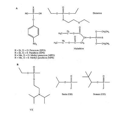
Organophosphate triesters are synthetic and do not occur naturally in the environment. They are used extensively in the production of insecticides and chemical warfare agents. In both applications, OP triesters act as potent nerve agents by inhibiting acetylcholinesterase (AChE) and ultimately causing death. AChE is a serine esterase that catalyses the hydrolysis of the neurotransmitter acetylcholine into choline and acetic acid [1, 18, 19]. Figure 1.3 shows examples of OP triesters that are commonly used in pesticides and chemical warfare agents.
Mulathion R = Et, X = O: Paraoxon (EPO)
R = Et, X = S: Parathion (EPS) R = Me, X = O: Methyl paraoxon (MPO) R = Me, X = S: Methyl parathion (MPS)
[image:31.531.73.459.302.676.2]VX
Figure 1.3: Examples of phosphotriesters.
Examples of phosphotriesters that are commonly used on pesticides (A) and
Phosphodiesters andphosphomonoesters
While phosphotriesters are not naturally occurring compounds, diesters and monoesters are abundant in nature and have play important biochemical roles, including in genetic materials like DNA and RNA (Figure 1.4), enzyme co-factors like ADP, intracellular energy transfer (ATP) and as intermediates in biochemical reaction pathways [19]. Phosphodiesterases that specifically react upon the phosphodiester bonds in DNA are known as nucleases.
N Adenine triphosphate (dATP)
Cytosine
l triphosphate (dCTP)
Phosphodiester bond
Phosphodiester 0---P
bond
[image:32.531.26.516.191.798.2]O
Figure 1.4: The phosphodiester bonds in DNA.
o o
H3C--- P---CH3 Dimethyl
phosphate Ch3Ch2--- P---CH2CH3 Diethylphosphate
O
0 ©
O
O
► o
---o ©
Figure 1.5: Examples of phosphodiesters.
Chemical structures of the ionised forms of dimethyl phosphate, diethyl phosphate and bis (p-nitrophenyl) phosphate.
1.2.3 The toxicity of organophosphate triesters
As discussed earlier, the popularity and commercial success of OP triesters are mainly due to their inherent ability to deactivate AChE. Unfortunately, the toxicity of OP triesters extends beyond the targeted organisms to pose serious threats to the environment, ecology and health [18-20]. As a result, many scientific research groups have set out to develop methods to degrade or to remove OP triesters from the environment.
Although the use of OP triesters as pesticides and nerve agents has a history of less than a century, some bacteria have evolved enzymes that are capable of hydrolysing a wide range of these compounds. These phosphotriesterases (PTEs) include the identical organophosphate hydrolases from
Flavobacterium sp. [21] and Pseudomonas diminuta [22],
laboratory, the C-terminus extension was deleted from the opdA gene so that the expressed enzyme would have an increased similarity to OPH [27].
All these PTEs, due to their individual substrate specificities, catalyse the hydrolysis of different phosphotriesters with various efficiencies. For instance, OpdA activities with triesters like methyl derivatives of paraoxon and parathion are nearly diffusion limited whereas its methyl chloropyrifos activity is relatively low [28]. Various research groups, including the Ollis laboratory, have set out to engineer their respective PTEs towards higher activity with poor substrates, since PTEs provide a promising OP detoxification and bioremediation technology that is both efficient and safe [29-32].
1.3 GpdQ - A NOVEL GLYCEROPHOSPHODIESTER
PHOSPHODIESTERASE FROM ENTEROBACTER AEROGENES
This section 1.3 is a summary of previous work on GpdQ, including the isolation, purification, cloning of the gpdQ gene and characterisation of the enzyme activity.
1.3.1 Growth of Escherichia coli co-expressing OpdA and GpdQ using paraoxon as the sole phosphorus source
o
o ©
-o ©
°P dA Dimethyl GpdQ Methyl _____ p. . .
Methyl paraoxon --- ► phosphate phosphate ^ ' P
Phosphotriesterase Phosphodiesterase Phosphatase
Figure 1.6: The proposed catabolic pathway for the breakdown of methyl paraoxon (MPO).
This rationally designed pathway was for the mineralisation of MPO in an E. coli that
co-expresses recombinant OpdA and GpdQ. Phosphatases such as alkaline
phosphatase are naturally expressed by E. coli.
E. coli transformed with the recombinant opdK gene would express the PTE
to readily degrade MPO. However, it had been previously reported that E. coli was unable to use dimethyl phosphate (DMP), the hydrolysis product of MPO, as its sole phosphorus source [33]. Therefore, a phosphodiesterase from an external source that is capable of hydrolysing DMP was sought.
Enterobacter aerogenes was reported to be able to survive on DMP as its
sole phosphorus source [34]. Gerlt and co-workers had isolated and characterised the phosphohydrolase that was responsible for DMP hydrolysis [35, 36]. The enzyme was found to be hexameric with a subunit molecular weight of 29 kDa. The phosphohydrolase was also found to be capable of hydrolysing a range of monoesters and diesters of phosphoric acid.
fragments was used to transform DHlOß E. coli cells that were then selected on minimal medium supplemented with 0.1 dimethyl phosphate (DMP) as the sole phosphorus source. A single colony of E. coli grew significantly larger than the rest. The plasmid was isolated and characterised as described in the next section. This new plasmid was later used to transform fresh DHlOß E. coli cells that already bore the opdA gene. These cells were then tested positive for growth on medium with dimethyl paraoxon (MPO) as the sole phosphorus source. The ability of the cells to grow on MPO as its sole phosphorus source would signify the success of the catabolic pathway outlined in Figure 1.7, where the breakdown of methyl paraoxon (MPO) occurred in three catalysed hydrolysis steps. OpdA catalysed the hydrolysis of MPO to DMP that then served as a substrate for GpdQ to be hydrolysed to release methyl phosphate, a monoester. E. coli readily expressed phosphomonoesterases with the likes of alkaline phosphatase (encoded by the phoA gene) [37] that would hydrolyse methyl phosphate to phosphate, that could be directly utilised by the bacteria as an energy source.
1.3.2 Characteristics and properties of GpdQ
The phosphohydrolase gene was isolated as part of a putative operon
The plasmid from the single E. coli colony that grew using DMP as the sole phosphorus source was isolated and studied. The presence of a 5.6-kb fragment was revealed and its sequence was determined. BLAST analysis of the sequence indicated that the fragment was partially homologous to glycerol-3-phosphate (G3P) uptake (ugp) operon of E. coli (Figure 1.7) [32]. The ugp operon contained four genes (A, B, C and E) that code for an ABC transporter that utilises the energy of adenosine triphosphate (ATP) hydrolysis to transport molecules across the cell wall [38, 39]. The E.
aerogenes DNA fragment could be divided into the promoter region and
aerogenes ugp operon has a 78% sequence match to the E. coli phosphate box, which is where the transcription activator PhoB binds when phosphate is limited [32, 40, 41].
I K f\ K
I OR1-3 \ OR.F2 ORF3 \
, / ORF4 ) ORF5
V '1 /
V
A. aerogenes 5.ft kb DNA trapir.cn:
K _ Jn l\ I L
p-I UfZP B ) »HP* ugpt . ugpC J
y i'vV A
V
V
V
V
VA. coii uf(p operon
Figure 1.7: Comparison of the 5.6-kb DNA fragment isolated from E. aerogenes
and the E. coli ugp operon.
ORFs in the E. aerogenes DNA fragment and their respective homologous genes in
the E. coli ugp operon are shaded in the same colours, eg. ORF1 and ugpA are shaded in purple. P indicates the promoter region of each operon while PB indicates
phosphate box. ORF3 is the 825-bp long gpdQ gene.
The remaining gene in the ugp operon codes for UgpQ, a glycerophosphodiesterase (GDPD) that had a high level of sequence similarity to GlpQ, a periplasmic GDPD whose sequence and structure is known [42-44]. GlpQ was one of more than 10 bacterial GDPD proteins whose structures were to be found in the Protein Data Bank (http://www.rcsb.org/pdb/). The GDPDs from Thermotoga maritima [45],
Agrobacterium tumefaciens [46] and Thermoanaerobacter tengcongensis
* R can be ethanolamine, choline, glycerol or glycerophosphoglycerol.
.rn-glycemphosphoethanolamine (GPE) Ethanolamine
Figure 1.8: Glycerophosphodiesters and their hydrolysis.
(A) The general chemical structure of glycerophosphodiesters; (B) Hydrolysis of s/>
glycerophosphoethanolamine (GPE) catalysed by GDPDs.
glycerophosphoethanolamine (GPE), DMP, bis (p-nitrophenyl) phosphate (b/?NPP) and p-nitrophenyl phosphate (pNPP). Additionally, the protein exhibited very low activity with paraoxon, a phosphotriester.
GpdQ, a highly promiscuous enzyme
GpdQ is a catalytically promiscuous enzyme with an exceptionally broad substrate specificity. All other known GDPDs generally show activity with a range of GPDs [45-49]. With GpdQ, the enzyme’s substrate repertoire is more extensive than the majority of GDPDs. GpdQ has primarily phosphodiesterase activity, capable of hydrolysing the phosphodiester bonds in simple alkyl phosphodiesters like dimethyl phosphate (DMP) and diethyl phosphate (DEP), other model phosphodiester substrates like bis (p- nitrophenyl) phosphate (bpNPP) and O-ethyl (p-nitrophenyl) phosphate (O- Et-pNPP). Additionally, GpdQ shows low activity towards phosphomonoesters, phosphotriesters, phosphonate monoesters and notably, an analogue of EA 2192, a highly toxic and stable hydrolysis product of the nerve agent VX [32, 51, 52].
1.4 THE STRUCTURE OF GpdQ
Three crystal structures of native GpdQ have been deposited in the Protein Data Bank (http://www.rcsb.org/pdb/). Jackson et al. first solved the structure in 2006 (PDB accession codes 2DXL and 2DXN) at 2.9 Ä and 3.0 Ä respectively [53]. Another structure of a higher resolution (1.9 Ä) was obtained from this study with the PDB code 3D03 [52]. Structural resolution analysis confirmed the a/ß sandwich tertiary fold.
1.4.1 Overall quaternary structure
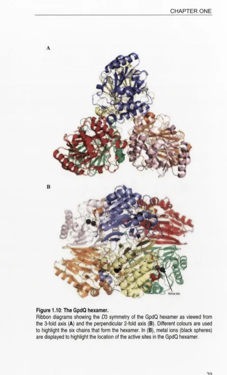
belonging to the P2i3 space group. The GpdQ molecule was found to have D3 (dihedral hexamer) symmetry like aspartate carbamoyl transferase.
The quaternary organisation of GpdQ can be described as a trimer of dimers:
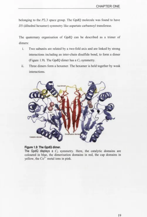
i. Two subunits are related by a two-fold axis and are linked by strong interactions including an inter-chain disulfide bond, to form a dimer (Figure 1.9). The GpdQ dimer has a C2 symmetry.
[image:40.531.21.522.28.766.2]ii. Three dimers form a hexamer. The hexamer is held together by weak interactions.
Figure 1.9: The GpdQ dimer.
The GpdQ displays a C2 symmetry. Here, the catalytic domains are
Figure 1.10: The GpdQ hexamer.
Ribbon diagrams showing the D3 symmetry of the GpdQ hexamer as viewed from
the 3-fold axis (A) and the perpendicular 2-fold axis (B). Different colours are used
to highlight the six chains that form the hexamer. In (B), metal ions (black spheres)
Protein oligomerisation
It is widely believed that protein oligomerisation happens as a result of the evolutionary process. The importance of protein oligomerisation is evidenced by its prevalence in nature [54-56]. It has been proposed that protein oligomerisation plays many roles that include foremostly, the functionality of a protein. Oligomerisation enables allosteric regulation involving cooperativity between subunits, a complete active catalytic site between subunits, or binding of substrate/product/effector at an additional regulatory site that exists between subunits. Oligomerisation also contributes to conformational and thermal stability, thus making the protein less susceptible to degradation. The reduced surface area also protects the protein from degradation and is thought to enhance the diffusion of substrates to the enzyme active site. Additionally, the increased complexity of an oligomer allows a more efficient control over the accessibility and specificity of the active site [54-57].
On the molecular level, oligomerisation reduces translation errors by providing an extra proofreading step in rejecting defective subunits. Moreover, association of homo-oligomers provides a compact and efficient way for large proteins to be encoded, thus minimising the genome size. In other words, only a small genetic or DNA space is required to encode small protein subunits which will later self-assemble to form a large protein complex [54-57].
Dissociation of protein oligomers
Protein oligomers are also known to dissociate, along with a concomitant change in enzyme activity. The process is often reversible; therefore these enzymes are able to re-associate to form the oligomer, as influenced by regulatory effectors [57]. Dissociation could lead to both positive and negative changes towards toward the enzyme activity, depending on individual enzymes.
It has also been shown that dilution has an effect on the specific activities of purine nucleoside phosphorylase with 50 pM inosine and 50 mM phosphate, respectively [59]. It is also likely for enzyme concentration to shift the equilibrium of the associated/dissociated enzyme mixture.
Despite the numerous advantages that oligomeric proteins have over their individual subunits, monomers also have a few functional niches. Many transfer and transport proteins like Cytochrome c are small and they diffuse rapidly to their sites of action within the cell. Small proteins are also more stable at low concentrations. Furthermore, unwanted oligomerisation of proteins can result in the formation of pathogenic aggregates [54, 55, 60].
Advances in protein engineering have enabled enzymes with stable quaternary structures to dissociate [61, 62], including GpdQ in this thesis. A brief review on protein engineering is given in section 1.6 of this chapter.
1.4.2 Structural domains in a GpdQ monomer
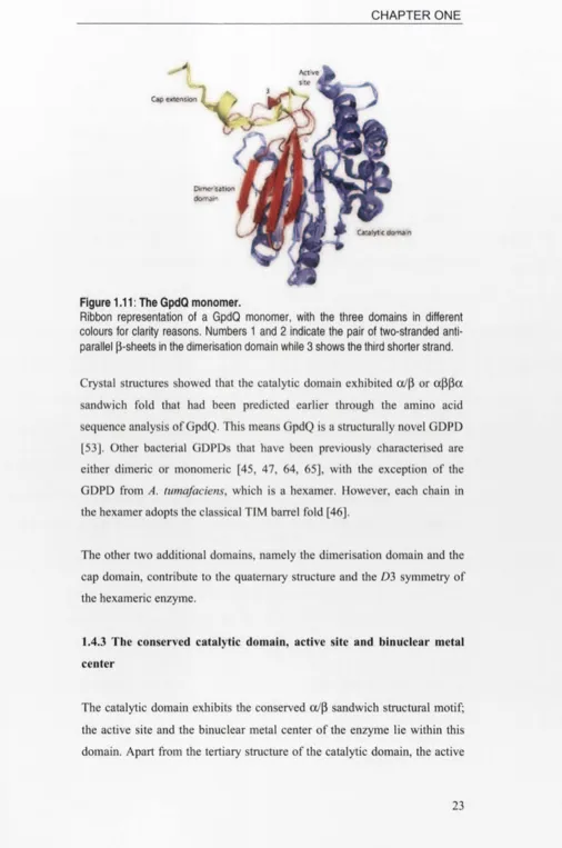
Jackson et at. divided the 274-residue GpdQ monomer into the following three domains [53] (Figures 1.11 and 1.12):
i. Catalytic (residues 1-195) ii. Dimerisation (residues 196-255) iii. Cap (residues 256-274)
Figure 1.11: The GpdQ monomer.
Ribbon representation of a GpdQ monomer, with the three domains in different colours for clarity reasons. Numbers 1 and 2 indicate the pair of two-stranded anti-parallel ß-sheets in the dimerisation domain while 3 shows the third shorter strand.
Crystal structures showed that the catalytic domain exhibited ot/ß or aßßa sandwich fold that had been predicted earlier through the amino acid sequence analysis of GpdQ. This means GpdQ is a structurally novel GDPD [53]. Other bacterial GDPDs that have been previously characterised are either dimeric or monomeric [45, 47, 64, 65], with the exception of the GDPD from A. tumafaciens, which is a hexamer. However, each chain in the hexamer adopts the classical TIM barrel fold [46].
The other two additional domains, namely the dimerisation domain and the cap domain, contribute to the quaternary structure and the D3 symmetry of the hexameric enzyme.
1.4.3 The conserved catalytic domain, active site and binuclear metal center
site residues of this superfamily of oc/ß sandwich phosphohydrolases are also conserved, including GpdQ and kidney bean purple acid phosphatase [66]. Additionally, two phosphodiesterases, whose structures have been solved, are known to adopt this oc/ß sandwich and the active residues are conserved too. The two proteins are the Rv0805 Class III 3’,5’-cyclic
nucleotide phosphodiesterase from Mycobacterium tuberculosis [67] and the and the MJ0936 phosphodiesterase from Methanococcus jannaschii [68]. The binuclear metal center at the active sites of these enzymes is capable of coordinating a wide range of divalent metal ions including Mn2t, Co2+’Ni2t, Zn2< and also Fe3' at the site with a higher metal binding affinity.
H10
H197
H10
H122
D8 H156 D21 H169
i D5Q N80 H23v ! P^3 :
I X / ; ' \ !
■y j
a/ /IK / a-
x
\ y
s ' y
b k H195 H209 OH
GpdQ Rv0805
D8 H97 D135 H286
! D3.6 N59 Y167 : P\64 !
/ ' ; y ! \ j
' ' v 1 • *' v\ 1 y '' *
a ,K ' X
/ y \ y
'ö t f >H122 H325 ÖH
MJ0936
N97
H207
N201
H323
Kidney bean purple acid phosphatase
Figure 1.12: The active sites of oc/ß sandwich dimetallo-phosphohydrolases. Simplified diagrams showing ligand coordination geometries at the active sites of GpdQ, Rv0805, MJ0936 phosphodiesterases and kidney bean protein acid phosphatase.
[66]. The active sites of other related phosphatases like the Ser/Thr protein phosphatase from bacteriophage X and 5’-nucleotidase from E. coli also vary slightly [69, 70].
Figure 1.13 shows the active site architecture of GpdQ.
Figure 1.13: The active site of GpdQ.
It has been shown that the active site of GpdQ was able to coordinate Mn2\ Co2+, Zn2+, Cd2+ and Fe2+ [51-53, 71-73], The bpNPP activities of GpdQ reconstituted with the first four metals have been characterized. The order of activities is Mn2+ > Cd2+> Co2* > Zn2+.
Non-identical metal binding sites
While analysing the structure of GpdQ, Jackson et al. observed full and partial metal occupancies at the a- and ß-sites respectively [53]. This observation will be discussed in more detail in Chapter 3. The observation agreed well with the ligand coordination geometries at the GpdQ active site: although both a- and ß-metal ions are coordinated by two histidine ligands each and jointly, the bridging D50 ligand, the a-metal has another strong acidic ligand in aspartate (D8) whereas the ß-metal is additionally coordinated by an asparagine residue (N80) (Figure 1.13).
The different metal binding affinities of the a- and ß-sites play a pivotal role in the catalytic mechanism of binuclear metallohydrolases. In PAPs for instance, it has been proposed that the loose-binding metal ion is responsible for substrate binding [77, 82, 83] while the nucleophile has been shown to bind to the tight binding trivalent one [84, 85].
1.4.4 The dimerisation domain
The dimerisation domain of GpdQ consists of a pair of two-stranded, anti- parellel ß-sheets and a third shorter strand between them. The shorter strand is not directly involved in the dimeric interface between two GpdQ subunits. (Figure 1.11) [53]. This domain is made up of approximately 60 residues (residues 196-255) and a significant number of them are large aromatic residues like trytophan, tyrosine and phenylalanine, which are favoured in ß-sheet secondary structures as well as other non-polar residues like methionine and valine [86]. The predominance of these non-polar residues creates a hydrophobic core within the dimeric interface (Figure 1.14).
Each GpdQ subunit most likely folds into a globular subunit and the protein-protein interface is formed by very specific interactions (eg.
Figure 1.14: The dimerisation domain of GpdQ.
A diagram of the dimerisation domain of GpdQ, highlighting the lack of water molecules (pink dots) in the hydrophobic core of the interface (centre) and the predominance of non-polar and aromatic amino acids in the domain. The dimeric interface results in 1667 A2 of the total surface area of each chain [53].
monomer (Figure 1.15). Specific interacting residues are typically complementary in shape and chemical properties (Figures 1.14 and 1.16).
Figure 1.15: Electrostatic potential mapped to the molecular surface of the GpdQ monomer.
Figure 1.16: The GpdQ dimer and the intertwining dimerisation domains.
A diagram of a GpdQ dimer demonstrating how the ß-strands donated from the dimerisation domain of each monomer (in blue) form a barrel at the interface. Catalytic and cap domains are in pink and green respectively.
1.4.5 The cap domain
The cap domain of GpdQ is made up of the last 19 (256-274) residues of the C-terminus. This domain is a relatively small piece of peptide with a short ß-sheet followed by a short oc-helix. Despite its size, the cap domain plays important structural roles in GpdQ and is also possibly involved in the catalytic mechanism of the enzyme.
Formation o f the GpdQ dimer through 3D domain swapping
Domain swapping is a common structural phenomenon in protein dimers. This mechanism is defined by the replacement of one portion or domain of a protein subunit by the same domain from another identical chain [87, 88]. In general, exchanged domains can vary from a single ß-strand to a globular, tertiary structural domain that may cause a sizable burial area in the ‘closed’ interface [87, 89].
like the bovine pancreatic ribonuclease (RNAse A), human alpha-crystallins and the capsid of the HIV-1 virus [90-93].
This cap structure in GpdQ extends from the dimerisation domain and sits above the active site of the other dimerising chain. The stability of this domain is strengthened by an inter-chain disulfide bond (between C54 of one monomer and C269 of the other monomer) (Figure 1.16). Intramolecular disulfide bonds play an important role in regulating protein activity, as well as maintaining the stability and folding of the tertiary structure [94]. Intermolecular disulfide bonds, on the other hand, usually take part in the quaternary structure of a protein, by stabilising the assembly of subunits to fonn an oligomer [95-97].
Chain A Chain B
Figure 1.17: The domain swapping mechanism by the cap domain and the inter-chain disulfide bond.
The cap domains are highlighted in yellow while the disulfide bond is in green.
C26! I Disulfide
bond >4 Cap don',air, chain 8
Chain A NS3
Figure 1.18: Inter-chain interactions by the cap domain of GpdQ.
Diagram showing the three inter-chain bonds contributed by the cap domain of GpdQ towards the formation of the dimer.
Formation of the GpdQ hexamer
In the GpdQ hexamer, the cap domains of all the six chains are located at the interfaces between one GpdQ dimer and another (Figure 1.18). The hexamer is held together weakly with electrostatic interactions, hydrogen bonds and an inter-chain hydrophobic patch that is explained in detail in Chapter 5.
Figure 1.19: The GpdQ hexamer with the cap domains highlighted.
The cap domains of all the six subunits in red colour to highlight their locations at the interface between the three dimeric units. The interaction between the three dimeric units results in the burial of 1131 Ä2 of the total surface area of each chain [53].
Controlling access into the active site
to the structurally conserved catalytic domain (primary domain), is often smaller and located near the active site. Examples of enzymes with this fold include the GDPD from Thermotoga maritima [45] (Figure 1.19), periplasmic 5’-nucleotidase from Escherichia coli [70] and Mrel 1 nuclease from Pyrococcus furiosus [98].
Cap secondary aoma*"
[image:52.531.10.519.16.808.2]Catalytic domain with TIM oairel fold
Figure 1.19: The cap domain in a TIM barrel GDPD.
A diagram of the GDPD from
T. maritima
(PDB accession code 101Z), with the capdomain coloured in red.
The cap domains of GpdQ (Figure 1.16) and the GDPD from T. maritima
shown in Figure 1.19 are both located at the entrance to the active site. It is widely proposed that these domains have evolved to constrain the accessibility of the active site in order to enhance the substrate specificity of the enzymes [45, 70, 98]. In particular the complexity of the cap domain seemingly correlates with the size and other chemical properties of the physiological substrate.
Interestingly, a cap domain is not found in the Rv0805 3’,5’-cyclic nucleotide phosphodiesterase from Mycobacterium tuberculosis [67] and the MJ0936 phosphodiesterase from Methanococcus janaaschii [68]. These two phosphodiesterases have been shown previously in section 1.4.3 to be structurally related to GpdQ. The homology exists only between the catalytic domains, where (a/ß) sandwich fold and all the active residues are conserved.
I. 5 Rv0805 - A 3 \5 ’-CYCLIC NUCLEOTIDE
PHOSPHODIESTERASE FROM MYCOBACTERIUM
TUBERCULOSIS
3’,5’-cyclic nucleotide phosphodiesterases (PDEs) catalyse the hydrolysis of the phosphodiester bond of second messenger cAMP to yield the biologically inert 5’-AMP, as a form of homeostatic regulation of signal transduction. Previously, this superfamily of enzymes was divided by their amino acid sequences and tertiary structure into two major classes of I and II. A third class was reported by Richter and the catalytic domain of this PDE class III of enzymes closely resembles that of dimetallophosphatases like purple acid phosphatase and calcineurin [99]. Examples of PDEs class III enzymes have been found in E. coli (CpdA) [100], Vibrio fischeri [101]
and Mycobacterium tuberculosis (Rv0805) [66, 102],
A search on the DALI server [103] nominated the PDE from M.
tuberculosis, Rv0805, as the enzyme with the highest sequence similarity to