RESEARCH NOTE
Candida vaginitis among symptomatic
pregnant women attending antenatal clinics
in Mwanza, Tanzania
Martha F. Mushi
*, Amani Mmole and Stephen E. Mshana
Abstract
Objective: This study was done to determine the patterns of Candida spp. causing vaginitis and associated factors among pregnant women attending antennal clinic in Mwanza, Tanzania.
Results: A total of 197 (65.6%) out of 300 non-repetitive swabs had positive growth of Candida spp. Candida albicans 125 (63.4%) was the most predominant isolated specie followed by C. tropicalis 35 (17.8%) and C. glabrata 33 (16.8%). Laboratory confirmed candida vaginitis was independently predicted by douching practices (OR 3.2, 95% CI 1.3–7.5 P = 0.007), history of antibiotics use (OR 1.8, 95% CI 1.02–3.0, P = 0.04) and low social economic status (OR 2.04, 95% CI 1.1–3.7 P = 0.02). About two-third of pregnant women with clinical features of vaginitis attending antenatal clinic in Mwanza, Tanzania were confirmed to have Candida vaginitis mainly caused by Candida albicans.
Keywords: Candida vaginitis, Candida albicans, Douching, Antibiotic use, Low social economic status
© The Author(s) 2019. This article is distributed under the terms of the Creative Commons Attribution 4.0 International License (http://creat iveco mmons .org/licen ses/by/4.0/), which permits unrestricted use, distribution, and reproduction in any medium, provided you give appropriate credit to the original author(s) and the source, provide a link to the Creative Commons license, and indicate if changes were made. The Creative Commons Public Domain Dedication waiver (http://creativecommons.org/ publicdomain/zero/1.0/) applies to the data made available in this article, unless otherwise stated.
Introduction
Vulvo vaginal candidiasis or Candida vaginitis is the fun-gal infection of the female lower genital tract (vagina and vulva) caused by Candida spp. [1]. Candida vagini-tis is the second most complain among women attending obstetrics and gynecological clinics worldwide [2, 3]. It is estimated that about 75% of women are affected with Candida vaginitis at least once during their life time with 15% of these cases present with a “cyclic recurrent type” which is defined as four or more episodes of Candida vaginitis in a year [4, 5].
Studies have shown that Candida albicans accounts for 80–95% of all episodes of candida vaginitis world-wide [4, 6]. However, there is an increase of cases due to non-Candida albicans species led by Candida glabrata [7]. Other non-Candida albicans species reported to be associated with Candida vaginitis include: Candida tropi-calis, Candida parapsilosis, Candida lusitaniae, Candida famata, Candida kefyr, Candida sake, Candida incon-spicua, Candida valida, Candida colliculosa, Candida
utilis, Candida catenulata, Candida lipolytica, Candida membranaefaciens, Candida intermedia and Candida globosa [7–9].
In East Africa, a study done in Aghakhan hospital-Kenya reported C. albicans as the prominent species with prevalence of 69.3% followed by C. glabrata 12.9% [10]. In Tanzania, non-Candida albicans species were reported to contribute about 37% of Candida vaginitis cases [11]. However, data on azole susceptibility patterns and factors associated with Candida vaginitis are still lim-ited. Here, we report the prevalence and factors associ-ated with laboratory confirmed Candida vaginitis among pregnant women with symptoms of vaginitis attending antenatal clinics in Mwanza, Tanzania. Furthermore, data on the azole susceptibility patterns of these Candida spp. are reported.
Main text
This was a cross section study conducted from Febru-ary to July 2016. The study was conducted at antenatal clinics of Nyamagana district hospital and Makongoro reproductive and child health clinic in Mwanza, North-western Tanzania. The selected clinics are representative
Open Access
*Correspondence: mushimartha@gmail.com
of antenatal clinics that are highly populated in Mwanza serving more than 100 pregnant women per day.
Fungal isolation and fungal speciation by chromogenic agar were carried in the CUHAS microbiology labora-tory and specie confirmation and antifungal susceptibil-ity testing was done at Institute of Medical Microbiology, Gottingen, Germany.
All pregnant mothers attending antenatal clinics sus-pected of having Candida vaginitis and consented were recruited serially until the sample size was reached. For this study clinical Candida vaginitis was defined as having two or more of the following symptoms vaginal pruritus (itching), a thick odorless cottage cheese-like discharge and soreness [1]. The minimum sample size was obtained by the use of Kish Leslie formula [4].
Culture for isolation and identifications of Candida spp. High vaginal swabs were cultured on Sabouraud’s dextrose agar (SDA) supplemented with 50 µg/ml gentamicin and 50 µg/ml chloramphenicol (HiMedia-Mumbai, India) as previously described [12]. All yeasts isolated were identified to species level using chromog-enic agar. Furthermore, 98 randomly picked isolates were confirmed by the use of matrix-assisted laser desorption ionization-time of flight (MALDI-TOF) mass spectrom-etry (Bruker Daltonics, Bremen, Germany) on extracted cells harvested from SDA as previous described [12, 13].
In vitro susceptibility assays
Antifungal susceptibility testing was done by establishing minimum inhibitory concentration (MIC) of fluconazole, voriconazole, posaconazole (Discovery Fine Chemicals, Bournemouth, United Kingdom), micafungin (Roth, Ger-many), caspofungin (Merck, US), and 5-fluorocytosine (Sigma Aldrich, US) following the guidelines laid down by the EUCAST [13].
Data analysis and management
All data collected were entered into Microsoft excel sheet for cleaning and coding then transferred to STATA ver-sion 13 for analysis according to the objectives of the study. Data were summarized into percentage for cat-egorical variables while continuous variables (age and gestation age) were summarized as median with inter-quartile range (IQR). Logistic regression analysis was done to determine predictors of candida vaginitis. Sta-tistical significant was considered when P value was less than 0.05 with 95% confidence interval.
Ethical considerations
The protocol to conduct this study was approved by the joint Catholic University of Health and Allied Sci-ences/Bugando Medical Centre research ethics and
review committee (CREC) with certificate number CREC/045/2014. Permission to conduct the study was sought from all hospital administrations. All patients were requested to sign the written informed consent before recruitment and patients’ data were treated as confidential.
Results
A total of 300 pregnant women with mean age of 27 ± 6.2 years were recruited. The majority of women were married 275 (91.7%), resided in urban areas 262 (87.3%) and had primary school education 201 (67%). About half of studied participants 168 (56%) booked their first antenatal clinic on third trimester while 106 (35.3%) and 26 (8.7%) booked on second and first trimester, respectively. Their median gestation age at recruitment was 28 with interquartile range of 20–32 weeks. Most of the pregnant women had low social economic status 221 (74%) as defined by having a fridge and television.
Laboratory confirmed Candida vaginitis was detected in 65.7% (197/300) of symptomatic pregnant women. Candida albicans was the most predominant detected Candida spp. 125 (63.4%). A total of 72 (24%) patients were diagnosed to have Candida vaginitis caused by non-Candida albicans spp. The predominant non Candida albicans spp. detected was Candida tropicalis 35 (17.8%), Fig. 1. All isolated Candida albicans were highly suscep-tible to azole antifungal agents. However, Candida kru-sei were highly resistant to fluconazole and susceptible to other azole agents, Additional file 1.
Factors associated with Candida vaginitis among pregnant women
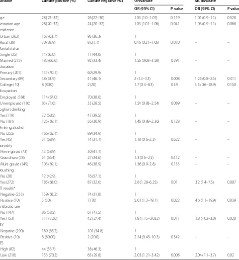
On univariable analysis, increase in gestation age OR 1.03, 95% CI 1.01–1.06, P = 0.04, not having a second-ary school education OR 2, 95% CI 1.3–3.3, P = 0.008, practicing douching OR 2.8, 95% CI 1.28–6.25, P = 0.01, history of antibiotic use in the past 2 weeks OR 1.8, 95% CI 1.15–3.03, P = 0.011 and low social economic status OR 2.03, 95% CI 1.21–3.42, P = 0.008 were found to be associated with laboratory confirmed Candida vaginitis, Table 1.
On multivariable logistic regression analysis, having douching practices OR 3.2, 95% CI 1.4–7.5, P = 0.007, history of antibiotic use in the past 2 weeks OR 1.8, 95% CI 1.02–3.0, P = 0.02 and low social economic status OR 2.04, 95% CI 1.1–3.7, P = 0.02 were independent predic-tors of laboratory confirmed Candida vaginitis, Table 1.
Discussion
observed in the current study were by the median age of the pregnant women with clinical Candida vaginitis was 28 with interquartile range of 22–32 years. Physiological and tissue changes, due to reproductive hormones, which happen in young women especially during pregnancy, increase their susceptibility to Candida infection, in addi-tion to adverse factors such as risky sexual behaviors. Previous study conducted in North America suggested that 70–75% of women can get at least one episode of Candida vaginitis in life time [6].
The current study established the prevalence of labo-ratory Candida vaginitis to be 65.7% among symp-tomatic pregnant women. This finding is similar to previous report from Nigeria which reported the preva-lence of 62.2% [4] and slightly higher than 55.4% that was reported in Cameroon [14]. The different in study population could explain the differences. The study in Cameroon involved health non-pregnant women while the current study and the one from Nigeria involved pregnant women. The changes in sex hormones during pregnancy has been highly associated with the increases chances of Candida vaginitis [3].
As previous reported from different studies [2, 3, 11], the current study found C. albicans to be the most pre-dominant specie causing Candida vaginitis. The virulence nature of Candida albicans in comparison to other Can-dida spp. could explain the findings [13]. Furthermore,
Candida albicans has been reported to be the most com-mon Candida spp. colonizing the vaginal mucosa giving it high chance of causing infections in case of the pres-ence of favorable conditions [10].
The use of antibiotic is known to suppress the bacte-rial normal flora and allow the overgrowth of yeast cells hence causing Candida vaginitis [15]. This was proven in the current study whereby the use of antibiotic was found to be an independent predictor of laboratory confirmed Candida vaginitis. Additionally in the current study douching practices which is also known to impair the growth of the vaginal microbiota [16–18] was found to independently predict the laboratory confirmed Candida vaginitis.
In the current study having low social economic status was also found to predict laboratory confirmed Candida vaginitis. Low social economic status is associated with poor hygiene [19]. Inability of the women to access basic needs include clean water and health care affect their hygienic practices. The poor hygienic practices can easily lead to vaginal candidiasis as previous observed in Cam-eroon [3].
About two-third of pregnant women with clinical fea-tures of vaginitis attending antenatal clinic in Mwanza, Tanzania were laboratory confirmed to have Candida vaginitis mainly caused by Candida albicans. Pregnant women with low social economic status (SES) with
[image:3.595.58.540.86.370.2]history of antibiotic use and who are practicing douching are more likely to suffer Candida vaginitis. A large cohort study among the high risk groups is recommended to
determine the effect of the vaginal candidiasis to the pregnancy.
Table 1 Factors associated with vaginal Candida vaginitis among 300 studied pregnant women
The box bracket [] is inter quartile range for gestation age while the curved brackets () is percentage *Only 243 women tested for VDRL
SES Social economic status
Variable Culture positive (%) Culture negative (%) Univariate Multivariate
OR (95% CI) P value OR (95% CI) P value
Age 28 [22–32] 26 [22–30] 1.03 (1.0–1.07) 0.119 1.01 (0.9–1.1) 0.528
Gestation age 28 [20–32] 24 [20–32] 1.03 (1.01–1.06) 0.041 1.03 (0.9–1.1) 0.068
Residence
Urban (262) 167 (63.7) 95 (36.3) 1
Rural (38) 30 (78.9) 8 (21.1) 0.46 (0.21–1.06) 0.070 – –
Marital status
Single (25) 14 (56.0) 11 (44.0) 1
Married (275) 183 (66.6) 92 (33.4) 1.56 (0.68–3.58) 0.291 – –
Education
Primary (201) 141 (70.1) 60 (29.9) 1
Secondary (89) 48 (53.9) 41 (46.1) 2 (1.3–3.3) 0.008 1.25 (0.8–2.5) 0.411
College (10) 8 (80.0) 2 (20) 1.7 (0.4–8.3) 0.5.9 3.5 (0.6–18.9) 0.150
Occupation
Employed (184) 114 (67.0) 70 (38.0) 1
Unemployed (116) 83 (71.6) 33 (28.5) 1.54 (0.93–2.54) 0.089 – –
Yoghurt drinking
Yes (119) 72 (60.5) 47 (39.5) 1
No (181) 125 (69.1) 56 (30.9) 1.46 (0.89–2.36) 0.128 – –
Drinking alcohol
No (255) 166 (65.1) 89 (34.9) 1
Yes (45) 31 (68.9) 14 (31.1) 1.18 (0.6–2.3) 0.622 – –
Gravidity
Prime gravid (73) 43 (58.9) 30 (41.1) 1
Gravid two (78) 51 (65.4) 27 (34.6) 1.3 (0.6–2.5) 0.412 – –
Multi gravid (149) 103 (69.1) 46 (38.9) 1.56 (0.9–2.8) 0.133 – –
Douching
No (28) 12 (42.9) 16 (57.1) 1
Yes (272) 185 (68.0) 87 (32.0) 2.8 (1.28–6.25) 0.01 3.2 (1.4–7.5) 0.007
STI results*
Negative (233) 159 (68.2) 74 (31.8) 1
Positive (10) 3 (30) 7 (70) 5.01 (1.3–19.7) 0.022 4.6 (1.1–19.9) 0.039
Antibiotic use
No (147) 86 (58.5) 61 (41.5) 1
Yes (153) 111 (72.6) 42 (27.4) 1.8 (1.15–3.032) 0.011 1.8 (1.02–3.0) 0.020
HIV
Negative (290) 189 (65.2) 101 (34.8) 1
Positive (10) 8 (80.00) 2 (20.0) 2.14 (0.45–10.3) 0.342 – –
SES
High (82) 44 (53.7) 38 (46.3) 1
[image:4.595.61.536.107.624.2]Limitation
The prevalence of laboratory Candida vaginitis in the current might been under estimated because the fea-tures used are not specific for Candida vaginitis.
Supplementary information
Supplementary information accompanies this paper at https ://doi. org/10.1186/s1310 4-019-4793-z.
Additional file 1: Table S1. Antifungal susceptibility test data.
Abbreviations
BMC: Bugando Medical Centre; CUHAS: Catholic University of Health and Allied Sciences; EUCAST: European Committee of Antimicrobial Susceptibility Testing; OR: odd ratio; MALDI-TOF: Matrix-assisted Laser Desorption Ionization-Time of Flight; SDA: Sabouraud’s dextrose agar.
Acknowledgements
Authors would like to acknowledge the support provided by department of Microbiology and immunology of the Catholic University of Health and Allied Sciences, Mwanza, Tanzania.
Authors’ contributions
MFM and SEM designed the work. AM & MFM recruited patients, performed laboratory investigations and results interpretations. MFM and SEM analyzed and interpreted the data. MFM wrote the first draft of the manuscript which was critically reviewed by SEM. All authors read and approved the final manuscript.
Funding None.
Availability of data and materials
The datasets used and/or analyzed during the current study available from the corresponding author on reasonable request.
Ethics approval and consent to participate
The protocol to conduct this study was approved by the joint CUHAS/BMC research ethics. All patients were requested to sign the written informed consent before recruitment was done. All patients’ data were treated as confidential.
Consent for publication None applicable.
Competing interests
The authors declare that they have no competing interests.
Received: 8 October 2019 Accepted: 11 November 2019
References
1. Sobel JD. Vulvovaginal candidosis. Lancet. 2007;369(9577):1961–71.
2. Foxman B, Muraglia R, Dietz J-P, Sobel JD, Wagner J. Prevalence of recur-rent vulvovaginal candidiasis in 5 European countries and the United States: results from an internet panel survey. J Lower Genit Tract Dis. 2013;17(3):340–5.
3. Toua V, Djaouda M, Gaké B, Menye DE, Christie E, Tambe E, Akindoh VV, Njiné T. Prevalence of vulvovaginal candidiasis amongst pregnant women in Maroua (Cameroon) and the sensitivity of Candida albicans to extracts of six locally used antifungal plants. Int Res J Microbiol. 2013;4(3):89–97.
4. Akah PA, Nnamani CE, Nnamani PO. Prevalence and treatment outcome of vulvovaginal candidiasis in pregnancy in a rural community in Enugu State, Nigeria. J Med Med Sci. 2010;1(10):447–52.
5. Sobel JD. Management of patients with recurrent vulvovaginal candidi-asis. Drugs. 2003;63:1059–66.
6. Sobel J, Faro S, Force R. Vulvovaginal candidiasis: epidemiologic, diagnostic, and therapeutic considerations. Am J Obstet Gynecol. 1998;178:203–11.
7. Mohanty S, Xess I, Hasan F, Kapil A, Mittal S, Tolosa JE. Prevalence & susceptibility to fluconazole of Candida species causing vulvovaginitis. Indian J Med Res. 2007;12:216–9.
8. Sobel JD. Genital candidiasis. Medicine. 2005;3(10):62–5. 9. Singh S. Treatment of vulvovaginal candidiasis. Clin Rev.
2003;136(9):26–30.
10. Mutua F, Revathi G, Machoki J. Species distribution and antifungal sensi-tivity patterns of vaginal yeasts. East Afr Med J. 2010;87(4):156–62. 11. Namkinga L, Matee M, Kivaisi K, Kullaya A, Mneney E. Identification of
Candida strains isolated from Tanzanian pregnant women with vaginal candidiasis. East Afr Med J. 2005;82(5):226–34.
12. Mushi MF, Mtemisika CI, Bader O, Bii C, Mirambo MM, Groß U, Mshana SE. High oral carriage of non-albicans Candida spp. among HIV-infected individuals. Int J Infect Dis. 2016;49:185–8.
13. Mushi MF, Bader O, Bii C, Groß U, Mshana SE. Virulence and susceptibility patterns of clinical Candida spp. isolates from a tertiary hospital, Tanzania. Med Mycol. 2018;57(5):566–72.
14. Kengne M, Shu SV, Nwobegahay JM, Achonduh O. Antifungals suscep-tibility pattern of Candida spp. isolated from female genital tract at the Yaoundé Bethesda Hospital in Cameroon. Pan Afr Med J. 2017;28(1):294. 15. Mushi MF, Ngeta N, Mirambo MM, Mshana SE. Predictors of esophageal
candidiasis among patients attending endoscopy unit in a tertiary hospital, Tanzania: a retrospective cross-sectional study. Afr Health Sci. 2018;18(1):66–71.
16. Heng LS, Yatsuya H, Morita S, Sakamoto J. Vaginal douching in Cambo-dian women: its prevalence and association with vaginal candidiasis. J Epidemiol. 2010;20(1):70–6.
17. Spence D. Candidiasis (vulvovaginal). BMJ Clin Evid. 2010;2010:0815. 18. Amaral R, Giraldo PC, Gonçalves AK, Junior J-E, Santos-Pereira S, Linhares
I, Passos MR. Evaluation of hygienic douching on the vaginal microflora of female sex workers. Int J STD AIDS. 2007;18(11):770–3.
19. Das P, Baker KK, Dutta A, Swain T, Sahoo S, Das BS, Panda B, Nayak A, Bara M, Bilung B. Menstrual hygiene practices, WASH access and the risk of urogenital infection in women from Odisha, India. PLoS ONE. 2015;10(6):e0130777.
Publisher’s Note