R E S E A R C H
Open Access
Anaplasma
spp. in dogs and owners in
north-western Morocco
Sarah Elhamiani Khatat
1,2*, Sylvie Daminet
2, Malika Kachani
3, Christian M. Leutenegger
4, Luc Duchateau
2,
Hamid El Amri
5, Mony Hing
6, Rahma Azrib
1and Hamid Sahibi
1Abstract
Background:Anaplasma phagocytophilumis an emerging tick-borne zoonotic pathogen of increased interest
worldwide which has been detected in northern Africa.Anaplasma platysis also present in this region and could possibly have a zoonotic potential. However, only one recent article reports on the human esposure toA. phagocytophilum
in Morocco and no data are available on canine exposure to both bacteria. Therefore, we conducted a cross-sectional epidemiological study aiming to assess both canine and human exposure toAnaplasmaspp. in Morocco. A total of 425 dogs (95 urban, 160 rural and 175 working dogs) and 11 dog owners were sampled from four cities of Morocco. Canine blood samples were screened forAnaplasmaspp. antibodies by an enzyme-linked immunosorbent assay (ELISA) and forA. phagocytophilumandA. platysDNA by a real-time polymerase chain reaction (RT-PCR) targeting the
msp2 gene. Human sera were tested for specificA. phagocytophilumimmunoglobulin G (IgG) using a commercial immunofluorescence assay (IFA) kit.
Results:Anaplasmaspp. antibodies andA. platysDNA were detected in 21.9 and 7.5% of the dogs, respectively.
Anaplasma phagocytophilumDNA was not amplified.Anaplasma platysDNA was significantly more frequently amplified for working dogs. No statistically significant differences in the prevalence ofAnaplasmaspp. antibodies orA. platysDNA detection were observed between sexes, age classes or in relation to exposure to ticks. A total of 348Rhipicephalus sanguineus(sensu lato) ticks were removed from 35 urban and working dogs. The majority of dog owners (7/10) were seroreactive toA. phagoyctophilumIgG (one sample was excluded because of hemolysis).
Conclusions:This study demonstrates the occurrence ofAnaplasmaspp. exposure andA. platysinfection in dogs,
andA. phagocytophilumexposure in humans in Morocco.
Keywords:Anaplasma phagocytophilum,Anaplasma platys, PCR, Serology, Dogs, Humans,Rhipicephalus
sanguineus, Morocco
Background
Ticks are considered to transmit the widest number of pathogens when compared to other arthropod vectors, and several of these pathogens are of veterinary and medical importance [1]. Some tick-borne pathogens (TBPs) are considered to be emerging because of several factors that play a crucial role in ticks multiplication and expansion, increasing the likelihood of ticks feeding on humans and animal and transmitting pathogens [2]. Among these emerging TBPs of zoonotic relevance,
Anaplasma phagocytophilum (formerly Ehrlichia equi,
Ehrlichia phagocytophila, and the human granulocytic ehrlichiosis agent) is an obligate intracellular
gram-negative bacterium belonging to the family of
Anaplasmataceae [3]. This bacterium causes a wide-spread disease called granulocytic anaplasmosis and is commonly transmitted by Ixodes tick species [4]. In the past decades, both human and animal exposure to
A. phagocytophilum has continuously increased in the USA, Europe and some Asian countries [4–8]. The clinical presentation of human granulocytic anaplas-mosis is a non-specific flu-like disease potentially fatal with severe complications, high hospitalization rates and difficult diagnosis [7–9]. Dogs are mostly recog-nized as incidental hosts and their role as potential * Correspondence:elhamianis@yahoo.fr
1Institut Agronomique et Vétérinaire Hassan II, Rabat, Morocco 2Faculty of Veterinary Medicine of Ghent University, Ghent, Merelbeke,
Belgium
Full list of author information is available at the end of the article
reservoir hosts for A. phagocytophilum infection is still controversial [10]. However, some authors sug-gested that dogs may be considered as potential reser-voir hosts for A. phagocytophilum in some regions, especially in urban environments [11–14], or at least as effective sentinels to assess the risk for human infection [15].
Anaplasma platys is another species of Anaplasma
known to infect dogs, which are considered the main
reservoir hosts. This bacterium is most likely
transmitted byRhipicephalus sanguineus(s.l.) ticks and is responsible for infectious canine cyclic thrombocytopenia [16]. Anaplasma platys is not considered as zoonotic although infection of other domestic animals [17–22] and humans [23–27] have been reported. BothA. platysandA. phagocytophilum infections remain usually asymptomatic or subclinical in dogs. When present, clinical signs are unspecific and include fever, lethargy, anorexia, lymphaden-opathy, lameness, thrombocytopenia and anemia [15, 16].
In Morocco, both Ixodes spp. and R. sanguineus (s.l.) ticks are present [28–30]. In addition,A. phagocytophilum
andA. platyswere reported in domestic animals and ticks in North Africa [31–36]. However, only one recent report described human exposure to A. phagocytophilum in Morroco [37] and no data are available on the canine ex-posure to bothA. phagocytophilumandA. platys. There-fore, the aim of this study was to assess the occurrence of
Anaplasma spp. infection and/or exposure in different groups of dogs and dog owners in Morocco.
Methods
Dogs
Between December 2013 and May 2015, 425 dogs were sampled from four Moroccan cities and divided in 3 groups. The first group (Group I) included 95 client-owned dogs sampled in the Veterinary Teaching Hospital (VTH) of the Institut Agronomique et Vétérinaire Hassan II, Rabat (34°01'31"N, 06°50'10"W). These dogs were clustered in two subgroups: Group Ia included 63 dogs without clinical signs compatible with tick-borne diseases (TBDs) and brought to the VTH for vaccination, surgery or post-surgical follow up, dermatology, cardiology or orthopedic consultations, and Group Ib included 32 dogs with clinical signs compatible with TBDs (fever, inape-tence or anorexia, lethargy and lameness without ortho-pedic origin). For each dog of the first group, an epidemiological questionnaire was completed describing the date of sample collection, age, sex, breed, outdoor activities, ectoparasite prophylaxis, exposure to ticks, travel history outside Morocco during the previous year, vaccination status, presenting complains and physical examination. The second group (Group II) was composed of 160 client-owned dogs from the rural region of Sidi Kacem (34°13'00"N, 5°42'00"W). These dogs behave like
stray or roaming dogs because of their outdoor living, close contact with other domestic of feral animals, and low health and or wellness care (absence or irregular vaccination and/or, parasite prevention). Information available on this group included age, sex and breed. The third group (Group III) contained 170 military and gendarmerie working dogs sampled in the first kennel of the Royal Army Forces of Benslimane (33°36'44"N, 7°07'16"W) and the kennel of the Royal Gendarmerie of Temara (33°55'36"N, 6°54'44"W), respectively. Data available on these dogs were age, sex and breed. Groups II and III included apparently healthy dogs considered at high risk for acquiring TBPs because of their regular outdoor activities or permanent outdoor living conditions and irregular ectoparasites prevention. All owners gave their consent for enrollment of their dogs.
For each dog, 8 ml of non-anticoagulated blood were collected from the cephalic vein. Blood was centrifuged at 3,500×rpmfor 10 min and serum was separated, ali-quoted and frozen at -32 °C. In addition, 2 ml of whole blood collected on ethylenediaminetetra-acetic acid (EDTA) anticoagulant tubes were sampled and frozen at -32 °C. The frozen sera and whole blood samples were sent to the IDEXX Laboratories (Sacramento, California, USA) to be tested for for anti-Anaplasma
spp. antibodies and for A. phagocytophilum and A. platysusing PCR.
Ticks
A total of 348 ticks were removed manually from the dogs included in this study, identified (species, stage, sex) [38] and conserved in 70% ethanol at 4 °C until shipment to the IDEXX Laboratories (Sacramento, California, USA).
Owners
All dog owners of the dogs included in Group I were contacted by phone to be sampled for A. phagocyto-philum antibodies testing. Only eleven accepted to be enrolled in this study and signed an informed consent forms. An epidemiological report was completed for each owner. Age, city of residence, occupational activ-ity, travels outside Morocco during the previous year, outdoor activities, tick exposure and potential contact with dogs and other domestic animals (cats, horses and ruminants) were recorded.
Laboratory procedures
Serological analysis of canine sera (ELISA)
TheAnaplasma spp. antibody ELISA utilizes orthogonal assay protocols to screen and subsequently confirm the presence ofAnaplasma antibodies in a serum or plasma sample. The protocols employ microwells coated with
Anaplasma p44 peptide and Anaplasma peptide conju-gated to Horseradish peroxydase (HRPO) [39]. Briefly, 50 μl of sample was added to a microtiter plate well, followed by 50 μl of conjugate. The plate was incubated for 30 min at room temperature. Wells were washed 5 times with a PBS Tween wash solution, followed by adding 100 μl of TMB substrate and a 15-min incuba-tion step at room temperature. The assay is stopped by adding a stop solution and read at 650 nm using a plate reader spectrophotometer. Positive and negative controls were run in parallel on each plate.
DNA extraction and real-time PCR assays on dogs
EDTA blood samples were used to extract total nucleic acid following a protocol adapted from Boom et al. [40]. Briefly, 180 μl whole blood were resuspended in a lysis solution and incubated for 10 min. Lysates were extracted using Whatman binding plates (Thermo Fisher Scientific, Whatham, Massachusetts, USA) on a Corbett X-Tractor platform (Qiagen, Valencia, CA, USA). Nucleic acids were eluted into 150 μl of PCR-grade nuclease-free water (Thermo Fisher Scientific, Whatham, Massachusetts, USA) and 5 μl amplified in subsequent real-time PCR reactions. Analysis was performed on a Roche LightCycler 480 (Roche Applied Science, Indianapolis, USA) and raw data analyzed using the second derivative maximum method with the ‘high sensitivity’ setting to generate crossing points (CP values).
Whole blood samples for PCR testing were available only for 362 dogs including 59 from Group Ia, 32 from Group Ib, 104 from Group II and 167 from Group III. Anaplasma spp. real-time PCR assays were used from a commercial source (IDEXX Laboratories, Inc., Westbrook, Maine, USA; test code 2824 RealPCRTM test). Real-time PCR tests were designed using a commer-cially available software (PrimerExpress 3.0) according to the published guidelines [41]. The test was adapted from previous publications [42, 43] and consisted of a mixture of two strain specific tests including A. phagocytophilum
(msp2 gene, GenBank accession no. DQ519570) and A. platys(AY848753). PCR tests positive forAnaplasmaspp. were then screened at the species level using the individ-ual strain specific real-time PCR tests. The internal sample control real-time PCR test was designed using 18S rRNA (DQ287955). All assays were designed and validated according to industry standards (Thermo Fisher Scientific, Whatham, Massachusetts, USA; User Bulletin #3).
Real-time PCR was run with 6 quality controls includ-ing (i) PCR positive controls (quantitatively); (ii) PCR negative controls; (iii) negative extraction controls; (iv) DNA pre-analytical quality control targeting canine 18S rRNA gene complex; (v) environmental contamination monitoring control; and (vi) spike-in internal positive control. These controls assessed the functionality of the PCR test protocols for the (i), absence of contamination in the reagents (ii) and laboratory (v), absence of cross-contamination during the extraction process (iii), quality and integrity of the DNA as a measure of sample quality (iv), reverse transcription protocol (v and vi) and absence of PCR inhibitory substances as a carryover from the sample matrix (vi).
Real-time PCR tests were validated analytically and clinically. For the analytical validation, each assay had to pass 6 validation criteria including amplification effi-ciency, linearity, reproducibility intra-run, reproducibility inter-run, r-square value and signal to noise ratio of the fluorescent signal. Clinical samples were used to repeat standard curves and to confirm PCR positive results by sequencing with outside flanking primers. A total of 4,125 clinical samples were used during the clinical validation of this panel and test results were compared to either alternative PCR test systems or immunofluores-cence assay (IFA) methods.
Serological analysis of human sera (IFA)
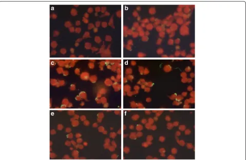
cytoplasmic morulae (Fig. 1). A serum titer of≥1:64 was considered as positive for A. phagocytophilum IgG according to the manufacturer’s instructions. Samples that were positive at the first dilution of 1:64 under ultraviolet light microscopy (×400) were then further diluted to 1:128 and those remaining positive at the second dilution were then tittered at 1:256 and 1:512.
Statistical analysis
Statistical analysis was performed using SAS version 6.4 (SAS Institute Inc., Car, NC, USA). The exact logistic regression model was fitted to compare seroreactivity and PCR positive rates between the different groups, age classes, sex and in relation to the presence of ticks. First, global hypothesis tests were performed, comparing all dog groups, based on the likelihood ratio test (LRT). With an overall significant test, groups were com-pared pairwise using Bonferroni’s multiple compari-sons technique at a global significance level of 5%. Significant pairwise comparisons were summarized in terms of the odds ratio (OR) with a 95% confidence interval (95% CI). Other risk factors (sex, tick expos-ure, age groups) were analysed in the same way.
Results
Serological and molecular screening of dogs
Off the 425 dogs, breed, sex and age were available for 299 (70.3%), 398 (93.6%) and 402 (94.6%) dogs, respect-ively. Dogs belonged to 23 different breeds with German and Belgian Shepherds (n= 122), Retrievers (n= 58), Saluki (n= 36), Cocker and English Spaniel (n= 27), mixed breeds (n= 19) and Pointers dogs (n= 10) the most frequently found during sampling. Other breeds included Poodles (n= 4), Rottweilers (n= 3), Pekingese (n= 3), Aidi (n= 2), Border Collie (n= 2), Pitbull (n= 2), Setters dogs (n= 2) and one dog for Drahthar, Saint
Hubert, German Mastiff, Argentin dogo, Dalmatian, Akita Inu, Husky, Havanese and Chihuahua. The age of dogs ranged from 3 months to 14 years-old (mean age 3.2 years-old) and males (n= 257) were more frequently sampled than females (n= 141). Previous ticks bites were available for 226 dogs (53.2%) from Group I (n= 40) and Group III (n= 18).
Table 1 summarises the results of Anaplasma spp. antibodies and A. platys DNA detection in the three groups of dogs. There were significant differences between dog groups (χ2= 10.28, df= 3, P= 0.016). Group Ia differed significantly from Group II (OR = 0.32, 95% CI: 0.14–0.75, P= 0.009). None of the 362 dogs screened forA. phagocytophilumDNA by PCR was found positive whereas 7.5% (95% CI: 0.05–0.11) of them were positive to A. platys (Table 1). There were globally significant differences between dog groups (χ2= 9.44,
df= 3, P= 0.024). The highest prevalence of A. platys
DNA detection was found in Group III but none of the pairwise comparisons was significant (Table 1). Table 2 summarizes the prevalence of positivity rates to Anaplasma spp. antibodies and A. platys DNA detection according to sex, age and exposure to ticks. No statistically significant differences were found in sero-positivity rates for the sex (χ2= 2.161, df= 1, P= 0.142), the age groups (χ2= 1.75, df= 2, P= 0.416) and exposure to ticks(χ2= 0.83, df= 1,P= 0.363). Similarly, no statisti-cally significant differences were found in positivity rates to A. platys DNA detection for sex (χ2= 2.88,
df= 1, P= 0.090), the exposure to ticks and age groups (χ2= 5.05, df= 2, P= 0.080).
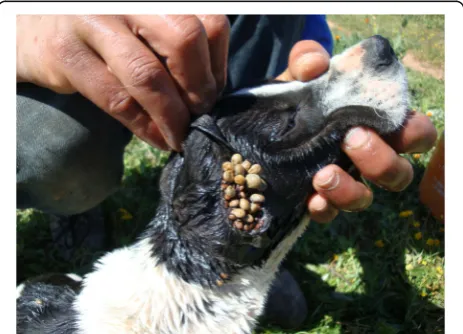
A total of 348 ticks were removed from 35 dogs and all belonged to R. sanguineus (s.l.). Two ticks were nymphs, 284 adult females and 63 adult males. The number of ticks removed from one dog ranged from 1 to 54 (mean number 9.9) (Fig. 1). Among the 35 infested dogs, 15 belonged to Group I, 2 to Group II and 18 to Group III. The number of dogs infested by ticks and positive toAnaplasma spp. antibodies only, toA. platys
DNA only or to both tests were eight, three and one, respectively. The only dog infested by ticks and positive for both tests was from Group II.
Serological screening of owners
Among the eleven dog owners sampled, three were women and eight were men. Ages ranged from 23 to 66 years, with an average of 51 years. Most lived in Rabat (9/11) and two in surrounding cities (Salé and Arjat). Seven mentioned having leisure outdoor activities in forest or rural areas and one farmer lived in a rural area (Arjat). Five owners reported to have contact with other domestic animals including cats, horses and rumi-nants. Five owners had additional dogs. Only one owner
[image:4.595.58.290.535.702.2]reported previous exposure to ticks and two traveled to foreign countries during the year.
One sample was excluded due to hemolysis that could interfere with the results according to the manufacturer’s instructions. Seven out of the ten remaining sera were positive to A. phagocytophilum IgG at the first dilution (1:64) (Fig. 2). Among the seropositive owners, three were women and four were men. Four reported regular outdoor activities in the forests of Rabat or the vicinity (Maamora forest, Khémisset, Bouznika and Benslimane). Four owners mentioned to have contact with domestic animals other than dogs. None of the seropositive owners had a travel history outside Morocco during the previous year and two mentioned to be regular blood donors. When further diluted, six, two and one samples remained positive at 1:128, 1:256 and 1:512, respectively (Fig. 2). The only sample that remained positive at 1:512 was from a farmer.
Discussion
To our knowledge, the results of this cross-sectional study demonstrated for the first time in Morocco a prevalence of 21.9 and 7.5% of Anaplasma spp. anti-bodies and A. platys DNA detection in dogs, respect-ively. It also showed that 7 among 10 dog owners were seroreactive to A. phagocytophilum IgG. Currently the
two most importantAnaplasma species known to infect dogs and humans are A. platysandA. phagocytophilum
[27]. Infection by both species have already been de-tected in dogs and ticks in North Africa [28, 31, 33–36]. Our study detected A. platys infection in dogs with a prevalence similar to what has been published in Algeria (5.4%) [36]. Although not statistically significant, work-ing dogs tested more frequently positive to A. platys
[image:5.595.57.539.111.225.2] [image:5.595.58.538.622.732.2]DNA than rural dogs. Therefore, although considered as a major risk factor for acquiring tick-borne infections [44–46], outdoor access alone cannot explain the high prevalence in working dogs. Similarly, a study on Sene-galese gendarmerie and private kennel living dogs showed a high prevalence of E. canis infection [47], another R. sanguineus (s.l.)-transmitted pathogen, prob-ably because this tick species can complete its entire life-cycle either indoor (in houses, kennels and veterin-ary hospitals where it readily colonizes the infrastruc-ture) or in outdoor environments (peri-urban and rural) [25, 47, 48]. Other factors explaining the higher preva-lence in working dogs in our study can be the absence of efficient ectoparasite control programs in this group or the access to areas with higher burdens ofA. platys.
Our study detected both Anaplasma spp.
anti-bodies and A. platys DNA in dogs but failed to iden-tify A. phagocytophilum DNA. This discrepancy has
Table 1Number and prevalence (%) of positive and negative dogs toAnaplasmaspp. antibodies (by ELISA) andA. platysDNA
detection (by PCR), and positive to both methods in the different groups
Groupa Anaplasmaspp. antibodies (%) (n= 425) A. platys(%) (n= 362) Anaplasmaspp.
andA. platys(%) (n= 362)
Positive Negative Positive Negative Not available
Group I (n= 95) 11 (2.6) 84 (19.8) 3 (0.8) 88 (24.3) 4 1 (0.3)
Group Ia (n= 63) 7 (1.6) 56 (13.2) 2 (0.5) 57 (15.7) 4 0 (0.0)
Group Ib (n= 32) 4 (0.9) 28 (6.6) 1 (0.3) 31 (8.6) 0 1 (0.3)
Group II (n= 160) 45 (10.6) 115 (27.1) 4 (1.1) 100 (27.6) 56 1 (0.3)
Group III (n= 170) 37 (8.7) 133 (31.3) 20 (5.5) 147 (40.7) 3 9 (2.3)
Total (n= 425) 93 (21.9) 332 (78.1) 27 (7.5) 335 (92.5) 63 11 (3.0)
a
Group I: urban client-owned dogs sample in the VTH; Group Ia: urban client-owned dogs sample in the VTH without clinical signs compatible with a TBD; Group Ib: urban client-owned dogs sample in the VTH with clinical signs compatible with a TBD; Group II: rural client-owned dogs; Group III: military and gendarmerie working dogs
Table 2Number and prevalence of (%) positive and negative dogs toAnaplasmaspp. antibodies (by ELISA) andA. platysDNA
detection (by PCR) according to the sex, the age and the exposure to ticks
Variable Anaplasmaspp. antibodies (%) (n= 425) A. platysDNA (%) (n= 362)
Positive Negative Not available Positive Negative Not available
Sex Male 59 (13.9) 198 (46.6) – 20 (5.5) 187 (51.7) 50
Female 23 (5.4) 118 (27.8) – 5 (1.4) 123 (34.0) 13
Age (yrs) < 1 9 (2.1) 52 (12.2) – 3 (0.8) 52 (14.4) 6
1–5 56 (13.2) 194 (45.6) – 21 (5.8) 183 (50.6) 46
≥6 13 (3.0) 61 (14.3) – 2 (0.5) 62 (17.1) 10
also been reported in other African, European and
American studies [31, 49–51]. Cross-reactivity
between Anaplasma spp. pathogens, especially
between A. phagocytophilum and A. platys, has been reported to occur. Therefore, in regions were both pathogens could co-exist, seropositivity may not enable the distinction at the species level [16]. In areas where Ixodes spp. ticks, are less prevalent or absent, a positive Anaplasma spp. serology could be the result of A. platys exposure [52]. Consequently, the fact that we detected exclusively R. sanguineus
(s.l.) ticks infesting dogs can be supportive of the potential predominance of A. platys in Morocco. However, Ixodes spp. ticks are also present in this country [28–30] and could have infected these dogs previously. On the other hand, infection with A. pha-gocytophilum in Rhipicephalus spp. has also been reported especially in the Mediterranean countries, and these ticks have been suggested as potential competent vectors of this bacterium in this part of the world [33, 53–56]. In a study from Jordan, a high prevalence of A. phagocytophilum infection (39.5%) was found in dogs and the most abundant tick spe-cies removed was R. sanguineus (s.l.) (95.1%) followed
by two Haemaphysalis species, whereas no I. ricinus
was collected from these dogs. The authors suggested that the ticks found in their study could be a possible competent vector of the pathogens detected including A. phagocytophilum [57]. Further studies are necessary to evaluate the ability of Rhipicephalus
ticks in transmitting A. phagocytophilum.
In regions where both A. platys and A. phagocytophi-lumare present, a PCR-based assay is required to deter-mine which of the two agents is responsible for positive serological test [16]. Nevertheless, false-negative results are reported to occur with PCR, mainly due to low template concentrations [27, 58], the short duration of
A. phagocytophilum bacteremia in dogs and the varia-tions in the levels of circulating bacteria [15, 58]. In addition, selective amplification of the predominant organism can occur in patients coinfected with genetic-ally similar organisms [27, 59] such as A. phagocytophi-lumandA. platys,which could be the case in our study. As DNA-based diagnostic tool enables the early detection of the infection byA. phagocytophilum, the bacteriemia is of short duration and is usually present transiently during the acute phase of the infection [15, 60, 61], negative PCR results might be more difficult to interpret in healthy dogs.
[image:6.595.59.539.86.398.2]Therefore, negative PCR results only indicate that the respective nucleic acid sequence was not detected in the sample evaluated under the assay conditions used and should not be interpreted as evidence of absence of infec-tion [58]. In addiinfec-tion, other factors could explain the nega-tive results in our study mainly the likely degradation of the DNA due to the transport conditions from Morocco to the USA and the selected region of sampling. Indeed, our dogs were sampled exclusively from the western part of Morocco but previous studies detectedI. ricinus ticks in the eastern regions [28, 29]. In addition, Borrelia burgdorferi (s.l.), that is transmitted by Ixodes spp. ticks, was reported in dogs in Algeria [31], a neighbour country of Morocco, and ticks in north-eastern Morocco [30], suggesting that these ticks could be more prevalent in eastern regions.
Consistently with our previous report that detected high prevalence rates ofA. phagocytophilumexposure in humans in northwestern regions of Morocco [37], the majority of dog owners sampled were found positive to
A. phagocytophilumIgG. In our previous study, the con-tact with dogs or other domestic animals was not a risk factor for the seropositivity [37], suggesting that other factors such as outdoor activities might be incriminated. Indeed, outdoor activities especially related to forests, meadow habitats and grasslands are considered as a major risk factor for acquiring a tick-borne infection due to the increase risk of contact with infected ticks [62]. Another study has found no significant difference in the seroprevalence of A. phagocytophilumamong owners of seropositive pets and owners without pets, suggesting that dog ownership may not be a risk factor [63].
Anaplasma platys was known to infect dogs exclu-sively, and they are are recognized as the main reservoir hosts. However, recent reports described the infection in domestic ruminants, cats and even in humans [17–27]. In addition, human infestation with R. sanguineus (s.l.) has also been reported [47, 57, 59], suggesting that A. platys could be transmitted to humans through the bite of this tick species. Moreover, all human cases infected with A. platyshad regular contact with dogs and/or re-ported infestation of their dogs with R. sanguineus(s.l.) [25–27]. In addition, in two human cases, the A. platys
sequence was identical to the sequence found in their dog [27]. This is in contrast to our current and previous study that both failed to detect a relationship between contact with dogs and human seropositivity toA. phago-cytophilumpossibly suggesting that humans in Morocco could be more likely to exposed to this bacterium than to A. platys. All previously reported cases of human A. platys infection were diagnosed by DNA detection or microscopic identification of morulae within platelets [25–27] and hence, the occurrence of immunological response to this bacterium is unknown. Moreover, to the
authors’ knowledge, the possible occurrence of cross-reaction between A. platys and A. phagocytophilum
antibodies has not been evaluated in humans. The IFA based on HL60-cells infected with a human iso-late of A. phagocytophilum, such as the one used in our study, are considered to be both sensitive [64] and highly specific for the investigation of seroreactiv-ity to this bacterium [9] with a specificseroreactiv-ity of 100%, according to the manufacturer.
Rhipicephalus sanguineus (s.l.) is the most common tick in the Mediterranean region [57]. It is known to transmit several pathogens includingRickettsia conorii, Babesia canis, Hepatozoon canis and E. canis and probablyBartonellaspp.,Mycoplasma haemocanisand
A. platys [46]. This tick has the particularity to be active during almost all the year and to achieve two or more generations per year. Warmer temperature may contribute to an increased tick abundance by a more rapid development. Although R. sanguineus (s.l.) ticks usually feed on dogs, they can feed on a wide variety of animal species including humans [48, 65]. Therefore, due to its high degree of adaptability, R. sanguineus
(s.l.) represents a major threat not only to dogs, but also to humans. Furthermore, the report of E. canis
andA. platyshuman infections [23–27, 66, 67]
empha-sizes the importance of R. sanguineus (s.l.) and the zoonotic potential of these two infections, and further investigation should be carried out to assess the public health implication [48].
The major limitations of this study are the restricted area of sampling, the absence of PCR performed on the ticks sampled from dogs, and the small number of owners and dogs with clinical signs compatible with a TBD. Unfortunately, DNA from the ticks collected was too degraded to perform PCR analysis, most probably due to the shipping conditions from Morocco to the USA.
Conclusions
This study demonstrates the Anaplasma spp. exposure in humans and dogs in Morocco. To our knowledge, it is also the first report on the occurrence of A. platys
main reservoir hosts, it is important to evaluate if this bacterium can cause human disease in Morocco and if the infection is associated with an immunological re-sponse. This study should serve as an indicator to Moroccan physicians and veterinarians thatA. phagocy-tophilum and A. platys exposure and infection are not rare, and it will help raise awareness on the potential occurrence of TBDs more generally in this country. Since we reported results in a limited area of the country and on a very limited number of humans, larger and more represeantative surveys are recommended.
Abbreviations
EDTA:Ethylenediaminetetra-acetic acid; ELISA: Enzyme-linked immunosorbent assay; IFA: Immunofluorescence assay; IgG: Immunoglobulin G; RT-PCR: Real-time polymerase chain reaction; TBD: Tick-borne disease; TBPs: Tick-borne pathogens
Acknowledgements
We would like to address our gratitude to the General Hosni Benslimane for the authorization to sample military and Gendarmerie dogs. We are also grateful to Dr Nourredine Tazi, Dr Hassan Fassil, Pr Ikhlass El Berbri and Mr Mohamed El Mjiyad for their contribution to dogs sampling and to Dr Iraqui for owners sampling. We would like to thank Dr Walter Heuninckx and Mrs Pierrette Parmentier for their help in IFA dosage. We also would like to thank Dr Souad Boutayeb and her team from the Laboratory of the Royal Gendarmerie of Rabat, Pr Mohamed Amar and his team and Mrs Rabiaa El Guennouni for their support in samples processing.
Funding
This research received no specific grant from any funding agency in the public, commercial, or not-for-profit sectors.
Availability of data and materials
The datasets supporting the conclusions of this article are included within the article.
Authors’contributions
Conception and design of data: SEK, LD and SD. Data acquisition: SEK and CL. Analysis and interpretation of data: SEK and LD. Reagents/materials/ analysis tools contribution: SEK, CL, HE, MH and LD. Drafting or revising the manuscript: SEK, SD, MK, CL, LD, HE, MH, HS and RA. All authors read and approved the final manuscript.
Competing interests
The authors declare that they have no competing interests.
Consent for publication
Not applicable.
Ethics approval
The study protocol was approved by the Ethical Committee for Biomedical Research of the Mohammed V University of Rabat (n°698, July, 10th2014) and the Ministry of Health of Morocco (n°965, June, 12th2014).
Publisher’s Note
Springer Nature remains neutral with regard to jurisdictional claims in published maps and institutional affiliations.
Author details
1
Institut Agronomique et Vétérinaire Hassan II, Rabat, Morocco.2Faculty of Veterinary Medicine of Ghent University, Ghent, Merelbeke, Belgium.3College of Veterinary Medicine, Western University of Health Sciences, Pomona, CA, USA.4Molecular Diagnostics IDEXX Laboratories, Inc. West Sacramento, Sacramento, CA 95605, USA.5Laboratory of the Royal Gendarmerie, Rabat, Morocco.6National Reference Laboratory for Anaplasma phagocytophilum, Laboratory of Clinical Biology, Queen Astrid Military Hospital, Brussels, Belgium.
Received: 16 February 2017 Accepted: 19 April 2017
References
1. Anderson JF, Magnarelli LA. Biology of ticks. Infect Dis Clin North Am. 2008;22:195–215.
2. Baneth G. Tick-born infections of animals and humans: a common ground. Int J Parasitol. 2014;44:591–6.
3. Dumler JS, Barbet AF, Bekker CPJ, Dasch GA, Palmer GH, Ray SC, et al. Reorganization of genera in the familiesRickettsiaceaeandAnaplasmataceae in the order Rickettsiales: Unification of some species ofEhrlichiawith Anaplasma,CowdriawithEhrlichiaandEhrlichiawithNeorickettsia, descriptions of six new species combinations and designation ofEhrlichia equiand‘HGE agent’as subjective synonyms ofEhrlichia phagocytophila. Int J Syst Evol Microbiol. 2001;51:2145–65.
4. Stuen S.Anaplasma phagocytophilum- the most widespread tick-borne infection in animals in Europe. Vet Res Commun. 2007;1:79–84. 5. Qurollo AB, Chandrashekar R, Hegarty BC, Beall MJ, Stillman BA, Liu J, et al.
A serological survey of tick-borne pathogens in dogs in North America and the Caribbean as assessed byAnaplasma phagocytophilum,A. platys, Ehrlichia canis,E. chaffeensis,E. ewingii, andBorrelia burgdorferi species-specific peptides. Infect Ecol Epidemiol. 2014;4:24699.
6. Centers for Disease Control and Prevention. Summary of notifiable diseases –United States. MMWR 2011;60:1–117 (cited January 2015), available from: https://www.cdc.gov/mmwr/preview/mmwrhtml/mm6053a1.htm. 7. Cochez C, Ducoffre G, Vandenvelde C, Luyasu V, Heyman P. Human
anaplasmosis in Belgium: a 10-years seroepidemiological study. Ticks Tick Borne Dis. 2011;2:156–9.
8. Zhang L, Liu H, Xu B, Zhang Z, Jin Y, Li W, et al. Rural residents in China are at increased risk of exposure to tick-borne pathogensAnaplasma phagocytophilumandEhrlichia chaffeensis. Biomed Res Int. 2014;2014:313–867. 9. Heyman P, Cochez C, Bigaignon G, Guillaume B, Zizi M, Vandenvelde C.
Human granulocytic ehrlichiosis in Belgium: an underestimated cause of disease. J Infect. 2003;47:129–32.
10. Schorn S, Pfister K, Reulen H, Mahling M, Manitz J, Thiel C, et al. Prevalence ofAnaplasma phagocytophiluminIxodes ricinusin Bavarian public parks, Germany. Ticks Tick Borne Dis. 2011;2:196–203.
11. Silaghi C, Gilles J, Hohle M, Fingerle V, Just FT, Pfister K.Anaplasma phagocytophiluminfection inIxodes ricinus, Bavaria, Germany. Emerg Infect Dis. 2008;14:972–4.
12. Hornok S, Dénes B, Meli ML, Tánczos B, Fekete L, Gyuranecz M, et al. Non-pet dogs as sentinels and potential synanthropic reservoirs of tick-borne and zoonotic bacteria. Vet Microbiol. 2013;167:700–3. 13. Torina A, Vicente J, Alongi A, Scimeca S, Turlá R, Nicosia S, et al.
Observed prevalence of tick-borne pathogens in domestic animals in Sicily, Italy during 2003–2005. Zoonoses Public Health. 2007;54:8–15. 14. Santos HA, Pires MS, Vilela JAR, Santos TM, Faccini JLH, Baldani CD, et al.
Detection ofAnaplasma phagocytophilumin Brazilian dogs by real-time polymerase chain reaction. J Vet Diagn Invest. 2011;23:770–4. 15. Carrade DD, Foley JE, Borjesson DL, Sykes JE. Canine granulocytic
anaplasmosis: a review. J Vet Intern Med. 2009;23:1129–41.
16. Harvey JW.Anaplasma platysinfection (thrombocytotropic anaplasmosis). In: Infectious diseases of the Dog and Cat, chapter 26,Ehrlichiaand Anaplasmainfections. St. Louis: Saunders Elsevier; 2012. p. 241–4. 17. Santarem VA, Laposy CB, Farias MR.Anaplasma platys(Ehrlichia platys)-like
inclusion bodies in platelets of a cat. Colloq Agrariae. 2005;1:60–6. 18. Chochlakis D, Ioannou I, Sharif L, Kokkini S, Hristophi N, Dimitriou T, et al.
Prevalence ofAnaplasmasp. in goats and sheep in Cyprus. Vector Borne Zoonotic Dis. 2008;9:457–63.
19. Djiba ML, Mediannikov O, Mbengue M, Thiongane Y, Molez JF, Seck MT, et al. Survey ofAnaplasmataceaebacteria in sheep from Senegal. Trop Anim Health Prod. 2013;45:1557–61.
20. Zobba R, Anfossi AG, Pinna Parpaglia ML, Dore GM, Chessa B, Spezzigu A, et al. Molecular investigation and phylogeny ofAnaplasmaspp. in Mediterranean ruminants reveal the presence of neutrophil-tropic strains closely related toA. platys. Appl Environ Microbiol. 2014;80: 271–80.
22. Salakij C, Lertwatcharasarakul P, Salakij J, Nunklang K, Rattanakunuprakarn J. Molecular characterization ofAnaplasma platysin a domestic cat from Thailand. Comp Clin Pathol. 2012;21:345–8.
23. Arraga-Alvarado C, Montero-Ojeda M, Bernardoni A, Anderson BE, Parra O. Human ehrlichiosis: report of the 1st case in Venezuela. Invest Clin. 1996;37:35–49. Abstract.
24. Arraga-Alvarado C, Palmar M, Parra O, Salas P. Fine structural characterization of aRickettsia-like organism in human platelets from patients with symptoms of ehrlichiosis. J Med Microbiol. 1999;48:991–7. 25. Maggi RG, Mascarelli PE, Havenga LN, Naidoo V, Breitschwerdt EB.
Coinfection withAnaplasma platys BartonellahenselaeandCandidatus mycoplasma haematoparvum in a veterinarian. Parasit Vectors. 2013;6:103. 26. Arraga-Alvarado CM, Qurollo BA, Parra OC, Berrueta MA, Hegarty BC,
Breitschwerdt EB. Molecular evidence ofAnaplasma platysinfection in two women from Venezuela. Am J Trop Med Hyg. 2014;91:1161–5.
27. Breitschwerdt EB, Hegarty BC, Qurollo BA, Saito TB, Maggi RG, Blanton LS, et al. Intravascular persistence ofAnaplasma platys,Ehrlichia chaffeensis, and Ehrlichia ewingiiDNA in the blood of a dog and two family members. Parasit Vectors. 2014;7:298.
28. Sarih M, M’Ghirbi Y, Bouattour A, Gern L, Baranton G, Postic D. Detection and identification ofEhrlichiaspp. in ticks collected in Tunisia and Morocco. J Clin Microbiol. 2005;43:1127–32.
29. Seng P, Sarih M, Socolovschi C, Boudebouch N, Hassar M, Parola P, et al. Detection of Anaplasmataceae in ticks collected in Morocco. Clin Microbiol Infect. 2009;15 Suppl 2:86–7.
30. Sarih M, Jouda F, Gern L, Postic D. First isolation ofBorrelia burgdorferi sensu lato fromIxodes ricinusticks in Morocco. Vector Borne Zoonotic Dis. 2003;3:133–9.
31. Azzag N, Petit E, Gandoin C, Bouillin C, Ghalmi F, Haddad N, et al. Prevalence of select vector-borne pathogens in stray and client-owned dogs from Algiers. Comp Immunol Microbiol Infect Dis. 2015;38:1–7. 32. Ben Said M, Belkahia H, Sayahi L, Aloui M, Jemli MH, Hadj Mohamed B, et al. First
serological study of the prevalence ofAnaplasma phagocytophilumin dromedary (Camelus dromedarius) in Tunisia. Bull Soc Pathol Exot. 2014;107:1–6. 33. Ghafar MW, Amer SA. Prevalence and first molecular characterization of
Anaplasma phagocytophilum, the agent of human granulocytic anaplasmosis, inRhipicephalus sanguineusticks attached to dogs from Egypt. J Adv Res. 2012;3:189–94.
34. M’ghirbi Y, Ghorbel A, Amouri M, Nebaoui A, Haddad S, Bouattour A. Clinical, serological, and molecular evidence of ehrlichiosis and anaplasmosis in dogs in Tunisia. Parasitol Res. 2009;104:767–74.
35. M’ghirbi Y, Yaïch H, Ghorbel A, Bouattour A.Anaplasma phagocytophilumin horses and ticks in Tunisia. Parasit Vectors. 2012;5:180–6.
36. Dahmani M, Loudahi A, Mediannikov O, Fenollar F, Raoult D, Davoust B. Molecular detection ofAnaplasma platysandEhrlichia canisin dogs from Kabylie, Algeria. Ticks Tick Borne Dis. 2015;6:198–203.
37. Elhamiani Khatat S, Sahibi H, Hing M, Alaoui Moustain I, El Amri H, Benajiba M, et al. Human exposure toAnaplasma phagocytophilumin two cities of northwestern Morocco. PLoS One. 2016;11:e0160880.
38. Walker AR, Bouattour A, Camicas J, Estrada-Pena A, Horak I. Ticks of domestic animals in Africa: a guide to identification of species. UK: Bioscience reports Edinburgh; 2003.
39. Stillman BA, Monn M, Liu J, Thatcher B, Foster P, Andrews B, et al. Performance of a commercially available in-clinic ELISA for detection of antibodies againstAnaplasma phagocytophilum,Anaplasma platys,Borrelia burgdorferi,Ehrlichia canis, andEhrlichia ewingiiandDirofilaria immitis antigen in dogs. J Am Vet Med Assoc. 2014;245:80–6.
40. Boom R, Sol CJ, Salimans MM, Jansen CL, Wertheim-van Dillen PM, van der Noordaa J. Rapid and simple method for purification of nucleic acids. J Clin Microbiol. 1990;28:495–503.
41. Livak K, Marmar J, Flood S. Guidelines for designing TaqMan fluorogenic probes for 5’nuclease assays. PE Appl Biosyst Res News. 1995. 42. Pusterla N, Leutenegger CM, Chae JS, Lutz H, Kimsey RB, Dumler JS, et al.
Quantitative evaluation of ehrlichial burden in horses after experimental transmission of human granulocyticEhrlichiaagent by intravenous inoculation with infected leukocytes and by infected ticks. J Clin Microbiol. 1999;37:4042–4.
43. Foley JE, Leutenegger CM, Dumler JS, Pedersen NS, Madigan JS. Evidence for modulated immune response toAnaplasma phagocytophilasensu lato in cats with FIV-induced immunosuppression. Comp Immunol Microbiol Infect Dis. 2003;26:103–13.
44. Lim S, Irwin PJ, Lee SR, Oh MH, Ahn KS, Myung BY, Shin SS. Comparison of selected canine vector-borne diseases between urban animal shelter and rural hunting dogs in Korea. Parasit Vectors. 2010;3:32.
45. Alho AM, Pita J, Amaro A, Amaro F, Schnyder M, Grimm F, et al. Seroprevalence of vector-borne pathogens and molecular detection ofBorrelia afzeliiin military dogs from Portugal. Parasit Vectors. 2016;9:225.
46. Pennisi MG, Caprì A, Solano-Gallego L, Lombardo G, Torina A, Masucci M. Prevalence of antibodies againstRickettsia conorii,Babesia canis,Ehrlichia canis, andAnaplasma phagocytophilumantigens in dogs from the Stretto di Messina area (Italy). Ticks Tick Borne Dis. 2012;3:314–7.
47. Davoust B, Mediannikov O, Chene J, Massot R, Tine R, Diarra M, et al. Study of ehrlichiosis in kennel dogs under treatment andprevention during 7 months in Dakar (Senegal). Comp Immunol Microbiol Infect Dis. 2013;36:613–7.
48. Ebani VV, Bertelloni F, Torracca B, Cerri D. Serological survey ofBorrelia burgdorferi sensu lato,Anaplasma phagocytophilum, andEhrlichia canis infections in rural and urban dogs in Central Italy. Ann Agric Environ Med. 2014;21:671–5.
49. Clarke LL, Ballweber LR, Allen K, Little SE, Lappin MR. Prevalence of select vector-borne disease agents in owned dogs of Ghana. J S Afr Vet Assoc. 2014;85:1–2.
50. Diniz P, Beall MJ, Omark K, Chandrashekar R, Daniluk DA, Cyr KE, et al. High prevalence of tick-borne pathogens in dogs from an Indian Reservation in northeastern Arizona. Vector Borne Zoonotic Dis. 2010;10:117–23. 51. Santos AS, Alexandre N, Sousa R, Núncio MS, Bacellar F, Dumler JS.
Serological and molecular survey ofAnaplasmaspecies infection in dogs with suspected tick-borne disease in Portugal. Vet Rec. 2009;164: 168–71.
52. Mircean V, Dumitrache MO, Gyöke A, Pantchev N, Jodies R, Mihalca AD, et al. Seroprevalence and geographic distribution of Dirofilaria immitis and tick-borne infections (Anaplasma phagocytophilum,Borrelia burgdorferisensu lato, andEhrlichia canis) in dogs from Romania. Vector Borne Zoonotic Dis. 2012;12:595–604.
53. Keysary A, Massung RF, Inbar M, Wallach AD, Shanas U, Mumcuoglu KY, Waner T. Molecular evidence forAnaplasma phagocytophilumin Israel. Emerg Infect Dis. 2007;13:1411–2.
54. Psaroulaki A, Chochlakis D, Ioannou I, Florentia A, Gikas A, Tselentis Y. Acute anaplasmosis in human in Cyprus. Clin Microbiol Infect. 2008;15:10–1. 55. Chastagner A, Bailly X, Leblond A, Pradier S, Vourc’h G. Single Genotype of
Anaplasma phagocytophilumidentified from ticks, Camargue, France. Emerg Infect Dis. 2013;19:825–6.
56. Dugat T, Chastagner A, Lagrée AC, Petit E, Durand B, Thierry S, et al. A new multiple-locus variable-number tandem repeat analysis reveals different clusters forAnaplasma phagocytophilumcirculating in domestic and wild ruminants. Parasit Vectors. 2014;7:439.
57. Qablan MA, Kubelová M, Siroký P, Modrý D, Amr ZS. Stray dogs of northern Jordan as reservoirs of ticks and tick-borne hemopathogens. Parasitol Res. 2012;111:301–7.
58. Allison RW, Little SE. Diagnosis of rickettsial diseases in dogs and cats. Vet Clin Pathol. 2013;42:127–44.
59. Dong J, Olano JP, McBride JW, Walker DH. Emerging pathogens: challenges and successes of molecular diagnostics. J Mol Diagn. 2008;10:185–97. 60. Lilliehöök I, Egenvall A, Tvedten HW. Hematopathology in dogs
experimentally infected with a Swedish granulocyticEhrlichiaspecies. Vet Clin Pathol. 1998;27:116–22.
61. Egenvall A, Bjoersdorff A, Lilliehook I, Olsson Engvall E, Karlstam E, Artursson K, et al. Early manifestations of granulocytic ehrlichiosis in dogs inoculated experimentally with a SwedishEhrlichiaspecies isolate. Vet Rec. 1998;143:412–7.
62. Otranto D, Dantas-Torres F, Giannelli A, Latrofa MS, Cascio A, Cazzin S, et al. Ticks infesting humans in Italy and associated pathogens. Parasit Vectors. 2014;7:328.
63. Skerget M, Wenisch C, Daxboeck F, Krause R, Haberl R, Stuenzner D. Cat or dog ownership and seroprevalence of ehrlichiosis, Q fever, and cat-scratch disease. Emerg Infect Dis. 2003;9:1337–9.
64. Walder G, Tiwald G, Dierich MP, Würzner R. Serological evidence for human granulocytic ehrlichiosis in Western Austria. Eur J Clin Microbiol Infect Dis. 2003;22:543–7.
66. Perez M, Bodor M, Zhang M, Xiong Q, Rikihisa Y. Human infection with Ehrlichia canisaccompanied by clinical signs in Venezuela. Ann N Y Acad Sci. 2006;1078:110–7.
67. Bouza-Mora L, Dolz G, Solórzano-Morales A, Romero-Zuniga JJ, Salazar-Sánchez L, Labruna MB, et al. Novel genotype ofEhrlichia canisdetected in samples of human blood bank donors in Costa Rica. Ticks Tick Borne Dis. 2016;pii: S1877-959X(16)30150-9. doi: 10.1016/j.ttbdis.2016.09.012.
• We accept pre-submission inquiries
• Our selector tool helps you to find the most relevant journal • We provide round the clock customer support
• Convenient online submission • Thorough peer review
• Inclusion in PubMed and all major indexing services • Maximum visibility for your research
Submit your manuscript at www.biomedcentral.com/submit