R E S E A R C H
Open Access
A system to simultaneously detect tick-borne
pathogens based on the variability of the 16S
ribosomal genes
Jana Melni
č
áková
1, Marketa Derdáková
2,3and Imrich Barák
1*Abstract
Background:DNA microarrays can be used to quickly and sensitively identify several different pathogens in one step. Our previously developed DNA microarray, based on the detection of variable regions in the 16S rDNA gene (rrs), which are specific for each selected bacterial genus, allowed the concurrent detection ofBorreliaspp., Anaplasmaspp., Francisellaspp., Rickettsiaspp.andCoxiellaspp.
Methods:In this study, we developed a comprehensive detection system consisting of a second generation DNA microarray and quantitative PCRs. New oligonucleotide capture probes specific forBorrelia burgdorferis.l.
genospecies andCandidatusNeoehrlichia mikurensis were included. This new DNA microarray system required substantial changes in solution composition, hybridization conditions and post-hybridization washes.
Results:This second generation chip displayed high specificity and sensitivity. The specificity of the capture probes was tested by hybridizing the DNA microarrays with Cy5-labeled, PCR-generated amplicons encoding therrsgenes of both target and non-target bacteria. The detection limit was determined to be 103genome copies, which corresponds to 1–2 pg of DNA. A given sample was evaluated as positive if its mean fluorescence was at least 10% of the mean fluorescence of a positive control. Those samples with fluorescence close to the threshold were further analyzed using quantitative PCRs, developed to identifyFrancisellaspp.,Rickettsiaspp. andCoxiellaspp. Like the DNA microarray, the qPCRs were based on the genus specific variable regions of therrsgene. No unspecific cross-reactions were detected. The detection limit forFrancisellaspp. was determined to be only 1 genome copy, forCoxiellaspp. 10 copies, and forRickettsiaspp., 100 copies.
Conclusions:Our detection system offers a rapid method for the comprehensive identification of tick-borne bacteria, which is applicable to clinical samples. It can also be used to identify both pathogenic and endosymbiontic bacteria in ticks for eco-epidemiological studies, tick laboratory colony testing, and many other applications.
Keywords:Tick-borne bacteria, DNA microarray, Quantitative PCR
Background
Tick transmitted diseases are a serious and permanent public health problem. In Europe, the most frequent and most epidemiologically important vector is the hard tick Ixodes ricinus. It transmits viral, bacterial and protozoan agents to humans and animals. The most common and important tick-transmitted disease in the northern hemi-sphere, Lyme borreliosis, is caused by spirochetes from
the Borrelia burgdorferisensu lato (s.l.) complex. It cur-rently includes 19 different genospecies [1]. The consid-erable genotypic and phenotypic heterogeneity of the B. burgdorferis.l. complex has been linked to differences in pathogenicity, clinical symptoms and ecology [2-4]. The Borrelia genus also includes a second group of spiro-chetes, called the relapsing fever group. The spirochetes of this group are transmitted mainly by soft ticks, but can also utilize some hard ticks as vectors [5].
Anaplasmoses are also common tick-borne, zoonotic bac-terial diseases. The causative agents are intracellular gram-negative bacteria that belong to the family Anaplasmataceae
* Correspondence:[email protected] 1
Institute of Molecular Biology, Slovak Academy of Sciences, Dúbravska cesta 21, 845 51, Bratislava, Slovak Republic
Full list of author information is available at the end of the article
[6]. The genus Anaplasmaconsists ofAnaplasma margi-nale, Anaplasma ovis, Anaplasma bovis and Anaplasma platys. While they are primary of veterinary significance, A. phagocytophilum can cause granulocytic anaplasmosis in humans as well as horses and dogs and tick-borne fever in ruminants [7]. A relatively new member of the family Anaplasmataceae is Candidatus Neoehrlichia mikurensis [8]. It infects endothelial cells and most infection symp-toms depend on the physical status of the patient. The ill-ness predominantly develops in immunocompromised patients [9-12].
Less common bacteria that may be transmitted by Ixodes ricinus amongst other tick species include Fran-cisella spp. and Coxiella spp. Coxiella burnetii is the causative agent of Q-fever, which can be either an acute or chronic disease.Francisella tularensis causes tularemia, a febrile disease with myalgia and headache and when left untreated, it can cause a high mortality rate [13]. Most cases of disease caused by bothC. burnetiiandF. tularensis result from non-vector transmission.
Another European tick-borne obligate intracellular parasite, which is also globally distributed, is Rickettsia spp. The genus Rickettsia contains many species which form several biogroups, including the typhus fever group, the spotted fever group and the group causing
tick-borne lymphadenopathy or Dermacentor spp.
-borne necrosis - erythema - lymphadenopathy (TIBOLA or DEBONEL) [14]. Many otherRickettsia species have been recently identified, but are not yet well described,
including the human pathogens R. helvetica and R.
aeschlimannii[15,16].
Considering all the serious diseases that humans can potentially be exposed to after a tick-bite, an unambigu-ous diagnostic tool is essential for identifying them. The most reliable modern diagnostic tools employ serological tests, including ELISA (enzyme linked immunoabsorbent assay), Western blot, indirect immunofluorescence assay (IFA), a microagglutination test, and in the case of rick-ettsial infection, the Weil-Felix test [17]. Unfortunately, these methods are only indirect and do not allow ill-nesses to be diagnosed in the early stages of infection. Another major limitation of serology is cross-reactivity [18], application of the non-standardized antigen prepa-rations and discrepancies in test procedures among la-boratories can lead to different test results. Furthermore, identification of Candidatus N. mikurensis using ser-ology is presently not possible and A. phagocytophilum and E. chaffeensis antigens do not interact with Candidatus N. mikurensis antibodies [19]. The primary approach for detecting CandidatusN. mikurensis there-fore relies on PCR-based methods.
Molecular biology approaches offer the advantages of directly detecting these pathogens during early infection along with better taxonomic classification. The most
common techniques employ conventional, nested, or quantitative PCR (qPCR) targeted to a genus or species specific gene, such as 16S rDNA gene (rrs), gltA, omp,
ospAorospC[20-23]. Another method, commonly used
for identifying B. burgdorferi s.l., targets the 5S-23S rDNA (rrfA-rrlB) intergenic spacer followed by genotyp-ing usgenotyp-ing RFLP or SSCP [24,25]. These tests target the rDNA genes because they are minimally affected by horizontal gene transfer. Typically, these genes have hy-pervariable regions, specific for each bacterial genus, which are flanked by conserved regions [26].
The more recent, microarray-based techniques are high-throughput large-scale screening systems for the simultaneous identification of several target amplicons. DNA microarrays are used in many fields of research, including transcription profile analysis and DNA-DNA or protein–protein interactions. Microarrays have been developed for the identification of microorganisms in soil extracts [27], for the detection of multiple pathogens [28-30] and for differentiating between differentBorrelia genospecies [31]. These techniques employ DNA or RNA as a template for the preparation of a target prod-uct which is suitable for passive hybridization with com-plementary DNA fragments or oligonucleotides bound
to the surface of a slide. The stringency and
hybridization efficiency is regulated by solution compos-ition and temperature.
An alternative to the DNA microarray is an electronic microarray - biosensor, which can be prepared using standard complementary metal oxide semiconductor
(CMOS) technology. This “smart” biosensor uses an
electric field to regulate the stringency, transport and ac-tive hybridization of nucleic acids [32,33]. An electronic microarray based on the genus-specific variability of the rrs gene has already been developed for the detection of marine bacterial species [34].
In this study, we report the development of a detection system combining a second generation DNA microarray with qPCR for the detection of pathogens in vectors or in clinical samples. A second generation DNA micro-array is basically an epoxy glass slide with bound capture oligonucleotides, which code for the hypervariable re-gions of the rrs gene, specific for each bacterial genus. The target DNA is amplified, Cy5-labeled using nested PCR and passively hybridized with capture probes on the microarray. We also developed qPCRs employing the genus-specific, hypervariable regions of rrs for Coxiellaspp.,Francisellaspp. andRickettsiaspp. to con-firm the DNA microarray results.
Methods
Bacterial isolates and genomic DNA preparation
spp., Coxiella spp. and Candidatus N. mikurensis. The DNA of A. phagocytophilum, R. africae, R. slovaca, F. tularensis subsp. holarctica and C. burnetii Nine Mile phase II were from laboratory stocks [28]. The DNA from different Borrelia species and Candidatus N. mikurensis was isolated from questing ticks collected in Slovakia using the Qiagen DNeasy Blood and Tissue kit (Qiagen, Hilden Germany). Positive samples were identified using previously described PCR methods and sequenced [19,25]. Borrelial DNA was also isolated from cultures kindly sup-plied by Dr. Ian Livey (Baxter, Orth, Austria). DNA
sam-ples from non-targeted bacteria used as negative
hybridization controls [28] were also taken from labora-tory stocks.
Sequence selection of capture probes
The sequences of the DNA microarray capture probes Bv, Be, Bg1 used for the detection of Borrelia spp, C1 and Cv for detection ofCoxiellaspp., Av and A3 for de-tection of Anaplasma spp., F1v, F2v, Fa and F2 for
de-tection of Francisella spp. and R1, Rv and Re for
detection ofRickettsiaspp. were previously published in Blaškovič and Barák [28]. New probes for detecting the
DNA of Borrelia spp. and Candidatus N. mikurensis
were designed (Table 1). The sequences for the new cap-ture probes were chosen based on the hypervariable re-gions of the 16S rDNA genes (rrs). These were identified
by an alignment of 16S rDNA (rrs) sequences from
GenBank and the Ribosomal Database Project II (RDPII) [35]. The sequences of the new capture probes were tested based on melting temperature and secondary structure prediction by Integrated DNA Technologies’ OligoAnalyzer 3.1 online software [36]. The hybridi-zation specificity of the designed probes was also ana-lyzed using a Blast search [37].
For qPCR, three genus-specific oligonucleotides and dual-labeled probe sets were designed. The first set bound exclusively to theCoxiella spp. 16S rDNA gene (rrs), the second set was specific for the Rickettsia spp. 16S rDNA gene (rrs), and the last set was designed to bind the Francisella spp. 16S rDNA gene (rrs). The unique region of theCoxiellaspp. 16S rDNA gene (rrs) was identified by aligning the 16S rDNA (rrs) sequences and comparing them to the previously published primers and probes for the 16S rDNA gene (rrs) ofCoxiella burnetii[22]. This re-gion was used to design oligonucleotides and dual-labeled probes using GenScript (GenScript USA Inc., Piscataway, NJ, USA). The same strategy was used to generate oligo-nucleotides and dual-labeled probes for the Francisella spp. andRickettsiaspp. [39,40]. Like the DNA microarray capture probes, the qPCR probes and oligonucleotides were validated based on melting temperature, predicted secondary structure folding and hybridization specificity as described above.
PCR amplification
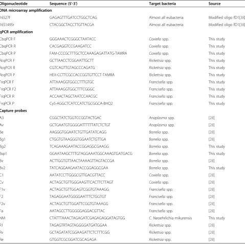
The sequences of all oligonucleotides and the probes used in this study are listed in Table 1.
PCR amplification for DNA microarray
The 16S rDNA (rrs) target genes of the targeted bacteria were amplified by nested PCR. The first cycle used 3 μl of genomic DNA, 1× high yield buffer complete with 2 mM MgCl2(Jena Bioscience, Germany), 200μM of each dNTP, 1μM of 16S27f (forward) and 16S1495r (reverse) primers (Table 1) and 1U Taq Pol (Jena Bioscience, Germany). The primers 16S27f and 16S1495r are slightly modified fD1 and rP2 general eubacterial primers [38]. The second cycle was used to incorporate the fluorescent labeled Cy5-dUTP into the PCR product of the first amp-lification. The incorporation was performed as
recom-mended by the manufacturer. Thus, the total 20 μl
reaction volume contained 1× high yield buffer complete with 2 mM MgCl2(Jena Bioscience, Germany), 100μM of
dATP, dCTP, dGTP, 50 μM of dTTP, 50 μM Cy5-dUTP
(Jena Bioscience, Germany), 0.5μM 16S27f and 16S1495r primers (Table 1) and 1U Taq Pol (Jena Bioscience, Germany). 1 μl of the PCR product from the first cycle was used as the template for the second cycle. The cycling conditions in both PCRs were the same. The initial de-naturation was performed for 2 minutes at 94°C followed by 30 cycles of 94°C for 30 seconds, 52°C for 30 seconds, 72°C for 1 minute and 30 seconds. The program ended with final elongation at 72°C for 5 minutes.
PCR amplification for quantitative PCRs (qPCRs)
TaqMan probes for qPCRs were synthetized by Microsynth AG, Austria. The CbPr probe was covalently bound at the 5′end with a FAM fluorophore and at the 3′end with a TAMRA quencher; the RLOqPCRPr probe was covalently bound at the 5′end with a HEX fluorophore and at the 3′ end with a TAMRA; and the FrqPCRPr probe was cova-lently bound with Cy5 at the 5′end and BHQ-2 at the 3′ end. The cycling conditions for all qPCRs were the same. The initial denaturation was performed for 2 minutes at 95°C, followed by 40 cycles at 95°C for 25 seconds and 50°C for 1 minute. The reaction mixture consisted of 300 nM of forward and reverse primes, 200 nM dual-labeled probes, 1×TaqMan Master Mix (Bioron, Germany), 4 mM MgCl2and 5μl of template DNA.
DNA microarray preparation and scanning
room temperature in 55–70% relative humidity and stored overnight at room temperature. All capture probes were spotted onto slides in triplicate. These prefabricated slides were blocked in a prehybridization solution (5× SSC, 0,1% SDS and 0.1 mg/ml BSA) at 42°C for 1 hour, washed 3 times in 0.1× SSC for 5 minutes and once more in purified water for 30 seconds. After washing, the slides were dried by centrifugation at 1 600 × g for 2 minutes. The Cy5-labelled target PCR products from the nested PCRs were diluted in a hybridization solution consisting of 5× SSC, 10% formamide, 0.1% SDS and 0.1 mg/ml of sonicated sal-mon sperm DNA, denaturated for 5 minutes in boiling
[image:4.595.58.538.100.577.2]water, shortly spun down, and cooled to room temperature. The target PCR products were pipetted onto microarray slides and covered with cover slips. The hybridization was performed at 42°C for 12–16 hours. After hybridization, the microarray slides were immersed in 2× SSC and 0.1% SDS at 42°C to gently release the cover slips from the slides and washed again for 5 minutes in 2× SSC and 0.1% SDS at 42°C. The final washing consisted of two washes in 1× SSC for 2 minutes at room temperature and two washes in 0.1× SSC for 1 minute at room temperature. The slides were dried by centrigufation at 1 600 × g for 2 minutes and scanned at 635 nm on a MARs Micro Array Scanner Table 1 Nucleotide sequences of PCR primers and probes
Oligonucleotide Sequence (5′-3′) Target bacteria Source DNA microarray amplification
16S27f GAGAGTTTGATCCTGGCTCAG Almost all eubacteria Modified oligo fD1[38]
16S1495r CTACGGCTACCTTGTTACGA Almost all eubacteria Modified oligo fD1[38]
qPCR amplification
CbqPCR F GGGAAACTCGGGCTAATACC Coxiellaspp. This study
CbqPCR R CACGAGGTCCGAAGATCC Coxiellaspp. This study
CbqPCR P FAM-CCCGCTTTGCTCCAAAGAGATTATG-TAMRA Coxiellaspp. This study
RcqPCR F GCTTAACCTCGGAATTGCTT Rickettsiaspp. This study
RcqPCR R CGTCAGTTGTAGCCCAGATG Rickettsiaspp. This study
RcqPCR P HEX-CCTTCGCCACCGGTGTTCCT-TAMRA Rickettsiaspp. This study
FrqPCR F ATTAAAGGTGGCCTTTGTGC Francisellaspp. This study
FrqPCR F2 ATTAAAGGTGGCTTTCGGGC Francisellaspp. This study
FrqPCR R ACCAACTAGCTAATCCAACGC Francisellaspp. This study
FrqPCR P Cy5-AGGCTCATCCATCTGCGGCA-BHQ2 Francisellaspp. This study
Capture probes
A3 CGGCTATCTGGTCCGGTACTGAC Anaplasmaspp. [28]
Av GCTGAATGTGGGGATTTTTTATCTCTGT Anaplasmaspp. [28]
Be AAGGGTGGAATCTGTTGATATCAGG Borreliaspp. [28]
Bg1 CTGGTGTAAGGGTGGAATCTGTTGA Borreliaspp. [28]
Bg2 TCAGAAAGAATACCGGAGGCGAAGG Borreliaspp. This study
Bsp1 GGAATAAGCTTTGTAGGAAATGGCAAAGTGATGACG Borreliaspp. This study
Bv ACTTGGTGTTAACTAAAAGTTAGTACCGA Borreliaspp. [28]
Bv2 TATCAGGAAGAATACCGGAGGCGAA Borreliaspp. This study
C1 AATATCCTTGGGCGTTGACGTTACC Coxiellaspp. [28]
Cv ACTAGCTGTTGGGAAGTTCACTTCTTAGT Coxiellaspp. [28]
F1v ACTAGCTGTTGGAGTCGGTGTAAAGG Francisellaspp. [28]
F2 TAGAGGAATGGGGAATTTCTGGTGT Francisellaspp. [28]
F2v ACTAGCTGTTGGATTCGGTGTAAAGG Francisellaspp. [28]
Fa AATAGCCTTGGGGGAGGACGTTAC Francisellaspp. [28]
NM CTATTTAAACTAGAGATCGAGAGAGGATAGTGG C.Neoehrlichia mikurensis This study
R1 TAGAGTRTAGTAGGGGATGATGGAA Rickettsiaspp. [28]
Rv GCTAGATATCGGAAGATTCTCTTTCGG Rickettsiaspp. [28]
(DITABIS - Digital Biomedical Imaging Systems AG, Germany) with SpotScout Pro at 50 μm resolution. The fluorescence intensity for a given spot is represented by the mean feature pixel intensity at 635 nm minus the me-dian background intensity at this wavelength (F635 Mean– B635). Since all slides were spotted in triplets, the reported measurements are the mean values of three measurements. An“M probe”consisting of mixed PCR fragments from the first PCRs was used as a positive control (details were previ-ously published in Blaškovičand Barák [28]. M probe frag-ments were spotted onto the microarray plates in three places as capture probes. This was done in triplicate. In addition to their use as a positive control, the M probes also aided in grid location during the scanning and made it pos-sible to performing the final negative sample evaluation. The lower border of a saturated spot was defined as 25% of the saturated spot area. All fluorescence intensity values were normalized with respect to the M probe and are reported as percentages of the M probe intensity (defined to be 100%).
Determination of the limit of detection (LOD)
The concentration of genomic DNA isolated from the targeted tick-borne bacteria was quantified using a GeneQuant spectrophotometer (LABFISH, Germany) and the number of copies perμl was calculated using an on-line DNA copy number calculator [41]. The DNA was diluted to a starting concentration of 105copies/μl for Coxiella spp. and Rickettsia spp. and 104 copies/μl for Francisellaspp. This starting solution was then seri-ally diluted 10-fold to prepare a series of solutions from 105or 104copies of genomic DNA (gDNA) perμl down to 1 copy/μl (that is, there were six dilutions: 105
, 104, 103, 102, 101, and 100). To determine the microarray LOD, 1μl of these diluted DNAs were used as templates for the amplifications of therrsgene and the products of these first PCRs were Cy5-labeled using nested PCRs as described above. The labeled amplicons were then hybridized with the prefabricated microarray and scanned and evaluated.
A very similar approach was used to determine the de-tection limit of qPCR. The same DNA dilutions were used as the templates and genus specific oligonucleotides and probes were used to amplify therrsgenes.
The highest target DNA dilution which still returned a positive result was determined the detection limit of the DNA microarray or qPCR. Three independent experi-ments were run for each dilution series. Each qPCR ex-periment consisted of one of the target gDNA dilutions and a mix of non-target gDNAs as negative control. To determine the detectable copy number, an absolute quantification method was employed. The mean quanti-fication cycle (Cq) was converted to a log starting quan-tity using a linear equation derived from the standard curves.
Results
Development of a DNA microarray for the detection of tick-borne pathogens
A DNA microarray is an efficient and simple tool for detecting a wide spectrum of tick-borne pathogens in a single step. A first generation DNA microarray for detecting tick-borne bacteria was developed previously by Blaškovičand Barák [28]. In that study, the amplifica-tion of target DNA by symmetric/asymmetric PCR appeared to be quite complicated. The authors were not able to obtain a single stranded amplicon using asym-metric PCR when DNA from B. burgdorferi,C. burnetii and R. africae was used as a template. They therefore recommended using symmetric PCR for the amplifica-tion of all targets involved in the study. The limit of de-tection (LOD) was also not determined for this assay.
The first steps for upgrading this DNA microarray were to improve the amplification of the 16S rDNA (rrs) gene to enable detection of all bacteria present in the analyzed samples and to increase the efficiency of Cy5-dUTP in-corporation into the PCR products. A nested PCR was designed whose first cycle was used to amplify the target gene in high yield. In the second cycle, Cy5-dUTP was in-corporated into the PCR amplicon. Crucial for the success of this PCR was the selection of a Taq-polymerase, which was able to both efficiently incorporate modified nucleo-tides into PCR fragments and to amplify the gene in high yield. Several types of Taq-polymerases, including DyNAzyme EXT DNA Polymerase, Taq DNA Polymerase, (Thermo Fisher Scientific, USA),TaqDNA Polymerase with Stand-ard Taq (Mg-free) Buffer (New England Biolabs, USA), and many cycling conditions were tested (not shown). The best Taq-polymerase appeared to be Taq Polymerase/ high yield from Jena Bioscience (Germany). The final annealing temperature was 52°C. In both nested PCR cy-cles, the same, so-called “catch-all” primers 16S27f and 16S1495r were used. These primers are slightly modified versions of the previously published eubacterial primers fD1 and rP2 [38]. 16S27f is the same as fD1 but with an additional G at the 5′ end. Modification of rP2 included CT addition to the 5′end of 16S1495r and truncation of three nucleotides from the 3′ end (Table 1). The Cy5-labeled PCR products were precipitated in the presence of ammonium sulphate and ethanol [42] and the pellets were resuspended in the required volume of hybridization solu-tion (see Methods).
Coupling of target Cy5-labeled amplicons with capture probes
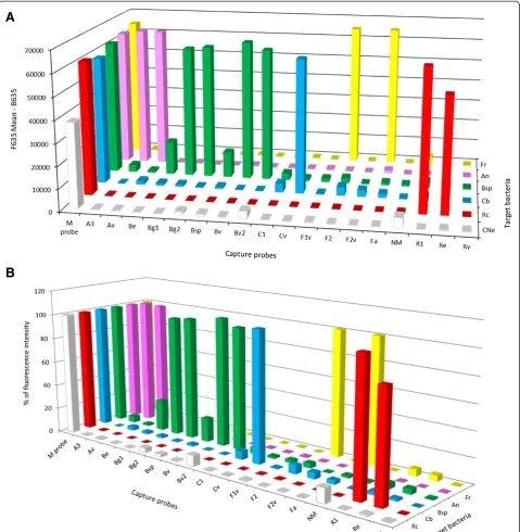
Even though the hybridization conditions and post-hybridization washes had been changed in the present study, they still reacted specifically. To increase the ability of the assay to detect the targeted bacteria, new capture probes for Borrelia spp. (Bg2, Bsp, Bv2) andCandidatus N. mikurensis (NM) were designed (see Table 1). All cap-ture probes were printed onto epoxy slides in triplets in final concentrations of 30μM. A Cy5-labeled PCR prod-uct encoding the 16S rDNA (rrs) ofBorreliaspp.,Coxiella spp.,Anaplasma spp.,Rickettsiaspp. andFrancisellaspp. was generated by amplifying purified DNA from bacterial stocks. These Cy5-amplicons were then hybridized with the capture probes. Cy5-amplicons encoding the 16S rDNA ofCandidatusN. mikurenses were generated using DNA isolated from ticks. The positivity of the tick for Candidatus N. mikurenses was analyzed in a previous study by qPCR [43].
The ability of the individual capture probes to bind the same target DNA differs depending on the status of the Cy5-amplicons or the quality of the spotted probes or epoxy slides. Our results clearly show that the coup-ling of capture probes with Cy5-PCR fragments gener-ated from DNA isolgener-ated from ticks was not as efficient as that with Cy5-PCR amplicons generated from purified DNA. Thus, the measured fluorescence intensity of the M probe (positive control) and positive capture probes had to be normalized after scanning. The fluorescence intensity is expressed as the mean feature pixel intensity of the positive spot at 635 nm minus the median
back-ground at 635 nm (F635 Mean–B635).
The fluorescence intensity of the M probe and the positive capture probes for Coxiella spp., Borrelia spp., Francisella spp., Rickettsia spp. and Anaplasma spp., gave much stronger positive signals than those obtained after the hybridization of a Cy5-labeled amplicon with DNA isolated from a tick coinfected with Borrelia spp. and CandidatusN. mikurensis (Figure 1A). In order to compare these different fluorescence values, the fluores-cence intensity of each positive signal was expressed as a percentage of the fluorescence of the M probe. The DNA microarray forCandidatusN. mikurensis revealed a possible co-infection with Borrelia spp. but the per-centage of fluorescence intensities was quite low, only 12% and 9% respectively of the fluorescence intensity of the M probe. Such a low signal was considered only am-biguously positive and required further analysis.
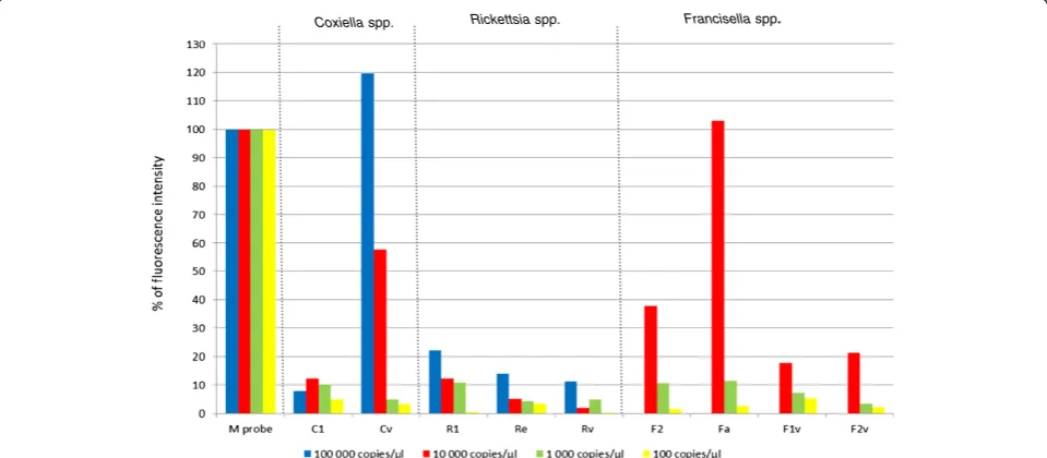
To determine if a sample with such a low fluorescence tests positive or negative, it was necessary to determine the limit of detection (LOD) of the DNA microarray. To do this, a series of 10-fold dilutions, ranging from 105 genome copies per μl down to 1 copy/μl, was prepared from DNA of the targeted tick-borne bacteria. These di-lutions were then used as templates for nested PCRs and DNA microarrays. The fluorescence intensities of the
capture probes were compared to that of the M probe (Figure 2). The fluorescence intensity of the Cv capture probe specific forCoxiellaspp. was 120% when 105
cop-ies were used as template DNA, 12% when 104 copies
and 10% when 103copies were used. 102copies of tem-plate DNA apparently did not bind the capture probes and no fluorescence intensity could be measured; the 10 copies and 1 copy dilutions were also negative. So, the maximal dilution of template DNA, which is detectable by the DNA microarray is 103 copies/μl, which gives a fluorescence intensity 10% of that of the M probe. Therefore, a sample should be evaluated as positive when the mean fluorescence intensity is at least 10% of the mean fluorescence intensity of the M Probe. Thus, sam-ples with values close to this cut off limit will require other analyses, such as some previously published qPCR proto-cols [20,43], to verify their results. An analysis using the protocols of Courtney et al. [20] and Jahfari et al. [43] confirmed that the signal at the 12% level for the Candidatus N. mikurensis specific probe, as well as the signal at 9% for theB.burgdorferis.l. probe both represent positive samples (data not shown).
The specificity of the DNA microarray was tested using mixed genomic DNAs from many bacterial spe-cies, including DNA from tick-borne pathogens, but excluding the target DNA for which the chip was designed. No unspecific cross-reactivity between the capture probes and genomic DNAs was detected (data not shown).
Development of qPCR based on the variability ofrrs specific for detection ofFrancisellaspp.,Rickettsiaspp. andCoxiellaspp.
FTT0523 real-time PCRs detect only the pathogenic but not the nonpathogenic Francisella subspecies, this ap-proach is not suitable as a confirmatory method for our DNA microarray analysis, since all Francisella species is our primary target. For this reason, we developed a qPCR employing the 16S rDNA (rrs) gene in order to detect all
[image:7.595.59.539.89.580.2]Francisella species, and two other qPCRs, to detect the Rickettsia spp. and Coxiella spp. The primers and oligo-nucleotide dual-labeled probes were designed using GenScript Real-time PCR (TaqMan) Primer Design online software [45] based on the alignment of therrs genes of all tick-borne bacteria of interest. These probes and Figure 1Specificity of capture probes and target bacteria detected by the DNA microarray. (A)Fluorescence intensity at 635 nm
primers are specific for the hypervariable region of therrs gene, which are different for every genus (Table 1). Their specificities were tested against mixed genomic DNA from all possible tick-borne bacteria and other bacterial gen-omic DNA present in our laboratory stocks.
Because of the slight variation in the hypervariable re-gion of the Francisella spp. rrs sequences, two forward primers were designed. Forward primer FrqPCRF prefer-entially binds F. piscicidaand F. philomiragia, while for-ward primer FrqPCRF2 is specific forF. tularensissubsp. holarctica, mediaasiatica, tularensisandnovicida. All qPCRs were specific when tested against mixed genomic DNA from other bacteria.
Determination of qPCR detection limit
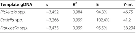
Limits of detection for all three qPCRs were determined based on the maximum dilution of genomic DNA from the target bacteria which still tested positive. To develop a qPCR specific for Coxiella spp. and Rickettsia spp., their genomic DNAs were diluted in a series of 10-fold dilutions, from 105 copies/μl to 1 copy/μl. Due to the low concentration of genomic DNA inFrancisellaspp., the starting copy number of the series was only 104copies/μl. The absolute quantifications of the detectable genome copy number from Francisella spp., Coxiella spp. and Rickettsia spp. is shown in Figure 3. The standard curve amplification efficiencies (E), regression coefficients (R2), slopes (s) andy-intercept (y-int) are listed in Table 2. The amplification efficiencies of all qPCRs were 95%, 102% and 96% forRickettsiaspp.,Coxiella spp. andFrancisella
spp., respectively. The sensitivity of qPCR forRickettsiaspp. was determined to be 102 genome copies; for Coxiella spp., 10 genome copies and for Francisella spp., only 1 genome copy. Considering the mean sizes of the genomes (2.2 Mb for Coxiella spp., 2 Mb forFrancisella spp. and 1.2 Mb for for Rickettsia spp.), the copy numbers deter-mined for the LOD correspond to approximately 22 fg of gDNA forCoxiellaspp., 2.1 fg gDNA forFrancisellaspp. and 140 fg gDNA forRickettsiaspp.
Discussion
Broad epidemiological studies of veterinary or human importance and clinical diagnostic laboratories require the application of high-throughput, large scale assays allowing the simultaneous detection of all possible mi-croorganisms present in a given sample. Such methods can involve broad range PCRs, multiplex quantitative PCR, molecular beacons or DNA microarrays. Usually these methods employ universal genes, such as the 16S
rDNA rrs gene, the 23S rDNA gene, or, occasionally,
[image:8.595.58.538.91.301.2]genes specific for each bacterial genus or species identi-fied in previous studies. In the last decade, DNA microarrays have become one of the most powerful ap-proaches for the simultaneous detection of several bac-terial species. They have many possible applications, including identifying bioterror agents [46] and detecting causative pathogens in clinical samples or epidemio-logical studies [31,47,48]. In clinical diagnostics, it is es-sential to know all possible co-infections of the patient in order to consider all potential complications, and thus Coxiella spp. Rickettsia spp. Francisella spp.
Figure 2The DNA microarray limit of detection (LOD).LOD forRickettsiaspp.,Coxiellaspp., andFrancisellaspp. was determined as the highest dilution of genomic DNA that still tested as positive. 103genome copies exhibited 10% of the fluorescence intensity of the M probe,
while 102genome copies produced no detectible signal; the LOD was therefore determined to be 103copies and 10% of the positive control. All
prescribe an effective treatment. Along with other methods, DNA microarrays allow the detection of all agents of a multiple infection in one step.
The major focus of this study was to develop an easy to use, second generation low-density DNA microarray for the simultaneous detection of the many kinds of bac-teria present in tick samples. This technique has poten-tial applications in clinical and veterinary laboratories and is also suitable for broad epidemiological studies. The DNA microarray consists of genus-specific capture probes for Borreliaspp.,Coxiella spp., Anaplasma spp., Francisella spp. and Rickettsia spp. that were designed for the first generation DNA microarray [28] along with new capture probes Bsp, Bg2 and Bv2 which were designed to increase the possibility of detecting all Borrelia species. In addition, a specific NM probe was designed to detect the newly emerged tick-borne bacterium Candidatus N. mikurensis. The modified solution com-positions, hybridization conditions and post-hybridization washes did not affect specificity either of the original or the new capture probes. Capture probe specificity was analyzed by hybridization with Cy5-dUTP labeled PCR fragments generated by PCR on a template containing mixed genomic DNA from other, non-target bacteria.
The sensitivity of the first generation DNA microarray was not tested [28], and thus it was necessary to test the
sensitivity of the second generation DNA microarray by determining the limit of detection (LOD). The LOD of our second generation DNA microarray was determined to be the highest dilution of the target genomic DNA that still tested as positive. The LOD was determined to be 103target genome copies based on hybridization with specific capture probes at different dilutions. Given the mean genome sizes of the targeted bacteria, the limit of detection is about ~ 1–2 pg of genomic DNA. This sen-sitivity is comparable to that of the low-density DNA microarrays developed to detect tick-borne bacteria such asBorreliaspp. [31] or other array techniques developed to detect potential biological weapons, with detection limits ~102-104target genome copies [29,30].
Differences were observed in the fluorescence signal intensities produced by the DNA microarray, depending on whether the target DNA was directly purified from bacteria, or was isolated from an infected tick. These dif-ferences are likely due to the presence of junk DNA from the tick and a low concentration of the target bac-terial DNA. The crucial steps of PCR amplification and Cy5-labeling on such targets are also more complicated. Both of these factors can lead to a low level of fluores-cence intensity following hybridization, making inter-pretation of a positive signal difficult. This situation was observed when genomic DNA isolated from a tick
co-infected with Candidatus N. mikurensis and Borrelia
[image:9.595.57.540.88.282.2]spp. was used as a template for DNA microarray ana-lysis. The mean fluorescence of the capture probes was at the detection limit. It was therefore necessary to develop qPCRs using dual labeled TaqMan probes (Table 1). Since duplex qPCR protocols have been devel-oped to detect A. phagoctytophilum and B. burgdorferi Figure 3Absolute quantification of the detectable genome copy numbers from tick-borne bacteria.Quantitative PCRs were developed forFrancisellaspp. (blue curve),Coxiellaspp. (red curve) andRickettsiaspp. (green curve). The trendlines and R2values were generated using
Microsoft Excel based on the average of the cycle of quantification values (Cq) and the genome copy numbers.
Table 2 The parameters of the standard curves of qPCRs
Template gDNA s R2 E Y-int
Rickettsiaspp. −3,452 0,984 94,8% 46,75
Coxiellaspp. −3,266 0,999 102,4% 41,2
[image:9.595.56.292.676.734.2]s.l. [20], and real-time PCR protocols exist forCandidatus N. mikurensis [43], the presence or absence of these pathogens could be verified. Quantitative PCR protocols were developed to detect Rickettsia spp., Coxiella spp. and Francisella spp. Oligonucleotide probes were designed according to the hypervariable regions of the rrs gene, which are specific for each bacterial genus; both the specificity and sensitivity of the procedure were evaluated. As for the DNA microarray, the qPCR specifi-city was determined by amplifying the target gene on a template containing mixed genomic, non-target DNAs other than the genomic DNA of the target bacteria. No cross-reactivity with any of these gDNAs was observed. To test the sensitivity of each qPCR, a series of 10-fold dilutions of the target genomic DNAs from Rickettsia spp.,Coxiellaspp. and Francisellaspp. were employed in the amplification, with the final efficiencies of 95%, 102% and 96%, respectively. The highest dilution that was evalu-ated as positive was 102 genome copies from Rickettsia spp., 10 genome copies fromCoxiellaspp. and only 1 gen-ome copy fromFrancisellaspp.; these corresponded to ap-proximately 140 fg ofRickettsiaspp. genomic DNA, 22 fg of Coxiella spp. genomic DNA and 2.1 fg of Francisella spp. genomic DNA. The limits of detection for all three qPCRs assays were at the breakpoint of qPCR detection and were very similar to those of multiplex qPCR for C. burnetii targeted to thecom1, icdandIS1111 genes [49], real-time PCR forF. piscicidatargeted to therrsgene [39], and qPCR developed to detect a Rickettsia-like micro-organism, which is responsible for strawberry disease in fish [40]. The qPCRs for the detection of all three path-ogens appeared to be more sensitive than the DNA microarray. The qPCR for the detection of Francisella spp. was more sensitive than that of eitherCoxiellaspp. or Rickettsia spp. This may be due to the existence of three copies of therrsgene in the Francisellagenomes present in the Ribosomal RNA Operon Copy Number Database [50] compared to only one copy of therrsgene inRickettsia prowazekii[51] andC. burnetii[52].
It should be noted that many ticks harbor non-pathogenic bacteria, including Coxiella-like [53-56], Rickettsia-like [57-59] andFrancisella-like [60,61] endosymbionts, which are in a mutualistic relationship with the tick. These pri-mary endosymbionts can provide nutrition to the host [62]. They likely evolved over a long time and they are characterized with a reduced genome [63]. However, they still retain ribosomal RNA genes which have a relatively high level of similarity to those found in pathogenic or-ganisms. For example, the rrs gene of the Amblyoma -associated,Coxiella-like endosymbiont has 93% indentity to theC. burnetii rrs[64]. The role of the secondary endo-symbionts is unknown, but they can serve as protection against other pathogens [65]. The DNA microarray devel-oped here, together with the qPCRs, is targeted to the
hypervariable regions ofrrsgenes. A positive signal gener-ated using this approach therefore does not necessarily in-dicate that the host vector contains a pathogenic bacteria. A sequence analysis of the final result is needed to dis-tinguish between endosymbiont and pathogen in these samples.
Conclusion
We have developed a sophisticated detection system for the simultaneous detection of bacteria present in reser-voir hosts, tick-vectors, and clinical specimens, based on a second generation DNA microarray employing the genus-specific, hypervariable regions of the rrs gene. These hypervariable sequences were used to design cap-ture probes for the DNA microarray as well as primers and TaqMan probes for qPCRs. The qPCRs can be used to verify the positive results from the DNA microarray. Both methods display a high level of specificity and sen-sitivity. The limit of detection for the DNA microarray was 103genome copies. Quantitative PCRs were devel-oped for Rickettsia spp, Coxiella spp. and Francisella spp. and the limits of detection were determined. Finally, previously developed and published qPCR procedures are available for the verification of presence of the other bacteria involved in this study.
Competing interests
The authors declare that they have no competing interests.
Authors’contributions
The study was designed by JM and IB. The DNA samples were provided by MD. JM and MD performed laboratory experiments. JM and IB conducted genetic analysis and evaluated microarray and qPCR data. The final manuscript was written by JM, IB and MD. All authors read and approved the final version of the manuscript.
Acknowledgements
We thank Jacob Bauer for helpful comments. This publication is the result of the project implementation: Development of diagnostic procedures for detection of tick-borne pathogens and procedures for the preparation of vaccines against ticks (ITMS code: 26240220044) supported by the Research & Development Operational Programme funded by the ERDF and by the Slovak Research and Development Agency under contract No. APVV–0267–10.
Author details
1Institute of Molecular Biology, Slovak Academy of Sciences, Dúbravska cesta
21, 845 51, Bratislava, Slovak Republic.2Institute of Zoology, Slovak Academy
of Sciences, Dúbravska cesta 9, 845 06, Bratislava, Slovak Republic.3Institute
of Parasitology, Slovak Academy of Sciences, Hlinkova 3, 040 01, Košice, Slovak Republic.
Received: 1 August 2013 Accepted: 12 September 2013 Published: 18 September 2013
References
1. Franke J, Hildebrandt A, Dorn W:Exploring gaps in our knowledge on Lyme borreliosis spirochaetes–updates on complex heterogeneity, ecology, and pathogenicity.Ticks Tick Borne Dis2013,4:11–25.
3. Gern L:Borrelia burgdorferisensu lato, the agent of lyme borreliosis: life in the wilds.Parasite2008,15:244–247. Review.
4. Margos G, Vollmer SA, Ogden NH, Fish D:Population genetics, taxonomy, phylogeny and evolution ofBorrelia burgdorferisensu lato.Infect Genet Evol2011,11:1545–1563.
5. Bunikis J, Tsao J, Garpmo U, Berglund J, Fish D, Barbour AG:Typing of Borreliarelapsing fever group strains.Emerg Infect Dis2004,10:1661–1664. 6. Dumler JS, Barbet AF, Bekker CPJ, Dasch GA, Palmer GH, Ray SC, Rikihisa Y,
Rurangirwa FR:Reorganization of Genera in the familiesRickettsiaceae andAnaplasmataceaein the orderRickettsiales; unification of some species ofEhrlichiawithAnaplasma,CowdriawithEhrlichia, andEhrlichia withNeorickettsia; description of six new species combinations; and designation ofEhrlichia equiand“HGE agent”as subjective synonyms of Ehrlichia phagocytophilum.Int J Syst Evol Microbiol2001,51:2145–2165. 7. Woldehiwet Z:The natural history ofAnaplasma phagocytophilum.Vet
Parasitol2010,167:108–122.
8. Kawahara M, Rikihisa Y, Isogai E, Takahashi M, Misumi H, Suto C, Shibata S, Zhang C, Tsuji M:Ultrastructure and phylogenetic analysis of‘Candidatus Neoehrlichia mikurensis’in the familyAnaplasmataceae, isolated from wild rats and found inIxodes ovatusticks.Int J Syst Evol Microbiol2004,
54:1837–1843.
9. Welinder-Olsson C, Kjellin E, Vaht K, Jacobsson S, Wennerås C:First case of human“CandidatusNeoehrlichia mikurensis”infection in a febrile patient with chronic lymphocytic leukemia.J Clin Microbiol2010,48:1956–1959. 10. Fehr JS, Bloemberg GV, Ritter C, Hombach M, Lüscher TF, Weber R, Keller
PM:Septicemia caused by tick-borne bacterial pathogenCandidatus Neoehrlichia mikurensis.Emerg Infect Dis2010,16:1127–1129.
11. Pekova S, Vydra J, Kabickova H, Frankova S, Haugvicova R, Mazal O, Cmejla R, Hardekopf DW, Jancuskova T, Kozak T:CandidatusNeoehrlichia mikurensis infection identified in 2 hematooncologic patients: benefit of molecular techniques for rare pathogen detection.Diagn Microbiol Infect Dis2011,69:266–270.
12. Von Loewenich FD, Geissdörfer W, Disqué C, Matten J, Schett G, Sakka SG, Bogdan C:Detection of“CandidatusNeoehrlichia mikurensis”in two patients with severe febrile illnesses: evidence for a European sequence variant.J Clin Microbiol2010,48:2630–2635.
13. Sjöstedt A:Tularemia: history, epidemiology, pathogen physiology, and clinical manifestations.Ann N Y Acad Sci2007,1105:1–29.
14. Rath N, Rath A:Rickettsial Infections: Indian perspective.Indian Pediatr 2010,47:157–164.
15. Beati L, Péter O, Burgdorfer W, Aeschlimann A, Raoult D:Confirmation that Rickettsia helveticasp. nov. is a distinct species of the spotted fever group of rickettsiae.Int J Syst Bacteriol1993,43:521–526.
16. Beati L, Meskini M, Thiers B, Raoult D:Rickettsia aeschlimanniisp. nov., a new spotted fever group rickettsia associated withHyalomma marginatumticks.Int J Syst Bacteriol1997,47:548–554.
17. Cowan G:Rickettsial diseases: the typhus group of fevers–a review. Postgrad Med J2000,76:269–272.
18. La Scola B, Raoult D:Laboratory diagnosis of rickettsioses: current approaches to diagnosis of old and new rickettsial diseases.J Clin Microbiol1997,35:2715–2727.
19. Li H, Jiang JF, Liu W, Zheng YC, Huo QB, Tang K, Zuo SY, Liu K, Jiang BG, Yang H, Cao WC:Human Infection withCandidatusNeoehrlichia mikurensis, China.Emerg Infect Dis2012,18:1636–1639.
20. Courtney JW, Kostelnik LM, Zeidner NS, Massung RF:Multiplex real-time PCR for detection ofAnaplasma phagocytophilumandBorrelia burgdorferi.J Clin Microbiol2004,42:3164–3168.
21. Rymaszewska A:PCR for detection of tick-borneAnaplasma phagocytophilumpathogens: a review.Vet Med2011,56:529–536. 22. Marmion BP, Storm PA, Ayres JG, Semendric L, Mathews L, Winslow W,
Turra M, Harris RJ:Long-term persistence ofCoxiella burnetiiafter acute primary Q fever.QJM2005,98:7–20.
23. Subramanian G, Sekeyova Z, Raoult D, Mediannikov O:Multiple tick-associated bacteria inIxodes ricinusfrom Slovakia.Ticks Tick Borne Dis2012,3:406–410. 24. Postic D, Assous M, Grimont PAD, Baranton G:Diversity ofBorrelia
burgdorferisensu lato evidenced by restriction fragment length polymorphism ofrrf (5S)-rrl(23S)intergenic spacer amplicons.Int J Syst Bacteriol1994,44:743–752.
25. Derdakova M, Beati L, Pet’ko B, Stanko M, Fish D:Genetic variability within Borrelia burgdorferisensu lato genospecies established by PCR-single -strand conformation polymorphism analysis of therrfA-rrlBintergenic
spacer inIxodes ricinusticks from the Czech Republic.Appl Environ Microbiol2003,69:509–516.
26. Doolittle WF:Phylogenetic classification and the universal tree.Science 1999,284:2124–2129.
27. Small J, Call DR, Brockman FJ, Straub TM, Chandler DP:Direct detection of 16S rRNA in soil extracts by using oligonucleotide microarrays.Appl Environ Microbiol2001,67:4708–4716.
28. Blaskovic D, Barák I:Oligo-chip based detection of tick-borne bacteria. FEMS Microbiol Lett2005,243:473–478.
29. Wilson WJ, Erler AM, Nasarabadi SL, Skowronski EW, Imbro PM:A multiplexed PCR-coupled liquid bead array for the simultaneous detection of four biothreat agents.Mol Cell Probes2005,19:137–144. 30. Deshpande A, Gans J, Graves SW, Green L, Taylor L, Kim HB, Kunde YA,
Leonard PM, Li PE, Mark J, Song J, Vuyisich M, White PS:A rapid multiplex assay for nucleic acid-based diagnostics.J Microbiol Methods2010,
80:155–163.
31. Houck JA, Hojgaard A, Piesman J, Kuchta RD:Low-density microarrays for the detection ofBorrelia burgdorferis.s. (the Lyme disease spirochete) in nymphalIxodes scapularis.Ticks Tick Borne Dis2011,21:27–36.
32. Barbaro M, Bonfiglio A, Raffo L, Alessandrini A, Facci P, Barák I:Fully electronic DNA hybridization detection by a standard CMOS biochip. Sens Actuators B2006,118:41–46.
33. Barbaro M, Bonfiglio A, Raffo L, Alessandrini A, Facci P, Barák I:A CMOS, fully integrated sensor for electronic detection of DNA hybridization.IEEE Electron Device Letters2006,27:595–597.
34. Barlaan EA, Sugimori M, Furukawa S, Takeuchi K:Electronic microarray analysis of 16S rDNA amplicons for bacterial detection.J Biotechnol2005,
115:11–21.
35. Cole JR, Chai B, Marsh TL, Farris RJ, Wang Q, Kulam SA, Chandra S, McGarrell DM, Schmidt TM, Garrity GM, Tiedje JM:The Ribosomal Database Project (RDP-II): previewing a new autoaligner that allows regular updates and the new prokaryotic taxonomy.Nucleic Acids Res2003,31:442–443. 36. Integrated DNA Technologie’s OligoAnalyzer 3.1.http://eu.idtdna.com/
analyzer/applications/oligoanalyzer/default.aspx.
37. Basic Local Alignment Search Tool.http://blast.ncbi.nlm.nih.gov/Blast.cgi? PROGRAM=blastn&BLAST_PROGRAMS=megaBlast&PAGE_TYPE=BlastSearch &SHOW_DEFAULTS=on&LINK_LOC=blasthome.
38. Weisburg WG, Barns SM, Pelletier DA, Lane DJ:16S ribosomal DNA amplification for phylogenetic study.J Bacteriol1991,173:697–703. 39. Ottem KF, Nylund A, Isaksen TE, Karlsbakk E, Bergh Ø:Occurrence of
Francisella piscicidain farmed and wild Atlantic cod, Gadus morhua L., in Norway.J Fish Dis2008,31:525–534.
40. Lloyd SJ, LaPatra SE, Snekvik KR, Cain KD, Call DR:Quantitative PCR demonstrates a positive correlation between aRickettsia-like organism and severity of strawberry disease lesions in rainbow trout,
Oncorhynchus mykiss (Walbaum).J Fish Dis2011,34:701–709. 41. DNA copy number calculation.http://www.thermoscientificbio.com/
webtools/copynumber/.
42. Sambrook J, Fritsch EF, Maniatis T:Molecular Cloning: A laboratory manual. 2nd edition. Cold Spring Harbor, NY: Cold Spring Harbor Laboratory Press; 1989.
43. Jahfari S, Fonville M, Hengeveld P, Reusken C, Scholte EJ, Takken W, Heyman P, Medlock J, Heylen D, Kleve J, Sprong H:Prevalence of Neoehrlichia mikurensisin ticks and rodents from North-west Europe. Parasit Vectors2012,5:74.
44. Mitchell JL, Chatwell N, Christensen D, Diaper H, Minogue TD, Parsons TM, Walker B, Weller SA:Development of real-time PCR assays for the specific detection ofFrancisella tularensisssp.tularensis, holarcticaand mediaasiatica.Mol Cell Probes2010,24:72–76.
45. GenScript Real-time PCR (TaqMan) Primer Design.https://www.genscript.com/ ssl-bin/app/primer.
46. Janse I, Bok JM, Hamidjaja RA, Hodemaekers HM, van Rotterdam BJ:
Development and comparison of two assay formats for parallel detection of four biothreat pathogens by using suspension microarrays. PLoS One2012,7:e31958.
47. Garaizar J, Rementeria A, Porwollik S:DNA microarray technology: a new tool for the epidemiological typing of bacterial pathogens?FEMS Immunol Med Microbiol2006,47:178–189.
49. de Bruin A, de Groot A, de Heer L, Bok J, Wielinga PR, Hamans M, van Rotterdam BJ, Janse I:Detection ofCoxiella burnetiiin complex matrices by using multiplex quantitative PCR during a major Q fever outbreak in The Netherlands.Appl Environ Microbiol2011,77:6516–6523.
50. Ribosomal RNA Operon Copy Number Database.http://rrndb.umms.med. umich.edu/search.php.
51. Pang H, Winkler HH:Transcriptional analysis of the 16s rRNA gene in Rickettsia prowazekii.J Bacteriol1996,178:1750–1755.
52. Afseth G, Mallavia LP:Copy number of the 16S rRNA gene inCoxiella burnetii.Eur J Epidemiol1997,13:729–731.
53. Bernasconi MV, Casati S, Péter O, Piffaretti JC:Rhipicephalusticks infected withRickettsiaandCoxiellain Southern Switzerland (Canton Ticino). Infect Genet Evol2002,2:111–120.
54. Lee JH, Park HS, Jang WJ, Koh SE, Park TK, Kang SS, Kim BJ, Kook YH, Park KH, Lee SH:Identification of theCoxiellasp detected fromHaemaphysalis longicornisticks in Korea.Micro Immun2004,48:125–130.
55. Mediannikov O, Ivanov L, Nishikawa M, Saito R, Sidelnikov YN, Zdanovskaya NI, Tarasevich IV, Suzuki H:Molecular evidence ofCoxiella-like
microorganism harbored byHaemaphysalis concinnaeticks in the Russian Far East.Ann N Y Acad Sci2003,990:226–228.
56. Clay K, Klyachko O, Grindle N, Civitello D, Oleske D, Fuqua C:Microbial communities and interactions in the lone star tick,Amblyomma americanum.Mol Ecol2008,17:4371–4381.
57. Mattila JT, Burkhardt NY, Hutcheson HJ, Munderloh UG, Kurtti TJ:Isolation of cell lines and a rickettsial endosymbiont from the soft tickCarios capensis(Acari: Argasidae: Ornithodorinae).J Med Entomol2007,
44:1091–1101.
58. Burgdorfer W, Hayes SF, Mavros AJ:Non-pathogenic rickettsiae inD. andersoni, a limiting factor for the distribution ofRickettsia rickettsii.In Rickettsiae and rickettsial diseases.Edited by Burgdorfer W, Anacker RL. New York: Academic Press, Inc; 1981:585–594.
59. Noda H, Munderloh UG, Kurti TJ:Endosymbionts of ticks and their relationship toWolbachiaspp. and tick-borne pathogens of humans and animals.Appl Environ Microbiol1997,63:3926–3932.
60. Scoles G:Phylogenetic analysis of theFrancisella-like endosymbionts of Dermacentorticks.J Med Entomol2004,41:277–286.
61. Sun LV, Scoles GA, Fish D, O’Neill SL:Francisella-like endosymbionts of ticks.J Invertebr Pathol2000,76:301–303.
62. Dale C, Moran NA:Molecular interactions between bacterial symbionts and their hosts.Cell2006,126:453–465.
63. Nakabachi A, Yamashita A, Toh H, Ishikawa H, Dunbar HE, Moran NA, Hattori M:
The 160-kilobase genome of the bacterial endosymbiontCarsonella. Science2006,314:267.
64. Klyachko O, Stein BD, Grindle N, Clay K, Fuqua C:Localization and visualization of aCoxiella-type symbiont within the lone star tick, Amblyomma americanum.Appl Environ Microbiol2007,73:6584–6594. 65. Moran NA, Degnan PH, Santos SR, Dunbar HE, Ochman H:The players in a
mutualistic symbiosis: insects, bacteria, viruses, and virulence genes. Proc Natl Acad Sci USA2005,102:16919–16926.
doi:10.1186/1756-3305-6-269
Cite this article as:Melničákováet al.:A system to simultaneously detect tick-borne pathogens based on the variability of the 16S ribosomal genes.Parasites & Vectors20136:269.
Submit your next manuscript to BioMed Central and take full advantage of:
• Convenient online submission
• Thorough peer review
• No space constraints or color figure charges
• Immediate publication on acceptance
• Inclusion in PubMed, CAS, Scopus and Google Scholar
• Research which is freely available for redistribution