R E S E A R C H
Open Access
Yin Yang 1 contributes to gastric carcinogenesis
and its nuclear expression correlates with shorter
survival in patients with early stage gastric
adenocarcinoma
Wei Kang
1,2,3,4, Joanna HM Tong
1,2,3, Anthony WH Chan
1, Junhong Zhao
2, Yujuan Dong
2, Shiyan Wang
2,
Weiqin Yang
5, Frankie MC Sin
1,2,3, Simon SM Ng
6, Jun Yu
2,5, Alfred SL Cheng
2,7and Ka Fai To
1,2,3,4*Abstract
Background:Yin Yang 1 (YY1) is a transcription factor that regulates diverse biological processes and increasing recognized to have important roles in carcinogenesis. The function and clinical significance of YY1 in gastric adenocarcinoma (GAC) have not been elucidated.
Methods:In this study, the functional role of YY1 in gastric cancer was investigated by MTT proliferation assays, monolayer colony formation, cell cycle analysis, signaling pathway analysis, Western blot analysis andin vivostudy through YY1 knockdown or overexpression. Immunohistochemical study with YY1 antibody was performed on tissue microarray consisting of 247 clinical GAC samples. The clinical correlation and prognosis significance were evaluated.
Results:YY1 expression was up-regulated in gastric cancer cell lines and primary gastric cancers. Knocking down YY1 by siYY1 inhibited cell growth, inducing G1 phase accumulation and apoptosis. Ectopic YY1 expression enhanced cell proliferationin vitroandin vivo. Knocking down YY1 in gastric cancer cells suppressed proliferation by inhibiting Wnt/ β-catenin pathway, whereas its overexpression exerted oncogenic property by activating Wnt/β-catenin pathway. In primary GAC samples, YY1 nuclear expression correlated with shorter survival and predicted poor prognosis in early stage GACs.
Conclusion:Our data demonstrated that YY1 contributes to gastric carcinogenesis in gastric cancer. In early stage GACs YY1 might serve as a poor prognostic marker and possibly as a potential therapeutic target.
Keywords:Yin Yang 1, Gastric adenocarcinoma, Prognostic marker, Oncogenic function
Background
Gastric adenocarcinoma (GAC) is one of the most com-mon malignancies in the world, with high rates of inci-dence in countries such as China, Japan, and South Korea [1]. Currently, GAC is one of the leading causes of cancer-related death worldwide, accounting for approximately 740,000 cases of cancer-related death annually [2]. While various factors such as H. pylori infection, genetic,
epigenetic and molecular alterations affecting signaling pathways as well as genetic instability have been impli-cated in gastric tumorigenesis, the mechanisms of GAC pathogenesis are still largely unknown [3].
Yin Yang 1 (YY1) is a ubiquitously distributed tran-scription factor belonging to the Gli-Kruepple class of Zinc-finger proteins [4]. YY1 has diverse and complex bio-logical functions and is involved in both repression and activation of numerous genes that play essential roles in a multitude of biological processes [5]. For example, YY1 has been shown to positively regulate several oncogenes, including c-Fos [6], c-Myc [7] and ERBB2 [8,9]. On the other hand, YY1 has also been found to negatively regulate * Correspondence:kfto@cuhk.edu.hk
1Department of Anatomical and Cellular Pathology, State Key Laboratory in
Oncology in South China, Prince of Wales Hospital, The Chinese University of Hong Kong, Hong Kong, SAR, PR China
2
Institute of Digestive Disease, Partner State Key Laboratory of Digestive Disease, The Chinese University of Hong Kong, Hong Kong, SAR, PR China Full list of author information is available at the end of the article
several tumor suppressor genes such as p27 [10], p16 [11], p73 [12] and p53 [13].
YY1 was implicated in the carcinogenesis of a number of malignancies [14]. For example, by binding to the Snail 3′ enhancer, YY1 regulates the transcription of Snail in human melanoma cells [15]. In osteosarcoma, YY1 appears to be responsible for the tumor cells’ability to invade and metastasize [16,17], and overexpression of YY1 in the primary site of osteosarcoma has shown to be associated with increased occurrence of metastasis and poor clinical outcome [18]. By affecting cell cycle and cellular motility, YY1 is involved in the transform-ation of non-neoplastic B cells to high grade B cell lymphomas [19]. In prostate cancer, YY1 physically in-teracts with androgen receptor (AR), which is required for the optimal transcriptional activity of AR in promo-ting the transcription of the prostate-specific antigen (PSA), a protein enhancing cell migration and metastasis [20]. YY1 promotes the expression of miR-190, a micro-RNA that is up-regulated in hepatic and pancreatic cancers and may play a role in AKT activation thus pro-motes growth factor-mediated cell survival [21,22].
In contrast, YY1 might serve as a tumor suppressor gene in several cancer types. In breast cancer, for in-stance, YY1 positively regulates the expression of breast cancer-associated gene 1 (BRCA1) [23] and heterochro-matin protein 1 (HP1) [24]. YY1 also enhances the tumor suppressor DnaJ-like heat shock protein 40 (HLJ1) expres-sion in a lung cancer cell model [25,26]. In follicular lymphoma, YY1 appears to act as a tumor suppressor and overexpression of YY1 is associated with favorable out-come with longer survival [27].
The expression and functional role of YY1 in gastric cancer is still unknown. In the current study, we aimed to investigate the functional role of YY1 in GAC and to examine its clinical significance in gastric cancer patients.
Methods
Cell line and cell culture
Ten gastric cancer cell lines (MKN28, KATOIII, MKN45, SNU16, SNU1, MKN7, MKN1, NCI-N87, AGS and MGC-803) were obtained from either the American Type Culture Collection (Rockville, MD) or RIKEN Cell Bank (Tsukuba, Japan), or received as a gift from Institute Di-gestive Disease of Prince Wales Hospital. These cell lines were grown in RPMI 1640 (GIBCO, Grand Island, NY) supplemented with 10% fetal bovine serum (FBS) (GIBCO, Grand Island, NY), 100 U/ml penicillin and 10 μg/ml streptomycin in a humidified atmosphere of 5% CO2 at 37°C.
Patients and clinical GAC samples
The study was approved by Joint Chinese University of Hong Kong–New Territories East Cluster Clinical
Research Ethics Committee, Hong Kong (CREC Ref. No. 2009.521) and all participants provided written informed consent for the collection of samples and subsequent ana-lysis. A total of 264 GAC samples were retrieved from the tissue bank of Anatomical and Cellular Pathology, Prince of Wales Hospital, Hong Kong from 1998 to 2006. The 264 GAC samples were embedded into tissue microarray blocks. Another 10 pairs of primary tumors and adjacent non-tumorous tissues were collected intra-operatively from patients with GAC who had not received radio-therapy or chemoradio-therapy prior to surgery. These
speci-mens were immediately snap frozen at −80°C for
molecular analysis.
Immunohistochemistry and scoring
Immunohistochemistry was performed according to methods described previously [28]. Briefly, 4-μm-thick sections were obtained from formalin-fixed and paraffin-embedded specimens. After de-waxing in xylene and graded ethanol, sections were incubated in 3% H2O2 solu-tion for 25 minutes to block endogenous peroxidase acti-vities and then subjected to microwaving in EDTA buffer for antigen retrieval. Next, the tissue sections were incu-bated with the primary monoclonal YY1 antibodies (1:50, H-10, sc-7341, Santa Cruz Biotechnology, Dallas, TX) overnight at 4°C, and chromogen development was per-formed using the Envision system (DAKO Corporation, Glostrup, Denmark). The slides were independently scored by two investigators. The nuclear expression of YY1 was scored by estimating the proportion of tumor cells with positive nuclear staining into 4 different groups (0, none; 1+, <=10%; 2+, 10 to < =25%; 3+, >25%).
RNA extraction and semiquantitative RT-PCR
Total RNA extraction was performed using TRIzol re-agent (Invitrogen, Grand Island, NY) according to manu-facturer’s instructions. RNA concentration was measured by NanoDrop 1000 (Thermo Fisher Scientific, Waltham, MA). High-Capacity cDNA Reverse Transcription Kits (Applied Biosystems, Grand Island, NY) were used for cDNA synthesis. For semiquantitative RT-PCR, 30-cycle touchdown PCR was applied for YY1 with sense primer GTCACCATGTGGTCCTCAGA and antisense primer CTGAGAGGTCAATGCCAGGT. The relative expression level was normalized withβ-actin.
Western blot analysis
YY1 protein was then detected using anti-YY1 antibody (1:1000, H-10, sc-7341, Santa Cruz Biotechnology, Dallas, TX). Other antibodies applied included cleaved-PARP (Asp214) (1:1000, #9541, Cell Signaling, Danvers, MA), active-β-catenin (1:1000, #05-665, Millipore, Billerica, MA), β-catenin (1:10000, #610154, BD Transduction La-boratories, San Jose, CA), CCND1 (1:1000, #2926, Cell Signaling, Danvers, MA ), c-Myc (1:1000, #9402, Cell Sig-naling, Danvers, MA), anti-Mouse IgG-HRP (1:30000, #00049039, Dako, Glostrup, Denmark) and anti-Rabbit IgG-HRP (1:40000, #00028856, Dako, Glostrup, Denmark).
Functional study assaysin vitro
For cell proliferation assays, transfection of YY1 siRNA (SI00051912, QIAGEN, Valencia, CA), siCTNNB1 (SI02662478, QIAGEN, Valencia, CA) and scramble controls was performed by Lipofectamine 2000 Transfec-tion Reagent (Invitrogen, Grand Island, NY). For the transfection of pcDNA3.1+ empty vector control and YY1, FuGENE HD transfection reagent (Roche, Basel, Switzerland) was employed. Cell proliferation was assessed using CellTiter 96 Non-Radioactive Cell Proliferation Assay (Promega, Madison, WI) according to manufac-turer’s instructions. For colony formation assays in monolayer cultures, cells transfected with YY1 siRNA or scramble control were seeded into 6-well plates and cul-tured for 8 days. For YY1 overexpression colony formation assays, the transfected cells were selected by G418 (100 ng/ml) for 2 weeks. Cells were fixed with 70% ethanol for 15 minutes and stained with 2% crystal violet. Colonies with more than 50 cells per colony were counted. The ex-periments were repeated in triplicate wells. For cell cycle analysis, AGS, MKN28 and NCI-N87 cells were collected 24 hours following transfection in 6-well plates. Before transfection with siYY1, the cells underwent serum-free starvation for 12 hours for synchronization. Cells were harvested using cold PBS and fixed in 70% cold ethanol overnight at 4°C and treated with 1 ng/ml RNase A for 10 minutes at 37°C. Cellular DNA was stained with 15 ng/ml propidium iodide (PI) for 30 minutes at 37°C in the dark. The cells then were sorted by FACS Cali-bur Flow Cytometer (Becton Dickinson, San Diego, CA) and cell-cycle profiles were determined using the Mo-dFitLT software (Becton Dickinson, San Diego, CA). The experiments were repeated twice.
Signaling pathway analysis and validation
Cancer 10-pathway Reporter Luciferase Kit (QIAGEN, Valencia, CA) was employed to investigate the possible signaling pathways in which YY1 might be involved in 4 gastric cancer cell lines, AGS, MKN28, NCI-N87 and MGC-803. These cell lines were transfected with siYY1 and seeded in the Kit plate for luciferase activity detec-tion. The Wnt/β-catenin signaling pathway was validated
by TOPflash (reporter plasmid containing multiple co-pies of wild-type Tcf-binding sites) luciferase assays.
In vivotumorigenicity study
For YY1 knockdown in vivo study, MKN45 cells were transfected with empty vector (pBABE) and with shYY1. After puromycin selection, the cells (1 × 10⁶ cells sus-pended in 0.1 ml PBS) were injected subcutaneously into the dorsal flank of eight 4-week-old male Balb/c nude mice (shYY1 on the right side and the negative control cells on the left). Tumor diameter was measured and documented every 3 days until the tumor reached 10 mm in diameter. For YY1 overexpression in vivo study, MKN45 was transfected with empty vector (pcDNA3.1+) and with YY1. After G418 selection, the pool with stable YY1 overexpression and their control counterparts were injected subcutaneously into the dor-sal flank of eight Balb/c nude mice (YY1 on the right side and the empty vector control cells on the left). Tumor diameter was measured and documented every 2 days until the tumors reached 10 mm in diameter. The mice were sacrificed and xenografts were removed for further validation. Tumor volume (mm3) was estimated by measuring the longest and shortest diameter of the tumor and calculated using the following formula: vol-ume = (shortest diameter)2× (longest diameter) × 0.5. All animal handling and experimental procedures were ap-proved by the Animal Ethics Committee of the Chinese University of Hong Kong.
Statistical analysis
The Mann–Whitney U test was used to compare the dif-ference in biological behavior between siYY1 knockdown cells and scramble siRNA transfected cells. Correlations between YY1 nuclear stain and clinicopathologic param-eters were assessed by the non-parametric Spearman’s rho rank test. The Kaplan-Meier method was used to es-timate the survival rates for each variable. The equiva-lences of the survival curves were tested by log-rank statistics. For those variables being statistically signifi-cant found in the univariate survival analysis (p< 0.05), the Cox proportional hazards model with the likelihood ratio statistics was employed to further evaluate them for multivariate survival analysis. All statistical analyses were carried out using the statistical program SPSS ver-sion 16.0. A two-tailedp-value of < 0.05 was regarded as statistically significant.
Results
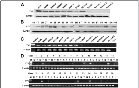
Up-regulation of YY1 in gastric cancer cell lines and primary GACs samples
showed no evidence of YY1 protein. In addition, up-regulated YY1 protein expression was observed in 9 out of 10 primary GACs but not seen in any of the adjacent non-tumorous gastric tissues (Figure 1B). YY1 mRNA also showed positive expression in 9 gastric cancer cell lines, but there was no obvious expression in 5 normal gastric tissues (Figure 1C). In 28 paired GAC cDNAs, 15 cases showed strong expression of YY1 in tumor tissue compared with adjacent non-tumorous tissue (Figure 1D).
YY1 knockdown has anti-oncogenic effect in GAC cells
in vitro
YY1 protein was knocked down in AGS, MKN28 and NCI-N87 cells by siRNA mediated degradation (Figure 2A). A significantly decreased cellular proliferation was ob-served in these cell lines when compared with scramble siRNA groups (p< 0.001, Figure 2B). Monolayer colony formation assay indicated that YY1 knockdown signifi-cantly reduced colony formation in these cell lines (p< 0.001, Figure 2C). The transfectants were then analyzed for cell cycle parameters using flow cytometry. Twenty-four hours after transfection, accumulation of G1 cells increased from 52.6% to 55.3% in AGS, from 26.2% to
36.0% in MKN28, and from 46.6% to 49.4% in NCI-N87 (Figure 2D). The same trends were observed in a re-peated set of experiments. Moreover, siYY1 induced late apoptosis, represented by an increase of cleaved-PARP, in three gastric cancer cell lines AGS, MKN28 and NCI-N87 (Figure 2E).
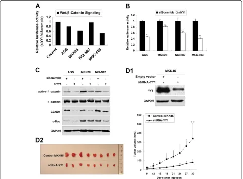
YY1 knockdown suppresses Wnt/β-catenin signaling pathway and inhibits tumor growthin vivo
By using Cancer 10-pathway Reporter Luciferase Kit, we found that siYY1 suppressed Wnt/β-catenin signaling path-way in AGS, MKN28 and MGC-803 cell lines (Figure 3A). We subsequently used TOPflash luciferase assays to con-firm that Wnt/β-catenin signaling pathway was indeed inhibited by siYY1 in AGS, MKN28, NCI-N87 and MGC-803 cells (Figure 3B). Suppression of Wnt/β-catenin sig-naling pathway by siYY1 as indicated by decreased active-β-catenin, CCND1 and c-Myc was observed in siYY1 transfected AGS, MKN28 and NCI-N87 cells (Figure 3C).
[image:4.595.55.539.89.395.2]Figure 2YY1 knockdown inhibited proliferation in gastric cancer cell lines. (A)Transfection with YY1 siRNA decreased YY1 protein expression in AGS, MKN28 and NCI-N87 cells.(B)5-day MTT assays revealed YY1 siRNA suppressed gastric cancer cell proliferation (**,p< 0.001).
(Figure 3D1). Then shYY1-MKN45 and vector control clones were injected into nude mice subcutaneously. The tumor growth in shYY1 expressing clones was sig-nificantly decreased compared with the vector control clones 30 days after injection (p< 0.001, Figure 3D2).
YY1 overexpression enhances tumor growth bothin vitro andin vivo
To further investigate the YY1 functions in gastric can-cer cells, we overexpressed YY1 into AGS, MKN28 and NCI-N87 cells. YY1 expression was first evaluated by Western blot after ectopic expression. Meanwhile active-β-catenin showed increased level (Figure 4A). YY1 over-expression increased cell proliferation in these three cell lines in 5-day MTT assays (p< 0.05, Figure 4B). To con-firm the proliferation-promoting effect by YY1 through Wnt/β-catenin signaling pathway, siCTNNB1 (25 nM) was transfected into the cultured cells to knockdown
Wnt pathway effector β-catenin. The β-catenin was ef-fectively knocked down and the same grow suppressive effect was observed (Figure 4C), indicating that Wnt/ β-catenin signaling pathway plays an important role in the YY1-induced tumor growth.
[image:6.595.57.540.90.446.2]We further examined the effect of YY1 on monolayer colony formation. Gastric cancer cells over-expressing YY1 formed more and larger colonies (2.4, 2.3 and 1.9 folds in AGS, MKN28 and NCI-N87) in comparison to vector control groups, (p< 0.001, Figure 4D). The acti-vated Wnt/β-catenin signaling pathway together with upregulation of its downstream effectors CCND1 and c-Myc were observed in YY1-MKN45 stable clones (Figure 4E1). The YY1-MKN45 stable clones were injected subcutaneously into the dorsal flank of 8 nude mice. Twenty days later, YY1 over-expressing clones formed larger xenografts than the control clones (p < 0.001, Figure 4E2).
Figure 3YY1 knockdown suppressed Wnt/β-catenin signaling pathway and inhibits tumor growthin vivo.(A)Relative luciferase activity of Wnt/β-catenin signaling pathway in 4 gastric cancer cell lines after siYY1 treatment.(B)Relative luciferase activity of TOPflash in AGS, MKN28, NCI-N87 and MGC-803 cells with siYY1 transfection (*,p< 0.05; **,p< 0.001).(C)Western blot of active-β-catenin, totalβ-catenin, CCND1 and c-Myc after siYY1 transfection in AGS, MKN28 and NCI-N87 cells.(D1)Western blot of YY1 expression in stable clones of shYY1-MKN45 and the vector control.
Nuclear expression of YY1 correlates with shorter survival time in patients with early stage GACs
YY1 was predominantly expressed in the nuclei of tumor cells (Figure 5A; left, case with negative YY1 expression; right, case with strong YY1 expression). Of the 247 GAC samples, 98 cases (39.7%) had a YY1 nuclear score 0, 3 cases (1.2%) had a score 1+, 5 cases (2%) had a score of 2+, and 141 cases (57.1%) had a score of 3+. In total, 101 of 247 samples (40.9%) of gastric cancer cases had negative/weak (0 to 1+) YY1 expression, while 146 of 247 samples (59.1%) had strong YY1 expression (2+ to 3+, Additional file 1: Figure S1). No survival difference was
observed between YY1 negative/weak and strong expres-sion by univariate analysis (p= 0.341).
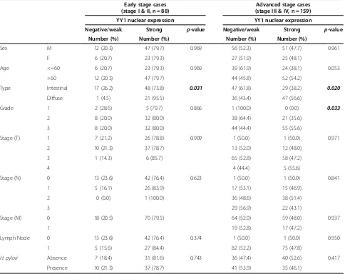
[image:8.595.58.540.356.686.2]We further investigated the clinicopathologic correl-ation of YY1 by stratifying the patients into early stage (stage I and II, n = 88) and advanced stage (stage III and IV, n = 159). Table 1 summarized the correlation of YY1 with clinicopathologic parameters in early stage and ad-vanced stage patients. In early stage patients, YY1 ex-pression correlated with diffuse type gastric cancer (p= 0.031). Univariate analysis (Table 2) indicated that YY1 strong expression in early stage GACs associated with poor disease specific survival (p= 0.045, Figure 5B). (See figure on previous page.)
Figure 4YY1 ectopic expression enhanced tumor growth bothin vitroandin vivo. (A)Western blot of YY1, active-β-catenin and totalβ-catenin after YY1 exogenous overexpression in AGS, MKN28 and NCI-N87 cell lines.(B)5-day MTT assays revealed YY1 enhanced gastric cancer cell proliferation (*,p< 0.05).(C)Upper: Western blot of active-β-catenin and totalβ-catenin under siCTNNB1 treatment. Lower: siCTNNB1 inhibits GAC cell proliferation (**,p< 0.001).(D)YY1 enhanced monolayer colony formation to 2.4, 2.3 and 1.9 folds in AGS, MKN28 and NCI-N87 cells, respectively, compared with vector controls (**,p< 0.001). The experiments were done in triplicates.(E1)YY1 ectopic expression activated Wnt/β-catenin signaling pathway in MKN45 cells by consecutively activating active-β-catenin, CCND1 and c-Myc.(E2)YY1-MKN45 formed bigger xenograft tumors than pcDNA3.1-MKN45 20 days after injection (**,p< 0.001).
Negative H. pylori infection also correlated with poor survival (p= 0.003). By multivariate Cox proportional hazards regression analysis (Table 2), only H. pylori in-fection was independently associated with disease spe-cific survival (p= 0.007).
In advanced stage GACs (Table 1), YY1 nuclear expres-sion correlated with histology with diffuse component (p= 0.020), higher tumor grade (p= 0.033). However, YY1 expression could not predict survival (Figure 5C, p= 0.958) by univariate analysis. Old age (age > 60,p= 0.006), advanced N stage (p= 0.002) and advanced M stage (p< 0.001) also predicted poor prognosis. By multivariate Cox proportional hazards regression analysis, age, N stage and M stage remained the independent prognostic factors for patients with advanced gastric cancer.
Discussion
[image:9.595.57.541.101.485.2]YY1 serves either as a tumor suppressor gene or onco-gene depending on the types of tumors it is expressed in [23,29-33]. The choice of its function and consequently
Table 1 Correlation of YY1 nuclear expression with clinicopathologic features (significantp-value in bold and italic format)
Early stage cases (stage I & II, n = 88)
Advanced stage cases (stage III & IV, n = 159)
YY1 nuclear expression YY1 nuclear expression
Negative/weak Strong p-value Negative/weak Strong p-value
Number (%) Number (%) Number (%) Number (%)
Sex M 12 (20.3) 47 (79.7) 0.969 56 (52.3) 51 (47.7) 0.961
F 6 (20.7) 23 (79.3) 27 (51.9) 25 (48.1)
Age <=60 6 (20.7) 23 (79.3) 0.969 39 (61.9) 24 (38.1) 0.053
>60 12 (20.3) 47 (79.7) 44 (45.8) 52 (54.2)
Type Intestinal 17 (26.2) 48 (73.8) 0.031 47 (61.8) 29 (38.2) 0.020
Diffuse 1 (4.5) 21 (95.5) 36 (43.4) 47 (56.6)
Grade 1 2 (28.6) 5 (79.7) 0.866 1 (100.0) 0 (0.0) 0.033
2 8 (20.0) 32 (80.0) 38 (64.4) 21 (35.6)
3 8 (20.0) 32 (80.0) 44 (44.4) 55 (55.6)
Stage (T) 1 7 (21.2) 26 (78.8) 0.909 1 (50.0) 1 (50.0) 0.971
2 10 (21.3) 37 (78.7) 13 (52.0) 12 (48.0)
3 1 (14.3) 6 (85.7) 65 (52.8) 58 (47.2)
4 4 (44.4) 5 (55.6)
Stage (N) 0 13 (23.6) 42 (76.4) 0.623 1 (50.0) 1 (50.0) 0.841
1 5 (16.1) 26 (83.9) 17 (53.1) 15 (46.9)
2 0 (0.0) 1 (100.0) 36 (48.6) 38 (51.4)
3 29 (56.9) 22 (43.1)
Stage (M) 0 18 (20.5) 70 (79.5) 64 (52.0) 59 (48.0) 0.937
1 19 (52.8) 17 (47.2)
Lymph Node 0 13 (23.6) 42 (76.4) 0.374 1 (50.0) 1 (50.0) 0.950
1 5 (15.6) 27 (84.4) 82 (52.2) 75 (47.8)
H. pylori Absence 7 (18.4) 31 (81.6) 0.743 36 (47.4) 40 (52.6) 0.417
Presence 10 (21.3) 37 (78.7) 41 (53.9) 35 (46.1)
Table 2 Univariate and multivariate Cox regression analysis of clinicopathologic factors in patients with gastric adenocarcinoma (significantp-value in bold and italic format)
Early stage cases (stage I & II, n = 88)
Advanced stage cases (stage III & IV, n = 159)
Univariate Multivariate Univariate Multivariate
Sex 0.900 0.318
Age 0.120 0.006 0.003
Type 0.342 0.654
Grade 0.594 0.747
Stage (T) 0.974 0.088
Stage (N) 0.656 0.002 <0.001
Stage (M) <0.001 <0.001
H. pylori 0.003 0.007 0.611
[image:9.595.57.291.571.733.2]the final outcome might be determined by multiple fac-tors such as cell context, oncogenic stimulation or the regulation of its upstream pathways. As a transcription factor, YY1 binds the promoter of associated oncogenes and exert its oncogenic property [34]. In prostate cancer, YY1 has two binding sites within the prostate stem cell antigen (PSCA) promoter facilitating development of malignant human prostate cancer [30]. YY1 also re-portedly activates the expression of human epidermal growth factor receptor 2 (ERBB2) [8,9], which is overex-pressed in approximately 30% of breast cancers and cor-relates with poor prognosis. YY1 induces expression of cyclooxygenase-2 (COX-2), which is overexpressed in 40% of human invasive breast cancers and mediates bone metastasis [35]. In Burkitt lymphoma, YY1 binds to this HS3 enhancer and recruits CBP to this region, which increases the histone acetylation of the c-Myc pro-moter and activates c-Myc gene expression [36]. YY1 forms an active complex with hypoxia inducible factor (HIF) 1α to activate vascular endothelial growth factor (VEGF) gene expression [37,38]. In gastric cancer, we pro-vide the first epro-vidence that YY1 also plays an oncogenic role. This was indicated by YY1 overexpression enhanced cell proliferation, monolayer colony formation and xeno-graft growth whereas YY1 knockdown inhibited gastric cancer cell proliferation bothin vitroandin vivo.
The current study also suggested that YY1 affected Wnt signaling cascades in gastric cancer cells. By using Cancer 10-pathway Reporter Luciferase Assay, we iden-tified that siYY1 inhibited Wnt/β-catenin signaling path-way in gastric cancer cell lines. The finding was further validated by the TOPflash luciferase assays. Suppression of Wnt/β-catenin pathway by siYY1, as evidenced by de-crease active-β-catenin, CCND1 and c-Myc level had been demonstrated. On the contrary, YY1 overexpres-sion promotes Wnt/β-catenin pathway and up-regulates its targets. These data were in keeping with a recent finding that YY1 activates Wnt signaling pathway through activatingβ-catenin in colon cancer [39]. Using microarray analysis, Zhang et al. found that YY1 regu-lated the expression of a number of Wnt-associate genes. It is plausible to speculate that YY1 might promote the Wnt signaling pathway by suppressing Wnt antagonists/ inhibitors, i.e. CSNK1A1, CTNNBIP1, SFRP1 and the deletion variant of LEF-1, and up-regulate Wnt initiators, i.e. CTNNB1, FZD4, Wnt1 and Wnt3a. Our data clearly indicated that YY1 acted to promote Wnt/β-catenin sig-naling in gastric carcinogenesis.
To determine the clinical relevance of YY1 in primary GACs, we examined the protein expression of YY1 in 247 clinical gastric cancer samples. The overexpression of YY1 was not correlated with TNM staging, this result was partly concordant with previous reports that there were no difference for the YY1 expression between
primary tumor and metastatic samples [40]. Neverthe-less, expression of YY1 associated with diffuse type hist-ology both in early-stage and advanced-stage GACs, suggesting that YY1 might be involved in the occurrence and development of diffuse type GACs. Comparing with intestinal type GACs, diffuse type GACs are less related to atrophy or intestinal metaplasia, occur more often in younger patients and are associated with a poorer prog-nosis. Our findings supported that YY1 overexpression is involved in the carcinogenesis diffuse type GACs. In early stage (stage I and II) GACs, YY1 nuclear expres-sion correlated with shorter survival and predicts poorer prognosis. This result was concordant with the clinical significance of YY1 in colon cancer [39].
Conclusion
In conclusion, we provided bothin vitroandin vivo evi-dence that YY1 contributes to gastric tumorigenesis through promoting cell survival in GAC cells. Functional studies demonstrated that downregulation of YY1 ex-pression by siYY1 quenched its oncogenic properties by inhibiting cell growth, inducing G1 phase accumulation and apoptosis. YY1 overexpression enhanced cell proli-feration by activating the Wnt/β-catenin signaling path-way. YY1 nuclear expression correlated with poor prognosis in patients with early stage GAC, suggesting that YY1 might potentially serve as a prognostic bio-marker and a therapeutic target in gastric cancer.
Additional file
Additional file 1: Figure S1.Representative figures of YY1
immunohistochemistry in 25 primary GACs with strong YY1 expression (2+ or 3+, original magnification × 100, insertion × 400). The up-regulated expression of YY1 mainly localized in the nuclei of the cancer cells.
Competing interests
The authors declare that they have no competing interests.
Authors’contributions
WK, JHMT, AWHC, JHZ, YJD, SYW, WQY and FMCS carried out the experimental studies, interpreted the data, performed the statistical analysis. SSMN, JY, ASLC provided experimental materials. WK, JHMT and KFT contributed to the study design, manuscript drafting and provided fund for this study. All authors read and approved the final manuscript.
Acknowledgements
This work was supported by National Natural Science Foundation of China (81201591). We thank Dr Elaine Chan for carefully reviewing the manuscript.
Author details
1Department of Anatomical and Cellular Pathology, State Key Laboratory in
Oncology in South China, Prince of Wales Hospital, The Chinese University of Hong Kong, Hong Kong, SAR, PR China.2Institute of Digestive Disease,
Partner State Key Laboratory of Digestive Disease, The Chinese University of Hong Kong, Hong Kong, SAR, PR China.3Li Ka Shing Institute of Health
Science, Sir Y.K. Pao Cancer Center, The Chinese University of Hong Kong, Hong Kong, SAR, PR China.4Shenzhen Research Institute, The Chinese
6Department of Surgery, The Chinese University of Hong Kong, Hong Kong,
PR China.7School of Biomedical Sciences, The Chinese University of Hong Kong, Hong Kong, PR China.
Received: 25 January 2014 Accepted: 24 March 2014 Published: 28 March 2014
References
1. Jemal A, Siegel R, Ward E, Murray T, Xu J, Thun MJ:Cancer statistics, 2007.
CA Cancer J Clin2007,57:43–66.
2. Vrdoljak E, Wojtukiewicz MZ, Pienkowski T, Bodoky G, Berzinec P, Finek J, Todorovic V, Borojevic N, Croitoru A:Cancer epidemiology in Central, South and Eastern European countries.Croat Med J2011,52:478–487. 3. Milne AN, Carneiro F, O’Morain C, Offerhaus GJ:Nature meets nurture:
molecular genetics of gastric cancer.Hum Genet2009,126:615–628. 4. Shi Y, Seto E, Chang LS, Shenk T:Transcriptional repression by YY1,
a human GLI-Kruppel-related protein, and relief of repression by adenovirus E1A protein.Cell1991,67:377–388.
5. Zaravinos A, Spandidos DA:Yin yang 1 expression in human tumors.
Cell Cycle2010,9:512–522.
6. Lee HY, Chaudhary J, Walsh GL, Hong WK, Kurie JM:Suppression of c-Fos gene transcription with malignant transformation of human bronchial epithelial cells.Oncogene1998,16:3039–3046.
7. Liao WR, Hsieh RH, Hsu KW, Wu MZ, Tseng MJ, Mai RT, Wu Lee YH, Yeh TS:
The CBF1-independent Notch1 signal pathway activates human c-myc expression partially via transcription factor YY1.Carcinogenesis2007,28:1867–1876. 8. Allouche A, Nolens G, Tancredi A, Delacroix L, Mardaga J, Fridman V, Winkler
R, Boniver J, Delvenne P, Begon DY:The combined immunodetection of AP-2alpha and YY1 transcription factors is associated with ERBB2 gene overexpression in primary breast tumors.Breast Cancer Res2008,10:R9. 9. Begon DY, Delacroix L, Vernimmen D, Jackers P, Winkler R:Yin Yang 1
cooperates with activator protein 2 to stimulate ERBB2 gene expression in mammary cancer cells.J Biol Chem2005,280:24428–24434.
10. Wan M, Huang W, Kute TE, Miller LD, Zhang Q, Hatcher H, Wang J, Stovall DB, Russell GB, Cao PD, Deng Z, Wang W, Zhang Q, Lei M, Torti SV, Akman SA, Sui G:Yin Yang 1 plays an essential role in breast cancer and negatively regulates p27.Am J Pathol2012,180:2120–2133. 11. Rayess H, Wang MB, Srivatsan ES:Cellular senescence and tumor
suppressor gene p16.Int J Cancer2012,130:1715–1725.
12. Wu S, Murai S, Kataoka K, Miyagishi M:Cooperative regulation of p73 promoter by Yin Yang 1 and E2F1.Nucleic Acids Symp Ser (Oxf)2007,51:347–348. 13. Gronroos E, Terentiev AA, Punga T, Ericsson J:YY1 inhibits the activation of
the p53 tumor suppressor in response to genotoxic stress.Proc Natl Acad Sci U S A2004,101:12165–12170.
14. Castellano G, Torrisi E, Ligresti G, Malaponte G, Militello L, Russo AE, McCubrey JA, Canevari S, Libra M:The involvement of the transcription factor Yin Yang 1 in cancer development and progression.Cell Cycle
2009,8:1367–1372.
15. Palmer MB, Majumder P, Cooper JC, Yoon H, Wade PA, Boss JM:Yin yang 1 regulates the expression of snail through a distal enhancer.Mol Cancer Res2009,7:221–229.
16. de Nigris F, Rossiello R, Schiano C, Arra C, Williams-Ignarro S, Barbieri A, Lanza A, Balestrieri A, Giuliano MT, Ignarro LJ, Napoli C:Deletion of Yin Yang 1 protein in osteosarcoma cells on cell invasion and CXCR4/ angiogenesis and metastasis.Cancer Res2008,68:1797–1808. 17. de Nigris F, Botti C, de Chiara A, Rossiello R, Apice G, Fazioli F, Fiorito C,
Sica V, Napoli C:Expression of transcription factor Yin Yang 1 in human osteosarcomas.Eur J Cancer2006,42:2420–2424.
18. de Nigris F, Zanella L, Cacciatore F, De Chiara A, Fazioli F, Chiappetta G, Apice G, Infante T, Monaco M, Rossiello R, De Rosa G, Alberghini M, Napoli C:YY1 overexpression is associated with poor prognosis and metastasis-free survival in patients suffering osteosarcoma.BMC Cancer2011,11:472. 19. Castellano G, Torrisi E, Ligresti G, Nicoletti F, Malaponte G, Traval S,
McCubrey JA, Canevari S, Libra M:Yin Yang 1 overexpression in diffuse large B-cell lymphoma is associated with B-cell transformation and tumor progression.Cell Cycle2010,9:557–563.
20. Deng Z, Wan M, Cao P, Rao A, Cramer SD, Sui G:Yin Yang 1 regulates the transcriptional activity of androgen receptor.Oncogene2009,28:3746–3757. 21. Zheng H, Chu J, Zeng Y, Loh HH, Law PY:Yin Yang 1 phosphorylation
contributes to the differential effects of mu-opioid receptor agonists on microRNA-190 expression.J Biol Chem2010,285:21994–22002.
22. Beezhold K, Liu J, Kan H, Meighan T, Castranova V, Shi X, Chen F: miR-190-mediated downregulation of PHLPP contributes to arsenic-induced Akt activation and carcinogenesis.Toxicol Sci2011,123:411–420.
23. Lee MH, Lahusen T, Wang RH, Xiao C, Xu X, Hwang YS, He WW, Shi Y, Deng CX:Yin Yang 1 positively regulates BRCA1 and inhibits mammary cancer formation.Oncogene2012,31:116–127.
24. Lieberthal JG, Kaminsky M, Parkhurst CN, Tanese N:The role of YY1 in reduced HP1alpha gene expression in invasive human breast cancer cells.Breast Cancer Res2009,11:R42.
25. Wang CC, Tsai MF, Dai TH, Hong TM, Chan WK, Chen JJ, Yang PC:
Synergistic activation of the tumor suppressor, HLJ1, by the transcription factors YY1 and activator protein 1.Cancer Res2007,67:4816–4826. 26. Wang CC, Tsai MF, Hong TM, Chang GC, Chen CY, Yang WM, Chen JJ, Yang PC:
The transcriptional factor YY1 upregulates the novel invasion suppressor HLJ1 expression and inhibits cancer cell invasion.Oncogene2005,
24:4081–4093.
27. Naidoo K, Clay V, Hoyland JA, Swindell R, Linton K, Illidge T, Radford JA, Byers RJ:YY1 expression predicts favourable outcome in follicular lymphoma.J Clin Pathol2011,64:125–129.
28. Kang W, Tong JH, Chan AW, Lee TL, Lung RW, Leung PP, So KK, Wu K, Fan D, Yu J, Sung JJ, To KF:Yes-associated protein 1 exhibits oncogenic property in gastric cancer and its nuclear accumulation associates with poor prognosis.Clin Cancer Res2011,17:2130–2139.
29. Zhang S, Jiang T, Feng L, Sun J, Lu H, Wang Q, Pan M, Huang D, Wang X, Wang L, Jin H:Yin Yang-1 suppresses differentiation of hepatocellular carcinoma cells through the downregulation of CCAAT/enhancer-binding protein alpha.J Mol Med (Berl)2012,90:1069–1077.
30. Tang S, Mishra M, Frazier DP, Moore ML, Inoue K, Deora R, Sui G, Dubey P:
Positive and negative regulation of prostate stem cell antigen expression by Yin Yang 1 in prostate epithelial cell lines.PLoS One2012,7:e35570. 31. Moseley SC, Rizkallah R, Tremblay DC, Anderson BR, Hurt MM, Chadwick BP:
YY1 associates with the macrosatellite DXZ4 on the inactive X chromosome and binds with CTCF to a hypomethylated form in some male carcinomas.Nucleic Acids Res2012,40:1596–1608.
32. Ishii H, Hulett MD, Li JM, Santiago FS, Parish CR, Khachigian LM:Yin Yang-1 inhibits tumor cell growth and inhibits p21WAF1/Cip1 complex formation with cdk4 and cyclin D1.Int J Oncol2012,40:1575–1580.
33. Nicholson S, Whitehouse H, Naidoo K, Byers RJ:Yin Yang 1 in human cancer.Crit Rev Oncog2011,16:245–260.
34. Zhang Q, Stovall DB, Inoue K, Sui G:The oncogenic role of Yin Yang 1.
Crit Rev Oncog2011,16:163–197.
35. Joo M, Wright JG, Hu NN, Sadikot RT, Park GY, Blackwell TS, Christman JW:
Yin Yang 1 enhances cyclooxygenase-2 gene expression in macrophages.
Am J Physiol Lung Cell Mol Physiol2007,292:L1219–L1226.
36. Hu HM, Kanda K, Zhang L, Boxer LM:Activation of the c-myc p1 promoter in Burkitt’s lymphoma by the hs3 immunoglobulin heavy-chain gene enhancer.Leukemia2007,21:747–753.
37. de Nigris F, Crudele V, Giovane A, Casamassimi A, Giordano A, Garban HJ, Cacciatore F, Pentimalli F, Marquez-Garban DC, Petrillo A, Cito L, Sommese L, Fiore A, Petrillo M, Siani A, Barbieri A, Arra C, Rengo F, Hayashi T, Al-Omran M, Ignarro LJ, Napoli C:CXCR4/YY1 inhibition impairs VEGF network and angiogenesis during malignancy.Proc Natl Acad Sci U S A2010,
107:14484–14489.
38. Wu S, Kasim V, Kano MR, Tanaka S, Ohba S, Miura Y, Miyata K, Liu X, Matsuhashi A, Chung UI, Yang L, Kataoka K, Nishiyama N, Miyagishi M:
Transcription factor YY1 contributes to tumor growth by stabilizing hypoxia factor HIF-1alpha in a p53-independent manner.Cancer Res
2013,73:1787–1799.
39. Zhang N, Li X, Wu CW, Dong Y, Cai M, Mok MT, Wang H, Chen J, Ng SS, Chen M, Sung JJ, Yu J:microRNA-7 is a novel inhibitor of YY1 contributing to colorectal tumorigenesis.Oncogene2012,32:5078–5088. 40. Monzon FA, Lyons-Weiler M, Buturovic LJ, Rigl CT, Henner WD, Sciulli C,
Dumur CI, Medeiros F, Anderson GG:Multicenter validation of a 1,550-gene expression profile for identification of tumor tissue of origin.J Clin Oncol
2009,27:2503–2508. doi:10.1186/1479-5876-12-80