RESEARCH
Endoplasmic reticulum stress
in perivascular adipose tissue promotes
destabilization of atherosclerotic plaque
by regulating GM-CSF paracrine
Ru Ying
1,2†, Sheng‑Wei Li
3†, Jia‑Yuan Chen
1†, Hai‑Feng Zhang
1, Ying Yang
1, Zhen‑Jie Gu
1, Yang‑Xin Chen
1*and Jing‑Feng Wang
1*Abstract
Background: Perivascular adipose tissue (PVAT) accelerates plaque progression and increases cardiovascular risk. We tested the hypothesis that PVAT contributed to plaque vulnerability and investigated whether endoplasmic reticulum stress (ER stress) in PVAT played an important role in vulnerable plaque.
Methods: We transplanted thoracic aortic PVAT or subcutaneous adipose tissue as a control, from donor mice to carotid arteries of recipient apolipoprotein E deficient (apoE−/−) mice after removing carotid artery collar placed for 6 weeks. Two weeks after transplantation, ER stress inhibitor 4‑phenyl butyric acid (4‑PBA) was locally administrated to the transplanted PVAT and then animals were euthanized after 4 weeks. Immunohistochemistry was performed to quantify plaque composition and neovascularization. Mouse angiogenesis antibody array kit was used to test the angiogenic factors produced by transplanted adipose tissue. In vitro tube formation assay, scratch wound migration assay and mouse aortic ring assay were used to assess the angiogenic capacity of supernatant of transplanted PVAT.
Results: Ultrastructural detection by transmission electron microscopy showed transplanted PVAT was a mixed population of white and brown adipocytes with abundant mitochondria. Transplanted PVAT increased the intra‑ plaque macrophage infiltration, lipid core, intimal and vasa vasorum neovascularization and MMP2/9 expression in plaque while decreased smooth muscle cells and collagen in atherosclerotic plaque, which were restored by local 4‑PBA‑treatment. Antibody array analysis showed that 4‑PBA reduced several angiogenic factors [Granulocyte Macrophage Colony Stimulating Factor (GM‑CSF), MCP‑1, IL‑6] secreted by PVAT. Besides, conditioned medium from 4‑PBA treated‑PVAT inhibited tube formation and migration capacity of endothelial cells and ex vivo mouse aortic ring angiogenesis compared to conditioned medium from transplanted PVAT. mRNA expression and protein levels of GM‑CSF were markedly elevated in adipocytes under ER stress which would be suppressed by 4‑PBA. In addition, ER stress enhanced NF‑κB binding to the promoter of the mouse GM‑CSF gene in adipocytes confirmed by Chromatin immunoprecipitation analyses.
Conclusions: Our findings demonstrate that ER stress in PVAT destabilizes atherosclerotic plaque, in part through increasing GM‑CSF paracrine via transcription factor NF‑κB.
© The Author(s) 2018. This article is distributed under the terms of the Creative Commons Attribution 4.0 International License (http://creativecommons.org/licenses/by/4.0/), which permits unrestricted use, distribution, and reproduction in any medium, provided you give appropriate credit to the original author(s) and the source, provide a link to the Creative Commons license, and indicate if changes were made. The Creative Commons Public Domain Dedication waiver (http://creativecommons.org/ publicdomain/zero/1.0/) applies to the data made available in this article, unless otherwise stated.
Open Access
*Correspondence: tjcyx1995@163.com; dr_wjf@hotmail.com †Ru Ying, Sheng‑Wei Li and Jia‑Yuan Chen contributed equally to this work
1 Department of Cardiology, Sun Yat‑sen Memorial Hospital of Sun Yat‑sen University, No.107, Yanjiang West Road, Yuexiu District, Guangzhou 510120, China
Background
Acute coronary syndrome (ACS) is the leading cause of cardiovascular morbidity and mortality. Most major adverse events occur independent of the plaque size and the degree of luminal stenosis and most ACS arises from vulnerable plaques which are more prone to rapid plaque progression [1]. The vulnerable lesion has several structural and functional hallmarks different from stable plaque including large necrotic core, inflammation, thin fibrous cap, depletion of smooth muscle cells and col-lagen, and increased formation of neo microvessels [2]. However, the precise mechanisms underlying plaque destabilization are still unclear.
Accumulating evidences have shown a strong associa-tion between obesity and cardiovascular disease [3]. Obe-sity is associated with the inflammation in perivascular adipose tissue (PVAT) which not only serves a structural support for most arteries but also secretes series of mol-ecules to actively modulate vascular function [4]. In 1991, it was the first time to report that PVAT had an anticon-tractile function [5]. In 2002, PVAT was found to release a vasoactive factor [6]. Since then, the list of factors released from PVAT has expanded and now includes adi-pokines (e.g., leptin and adiponectin), angiogenesis fac-tors and inflammatory facfac-tors (e.g., MCP-1, IL-6). From twentieth century on, scientists revealed that chemokines production from PVAT played a role in the pathogenesis of atherosclerosis [7]. These molecules might contribute to the alterations of the function and structure of vascu-lar wall, including chronic inflammation, alterations of vascular tone, smooth muscle cell dysfunction, neo-angi-ogenesis and hence to the development of atherosclero-sis. Epidemiological studies show that PVAT correlates with plaque burden in human [8–10]. Experimental animal studies also demonstrate that PVAT accelerated atherosclerosis in mice [11, 12]. Additionally, clinical observations suggest that coronary perivascular adipose tissue is related to the presence of lipid core, macrophage infiltration and severity of atherosclerotic plaque which are the characteristics of high risk plaque [13, 14]. These findings urged us to hypothesize that PVAT contributes to plaque vulnerability through paracrine effects on the vasculature from ‘outside to inside’.
There are compelling evidences that endoplasmic reticulum stress (ER stress) plays fundamental roles in atherogenesis and atherosclerotic progression [15, 16]. Endoplasmic reticulum (ER) is the cellular organelle in which protein folding, calcium homeostasis, and lipid
biosynthesis occurs [15]. Perturbations in ER homeo-stasis cause ER stress. High fat feeding and obesity [17,
18] and several other high-risk factors of atherosclerosis such as hypertension [19, 20], cigarette smoking [21], high glucose [22] and hyperhomocysteinemia [23], can lead to increased ER stress in adipose tissue. ER stress induces inflammatory phenotypic alteration of adipose tissue which plays an important role in atherogenesis [17]. Thus, we speculated that unresolved ER stress might induce the dysfunction of PVAT by leading to aberrant chemokines secretion and contributed to atherosclerotic plaque progression.
In this study, we found that transplanted PVAT pro-moted plaque vulnerability in the setting of high-fat diet (HFD) which could be ameliorated by 4-PBA at least in part dependent on decreased GM-CSF released locally by transplanted PVAT. These findings demonstrate a direct relationship between ER stress in PVAT and plaque destabilization and implicate GM-CSF secretion by PVAT as a mediator of this pathological process.
Methods
Animal model
Eight weeks old male apoE−/− mice (Guangdong medical laboratory animal center) were used. To investigate the impact of PVAT on atherosclerotic stability, we executed carotid collar placement as described [24, 25] and these mice were maintained on a HFD (Guangdong medi-cal laboratory animal center, 0.15% cholesterol and 21% fat). The carotid collar was removed after 6 weeks, fol-lowed by adipose tissue transplantation surgery. 2 weeks after transplantation, ER stress inhibitor 4-PBA (5 μg per mouse) was locally administrated using pluronic gel to the transplanted PVAT and then animals were eutha-nized 4 weeks later. The left common carotid arteries and the transplanted adipose tissue were collected for histo-logical and molecular biohisto-logical analysis.
All procedures were approved by the Institutional Ani-mal Care and Use committee of Sun Yat-sen University at Guangzhou.
Carotid collar placement
then tied with three circumferential silk ties at their axial edges. Then, the entry wound was closed.
Adipose transplantation to carotid artery
Thirty mg of PVAT was collected from the thoracic aorta of donor C57BL/6J mice (Guangdong medical labora-tory animal center) fed a HFD for 4 weeks. The col-lected PVAT, or inguinal subcutaneous adipose tissue (SQAT) from the same group of donor mice as a control, was implanted adjacent to left common carotid artery of apoE−/− mice. Sham-operated apoE−/− mice under-went the same surgery without a fat transplant. Skin was sutured with 6-0 nylon filament.
Angiogenesis‑related protein analysis
To analyze the expression profiles of angiogenesis-related proteins, we used the mouse angiogenesis antibody array (RayBiotech, USA), according to the manufacturer’s instructions. They can detect 24 antibodies directed to proteins involved in angiogenesis. Briefly, 10 mg tis-sue of PVAT and SQAT were incubated in 1 ml serum-free DMEM medium. After 24 h the supernatants from tissue cultures were centrifuged at 14,000 rounds/min for 3 min. The centrifuged supernatants were stored at
− 80 °C until further processing.
Supernatants from tissue cultures were mixed with 70 μl of biotin-conjugated detection antibodies for 2 h at room temperature with gentle shaking. Following a wash-ing step, streptavidin-fluor was added to each sub-array. Make sure the glass slides were absolutely dry before scanning or storage. Data were captured by GenePix 4000B Microarray Scanner (Molecular Devices, USA) and analyzed by analysis tool software for RayBio anti-body array (RayBiotech).
Cell culture
Mouse preadipocytes 3T3-L1 cells (Shanghai institute for biological sciences, Shanghai, China) were cultured in growth medium (DMEM supplemented with 10% FCS). Two days postconfluence, the cells were induced to dif-ferentiate with standard cocktail consisting of medium (DMEM supplemented with 10% FBS) with 1 μmol/l dex-amethasone, 10 μg/ml insulin and 0.5 mmol/l isobutyl-methylxanthine (Sigma). After 4 days in differentiation medium, the cells were treated with medium contain-ing 10 μg/ml insulin for 4 days and then maintained in DMEM supplemented with 10% FBS alone. Then, cells were considered mature adipocytes. ER stress was induced by tunicamycin (TM) and suppressed by 4-PBA.
Human umbilical vein endothelial cells (HUVECs) (Shanghai institute for biological sciences) were main-tained in endothelial cell growth medium-2 (EGM-2; Lonza) supplemented with 10% FBS (Biological
Industries, Israel), at 37 °C in humidified incubator (5% CO2). HUVECs were cultured to 2–6 passages for experiment.
Mouse aortic ring assay
Three-dimensional ex vivo mouse aortic ring angiogen-esis assay allows analysis of cellular proliferation, migra-tion, tube formamigra-tion, microvessel branching, perivascular recruitment and remodeling, providing a more complete picture of angiogenic processes compared with tradi-tional in vitro tube formation assays. We performed the aortic ring assay as previously described [26], with minor modifications. Briefly, the rings from the aor-tas of 4 weeks old C57BL/6 mice were inserted between two layers of matrigel basement membrane matrix (BD Biosciences) and cultured in EGM-2 in the presence of DMEM or supernatants from PVAT cultures for 7 days. Endothelial sprouts were stained with antibody to CD31 (abcam) by immunofluorescence and quantitative analy-sis was performed with software Image-Pro Plus 6.0. Three separate aortic sections were quantified for each aorta, and the results were averaged for each animal.
In vitro tube formation assay
96-well plates were coated with 50 μl Matrigel (Millipore) and incubated at 37 °C for 60 min to allow the Matrigel to solidify. HUVECs were plated at a density of 5 × 10⁴ cells/ well with supernatants from PVAT cultures or vehicle and incubated at 37 °C for 8 h. The cells were then pho-tographed using a Nikon digital camera. Five randomly selected view fields were photographed in each well. The average of five fields was taken as the value for each sample. Tube formation was quantified by measuring the length of capillary structures using the software Image-Pro Plus 6.0 [27].
Cell migration assay
The scratch wound migration assay was performed as described before [28]. Briefly, confluent HUVEC sheets were starved for 6 h before starting the experiments. Confluent cell monolayer was then scraped with a 200 μl pipette tip to generate scratch wounds and rinsed twice with PBS. Cells were photographed immediately and 24 h after the scratch with a Nikon digital camera (Nikon ECLIPSE Ti). The wound area was then measured to determine cell migration.
Chromatin immunoprecipitation (ChIP)
II antibody was used (Cell Signaling Technology). Corre-sponding normal IgG was used as negative control. ChIP was performed overnight at 4 °C with rotation. Input DNA was purified along with ChIP probes. Association with pNF-κB and GM-CSF promoters was estimated using SYBR-Green RT-PCR. Sequence of primers used for ChIP is given in Additional file 1: Table S1.
Ready‑To‑Glow™ NF‑κB Secreted Luciferase Reporter System
We used the Ready-To-Glow™ NF-κB Secreted Lucif-erase Reporter System (Clotech, Catalog No.631743), according to the manufacturer’s instructions. Mature 3T3-L1 cells were transiently transfected with pMetLuc2-control vector or pNFκB- MetLuc2-reporter vector. 24 h after transfection, the media was removed and replaced by media with vehicel or TM (1 μg/ml) or pretreatment of 4-PBA (5 mM) for half hour to activate the NF-κB sig-nal transduction pathway. After 8, 16 and 24 h, 160 μl media samples were removed and analyzed for Metridia luciferase activity in a luminometer (Molecular devices, SpectraMax M5/M5e). The fold induction was calculated for different time points following substrate addition.
Statistical analysis
The statistical significance of differences between groups was determined by one-way ANOVA for multiple com-parisons, or Student’s t test when comparisons were
made between two groups. Values are expressed as mean ± SEM, p < 0.05 are considered significant.
Results
Effects of adipose tissue transplantation
To determine whether adipose tissue could contribute to characteristics of atherosclerotic plaque, ApoE−/− mice underwent PVAT, SQAT transplant or sham operation to the left common carotid artery a site that is devoid of perivascular adipose tissue and typically does not develop spontaneous atherosclerosis. The sur-gical images showed that transplanted adipose tissue was healthy appearing and was incorporated into the carotid adventitia (Fig. 1).
HE staining demonstrated that SQAT was white adi-pocytes while PVAT was similar to brown adiadi-pocytes (Fig. 2A, B). However, ultrastructural detection by transmission electron microscopy showed PVAT was a mixed population of white and brown adipocytes (Fig. 2D–F) and the adipocytes had abundant mito-chondria with one big or several small lipid droplets in PVAT while SQAT was made up of a single lipid vacu-ole with rare mitochondria (Fig. 2C).
To exclude the systemic confounding factors, we com-pared mouse weight, systemic lipid levels and serum adipokines of PVAT-transplanted animals with mice in other groups and found that there were no signifi-cant difference between them (Additional file 1: Table
[image:4.595.58.541.447.697.2]S2). In addition, analysis of gene expression showed that mRNA expression of leptin and adiponectin in transplanted PVAT and SQAT were similar to endog-enous fat from the corresponding depots of the same mice, which suggested that adipose phenotype was not affected by the transplantation experiment (Additional file 1: Figure S1).
Effects of transplanted adipose tissue on destabilization of atherosclerotic plaque
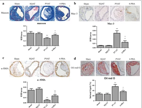
To characterize the effects of PVAT on plaque composi-tion, collagen, SMCs, macrophages and cholesterol core of carotid artery lesion were detected. Masson Trichrome is a three-color staining protocol used in histology, which
stains collagen and bone into blue or green, keratin and muscle fibers into red, cell nuclei into dark brown to black and cytoplasm into light red or pink. Oil red O is a fat-soluble dye used for staining of lipids into red color. PVAT transplantation markedly increased intraplaque macrophages number, enlarged lipid core and upregu-lated MMP2/9 expression compared to SQAT transplan-tation while reduced collagen and SMCs in plaque (Fig. 3
and Additional file 1: Figure S2). Especially, the fibrous cap mainly composed of collagen and SMCs in PVAT group was thinner than that in sham and SQAT group (Fig. 3a). Moreover, SQAT transplantation failed to sub-stantially change the plaque composition.
[image:5.595.60.538.87.502.2]Effects of 4‑PBA treatment on plaque composition
To investigate whether endoplasmic reticulum stress, a characteristic of obese adipose tissue, plays a role in the effects of PVAT determined above, ER stress inhibitor 4-PBA was locally administrated to the transplanted PVAT. After ER stress in PVAT was inhibited, intraplaque collagen and SMCs number increased compared to PVAT group without inhibiting ER stress while intraplaque macrophage infiltration, lipid core and MMP-2 expression decreased (Fig. 3 and Additional file 1: Figure S2). However, 4-PBA failed to decrease the expression of MMP-9 in plaque.
Effects of transplanted adipose tissue on intimal and vasa vasorum neovascularization
Intraplaque angiogenesis functions to sustain the growth of subintima and media from the artery lumen, and vasa vasorum can facilitate macrophage and erythrocyte entry into the plaque and cause inflammation and intraplaque
hemorrhage [29]. Therefore, intimal and vasa vasorum neovascularization would destabilize plaque. The mice in sham group and SQAT group had no obvious intraplaque microvessel development, while transplanted PVAT pro-moted intraplaque angiogenesis (Fig. 4a, c). When ER stress was inhibited in transplanted PVAT, microves-sels were significantly reduced in plaque (Fig. 4d). Like-wise, vasa vasorum (VV) were not present in mice of sham group and SQAT group, but transplanted PVAT increased VV neovascularization (Fig. 4d) which would be attenuated by 4-PBA.
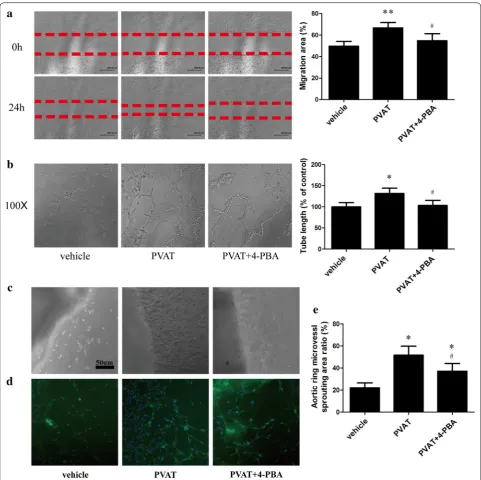
Conditioned medium from PVAT promoted tube formation and migration capacity of endothelial cells and ex vivo mouse aortic ring angiogenesis
Although PVAT transplantation increased in vivo intra-plaque and vasa vasorum neovascularization, whether it
[image:6.595.59.540.88.454.2]could up-regulate angiogenesis in vitro and ex vivo need to be further investigated. We used in vitro tube forma-tion assay and scratch wound migraforma-tion assay to assess tube formation and migration capacity of endothelial cells. The conditioned medium from transplanted adi-pose tissue were used to administrate endothelial cells. The migration of ECs treated with the supernatant of transplanted PVAT was faster than control group and the tube formation capacity of the former was also stronger than the latter. Moreover, supernatant of 4-PBA treated-PVAT would restored tube formation and migration function of ECs (Fig. 5a, b).
We next tested ex vivo angiogenesis via mouse aortic ring assay. The supernatant of transplanted PVAT mark-edly promoted the ex vivo mouse aortic ring angiogen-esis which was confirmed by immunostaining of CD31 (Fig. 5c, d). When ER stress in PVAT was inhibited by 4-PBA, the angiogenesis effect would become weaker.
Thus, from the in vitro, ex vivo and in vivo evidences, we concluded that PVAT could promote angiogenesis, which could be attenuated by ER stress inhibitor.
Mouse angiogenesis antibody array for angiogenic factors produced by transplanted adipose tissue
In spite of angiogenic effect of PVAT, it is still unkown about the related angiogenic factors playing an important
role in the angiogenic process. Therefore, we determined to screen out these factors by mouse angiogenesis anti-body array which could detect 24 antibodies directed to proteins involved in angiogenesis. The results suggested that PVAT increased several pro-angiogenic factor lev-els (MCP-1, IL-6, GM-CSF) and also up-regulated the expression of anti-angiogenic factor (PF-4) (Fig. 6).
ER stress upregulated GM‑CSF expression of adipocytes by a transcriptional mechanism
The results of angiogenesis antibody array revealed that 4-PBA reduced GM-CSF expression produced by PVAT. Then, we established the models of ER stress in adipo-cytes. We treated adipocytes with ER stress inducer tunicamycin (TM) (1 μg/ml) or vehicle (DMSO) in the presence or absence of 5 mM 4-PBA. QRT-PCR results showed that TM induced GM-CSF gene expression in 3T3-L1 adipocytes and peaked at the 4th hour (Fig. 7a). Elisa results suggested the supernatant of adipocytes treated by TM had higher GM-CSF level than control, and 4-PBA attenuated GM-CSF expression (Fig. 7b).
Next, we investigated the mechanism of ER stress regulating GM-CSF expression was transcriptional or posttranscriptional. For this purpose, adipocytes were administrated with the transcriptional inhibitor Actino-mycin D (5 μg/ml) in the presence or absence of 1 μg/ml
Fig. 4 Effects of transplanted adipose tissue on intimal and vasa vasorum neovascularization. a Immunostaining for CD31 was performed to evaluate intraplaque angiogenesis. b Statistical analysis for a (n = 6). **p < 0.01 compared with sham group, ##p < 0.01 compared with PVAT group.
[image:7.595.57.541.88.343.2]TM. RT-PCR results revealed that TM treatment failed to prolong the half-life of GM-CSF mRNA, which suggested that ER stress upregulated GM-CSF by a transcriptional mechanism (Fig. 7c).
One candidate transcription factor that could explain the effects of ER stress on GM-CSF is NF-κB. Therefore, we used Ready-To-Glow™ NF-κB Secreted Luciferase Reporter System to assess NF-κB activity. NF-κB was more active in TM treated-adipocytes than in control
group, but some of this activity was lost when the cells were pretreated with 4-PBA, which demonstrated that NF-κB might contribute to ER stress induced GM-CSF expression.
NF‑κB contributed to increasing GM‑CSF mRNA expression by ER stress
To verify whether NF-κB contribute to GM-CSF expres-sion induced by ER stress, we conducted studies with
[image:8.595.57.539.86.566.2]NF-κB inhibitor BAY11-7082 (10 μM) and confirmed it by western blot. Results showed that BAY11-7082 inhib-ited NF-κB phosphorylation, and attenuated GM-CSF expression by TM (Fig. 8a).
To further confirm whether NF-κB could bind to the promoter of GM-CSF, we measured ChIP at two pre-dicted binding sites. ChIP results showed that NF-κB could bind to the predicted region 1, but failed to bind-ing site 2 (Fig. 8b, c). TM treatment increased occupancy of NF-κB at the binding site 1 in GM-CSF promoter (Fig. 8d).
Discussion
A consensus is emerging that PVAT is related to plaque vulnerability, but direct evidence is lacking. Here, we have established a model in which PVAT was trans-planted to carotid artery with atherosclerosis to test the effects of PVAT transplantation on plaque vulnerability. In addition, we locally administrated 4-PBA to the trans-planted PVAT to investigate the role of ER stress in par-acrine effects of PVAT. Our data suggest that ER stress in PVAT could increase plaque vulnerability, partly by increasing GM-CSF paracrine via transcription factor NF-κB.
Adipose tissue not only stores energy but also secrets numerous growth factors, cytokines, and hormones. PVAT has recently been recognized as a novel factor in vascular biology implicated in the pathophysiology of cardiovascular disease. Emerging researches suggest that PVAT may contribute to the pathogenesis of vas-cular disease by producing large numbers of biologically active molecules [6]. Our study may explain its biologi-cal activity from the perspective of the microstructures. Even though PVAT was a mixed population of white and brown adipocytes, ultrastructural detection by transmis-sion electron microscopy showed that there were abun-dant big mitochondria no matter with small lipid droplets or one big lipid droplet in PVAT while SQAT was made up of a single lipid vacuole with rare mitochondria (Fig. 2). Therefore, the rich mitochondria in perivascular adipocytes might be the structure basis of its biological activity.
Vulnerable plaque is an important culprit mecha-nism for the development of acute coronary syndrome (ACS), which is influenced by several factors such as vasa vasorum and intraplaque neovascularization, large necrotic core, inflammation and intraplaque protease activity [2]. However, the underlying mecha-nism of plaque vulnerability remains unclear and need
[image:9.595.57.538.87.378.2]Fig. 7 ER stress upregulated GM‑CSF expression by a transcriptional mechanism. a GM‑CSF mRNA levels of adipocytes treated with TM (1 μg/ml) in different time. b Elisa results of supernatant of adipocytes treated with TM in the presence or absence of 4‑PBA. c RT‑PCR for GM‑CSF mRNA assess‑ ment. Adipocytes were administrated with 5 μg/ml Actinomycin D in the presence or absence of 1 μg/ml TM. d Ready‑To‑Glow™ NF‑κB Secreted Luciferase Reporter System to assess NF‑κB activity
[image:10.595.58.537.86.376.2] [image:10.595.58.541.430.686.2]to be further elucidated. Clinical observations sug-gest PVAT increases severity of atherosclerotic plaque and correlates with coronary plaque burden [13]. We demonstrated PVAT promoted plaque vulnerability in PVAT-transplanted carotid arteries (Figs. 3, 4 and Additional file 1: Figure S2). To examine the impact of PVAT, endogenous PVAT in femoral artery was removed and replaced with transplanted fat in some studies [30, 31], or exogenous PVAT was transplanted to the carotid artery without PVAT in other studies [11,
12]. Fat transplantation was used in our study to reduce injuring artery. Carotid collar placement was used to study plaque vulnerability and produced atherosclero-sis plaque in fixed location and facilitate administration to artery.
Several researches have also revealed the pathogenic role of PVAT in atherosclerosis [32–34]. David Manka et al. [11] transplanted PVAT to the mouse carotid artery and found that PVAT could enhance the neointimal response to vascular injury. On the contrary, some stud-ies suggest that PVAT is vasculoprotective. For instance, removal of endogenous PVAT enhanced the neointimal response to wire injury in the femoral artery [30]. Addi-tionally, research in the SMPG knockout mouse suggest an atheroprotective effect of PVAT at a cool tempera-ture [35]. However, the results in current study are not strictly comparable with the previous researches. Some of these researches transplanted visceral adipose tissue rather than real PVAT to the carotid artery. Some stud-ies removed endogenous PVAT, which might make the artery injured. Therefore, these conclusions might be dependent on the experimental model. However, the model of fat transplantation used in this research might be more proportionate to study the impact of PVAT.
Several high-risk factors of atherosclerosis can cause PVAT dysfunction and increase ER stress in adipose tis-sue [7]. In current research, we locally treated the trans-planted PVAT with ER stress inhibitor 4-PBA and found that reduction ER stress in PVAT would attenuate the effect of PVAT on plaque destabilization and angiogen-esis (Fig. 3, 4 and Additional file 1: Figure S2). Thus, we speculated that ER stress might contribute to the dys-function of adipose tissue and lead to aberrant adipokine secretion.
Intimal angiogenesis, as a source of intraplaque hem-orrhage, is associated closely with plaque vulnerabil-ity. From the in vitro, ex vivo and in vivo evidences, we concluded that PVAT could increase angiogenesis which would be attenuated by ER stress inhibitor. So then, we screened out the angiogenesis factors produced by trans-planted adipose tissue with mouse angiogenesis anti-body array kit. The results showed that several factors might play a role in the paracrine effect of PVAT, mainly
including MCP-1, IL-6 and GM-CSF (Fig. 6). We focused on GM-CSF, because there were several reports on the role of MCP-1 and IL-6 secretion in PVAT.
GM-CSF is a hematopoietic growth factor, stimulating hematopoietic stem cells to produce granulocytes and monocytes. It is also a proinflammatory and pro-angi-ogenic factor. Additionally, GM-CSF could recruit and activate M1 macrophages and thus contributes to adi-pose tissue inflammation in response to a HFD [36]. The current data indicate that 4-PBA impaired production of GM-CSF in PVAT accompanied by improved plaque stabilization. Interestingly, another study also found that GM-CSF promoted plaque vulnerability due to promo-tion of macrophage apoptosis and plaque necrosis [37]. In addition, several studies suggest GM-CSF is correlated with plaque burden [38–40]. Therefore, GM-CSF might play an important role in the impact of PVAT on plaque vulnerability. However, this conclusion was based on the previous study and direct evidence is needed for better understanding the link between GM-CSF and plaque vulnerability.
Next, we used the transcriptional inhibitor Actinomy-cin D to demonstrate that ER stress upregulated GM-CSF expression in adipocytes by a transcriptional mechanism. Previous researches found that ER stress activated tran-scriptional factor nuclear factor-κb (NF-κB) [41, 42] and other studies suggested NF-κB was a transcription fac-tor of GM-CSF [43, 44]. NF-κB is a transcription factor that controls the expression of genes involved in immune responses, inflammation, and cell cycle, which can be activated by a variety of stimuli, including cytokines, T and B cell mitogens, viral proteins, and stress inducers [45]. Therefore, we hypothesized that ER stress might increase NF-κB binding to the promoter of GM-CSF gene in adipocytes. Our present results showed that NF-κB could bind to the promoter of GM-CSF in the absence of stimulation, and the combination would be enhanced by ER stress inducer. Thus, NF-κB was a transcription factor of GM-CSF in adipocytes. However, 3T3-L1 cell line was used in this part of research, which might make the sig-nificance of the results limited.
Conclusion
therapeutic implication. In addition, nano-targeted delivery of 4-PBA to PVAT might be a novel method for the treatment of vulnerable plaque in the future.
Abbreviations
PVAT: perivascular adipose tissue; ER Stress: endoplasmic reticulum stress; SQAT: subcutaneous adipose tissue; apoE−/−: apolipoprotein E deficient;
4‑PBA: 4‑Phenyl butyric acid; GM‑CSF: Granulocyte Macrophage Colony Stimu‑ lating Factor; ChIP: Chromatin immunoprecipitation; ACS: acute coronary syndrome; ER: endoplasmic reticulum; HFD: high‑fat diet; HUVECs: human umbilical vein endothelial cells; TM: tunicamycin; EGM‑2: endothelial cell growth medium‑2.
Authors’ contributions
RY, SWL and JYC performed the experiments, analyzed the data, and drafted the manuscript. YY carried out q‑PCR. MXW carried out Western blotting. HFZ and ZJG participated in animal experiments. JFW and YXC conceived the study, analyzed the results and wrote the manuscript. All authors read and approved the final manuscript.
Author details
1 Department of Cardiology, Sun Yat‑sen Memorial Hospital of Sun Yat‑sen University, No.107, Yanjiang West Road, Yuexiu District, Guangzhou 510120, China. 2 Department of Cardiology, The First Affiliated Hospital of NanChang University, Nanchang 330006, China. 3 Department of Respiratory Medicine, The 94th Hospital of Chinese People’s Liberation Army, Nanchang 330026, China.
Acknowledgements None.
Competing interests
The authors declare that they have no competing interests.
Availability of data and materials Available under request.
Consent for publication Not applicable.
Ethics approval and consent to participate
The study was performed in accordance with the ethical guidelines of the Institutional Animal Care and Use committee of Sun Yat‑sen University at Guangzhou.
Funding
This research work was financially supported by grants: the National Natural Science Foundation of China (Nos.: 91439125, 81570213, 81100101, 81570329, 81770229), the Guangdong Province Natural Science Fund (No. 2016A030313263), Science and Technology Program of Guangdong Province (No. 2015B010131010), Science Technology Program of Guangzhou (Nos. 201707010206, 2014Y2‑00118) and Science and Technology Program of Jiangxi Province (No. 20171BAB215003).
Publisher’s Note
Springer Nature remains neutral with regard to jurisdictional claims in pub‑ lished maps and institutional affiliations.
Received: 17 November 2017 Accepted: 10 April 2018
Additional file
Additional file 1. PVAT promotes destabilization of atherosclerotic plaque.
References
1. Ahmadi A, Leipsic J, Blankstein R, Taylor C, Hecht H, Stone GW, Narula J. Do plaques rapidly progress prior to myocardial infarction? The interplay between plaque vulnerability and progression. Circ Res. 2015;117:99–104. 2. Chistiakov DA, Orekhov AN, Bobryshev YV. Contribution of neovasculari‑
zation and intraplaque haemorrhage to atherosclerotic plaque progres‑ sion and instability. Acta Physiol. 2015;213:539–53.
3. Hajer GR, van Haeften TW, Visseren FL. Adipose tissue dysfunction in obesity, diabetes, and vascular diseases. Eur Heart J. 2008;29:2959–71. 4. Szasz T, Webb RC. Perivascular adipose tissue: more than just structural
support. Clin Sci. 2012;122:1–12.
5. Soltis EE, Cassis LA. Influence of perivascular adipose tissue on rat aortic smooth muscle responsiveness. Clin Exp Hypertens A. 1991;13:277–96. 6. Lohn M, Dubrovska G, Lauterbach B, Luft FC, Gollasch M, Sharma
AM. Periadventitial fat releases a vascular relaxing factor. FASEB J. 2002;16:1057–63.
7. Henrichot E, Juge‑Aubry CE, Pernin A, Pache JC, Velebit V, Dayer JM, Meda P, Chizzolini C, Meier CA. Production of chemokines by perivascular adipose tissue: a role in the pathogenesis of atherosclerosis? Arterioscler Thromb Vasc Biol. 2005;25:2594–9.
8. Lehman SJ, Massaro JM, Schlett CL, O’Donnell CJ, Hoffmann U, Fox CS. Peri‑aortic fat, cardiovascular disease risk factors, and aortic calcification: the Framingham Heart Study. Atherosclerosis. 2010;210:656–61. 9. Mahabadi AA, Reinsch N, Lehmann N, Altenbernd J, Kalsch H, Seibel RM,
Erbel R, Mohlenkamp S. Association of pericoronary fat volume with ath‑ erosclerotic plaque burden in the underlying coronary artery: a segment analysis. Atherosclerosis. 2010;211:195–9.
10. Dong DD, Wang K, Wang D, Zhang T, Tu YF, Shen BZ. Relationship between epicardial adipose tissue volume measured using coronary computed tomography angiography and atherosclerotic plaque char‑ acteristics in patients with severe coronary artery stenosis. J Int Med Res. 2013;41:1520–31.
11. Manka D, Chatterjee TK, Stoll LL, Basford JE, Konaniah ES, Srinivasan R, Bogdanov VY, Tang Y, Blomkalns AL, Hui DY, Weintraub NL. Transplanted perivascular adipose tissue accelerates injury‑induced neointimal hyper‑ plasia: role of monocyte chemoattractant protein‑1. Arterioscler Thromb Vasc Biol. 2014;34:1723–30.
12. Ohman MK, Luo W, Wang H, Guo C, Abdallah W, Russo HM, Eitzman DT. Perivascular visceral adipose tissue induces atherosclerosis in apolipopro‑ tein E deficient mice. Atherosclerosis. 2011;219:33–9.
13. Verhagen SN, Vink A, van der Graaf Y, Visseren FLJ. Coronary perivas‑ cular adipose tissue characteristics are related to atherosclerotic plaque size and composition. A post‑mortem study. Atherosclerosis. 2012;225:99–104.
14. Mohar DS, Salcedo J, Hoang KC, Kumar S, Saremi F, Erande AS, Naderi N, Nadeswaran P, Le C, Malik S. Epicardial adipose tissue volume as a marker of coronary artery disease severity in patients with diabetes independent of coronary artery calcium: findings from the CTRAD study. Diabetes Res Clin Pract. 2014;106:228–35.
15. Minamino T, Komuro I, Kitakaze M. Endoplasmic reticulum stress as a therapeutic target in cardiovascular disease. Circ Res. 2010;107:1071–82. 16. Zhou AX, Tabas I. The UPR in atherosclerosis. Semin Immunopathol.
2013;35:321–32.
17. Kawasaki N, Asada R, Saito A, Kanemoto S, Imaizumi K. Obesity‑induced endoplasmic reticulum stress causes chronic inflammation in adipose tissue. Sci Rep. 2012;2:799.
18. Ozcan U, Cao Q, Yilmaz E, Lee AH, Iwakoshi NN, Ozdelen E, Tuncman G, Gorgun C, Glimcher LH, Hotamisligil GS. Endoplasmic reticulum stress links obesity, insulin action, and type 2 diabetes. Science. 2004;306:457–61.
19. Hasty AH, Harrison DG. Endoplasmic reticulum stress and hypertension— a new paradigm? J Clin Invest. 2012;122:3859–61.
20. Santos CXC, Nabeebaccus AA, Shah AM, Camargo LL, Filho SV, Lopes LR. Endoplasmic reticulum stress and nox‑mediated reactive oxygen species signaling in the peripheral vasculature: potential role in hypertension. Antioxid Redox Signal. 2014;20:121–34.
•fast, convenient online submission
•
thorough peer review by experienced researchers in your field
• rapid publication on acceptance
• support for research data, including large and complex data types
•
gold Open Access which fosters wider collaboration and increased citations maximum visibility for your research: over 100M website views per year
•
At BMC, research is always in progress.
Learn more biomedcentral.com/submissions
Ready to submit your research? Choose BMC and benefit from: 22. Alhusaini S, McGee K, Schisano B, Harte A, McTernan P, Kumar S, Tripathi
G. Lipopolysaccharide, high glucose and saturated fatty acids induce endoplasmic reticulum stress in cultured primary human adipo‑ cytes: salicylate alleviates this stress. Biochem Biophys Res Commun. 2010;397:472–8.
23. Li Y, Zhang H, Jiang C, Xu M, Pang Y, Feng J, Xiang X, Kong W, Xu G, Li Y, Wang X. Hyperhomocysteinemia promotes insulin resistance by inducing endoplasmic reticulum stress in adipose tissue. J Biol Chem. 2013;288:9583–92.
24. Zhang X, Zhu X, Chen B. Inhibition of collar‑induced carotid atheroscle‑ rosis by recombinant apoA‑I cysteine mutants in apoE‑deficient mice. J Lipid Res. 2010;51:3434–42.
25. Li JJ, Meng X, Si HP, Zhang C, Lv HX, Zhao YX, Yang JM, Dong M, Zhang K, Liu SX, Zhao XQ, Gao F, Liu XL, Cui TX, Zhang Y. Hepcidin destabilizes ath‑ erosclerotic plaque via overactivating macrophages after erythrophago‑ cytosis. Arterioscler Thromb Vasc Biol. 2012;32:1158–66.
26. Baker M, Robinson SD, Lechertier T, Barber PR, Tavora B, D’Amico G, Jones DT, Vojnovic B, Hodivala‑Dilke K. Use of the mouse aortic ring assay to study angiogenesis. Nat Protoc. 2012;7:89–104.
27. Menden H, Welak S, Cossette S, Ramchandran R, Sampath V. Lipopolysac‑ charide (LPS)‑mediated angiopoietin‑2‑dependent autocrine angio‑ genesis is regulated by NADPH oxidase 2 (Nox2) in human pulmonary microvascular endothelial cells. J Biol Chem. 2015;290:5449–61. 28. Jonkman JE, Cathcart JA, Xu F, Bartolini ME, Amon JE, Stevens KM,
Colarusso P. An introduction to the wound healing assay using live‑cell microscopy. Cell Adhes Migr. 2014;8:440–51.
29. Langheinrich AC, Michniewicz A, Sedding DG, Walker G, Beighley PE, Rau WS, Bohle RM, Ritman EL. Correlation of vasa vasorum neovascularization and plaque progression in aortas of apolipoprotein E(−/−)/low‑density lipoprotein(−/−) double knockout mice. Arterioscler Thromb Vasc Biol. 2006;26:347–52.
30. Takaoka M, Nagata D, Kihara S, Shimomura I, Kimura Y, Tabata Y, Saito Y, Nagai R, Sata M. Periadventitial adipose tissue plays a critical role in vascu‑ lar remodeling. Circ Res. 2009;105:906–11.
31. Tian Z, Miyata K, Tazume H, Sakaguchi H, Kadomatsu T, Horio E, Takahashi O, Komohara Y, Araki K, Hirata Y, Tabata M, Takanashi S, Takeya M, Hao H, Shimabukuro M, Sata M, Kawasuji M, Oike Y. Perivascular adipose tissue‑ secreted angiopoietin‑like protein 2 (Angptl2) accelerates neointimal hyperplasia after endovascular injury. J Mol Cell Cardiol. 2013;57:1–12. 32. Schroeter MR, Eschholz N, Herzberg S, Jerchel I, Leifheit‑Nestler M,
Czepluch FS, Chalikias G, Konstantinides S, Schafer K. Leptin‑dependent and leptin‑independent paracrine effects of perivascular adipose tissue on neointima formation. Arterioscler Thromb Vasc Biol. 2013;33:980–7. 33. Verhagen SN, Visseren FL. Perivascular adipose tissue as a cause of athero‑
sclerosis. Atherosclerosis. 2011;214:3–10.
34. Chang L, Milton H, Eitzman DT, Chen YE. Paradoxical roles of perivascular adipose tissue in atherosclerosis and hypertension. Circ J. 2013;77:11–8. 35. Chang L, Villacorta L, Li R, Hamblin M, Xu W, Dou C, Zhang J, Wu J, Zeng R,
Chen YE. Loss of perivascular adipose tissue on peroxisome proliferator‑ activated receptor‑gamma deletion in smooth muscle cells impairs intravascular thermoregulation and enhances atherosclerosis. Circulation. 2012;126:1067–78.
36. Kim DH, Sandoval D, Reed JA, Matter EK, Tolod EG, Woods SC, Seeley RJ. The role of GM‑CSF in adipose tissue inflammation. Am J Physiol Endo‑ crinol Metab. 2008;295:E1038–46.
37. Subramanian M, Thorp E, Tabas I. Identification of a non‑growth factor role for GM‑CSF in advanced atherosclerosis: promotion of macrophage apoptosis and plaque necrosis through IL‑23 signaling. Circ Res. 2015;116:e13–24.
38. Haghighat A, Weiss D, Whalin MK, Cowan DP, Taylor WR. Granulocyte colony‑stimulating factor and granulocyte macrophage colony‑stimulat‑ ing factor exacerbate atherosclerosis in apolipoprotein E‑deficient mice. Circulation. 2007;115:2049–54.
39. Shaposhnik Z, Wang X, Weinstein M, Bennett BJ, Lusis AJ. Granulocyte macrophage colony‑stimulating factor regulates dendritic cell content of atherosclerotic lesions. Arterioscler Thromb Vasc Biol. 2007;27:621–7. 40. Feng D, Tang Y, Kwon H, Zong H, Hawkins M, Kitsis RN, Pessin JE. High‑
fat diet‑induced adipocyte cell death occurs through a cyclophilin D intrinsic signaling pathway independent of adipose tissue inflammation. Diabetes. 2011;60:2134–43.
41. Willy JA, Young SK, Stevens JL, Masuoka HC, Wek RC. CHOP links endo‑ plasmic reticulum stress to NF‑kappaB activation in the pathogenesis of nonalcoholic steatohepatitis. Mol Biol Cell. 2015;26:2190–204. 42. Zha X, Yue Y, Dong N, Xiong S. Endoplasmic reticulum stress aggravates
viral myocarditis by raising inflammation through the IRE1‑associated NF‑kappaB pathway. Can J Cardiol. 2015;31:1032–40.
43. Kiyan Y, Tkachuk S, Hilfiker‑Kleiner D, Haller H, Fuhrman B, Dumler I. oxLDL induces inflammatory responses in vascular smooth muscle cells via urokinase receptor association with CD36 and TLR4. J Mol Cell Cardiol. 2014;66:72–82.
44. Schreck R, Baeuerle PA. NF‑kappa B as inducible transcriptional activator of the granulocyte‑macrophage colony‑stimulating factor gene. Mol Cell Biol. 1990;10:1281–6.