THE IMPACT OF THYROID HORMONE THERAPY ON THE
CHANGES IN ESTIMATED GLOMERULAR FILTRATION
RATE IN CHRONIC KIDNEY DISEASE PATIENTS WITH
SUBCLINICAL HYPOTHYROIDISM
DISSERTATION SUBMITTED FOR
M.D GENERAL MEDICINE
BRANCH – I
APRIL 2019
THE TAMILNADU
CERTIFICATE
This is to certify that the dissertation entitled “THE IMPACT OF
THYROID HORMONE THERAPY ON THE CHANGES IN ESTIMATED
GLOMERULAR FILTRATION RATE IN CHRONIC KIDNEY DISEASE
PATIENTS WITH SUBCLINICAL HYPOTHYROIDISM” is the bonafide
work of Dr. KANNAN R in partial fulfilment of the university regulations of the Tamil Nadu Dr.M.G.R Medical University, Chennai, for M.D General Medicine Branch I examination to be held in April 2019.
Dean,
CERTIFICATE
This is to certify that the dissertation entitled “THE IMPACT OF THYROID HORMONE THERAPY ON THE CHANGES IN ESTIMATED
GLOMERULAR FILTRATION RATE INCHRONIC KIDNEY DISEASE
PATIENTS WITH SUBCLINICAL HYPOTHYROIDISM” is the bonafide
work of Dr. KANNAN R in partial fulfilment of the university regulations of the Tamil Nadu Dr.M.G.R Medical University, Chennai, for M.D General Medicine Branch I examination to be held in April2019.
Dr.V. T. Premkumar, M.D.,
Professor and HOD,
Department of General Medicine,
Government Rajaji Hospital,
Madurai Medical College,
DECLARATION
I, Dr.KANNAN R solemnly declare that, this dissertation “THE
IMPACT OF THYROID HORMONE THERAPY ON THE CHANGES IN
ESTIMATED GLOMERULAR FILTRATION RATE IN CHRONIC
KIDNEY DISEASE PATIENTS WITH SUBCLINICAL
HYPOTHYROIDISM” is a bonafide record of work done by me at the Department of General Medicine, Govt. Rajaji Hospital, Madurai, under the guidance of Dr.V. T. Premkumar, M.D, Professor, Department of General Medicine, Madurai Medical College, Madurai.
This dissertation is submitted to The Tamil Nadu Dr. M.G.R Medical University, Chennai in partial fulfilment of the rules and regulations for the award of M.D Degree General Medicine Branch-I; examination to be held in April 2019.
Place: Madurai
Date:
ACKNOWLEDGEMENT
I would like to thank Dr. MARUTHU PANDIYAN M.S., Dean, Madurai Medical College, for permitting me to utilize the facilities of Madurai Medical College and Government Rajaji Hospital for this dissertation.
I wish to express my respect and sincere gratitude to my beloved teacher and head of department, Prof. Dr.V. T. PREMKUMAR, M.D., Professor of medicine for his valuable guidance and encouragement during the study and also throughout my course period.
I am greatly indebted to my beloved Professors Dr.R.BALAJINATHAN, M.D., Dr.M.NATARAJAN,M.D., Dr.G.BAGHYALAKSHMI, M.D.,
Dr.J.SANGUMANI,M.D., Dr.C.DHARMARAJ,M.D., Dr.R.PRABHAKARAN,
M.D., and Dr. RAVINDRAN M.D for their valuable suggestions throughout the course of study.
I express my special thanks to Prof. Dr.ARUL MD,DM. Associate Professor Department of Nephrology for permitting me to utilize the facilities in the Department, for the purpose of this study and guiding me with enthusiasm throughout the study period.
I express my special thanks to Assistant professor Dr. Sridhar MD, DM
I extend my sincere thanks to Prof. Dr.S.SUMATHY DGO, MDRD.,
Head of the department of Radiology, Prof. Dr.G.MEENA KUMARI MD.,
Head of the department of Biochemistry for their constant support, guidance, cooperation to complete this study.
I am extremely thankful to Assistant Professors of Medicine of my Unit, Dr.MURLAIDHARAN.K, Dr. MURUGESAN .S, Dr.RAJKUMAR .M,
Dr. KRISHNASAMY PRASAD, for their valid comments and suggestions
I sincerely thank all the staffs of Department of Medicine and Department of Nephrology, Department of Endocrinology, Department of Radiology and Department of biochemistry for their timely help rendered to me, whenever and wherever needed.
I extend my love and express my gratitude to my family and friends Co PGs Dr.Vinoj, Dr.Vinila, Dr.Sethupathy, Dr.Kishore, Dr.Madhu Sudhanan and Dr.Karthik Pandiyan, Dr.Arthy for their constant support during my study period in times of need.
CONTENTS
S.NO
CONTENTS
PAGE NO1
INTRODUCTION
12
AIM OF STUDY
33
REVIEW OF LITERATURE
44
MATERIALS AND METHODS
635
RESULTS AND OBSERVATIONS
666
DISCUSSION
727
CONCLUSION
748
SUMMARY
75BIBLIOGRAPHY PROFORMA ABBREVATIONS MASTER CHART
1
INTRODUCTION
Chronic Kidney disease is a clinical syndrome characterized by irreversible renal by dysfunction leading to excretory, metabolic and synthetic failure causing accumulation of non-protein nitrogenous substances and presents with various clinical manifestations.
It is a progressive loss in renal function over a period of months or years leading to progressive decline in glomerular filtration rate.
ESRD (End Stage Renal Disease) is a terminal stage of chronic kidney disease that without replacement therapy would result in death. Despite various etiologies, CKD is the final common pathway of irreversible destruction of nephrons resulting in alteration of ‘Milieu Interior’ that affects every system in the body. One such system is thyroid hormonal system.
Patients with chronic kidney disease may have manifestations of thyroid dysfunction like dry skin, cold intolerance, pedal edema, fatigue, constipation.
2
dysfunction i.e., alteration in thyroid hormone function in the absence of an underlying intrinsic disorder.
The prevalence of hypothyroidism in ESRD has been estimated between 0 – 9% when compared to 0.6 – 1.1% of general population TT3, TT4, FT3 are decreased more commonly in patients with Chronic renal failure.
When hypothyroidism becomes more severe, it can cause reduced cardiac function and lead to progressive worsening of renal function.
3
AIMS AND OBJECTIVES
4
REVIEW OF LITERATURE
KIDNEY
Each human kidney has approximately 1.3 million nephrons. Each individual renal tubule and its glomerulus is a unit (nephron). The size of the kidneys between species varies, as does the number of nephrons they contain.
Each nephron is partitioned during embryologic development into a proximal tubule, descending and ascending limbs of the loop of Henle, distal tubule, and the collecting duct. These classic tubular segments build from subsegments lined by highly unique epithelia serving regional physiology.
Between nephrons lies the renal interstitium. This region forms a functional space surrounding glomeruli and their downstream tubules.
In a resting adult, the kidneys receive 1.2 to 1.3 L of blood per minute, or just under 25% of the cardiac output. Renal blood flow can be measured with electromagnetic or other types of flow meters, or it can be determined by applying the Fick principle to the kidney.
5
normal urine volume is about 1 L/d. Thus, 99% or more of the filtrate is normally reabsorbed. At the rate of 125 mL/min, in 1 day the kidneys filter an amount of fluid equal to 4 times the total body water, 15 times the ECF volume, and 60 times the plasma volume.
FACTORS AFFECTING GFR
DISORDERS OF KIDNEY
Glomerular diseases
Tubular and interstitial diseases Vascular diseases
6 Neoplasmas of kidney
These can be divided as acute and chronic diseases of kidney.
CHRONIC DISEASES OF KIDNEY
Chronic kidney disease (CKD) encompasses a spectrum of different pathophysiologic processes associated with abnormal kidney function and a progressive decline in glomerular filtration rate (GFR).
DEFINITION
7
CLASSIFICATION
The latest 2012 KDIGO (Kidney Diseases International Guidelines Outcomes) CKD classification recommends detailing the cause of CKD and classifying into six categories related to glomerular filtration rate (G1 to G5, with G3 split into 3a and 3b) but also based on three levels of albuminuria (A1, A2, and A3), each assessed according to the urinary albumin-creatinine ratio (in mg/g or mg/mmol in an early morning “spot” urine sample). The term microalbuminuria is no longer incorporated into this classification; the term moderate level of albuminuria is used (30 to 300 mg/g or 2.5 to 30 mg/mmol)
8
EPIDEMIOLOGY
9
When no overt evidence for a primary glomerular or tubulointerstitial kidney disease process is present, CKD is often attributed to hypertension.
10
11
ETIOLOGY
Diabetic nephropathy
Glomerulonephritis
Hypertension-associated CKD (includes vascular and ischemic
kidney disease and primary glomerular disease with associated hypertension)
Autosomal dominant polycystic kidney disease
Other cystic and tubulointerstitial nephropathy
These are five major categories which leads to CKD in human
population.
RISK FACTORS
Older age Gender Ethnicity
Family history of chronic kidney disease Low nephron number
Diabetes mellitus Hypertension Obesity
12 Primary renal disease
Genetic renal disease Urologic disorders Acute kidney injury Cardiovascular disease Albuminuria
Hypoalbuminemia Anemia
Dyslipidemia Hyperuricemia ↑ ADMA level Hyperphosphatemia Smoking
13
RISK FACTORS AND MECHANISMS OF CKD PROGRESSION
14
PATHOPHYSIOLOGY OF UREMIC SYNDROME
1. Those consequent to the accumulation of toxins that normally undergo renal excretion, including products of protein metabolism; 2. Those consequent to the loss of other kidney functions, such as
fluid and electrolyte homeostasis and hormone regulation; and 3. Progressive systemic inflammation and its vascular and nutritional
consequences.
COMPLICATIONS OF CHRONIC KIDNEY DISEASE AND
UREMIA FLUID, ELECTROLYTE, AND ACID-BASE
DISORDERS
Sodium and Water Homeostasis:
In most patients with stable CKD, the total-body content of sodium and water is modestly increased, although this may not be apparent on clinical examination.
Potassium Homeostasis:
15
Metabolic Acidosis
Metabolic acidosis is a common disturbance in advanced CKD. The majority of patients can still acidify the urine, but they produce less ammonia and, therefore, cannot excrete the normal quantity of protons in combination with this urinary buffer. Hyperkalemia, if present, further depresses ammonia production.
DISORDERS OF CALCIUM AND PHOSPHATE METABOLISM
The principal complications of abnormalities of calcium and phosphate metabolism in CKD occur in the skeleton and the vascular bed, with occasional severe involvement of extra osseous soft tissues. It is likely that disorders of bone turnover and disorders of vascular and soft tissue calcification are related to each other.
Bone Manifestations of CKD
The major disorders of bone disease can be classified into those associated with high bone turnover with increased PTH levels (including osteitis fibrosa cystica, the classic lesion of secondary hyperparathyroidism) and low bone turnover with low or normal PTH levels (adynamic bone disease and osteomalacia)
Calcium, Phosphorus, and the Cardiovascular System
16
CKD. Hyperphosphatemia and hypercalcemia are associated with increased vascular calcification, but it is unclear whether the excessive mortality rate is mediated by this mechanism.
Calciphylaxis (calcific uremic arteriolopathy) is a devastating condition seen almost exclusively in patients with advanced CKD. It is heralded by livedo reticularis and advances to patches of ischemic necrosis, especially on the legs, thighs, abdomen, and breasts
CARDIO VASCULAR ABNORMALITIES
Cardiovascular disease is the leading cause of morbidity and mortality in patients at every stage of CKD. The incremental risk of cardiovascular disease in those with CKD compared to the age- and sex-matched general population ranges from 10- to 200-fold, depending on the stage of CKD. Between 30 and 45% of patients reaching stage 5 CKD already have advanced cardiovascular complications. As a result, most patients with CKD succumb to cardiovascular disease before ever reaching stage 5 CKD.
Ischemic Vascular Disease
17
factors. Traditional risk factors include hypertension, hypervolemia, dyslipidemia, sympathetic over activity, and hyperhomocysteinemia. The CKD-related risk factors comprise anemia, hyperphosphatemia, hyperparathyroidism, increased FGF-23, sleep apnea, and generalized inflammation.
Heart Failure
Abnormal cardiac function secondary to myocardial ischemia, left ventricular hypertrophy, and frank cardiomyopathy, in combination with the salt and water retention that can be seen with CKD, often results in heart failure or even pulmonary edema. Heart failure can be a consequence of diastolic or systolic dysfunction, or both.
Hypertension and Left Ventricular Hypertrophy
18
Pericardial Disease
Chest pain with respiratory accentuation, accompanied by a friction rub, is diagnostic of pericarditis. Classic electrocardiographic abnormalities include PR-interval depression and diffuse ST-segment elevation. Pericarditis can be accompanied by pericardial effusion that is seen on echocardiography and can rarely lead to tamponade.
HEMATOLOGIC ABNORMALITIES
Anemia
A normocytic, normochromic anemia is observed as early as stage 3 CKD and is almost universal by stage 4. The primary cause in patients with CKD is insufficient production of erythropoietin (EPO) by the diseased kidneys.
Abnormal Hemostasis
19
NEUROMUSCULAR ABNORMALITIES
Central nervous system (CNS), peripheral, and autonomic neuropathy as well as abnormalities in muscle structure and function are all well recognized complications of CKD. Subtle clinical manifestations of uremic neuromuscular disease usually become evident at stage 3 CKD. Early manifestations of CNS complications include mild disturbances in memory and concentration and sleep disturbance. Neuromuscular irritability, including hiccups, cramps, and twitching.
GASTROINTESTINAL AND NUTRITIONAL ABNORMALITIES
Uremic fetor, a urine-like odor on the breath, derives from the
breakdown of urea to ammonia in saliva and is often associated with an unpleasant metallic taste (dysgeusia). Gastritis, peptic disease, and mucosal ulcerations at any level of the GI tract occur in uremic patients and can lead to abdominal pain, nausea, vomiting, and GI bleeding. These patients are also prone to constipation, which can be worsened by the administration of calcium and iron supplements. The retention of uremic toxins also leads to anorexia, nausea, and vomiting.
ENDOCRINOLOGICAL COMPLICATIONS
20
a key modulator of endocrine function, and an important target for hormonal action. Thus, alterations in signal-feedback mechanisms and in production, transport, metabolism, elimination, and protein binding of hormones occur rather commonly in these conditions. As a direct consequence, the uremic state is associated with abnormalities in the synthesis or action of many hormones, including those of the pituitary gland (e.g., prolactin, growth hormone [GH]) and those of the pancreas
INSULIN RESISTANCE
Causes
Insulin secretion abnormalities
Hyperparathyroidism
Vitamin D deficiency
Insulin resistance
Anemia
Metabolic acidosis
Inflammation
Oxidativestress
Muscleloss
Increased fat mass
21
Consequences
Dyslipidemia
Sodiumretention
Vascularcalcifcation
Musclewasting
Hyperuricemia
Reninactivation
Hypertension and cardiovascular disease
HYPOTHALAMIC PITUTRIC AXIS
22
DERMATOLOGIC ABNORMALITIES
Abnormalities of the skin are prevalent in progressive CKD. Pruritus is quite common and one of the most vexing manifestations of the uremic state. In advanced CKD, even on dialysis, patients may become more pigmented, and this is felt to reflect the deposition of retained pigmented metabolites, or urochromes. A skin condition unique to CKD patients called nephrogenic fibrosing dermopathy consists of progressive subcutaneous induration, especially on the arms and legs.
APPROACH TO CKD
HISTORY AND CLINICAL
History of hypertension (which can cause CKD or more commonly be a consequence of CKD), diabetes mellitus, abnormal urinalyses, and problems with pregnancy such as preeclampsia or early pregnancy loss. A careful drug history should be elicited: patients may not volunteer use of analgesics, for example. Other drugs to consider include nonsteroidal anti-inflammatory agents, cyclooxygenase-2 (COX-2) inhibitors, antimicrobials, chemotherapeutic agents, antiretroviral agents, proton pump inhibitors, phosphate-containing bowel cathartics, and lithium.
23
auditory, visual, and integumentary, may lead to the diagnosis of a heritable form of CKD (e.g., Alport or Fabry disease, cystinosis)
Blood pressure Funduscopy
Precordial examination (left ventricular heave, a fourth heart sound) Funduscopy is important in the diabetic patient, because it may show evidence of diabetic retinopathy, which is associated with nephropathy.
Other physical examination manifestations of CKD include edema and sensory polyneuropathy. The finding of asterixis or a pericardial friction rub not attributable to other causes usually signifies the presence of the uremic syndrome.
LABORATORY INVESTIGATION
The Modification of Diet in Renal Disease GFR (MDRD GFR) calculation is a creatinine-based equation used to estimate a patient’s GFR.
eGFR = 175 x (SCr)-1.154 x (age)-0.203 x 0.742 [if Female] x 1.212 [if Black]
24
25
Urine protein excretion:
Heavy proteinuria is associated with progression of renal disease, and the amount of proteinuria serves as an indicator of the severity of disease in certain glomerulonephritis.
It can be assessed in a 24-hour urine collection or by calculating the urine protein to creatinine ratio in a spot collection (see chapter on proteinuria).
Evaluation of anemia:
Complete blood cell count
Ferritin and transferrin saturation levels are required to help guide the treatment of anemia. The Kidney Disease Outcomes Quality Initiative (KDOQI) work group recommends a ferritin level >100 ng/mL and a
transferrin saturation >20% in predialysis CKD patients.
26
Diabetic workup includes FBS, PPBS
THYROID FUNCTION TEST includes TSH, free T4, free T3, total T4, total T3. A lipid panel should be checked in all CKD patients at initial office visit and annually. Patients with nephrotic syndrome are particularly susceptible to dyslipidemia. It is generally recommended that triglyceride level is kept below 200 mg/dL.
Hepatitis B and C and HIV serologies may help diagnose the underlying cause of the patient’s CKD (e.g., membranoproliferative glomerulonephritis secondary to Hepatitis C-induced cryoglobulinemia or HIV nephropathy). Hepatitis B titers are also useful to determine which patients need to be immunized against the disease as CKD progresses.
An antinuclear antibody can suggest the presence of autoimmune disease as the cause of the patient’s CKD, and high titers may warrant additional workup such as complement levels and extractable nuclear antibody screen.
Serum and urine protein electrophoresis, along with
immunofixation, are useful diagnostic tools to evaluate for paraproteinemia and related renal disorders such light chain deposition disease, cast nephropathy, and amyloidosis.
27
It may detect abnormalities such as nephrocalcinosis/nephrolithiasis. It aids in the detection of cysts or renal cell cancers. Renal cell malignancies are more common in patients with CKD. Abnormally large kidneys (>13 cm) may suggest the presence of certain disease such as diabetic nephropathy, HIV-associated nephropathy, infiltrative disorders, interstitial nephritis, and so forth.
A Doppler study of the renal arteries can help rule out renal artery disease in a patient with resistant or difficult-to-control hypertension.
28
RECOMMENDATIONS TO SLOWER THE DISEASE
29
HYPERTENSION
The blood pressure goal is <130/80 mm Hg. Diuretics, beta-blockers, angiotensin-converting enzyme (ACE) inhibitors and/or angiotensin-receptor blockers (ARB), calcium channel blockers, alpha-blockers, alpha-2 agonists, aldosterone receptor antagonists, and vasodilators are all used to achieve blood pressure goals.
ACE inhibitors and/or ARBs are useful in patients with proteinuria. ACE inhibitors and ARBs classically lower intraglomerular pressure, thus protecting the kidneys from disease progression. A renal function panel should be checked 1 to 2 weeks after starting treatment with ACE inhibitors or ARBs. A 30% increase in creatinine is considered acceptable, although often hyperkalemia can limit the use of these medications.
Edema and Volume Overload
Edema and volume overload are frequently seen in CKD, especially in the setting of nephrotic range proteinuria, congestive heart failure, pulmonary dysfunction, sleep apnea, and GFR <40 mL/min.
30
patient with CKD is furosemide 40 mg PO bid or bumetanide 1 mg PO bid. A new steady state will be reached within 1 to 2 weeks.
Sodium restriction is also an essential component of management of edema
Anemia and Iron Deficiency
If iron deficiency is diagnosed, appropriate gastrointestinal workup should be considered. Iron deficiency, even in the absence of anemia, is associated with neurological dysfunction, restless legs syndrome, fatigue, hair loss, and even worsening of cardiac dysfunction.If no contraindications exist, iron replacement should begin via the oral route, which is the preferred first line of treatment. Commonly ferrous sulfate PO bid–tid used.
31
Erythropoiesis-stimulating agents (ESAs):
Erythropoietin replacement or ESA is necessary once hemoglobin levels fall to <10 g/dL. The current Food and Drug Administration guidelines recommend that ESA should be used only to prevent blood transfusion and not to treat symptoms such as fatigue and decreased energy. Currently approved ESAs include epoetin alfa and darbepoetin alfa. A typical starting dose of epoetin alfa is 50 to 100 units/kg subcutaneously every week or darbepoetin alfa 40 mcg subcutaneously every 2 weeks.
Higher hemoglobin levels are not necessarily beneficial to patients. As hemoglobin levels >13 g/dL during ESA therapy have been associated with increased rates of blood clots, heart attacks, strokes, and death, the complete blood cell count should be monitored at least monthly while administering an ESA to avoid exceeding that level.
Renal Osteodystrophy/Secondary Hyperparathyroidism
The management of ROD is complex and involves reduction of phosphorus in diet, use of phosphate binders, and appropriate use of vitamin D analogs.
Dietary restriction:
32
Phosphorus binders:
Phosphorus binders are primarily calcium compounds (calcium carbonate or acetate). Aluminium compounds are rarely used because of risk of toxicity. Other binders, such as sevelamer hydrochloride or carbonate, are not approved for use in predialysis CKD.
Because of concern of excessive calcium loading and vascular calcification, it is generally recommended to limit the amount of elemental calcium no more than 2000 mg/d.
Vitamin D analogs:
Active vitamin D (1,25-dihydroxycholecalciferol also known as “calcitriol”) provides a feedback mechanism to the parathyroid gland to
reduce PTH production and secretion. The typical starting dose for calcitriol is 0.25 mcg PO daily, for paricalcitol it is 1 mcg PO daily or 2 mcg PO three times a week, and for doxercalciferol it is 1 mcg/d. Serum calcium and phosphorus should be evaluated within 2 to 4 weeks of therapy initiationto check for hypercalcemia and hyperphosphatemia.
Metabolic Acidosis
33
preserve bone histology and lessen the impact of acidosis on protein catabolism. Correction of acidosis will also help in the management of hyperkalemia. A recent randomized controlled study also noted that treatment with sodium bicarbonate decelerated the rate of progression to ESRD, while improving nutritional parameters in CKD patients.
Hyperlipidemia
CKD patients should be considered to be in the highest risk group for coronary artery disease. The updated National Cholesterol Education Program Adult Treatment Program (NCEP ATP) III guidelines for cholesterol management recommend a low-density lipoprotein cholesterol
goal of <100 mg/dL for high-risk patients defined as those with established CHD or coronary heart disease risk equivalents.
Lifestyle modifications are recommended for LDL 100 to 130 mg/dL as initial line of therapy. Medications are recommended for those with LDL above 130 mg/dL or those failing lifestyle modifications. Statins are first-line agents in CKD.
34
Hyperkalemia
Hyperkalemia is a potentially life-threatening complication of CKD.
Dietary restriction: Patients should be counseled regarding the restriction of high-potassium foods in their diet. High potassium content is seen in all types of foods, but the common ones are citrus fruits/juices; bananas; watermelons; dried foods such as raisins, prunes, and dates; vegetables such as tomatoes, potatoes, and broccoli; and other food products such as milk.
Hyperuricemia
Hyperuricemia is associated with metabolic syndrome and increased risk for cardiovascular disease and all-cause mortality.27 Hyperuricemia is prevalent in CKD due to decreased uric acid excretion as well as the use of diuretics, which further decrease uric acid excretion.
Treatment of hyperuricemia and gout involves use of steroids or colchicine to treat acute flares. Once acute flare has been treated, it is recommended that a xanthine oxidase inhibitor such as allopurinol or febuxostat be used to lower uric acid levels.
35
THYROID
ANATOMY
The thyroid is an endocrine gland which consists of two lobes. They connected by an isthmus. It is located anterior to the trachea between the cricoid cartilage and the suprasternal notch. The normal weight of thyroid is 12–20 g . It is highly vascular, and soft in consistency.
PHYSIOLOGY
The thyroid gland produces two related hormones, thyroxine (T4) and triiodothyronine (T3). Acting through thyroid hormone receptors α and β,
36
PROPERTIES OF HORMONES
these hormones play a critical role in cell differentiation during development help maintain thermogenic and metabolic homeostasis in the adult.
SYNTHESIS
37
T4 and T3 are the two principle hormones produced by thyroid gland in our body. Initially iodine is absorbed in the gut and is converted
to iodide and transported in the blood. It is then actively transferred into the thyroid cell by “Iodide trapping”. The trapped iodide is
38
secreted. Once secreted in the blood, it is transported in two forms. One is bound form, in which T3 and T4 are bound form, in which T3 and T4 are bound to plasma proteins namely thyroid binding globulin, prealbumin and albumin.
Thyrotropin Releasing Hormone (TRH)
TRH is a very small tripeptide amide L-pyroglutamyl-Lhistidyl-L-prolineamide (L-PHP). TRH release is influenced by thyroid hormones in circulation and control from the hypothalamus. Once TRH is released from the hypothalamus, it travels to the anterior pituitary, where it interacts with TRH receptors (C-phosphoinositide pathway) to cause the release of thyroid stimulating hormone (TSH)
Thyroid Stimulating Hormone (TSH)
TSH is a 28 to 30 kDa glycoprotein subset of the cystineknot growth factor super family. It is produced from the basophilic cells of the anterior pituitary gland. After being released from the anterior pituitary, TSH travels to the thyroid gland and binds with TSH receptors on thyroid cells. Tis physiological action activates the second messenger pathway resulting in thyroid gene expression and the release of T3/T4.
THYROID BINDING PROTEINS
39
thyroxine- binding globulin (TBG), transthyretin (TTR, formerly known as thyroxinebinding prealbumin, or TBPA), and albumin. The plasma-binding proteins increase the pool of circulating hormone, delay hormone clearance, and may modulate hormone delivery to selected tissue sites. The concentration of TBG is relatively low (1–2mg/dL), but because of its high affinity for thyroid hormones (T4 > T3), it carries about 80% of the bound hormones. Albumin has relatively low affinity for thyroid hormones but has a high plasma concentration (~3.5 g/dL), and it binds up to 10% of T4 and 30% of T3. TTR carries about 10% of T4 but little T3.
Disorders of the thyroid gland
overproduction of thyroid hormones (thyrotoxicosis)
glandular destruction or/and hormone deficiency (hypothyroidism) neoplasm of thyroid gland
HYPOTHYROIDISM It is a condition caused by a structural or
functional derangement that interferes with the production of thyroid
40
41
42
Classification
Autoimmune hypothyroidism may be associated with a goiter (Hashimoto’s, or goitrous thyroiditis) or, at the later stages of the disease, minimal residual thyroid tissue (atrophic thyroiditis). Because the autoimmune process gradually reduces thyroid function, there is a phase of compensation when normal thyroid hormone levels are maintained by a rise in TSH. Although some patients may have minor symptoms, this state is called subclinical hypothyroidism. Later, unbound T4 levels fall and TSH levels rise further; symptoms become more readily apparent at this stage (usually TSH >10 mIU/L), which is referred to as clinical hypothyroidism or overt hypothyroidism.
Prevalence
43
CLINICAL FEATURES
SYMPTOMS
Tiredness,
Weakness
Dryskin
Feeling cold
Hair loss
Difficulty concentrating and poor memory
Constipation
Weight gain with poor appetite
Dyspnea
Hoarse voice
Menorrhagia(later oligo menorrhea or amenorrhea)
Paresthesia
Impaired hearing
SIGNS
Dry coarse skin
cool peripheral extremities
Puffy face, hands, and feet (myxedema)
Diffuse alopecia
44
Peripheral edema
Delayed tendon reflex relaxation
Carpal tunnel syndrome
Serous cavity effusions
Skin and Appendages
Hypothyroidism causes an accumulation of hyaluronic acid that alters the composition of the ground substance in the dermis and other tissues. This material is hygroscopic, producing the mucinous edema that is responsible for the thickened features and puffy appearance (myxedema) with full-blown hypothyroidism. Myxedematous tissue is characteristically boggy and non pitting and is apparent around the eyes.
Cardiovascular System
45
Respiratory System
Hypothyroidism affects breathing by actions on the central regulation of respiration as well as the innervation and function of the respiratory muscles, upper airways, and tongue. Pleural effusions usually are evident only on radiologic examination.
Alimentary System
Although most patients experience a modest gain in weight, appetite is usually reduced. The weight gain that occurs is caused partly by retention of fluid by the hydrophilic glycoprotein deposits in the tissues, but generally does not exceed 10% of body weight. Peristaltic activity is decreased and, together with the decreased food intake, is responsible for the frequent complaint of constipation.
Central and Peripheral Nervous Systems
Thyroid hormone is essential for the development of the central nervous system. Deficiency in fetal life or at birth impairs neurologic development, including hypoplasia of cortical neurons with poor development of cellular processes, retarded myelination, and reduced vascularity. If the deficiency is not corrected in early postnatal life, the damage is irreversible.
Muscular System
46
relaxation cause slowness of movement and delayed tendon jerks. Muscle mass may be reduced or enlarged due to interstitial myxedema.
Skeletal System: Calcium and Phosphorus Metabolism
Thyroid hormone is essential for normal growth and maturation of the skeleton, and growth failure is due both to impaired general protein synthesis and to a reduction in growth hormone, but especially of insulin-like growth factor 1.
Renal Function: Water and Electrolyte Metabolism
47
be increased. Urine flow is reduced, and delay in the excretion of a water load may result in of the normal diurnal pattern of urine excretion.
Hyponatremia is the commonest electrolyte derangement in hypothyroid patients. Hyponatremia appears in 45% of hypothyroid patients who have elevated serum creatinine, but in less than a quarter (21%) of those with normal creatinine levels. It is mainly due to a reduction in GFR causing diminished water delivery to the distal tubular segments. This becomes evident after water load, although ADH may be appropriately suppressed. Other possible mechanism of hypothyroidism induced hyponatremia is an inappropriate ADH secretion syndrome (SIADH)-like disorder
Hematopoietic System
In response to the diminished oxygen requirements and decreased production of erythropoietin, the red blood cell mass is decreased; this is evident in the mild normocytic, normochromic anemia that often occurs. Less commonly, the anemia is macrocytic, sometimes from deficiency of vitamin B12. Conversely, overt and subclinical hypothyroidism is present in 12% and 15% of patients, respectively, with pernicious anemia.
Pituitary and Adrenocortical Function
48
can be detected radiologically as an increase in the volume of the pituitary fossa. Rarely, the pituitary enlargement compromises the function of other pituitary cells and causes pituitary insufficiency or visual field defects. Patients with severe hypothyroidism may have increased serum prolactin levels, stimulated by the elevation in thyrotropin-releasing hormone (TRH).
Reproductive Function
In both sexes, thyroid hormones influence sexual development and reproductive function. Infantile hypothyroidism, if untreated, leads to sexual immaturity, and juvenile hypothyroidism causes a delay in the onset of puberty followed by anovulatory cycles. Paradoxically, primary hypothyroidism may also rarely cause precocious sexual development and galactorrhea, presumably due to “spillover” of elevated TSH stimulating the luteinizing hormone (LH) receptor and elevated TRH initiating excess prolactin release.
Energy Metabolism: Protein, Carbohydrate, and Lipid Metabolism
49
decrease in protein synthesis is reflected in retardation of both skeletal and soft tissue growth.
Hypothyroidism is associated with a reduction in glucose disposal to skeletal muscle and adipose tissue. Thyroid hormone has been shown to stimulate expression of the insulin-sensitive glucose transporter (GLUT4), and the levels of this transporter are reduced in hypothyroidism. Both the synthesis and the degradation of lipid are depressed in hypothyroidism. Degradation, however, is reduced to a greater extent, with a net effect of accumulation of LDL and triglycerides.
50
PHYSICAL EXAMINATION
In addition to the examination of the thyroid itself, the physical examination should include a search for signs of abnormal thyroid function and the extrathyroidal features of ophthalmopathy and dermopathy.
Examination of the neck begins by inspecting the seated patient from the front and side and noting any surgical scars, obvious masses, or distended veins. The thyroid can be palpated with both hands from behind or while facing the patient, using the thumbs to palpate each lobe.
51
LABORATORY EVALUATION
Measurement of Thyroid Hormones
The enhanced sensitivity and specificity of TSH assays have greatly improved laboratory assessment of thyroid function. Because TSH levels change dynamically in response to alterations of T4 and T3, a logical approach to thyroid testing is to first determine whether TSH is suppressed, normal, or elevated. With rare exception, a normal TSH level excludes a primary abnormality of thyroid function. This strategy depends on the use of immunochemiluminometric assays (ICMAs) for TSH that are sensitive enough to discriminate between the lower limit of the reference range and the suppressed values that occur with thyrotoxicosis. Extremely sensitive (fourth-generation) assays can detect TSH levels ≤0.004 mIU/L, but, for practical purposes, assays sensitive to ≤0.1 mIU/L are sufficient. The widespread availability of the TSH ICMA has rendered the TRH stimulation test obsolete, because the failure of TSH to rise after an intravenous bolus of 200–400 μg TRH has the same implications as a suppressed basal TSH measure
52
cause abnormalities in thyroid hormone binding proteins, and various drugs (phenytoin, carbamazepine, salicylates, and nonsteroidal anti inflammatory drugs [NSAIDs]) can interfere with thyroid hormone binding. Because unbound thyroid hormone levels are normal and the patient is euthyroid in all of these circumstances, assays that measure unbound hormone are preferable to those for total thyroid hormones d by ICMA.
The unbound T4 level is sufficient to confirm thyrotoxicosis, but 2–5% of patients have only an elevated T3 level (T3 toxicosis). Thus, unbound T3 levels should be measured in patients with a suppressed TSH but normal unbound T4 levels. suppressed TSH level, particularly <0.01 mIU/L, usually indicates thyrotoxicosis. However, subnormal TSH levels between 0.01 and 0.1 mIU/L may be seen during the first trimester of pregnancy (due to hCG secretion), after treatment of hyperthyroidism (because TSH can remain suppressed for several months), and in response to certain medications (e.g., high doses of glucocorticoids or dopamine). Importantly, secondary hypothyroidism, caused by hypothalamic-pituitary disease, is associated with a variable (low to high-normal) TSH level, which is inappropriate for the low T4 level. Thus, TSH should not be used as an isolated laboratory test to assess thyroid function in patients with suspected
53
Autoimmune thyroid disease is detected most easily by measuring circulating antibodies against TPO and Tg. Because antibodies to Tg alone are uncommon, it is reasonable to measure only TPO antibodies. About 5–15% of euthyroid women and up to 2% of euthyroid men have thyroid antibodies; such individuals are at increased risk of developing thyroid dysfunction. Almost all patients with autoimmune hypothyroidism, and up to 80% of those with Graves’ disease, have TPO antibodies, usually at high levels.
NUCLEAR IMAGING
54
Thyroid Ultrasound
55
56
THYROID FUNCTIONS IN CHRONIC RENAL FAILURE
Various studies of thyroid function in uremic patients have been carried out which have shown various conflicting results. Hypothyroidism, hyperthyroidism and euthyroid state have all been reported by various workers. CKD is associated with a higher prevalence of primary hypothyroidism both overt and sub clinical, but not with hyperthyroidism. In fact the prevalence of primary hypothyroidism, mainly in the sub clinical form, increases as GFR decreases. Most of the patients are euthyroid because of circulating free thyroid hormone levels. Thyroid hormone levels in CKD Serum triiodothyronine levels were consistently found to be low without any regard to treatment of CRF. Thyroid function studies conducted in clinically euthyroid uremic dialysis patients demonstrate decrease levels of triiodothyronine.
Effect of Hemodialysis on thyroid function
57
among the condition causing low syndrome. As with other low T3 syndromes, CRF produces decrease in T3 when GFR falls below 50%. There is marked decrease of T3 than T4 as the GFR deceases. In ESRD, on average, diminished T4 is found in 29% of the patients and diminished T3 in 55% of the patients.
CRF as low T3 syndrome differs from other conditions causing similar illness, by two unique features.
1. rT3 is usually low or normal in CRF due to redistribution into the extravascular compartment.
2. Increased incidence of goiter is present in CRF, probably due to decreased clearance of iodine by the kidney.
Despite decreased circulating T4 and T3, thyroid stimulating hormone (TSH) level in serum is not elevated. This absence of TSH elevation is not due to dysfunction of hypothalamo-pituitary axis, because truly hypothyroid renal failure patients can mount a high TSH response.
58
Thyroid hormone metabolism normality’s with renal transplantation. Goiter and hypothyroidism may be induced by iodide excess due to reduced iodide excretion by the kidneys.
PATHOPHYSIOLOGY OF LOW T3 SYNDROME
In CKD, there is initial decrease in total T3, later T4 despite normal or low TSH. Various mechanisms have been proposed for the change in Thyroid profile. According to the postulates, CKD affect thyroid at all levels.
A. Change in Hypothalamic – pituitary – thyroid axis
1. Sensitivity of TSH secretion to low thyroid hormone is decreased.
2. Limited TSH reserve.
3. Due to changes in thyrotrophs or to decreased TRH secretion, as manifested by decrease in nocturnal pulses of TSH secretion. 4. Tissue concentration of the hormone may be appropriate for the
patient, so the patient is in euthyroid state.
5. Serum FT3 and FT4 appears normal by sensitive methods.
B. Changes in hormone Transport
59
2. Acquired intrinsic structural alternation in the T4 binding site. 3. Decrease in concentration of thyroxin binding globulin.
C. Changes in metabolism
1. Decrease in the activity of lodothyronine 5 – Deiodinase leading to lowT3
2. Increase in Non-deiodinative pathways of iodothyronine degradation leading to increased serum T3 sulphate, diiodotyrosine, triiodothyroacetic acid and tetraiodothyroacetic acid. As previously stated, there is no increase in rT3 in CRF due to redistribution rT3 into extra vascular compartment.
D. Changes in plasma membrane Transport:
T3 and T4 may enter cells not only by diffusion but also by active energy dependent transport across plasma membrane. Accumulation of the following substances prevents uptake and subsequent deiodination. a)3 – carboxy 4 – methyl 5 – propyl 2 – Furane (CMPF) b)Indoxyl sulphate Complications of CKD worsened by coexisting hypothyroidism are,
I.Cardiovascular complications a) Secondaryhypertension
60 e) Pericarditis
f) Uremic cardiomyopathy
g) Hypothyroidism is usually associated with elevated LDL-c levels due to a reduction in hepatic LDL receptor formation and delayed clearance of LDL.
II. CNS and neuromuscular complications: Dementia
Uremic encephalopathy Peripheral neuropathy Proximal muscle weakness
III. Hematological complications like anemia
Hypothyroidism can cause normocytic normochromic, macrocytic anemia or iron deficiency (microcytic) anemia due to menorrhagia. IV. Electrolyte imbalance like hyponatremia
Hypothyroidism can cause euvolemic hypoosmolar hyponatremia. The occurrence of hyponatremia with hypothyroidism generally suggests severe disease, including myxedema coma.
V. Fluid overload – edema
61 VI. Effects on Vascular System:
1. Increased Systematic Vascular Resistance (SVR)
2. Increased arterial stiffness as indicated by increased brachial ankle
pulse wave velocity
3. Endothelial dysfunction because of impaired Nitric Oxide (NO) availability.
4. All these factors predispose to diastolic HTN.
62
DIAGNOSIS OF PRIMARY THYROID DISEASES IN CKD
63
MATERIALS AND METHODS
STUDY POPULATION:
The study will be conducted on 50 consecutive adult (age≥18 years) CKD patients attending outpatient and inpatient
department of Government Rajaji Hospital & Madurai Medical College during the study period.
INCLUSION CRITERIA:
Non diabetic chronic kidney disease patients of various etiology as diagnosed by biochemical and radiological investigations.
EXCLUSION CRITERIA:
Patients with
1) Thyroid treatment 2) Diabetes
3) concurrent treatment with lithium, amiodarone or iodin
DESIGN OF STUDY:
Prospective study
PERIOD OF STUDY:
64
METHODOLOGY:
50 CKD pts stage 1 – 4 (MDRD), >18 yrs, non DM ; ANA -ve , elev. TSH
↓
start levothyroxine 25 – 50 microg/d
↓
repeat T3,T4, TSH, urea and creatinine after 3 months
↓
titrate thyroxine dose to maintain TSH below lower half of reference range
↓
repeat after 3 months
LABORATORY INVESTIGATIONS:
Blood urea
Serum creatinine
Fasting and postprandial blood sugar Thyroid stimulating hormone
Free thyroxine
65 Serum uric acid
Urine albumin and sugar
RADIOLOGICAL INVESTIGATION
Ultrasound abdomen and pelvis
COLLABORATING DEPARTMENTS:
Department of Nephrology Department of Endocrinology Department of Biochemistry Department of Radiology
ETHICAL CLEARANCE: Clearance obtained
CONSENT: Individual written and informed consent obtained.
STATISTICAL ANALYSIS:
All data were entered in Excel 2007 and statistical analysis was performed using the statistical software SPSS 16.0.Data were expressed as frequency (with percentages), median values (with range (min, max)). For continuous variables, Mann Whitney U-test was performed to find the differences between two groups and for categorical variables Pearson’s chi-square test was performed.Results were defined as statistically significant when the P value (2-sided) was less than 0.05
CONFLICT OF INTEREST: NIL
66
[image:73.595.124.508.350.594.2]RESULTS AND OBSERVATIONS
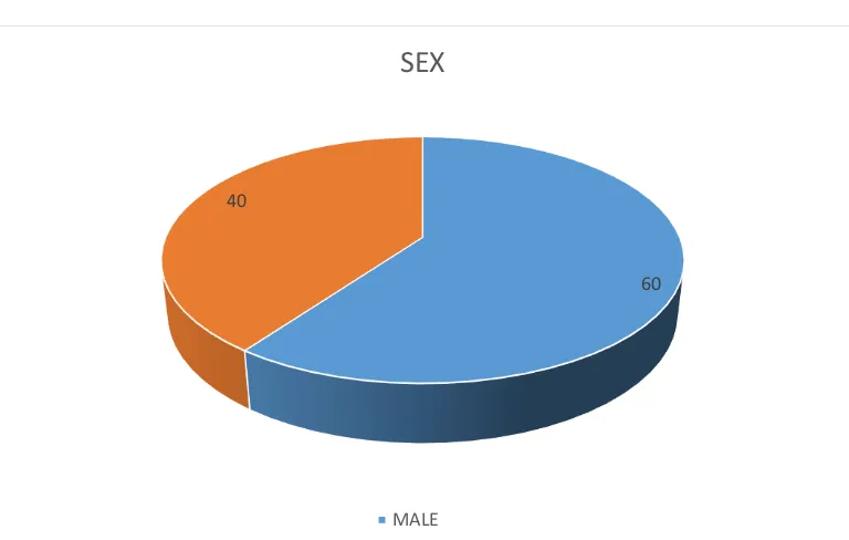
TABLE 1- COMPARISSION OF SEX
60 40
SEX
MALE
SEX MALE FEMALE
NO 30 20
67
TABLE 2 - COMPARISSION OF AGE
S.NO AGE (years) PERSON
1 <20 0
2 20 – 29 5
3 30 – 39 8
4 40 – 49 20
5 50 – 59 9
6 >59 8
0
5
8
20 9
8
AGE
68
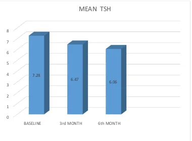
TABLE - 3 MEAN TSH COMPARISION
S.NO DURATION MEAN S.D. SIGNIFICANCE
1 BASELINE 7.28 1.06 <.001 2 3rd MONTH 6.47 1.01 <.001 3 6th MONTH 6.06 0.94 <.001
0 1 2 3 4 5 6 7 8
BASELINE 3rd MONTH 6th MONTH 7.28
6.47 6.06
69
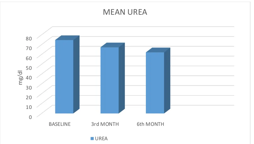
TABLE - 4 MEAN UREA COMPARISION
S.NO DURATION MEAN S.D. SIGNIFICANCE
1 BASELINE 74.72 21.64 <.001
2 3rd MONTH 67.34 19.17 <.001
3 6th MONTH 62.20 17.51 <.001
0 10 20 30 40 50 60 70 80
BASELINE 3rd MONTH 6th MONTH
m
g/d
l
MEAN UREA
70
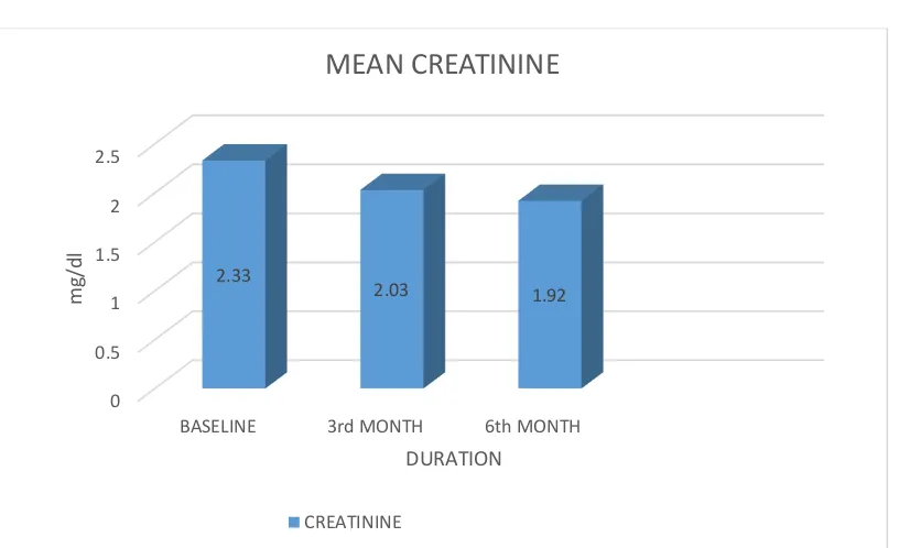
TABLE - 5 MEAN CREATININE COMPARISION
S.NO DURATION MEAN S.D. SIGNIFICANCE
1 BASELINE 2.33 0.71 <.001 2 3rd MONTH 2.03 0.61 <.001
3 6th MONTH 1.92 0.59 <.001
0 0.5 1 1.5 2 2.5
BASELINE 3rd MONTH 6th MONTH 2.33
2.03 1.92
m
g/d
l
DURATION MEAN CREATININE
71
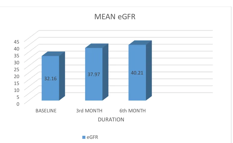
TABLE 6 – MEAN eGFR COMPARISION
0 5 10 15 20 25 30 35 40 45
BASELINE 3rd MONTH 6th MONTH
32.16 37.97 40.21
DURATION MEAN eGFR
eGFR
S.NO. DURATION MEAN S.D. SIGNIFICANCE
72
DISCUSSION
Even though previous studies have demonstrated that restoration of euthyroidism has beneficial effects on cardiac dysfunction in patients with SCH, the impact of THRT on renal function has not been extensively explored in these patients. The results of this study show that thyroid hormone treatment significantly abrogated the decrease in eGFR in CKD patients with SCH.
73
and 3% of men. This clearly demonstrates the higher prevalence of hypothyroidism in patients with chronic renal dysfunction. The effect of thyroid hormone replacement on renal function has not been widely investigated in hypothyroid CKD patients, especially in SCH. A recent study by Shin et al. demonstrated that thyroid hormone treatment not only preserved renal function but was also an independent predictor of renal outcome. However, they compared changes in eGFR in two different study populations. Thus, to clarify the direct impact of thyroid hormone treatment on the decline in renal function, it was imperative to compare decline in eGFR before and after L-thyroxine replacement in the same patient.
74
CONCLUSION
75
SUMMARY
BIBLIOGRAPHY
1. Vaidya B, Pearce SH. Management of hypothyroidism in adults. BMJ. 2008;337:a801.
2. Almandoz JP, Gharib H. Hypothyroidism: etiology, diagnosis, and management. Med Clin North Am. 2012;96:203-221
3. Caturegli P, De Remigis A, Rose NR. Hashimoto thyroiditis: clinical and diagnostic criteria. Autoimmun Rev. 2014;13:391-397
4. Vanderpump MP, Tunbridge WM, French JM, et al. The incidence of thyroid disorders in the community: a twenty-year follow-up of the Whickham Survey. Clin Endocrinol (Oxf). 1995;43:55-68.
5. Hollowell JG, Staehling NW, Flanders WD, et al. Serum TSH, T(4), and thyroid antibodies in the United States population (1988 to 1994): National Health and Nutrition Examination Survey (NHANES III). J Clin Endocrinol Metab.2002;87:489-499.
6. Cooper DS, Biondi B. Subclinical thyroid disease. Lancet. 2012;379:1142-1154.
7. Biondi B. Natural history, diagnosis and management of subclinical thyroid dysfunction. Best Pract Res Clin Endocrinol Metab. 2012;26: 431-446
9. Safer JD. Thyroid hormone action on skin. Curr Opin Endocrinol Diabetes Obes. 2012;19:388-393
10. 10. Kahaly GJ, Dillmann WH. Thyroid hormone action in the heart. Endocr Rev. 2005;26:704-728.
11. Danzi S, Klein I. Thyroid hormone and the cardiovascular system. Med Clin North Am. 2012;96:257-268.
12. Schlenker EH. Effects of hypothyroidism on the respiratory system and control of breathing: human studies and animal models. Respir Physiol Neurobiol. 2012;181:123-131
13. Williams GR. Neurodevelopmental and neurophysiological actions of thyroid hormone. J Neuroendocrinol. 2008;20:784-794.
14. Wood-Allum CA, Shaw PJ. Thyroid disease and the nervous system. Handb Clin Neurol. 2014;120:703-735
15. Tagoe CE, Zezon A, Khattri S. Rheumatic manifestations of autoimmune thyroid disease: the other autoimmune disease. J Rheumatol. 2012;39:1125-1129
16. Combs CE, Nicholls JJ, Duncan Bassett JH, Williams GR. Thyroid hormones and bone development. Minerva Endocrinol. 2011;36:71-85. 17. Wojcicka A, Bassett JH, Williams GR. Mechanisms of action of thyroid
hormones in the skeleton. Biochim Biophys Acta. 2013;1830:3979-3986. 18. Mintziori G, Anagnostis P, Toulis KA, Goulis DG. Thyroid diseases and
19. Mullur R, Liu YY, Brent GA. Thyroid hormone regulation of metabolism. Physiol Rev. 2014;94:355-382.
20. Pearce EN. Thyroid hormone and obesity. Curr Opin Endocrinol Diabetes Obes. 2012;19:408-413.
21. Iwen KA, Schroder E, Brabant G. Thyroid hormones and the metabolic syndrome. Eur Thyroid J. 2013;2:83-92
22. Stancu S, Barsan L, Stanciu A, et al. Can the response to iron therapy be predicted in anemic nondialysis patients with chronic kidney disease? Clin J Am Soc Nephrol. 2010;5(3): 409–416.
23. Wavamunno MD, Harris DC. The need for early nephrology referral.Kidney Int Suppl. 2005;(94):S128–S132.
24. Toto RD. Treatment of hypertension in chronic kidney disease. Semin Nephrol. 2005;25(6): 435–439.
25. Hou FF, Zhang X, Zhang GH, et al. Efficacy and safety of benazepril for advanced chronic renal insufficiency. N Engl J Med. 2006;354(2):131– 140.
26. Mann JF, Schmieder RE, McQueen M, et al. Renal outcomes with telmisartan, ramipril, or both, in people at high vascular risk (the ONTARGET study): a multicentre, randomised, double-blind, controlled trial. Lancet. 2008;372(9638):547–553.
Madero M, Sarnak MJ, Wang X, et al. Uric acid and long-term outcomes in CKD. Am J Kidney Dis. 2009;53(5):796–803.
28. Gaffo AL, Saag KG. Management of hyperuricemia and gout in CKD.Am J Kidney Dis. 2008;52(5):994–1009.
29. Goicoechea M, de Vinuesa SG, Verdalles U, et al. Effect of allopurinol in chronic kidney disease progression and cardiovascular risk. Clin J Am Soc Nephrol. 2010;5(8):1388–1393.
30. Okusa MD, Chertow GM, Portilla D. The nexus of acute kidney injury, chronic kidney disease, and World Kidney Day 2009. Clin Am Soc Nephrol. 2009;4(3):520–522. Kidney Int. 2006;70(12):2116– 2123.
31. Van der Velde M, Matsushita K, Coresh J, et al. Lower estimated glomerular fltration rate and higher albuminuria are associated with all-cause and cardiovascular mortality. A collaborative meta-analysis of high-risk population cohorts. Kidney Int. 2011;79(12):1341-1352.
32. Astor BC, Matsushita K, Gansevoort RT, et al. Lower estimated glomerular fltration rate and higher albuminuria are associated with mortality and end-stage renal disease. A collaborative meta-analysis of kidney disease population cohorts. Kidney Int. 2011;79(12):1331-1340. 33. Pan Y, Guo LL, Jin HM. Low-protein diet for diabetic nephropathy:
34. Lambers Heerspink HJ, Gansevoort RT, et al. Comparison of different measures of urinary protein excretion for prediction of renal events. J Am Soc Nephrol. 2010;21(8):1355-1360.
35. Stengel B, Tarver-Carr ME, Powe NR, et al. Lifestyle factors, obesity and the risk of chronic kidney disease. Epidemiology. 2003;14(4):479-487. 36. Yusuf S, Hawken S, Ounpuu S, et al. Effect of potentially modifable
risk factors associated with myocardial infarction in 52 countries (the INTERHEART study): case-control study. Lancet. 2004;364(9438):937-952.
37. O’Donnell MJ, Xavier D, Liu L, et al. Risk factors for ischaemic and intracerebral haemorrhagic stroke in 22 countries (the INTERSTROKE study): a case-control study. Lancet. 2010;376(9735):112-123.
38. Scott LJ, Warram JH, Hanna LS, et al. A nonlinear effect of hyperglycemia and current cigarette smoking are major determinants of the onset of microalbuminuria in type 1 diabetes. Diabetes. 2001;50(12):2842-2849
PROFORMA
Name: Age/ Sex: Occupation:
Presenting complaints:
Past history:
H/O Diabetes, Systemic Hypertension, Coronary artery disease, H/O thyroid disease in the past
H/O drug intake in the past
Personal history
H/O Alcohol consumption
General Examination
Vitals
BP
Pulse rate RR
System Examination
CVS:
RS:
Abdomen:
CNS:
LIST OF ABBREVIATION
GFR : Glomerular Filtration Rate ESRD : End Stage Renal Disease CKD : Chronic Kidney Disease
ESA : Erythropoiesis Stimulating Agent PCR : Protein Creatinine Ratio
DM : Diabetes Mellitus
TBG : Thyroxine Binding Globulin TTR : Transthyretin
TSH : Thyroid Stimulating Hormone TRH : Thyrotropin Releasing Hormone
MASTER CHART
AGE SEX eGFR 0 eGFR 3 eGFR 6 TSH 0 TSH 3 TSH 6 UREA 0 UREA 3 UREA 6 CREA 0 CREA 3 CREA 6
46 1 22.34 24.06 26.06 7.8 6.1 5.3 78 70 64 3.2 3 2.8
51 1 16.43 19.09 19.73 6.4 5.4 5 83 78 72 4.1 3.6 3.5
42 1 30.25 37 39.14 7.2 6.2 5.7 48 46 40 2.5 2.1 2
43 2 27.32 34.86 37.39 6.2 5.1 4.8 54 50 52 2.1 1.7 1.6
48 1 20.65 23.86 23.86 5.8 5.1 4.8 82 70 68 3.4 3 3
61 1 46.94 54.76 59.65 8.1 6.1 5.9 48 44 40 1.6 1.4 1.3
57 2 24.45 25.8 27.3 8.6 7.5 6.5 58 56 50 2.2 2.1 2
38 1 33.99 37.76 39.94 7.6 6.5 5.8 69 54 40 2.3 2.1 2
32 1 37.05 46.71 53.51 7.2 6.1 6 74 60 59 2.2 1.8 1.6
46 2 39.73 51.4 51.4 6.6 5.4 5.2 48 40 38 1.5 1.2 1.2
33 1 19.02 23.06 25.74 6.3 6.1 5.8 127 114 100 3.9 3.3 3
28 2 33.46 40.79 43.95 7.4 7 7 76 68 65 1.9 1.6 1.5
43 1 28.78 31.56 31.56 9.8 9.6 9.3 88 83 80 2.6 2.4 2.4
46 1 49.71 57.99 53.55 7.2 6.8 6.5 72 68 66 1.6 1.5 1.5
40 1 26.81 30.55 32.03 6.8 6.2 6 110 105 97 2.8 2.5 2.4
38 2 19.3 22.91 24.01 5.9 5.5 5.2 88 73 70 2.9 2.5 2.4
62 2 26.83 30.3 34.71 8.6 8.3 8 93 88 72 2 1.8 1.6
54 1 42 48.12 51.84 7.4 7.2 6.8 94 90 85 1.8 1.6 1.5
58 1 48.12 55.32 55.32 8.1 7.8 7.5 60 48 40 1.5 1.4 1.4
59 1 26.99 29.6 32.72 6.3 5.9 5.5 78 76 70 2.6 2.4 2.2
43 2 25.89 28.9 32.64 6.1 5.8 5.7 64 60 58 2.2 2 1.8
56 2 31.05 35.57 38.18 5.8 5.5 5.5 56 50 40 1.8 1.6 1.5
65 1 40.08 49.47 49.47 7.4 7 6.4 52 48 45 1.8 1.5 1.5
43 1 34.89 38.95 38.95 7.1 6.9 6.5 70 68 57 2.2 2 2
24 2 34.52 42.09 39.25 6.8 6.5 6.3 63 55 86 1.9 1.9 1.7
63 1 34.07 38.24 40.17 6.3 6 5.7 99 86 78 2.1 1.9 1.8
65 1 15.77 17.8 19.02 5.9 5.4 5.3 103 89 76 4 3.6 3.4
49 2 19.91 23.95 25.21 6.2 5.9 5.2 63 58 55 2.7 2.3 2.2
36 2 31.79 36.15 41.76 8.1 7.8 7.5 59 58 46 1.9 1.7 1.5
24 1 52.89 61.11 66.18 9.2 9 8.2 49 46 45 1.7 1.5 1.4
30 2 43.34 51.12 56.06 6.6 6.3 6.1 44 43 40 1.5 1.3 1.2
36 2 19.52 23.16 24.28 7.3 7 5.9 93 90 75 2.9 2.5 2.4
45 1 26.18 29.81 32.85 5.8 5.2 5 85 78 60 2.8 2.5 2.3
40 1 20.72 24.76 25.74 6 5.5 5.1 130 110 98 3.5 3 2.9
65 1 37.77 46.05 44.61 7.1 5.6 5 38 36 35 1.9 1.6 1.5
48 2 28.26 34.09 39.39 6.5 5.8 4.9 56 48 44 2 1.7 1.5
27 1 36.43 42.81 42.81 8.6 7.5 6.4 77 73 86 2.3 2 2
36 1 52.24 60.95 60.95 8.2 6.1 5.8 66 53 50 1.6 1.4 1.4
43 1 41.33 50.39 54.29 7.1 5.8 5.5 61 55 52 1.9 1.6 1.5
60 1 47.1 59.85 65.64 6.2 5.8 5.5 66 53 50 1.6 1.3 1.2
58 2 24.37 30.72 47.42 7.1 6.4 6.6 78 76 70 2.2 1.8 1.6
56 2 20.24 25.89 27.39 8.3 6.2 6 88 84 76 2.6 2.1 2
62 2 19.67 24.61 26.72 6.4 5.6 5.5 103 97 80 3.4 2.8 2.6
49 2 19.89 28.02 21.76 7.6 6.1 5.7 118 86 73 3.5 2.6 2.5
47 2 28.39 30.12 34.24 8.2 7.3 7.2 92 88 86 2 1.9 1.7
40 1 24.76 30.55 33.64 9.6 8.1 6.6 90 76 68 3 2.5 2.3
56 1 41.69 47.76 51.45 8 6.2 5.9 66 63 64 1.8 1.6 1.5
40 2 33.12 40.88 40.88 8.3 7.1 6.3 64 57 58 1.8 1.5 1.5
26 1 43.14 48.72 55.81 9.2 8 7.1 43 42 40 2 1.8 1.6
10/12/2018 Gmail - [Urkund] 21% similarity - drkannanraja@gmail.com
https://mail.google.com/mail/u/0?ik=22e0d06b9a&view=pt&search=all&permthid=thread-f%3A1614086861231973450&simpl=msg-f%3A16140868612… 1/1
kannan rajalingam <drkannanraja@gmail.com>
[Urkund] 21% similarity - drkannanraja@gmail.com
1 message
report@analysis.urkund.com <report@analysis.urkund.com> Fri, Oct 12, 2018 at 8:29 AM To: drkannanraja@gmail.com
Document sent by: drkannanraja@gmail.com
Document received: 10/12/2018 4:59:00 AM
Report generated 10/12/2018 4:59:43 AM by Urkund's system for automatic control.
Student message:
---
Document : kannan dissertation.docx [D42453214]
IMPORTANT! The analysis contains 1 warning(s).
About 21% of this document consists of text similar to text found in 123 sources. The largest marking is 234 words long and is 99% similar to its primary source.
PLEASE NOTE that the above figures do not automatically mean that there is plagiarism in the document. There may be good reasons as to why parts of a text also appear in other sources. For a reasonable suspicion of academic dishonesty to present itself, the analysis, possibly found sources and the original document need to be examined closely.
Click here to open the analysis:
https://secure.urkund.com/view/41487137-349694-448173
Click here to download the document: