A STUDY ON MICROBIOLOGICAL PROFILE OF VAGINITIS AND ITS ASSOCIATION WITH URINARY TRACT INFECTION
DURING PREGNANCY IN A TERTIARY CARE HOSPITAL
Dissertation submitted for
M.D. MICROBIOLOGY BRANCH – 1V DEGREE EXAMINATION
THE TAMILNADU DR.M.G.R.MEDICAL UNIVERSITY CHENNAI – 600 032
TAMILNADU
CERTIFICATE
This is to certify that this dissertation titled “A STUDY ON
MICROBIOLOGICAL PROFILE OF VAGINITIS AND ITS ASSOCIATION
WITH URINARY TRACT INFECTION DURING PREGNANCY IN A
TERTIARY CARE HOSPITAL” is a bonafide record of work done by
Dr.G.JABEEN FATHIMA, during the period of March 2017 to February 2018 under the guidance of Prof.Dr.S.THASNEEM BANU M.D., Professor of Microbiology, Institute of Microbiology , Madras Medical College and Rajiv
Gandhi Government General Hospital, Chennai - 600003, in partial fulfillment of
the requirement of M.D. MICROBIOLOGY Degree Examination of The
Tamilnadu Dr.M.G.R. Medical University to be held in May 2019.
Dr.R.JAYANTHI , M.D.FRCP (Glas )
Dean,
Madras Medical College &
Rajiv Gandhi Govt. General Hospital Chennai – 600003.
Dr.J.EUPHRASIA LATHA.,M.D.DGO
Director,
Madras Medical College &
DECLARATION
I,Dr.G.JABEEN FATHIMA, Post Graduate , Institute of Microbiology, Madras Medical College, solemnly declare that the dissertation titled “A STUDY
ON MICROBIOLOGICAL PROFILE OF VAGINITIS AND ITS ASSOCIATION
WITH URINARY TRACT INFECTION DURING PREGNANCY IN A
TERTIARY CARE HOSPITAL” is the bonafide work done by me at Institute of
Microbiology, Madras Medical College under the expert guidance and supervision of Prof.Dr.S.THASNEEM BANU,M.D., Professor, Institute of Microbiology, Madras Medical College. The dissertation is submitted to the Tamil Nadu Dr.
M.G.R Medical University towards partial fulfillment of requirement for the
award of M.D., Degree (Branch IV) in Microbiology.
Place: Chennai
Date: Dr.G.JABEEN FATHIMA
Signature of the Guide
Prof. Dr.S.THASNEEM BANU ,MD., Professor,
Institute of Microbiology
ACKNOWLEDGEMENTS
I wish to express my sincere thanks to Dr.R.JAYANTHI,M.D.FRCP (Glas ), Dean, Rajiv Gandhi Government General Hospital & Madras Medical College, Chennai-3 for permitting me to use the resources of this institution for
my study.
I express my thanks to Dr.J.EuphrasiaLatha, M.D.DGO, Director, Institute of Microbiology for her guidance and support.
Sincere thanks toFormer Professor Dr.MangalaAdisesh M.D., Institute of Microbiology for her constant encouragement and support during this work.
I owe my heartfelt gratitude and sincere thanks to my guide
Dr.S.THASNEEM BANU, M.D.,Professor, Institute of Microbiology for her valuable suggestions,guidance,constant support, motivation and encouragement
throughout this study.
My Sincere thanks to Dr.T.K.ShaathyGunasingh, M.D., Director , Institute of Obstetrics and Gynaecology for her constant encouragement and
I would like to thank all my Professors Dr.U.Umadevi M.D., Dr.R.Vanaja M.D., and Dr.C.P.Ramani M.D., for their support during this study.
I extend my gratitude to my co-guide DR.S.VINOTHA, M.D.,Assistant Professor, Institute of Microbiology for his valuable guidance and constant
support in this study.
I extend my gratitude to my former co-guide Dr.C.S.SripriyaM.D., Senior Assistant Professor, Institute of Microbiology for her valuable guidance
and support in this study.
I wish to extend my thanks to our Assistant Professors
Dr.Lakshmipriya.NM.D.DCH., Dr.K.G.Venkatesh M.D., Dr.P.Shanmugapriya M.D., Dr.C.Nithya M.D., Dr.J.PadmaKumariM.D., Dr.R.KesavanM.D.DCH., Dr.B.Gomathi Manu M.D., for their support.
I wish to extend my thanks to our former Assistant Professors Dr.Deepa.R M.D., Dr.K.Ushakrishnan M.D., Dr.N.RathnaPriya M.D., Dr.DavidAgatha M.D., Dr.B.Natesan M.D.DLO., for their support.
I would like to extend my thanks to all my postgraduate colleagues and
I thank my husband Dr.A.R.AKBARM.D.S.,for his constant motivation, emotional support and help in completing the dissertation work. I feel indebted to my father MR.YOUSUFF GHULAM MOHAMED and mother MRS.G.HALEEMA who had been solid pillars of everlasting support and encouragement and for their heartfelt blessings. I thank my inlaws for their
support, blessings and encouragement .I thank my lovable sons A.R.ASLAM and A.R.AKRAM for their love and sacrifice.
I would like to thank the Institutional Ethics Committee ,Madras Medical college for approving my study .
Last but not the least, I would like to thank the patients participated in this
TABLE OF CONTENTS
Sl.
No. TITLE Page No.
1 Introduction 1
2 Aims and Objectives 5
3 Review of Literature 6
4 Materials and Methods 37
5 Results 60
6 Discussion 81
7 Summary 90
8 Conclusion 92
9 Colour Plates 93
10 Bibliography
Appendix- I Abbreviations
Appendix- II Stains, Reagents, Media
Annexure –I Certificate of Approval
Annexure –II Proforma
Annexure –III Patients Consent Form
LIST OF TABLES
Sl.
No TITLE
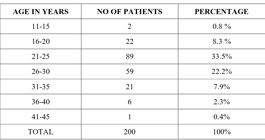
Page No. 1 Distribution of age (in years) of the patients included in the
study 60
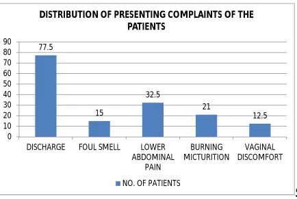
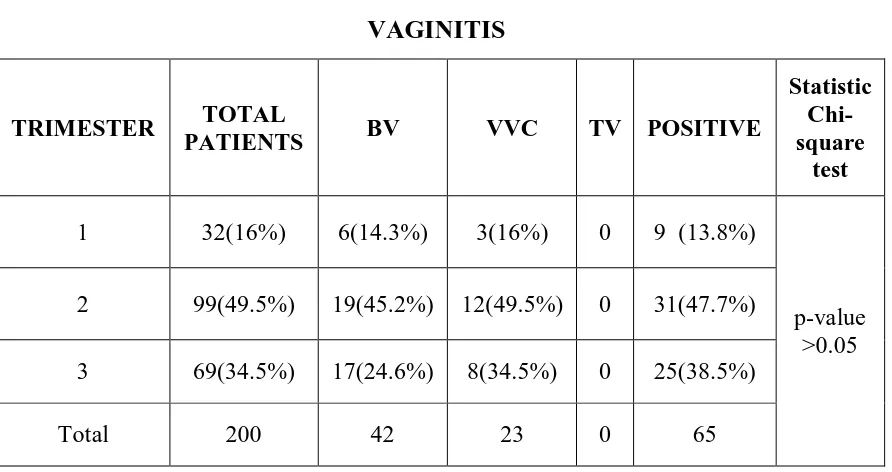
2 Distribution of presenting complaints of the patients 61 3 Age wise distribution of the presenting complaints 62 4 Trimester wise distribution of Infectious vaginitis 63
5 Trimester wise distribution of UTI 64
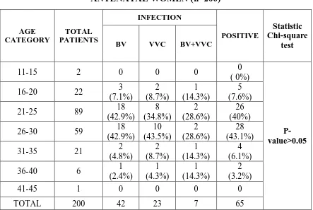
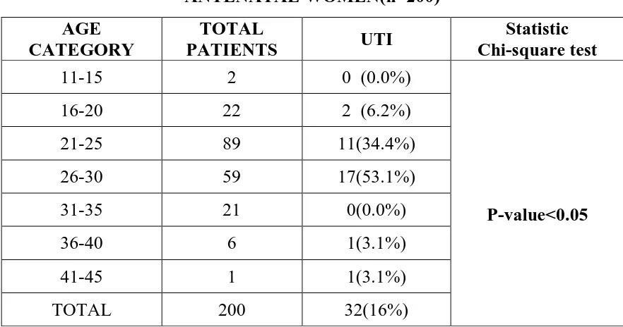
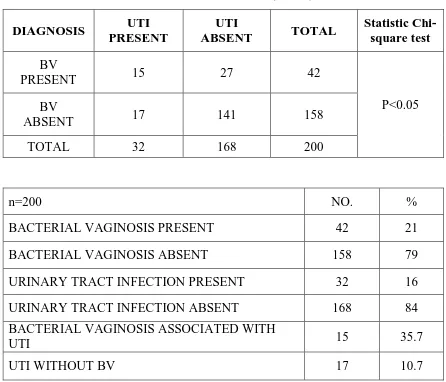
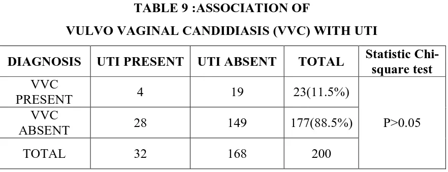
6 Age wise distribution of vaginitis among antenatal women 65 7 Age wise distribution of UTI among antenatal women 66 8 Association of bacterial vaginosis with UTI during pregnancy 67 9 Association of vulvo vaginal candidiasis with UTI 68 10 Amsel’s criteria for diagnosis of BV 68 11 Nugent’s scoring for diagnosis of BV 69 12 Diagnosis of BV by comparison between Amsel’s criteria and
Nugent’s scoring. 70
13 Bacterial isolates detected from HVS and urine culture 71 14 Antimicrobial susceptibility pattern for urinary isolates GNB 74 15 Antimicrobial susceptibility pattern for urinary isolates GPC 74 16 Antimicrobial susceptibility pattern for vaginal isolates GNB 75 17 Antimicrobial susceptibility pattern for vaginal isolates GPC 76
18 Candida species isolated from HVS 77
19 Antifungal susceptibility pattern of Candida isolates from HVS
samples 78
20 Candida species isolated from urine 79
21 Antifungal susceptibility pattern of candida isolates from urine
LIST OF FIGURES
S.No. TITLE Page No.
1 Distribution of age (in years) of the patients 61
2 Distribution of presenting complaints of the patients 62
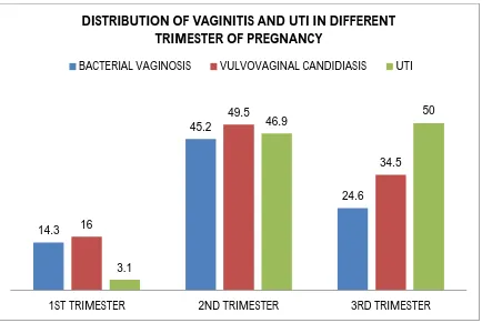
3 Distribution of vaginitis and UTI in different trimester 64
4 Distribution of vaginitis and UTI in different age group 66
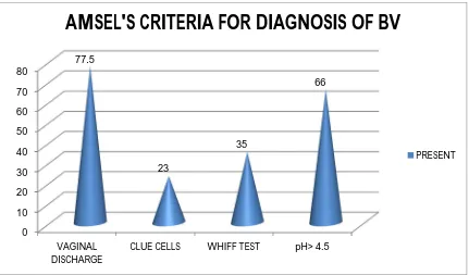
5 Amsel’s Criteria for diagnosis of BV 69
6 Nugent’s scoring for diagnosis of BV 70
7 Trimester wise distribution of isolates from urine 72
8 Trimester wise distribution of isolates from HVS 73
9 Distribution of Candida isolates from HVS 77
LIST OF COLOUR PLATES
Sl.
No. TITLE
1 Direct Gram stain showing normal vaginal epithelial cell 2 Direct Gram stain of HVS showing clue cells
3 Culture smear showing GPC in clusters from a patient with BV 4 Direct Gram stain picture of a patient with VVC
5 pH measurement for Amsel’s criteria
6
BAP showing βhemolytic colonies of S.agalactiae isolated from HVS sample.
7 S.agalactiae showing positive CAMP test
8 S.agalactiae showing resistance to bacitracin and cotrimoxazole disc 9 CONS speciation by using novobiocin and polymyxin B disc
10 Biochemical reactions and colonies of E.coli on CLED 11 ESBL production in E.coli
12 Biochemical reactions and colonies of K.pneumoniae on MAC 13 Vancomycin MIC by E test for MRSA strain
14 SDA culture of Candida albicans
15 Candida albicans showing positive Germ tube test
16 Sugar fermentation test showing reactions for C.albicans 17 Antifungal susceptibility by disc diffusion for C.albicans 18 CHROM agar showing colonies of C.tropicalis
CERTIFICATE II
This is to certify that this dissertation work titled “A STUDY ON MICROBIOLOGICAL PROFILE OF VAGINITIS AND ITS ASSOCIATION WITH URINARY TRACT INFECTION DURING PREGNANCY IN A TERTIARY CARE HOSPITAL” of the candidate Dr.G.JABEEN FATHIMA with registration number 201614005 for the award
of M.D.,Degree in the branch of Microbiology.I personally verified the
urkund.com website for the purpose of plagiarism check. I found that the
uploaded thesis file contains from introduction to conclusion pages and result
shows 1 percentage of plagiarism in the dissertation.
1
INTRODUCTION
There is an increasing global burden on lower genital tract infections
among women of reproductive age group (both pregnant and non pregnant) . Some of the consequences of these infections are pelvic inflammatory disease,
abortions, preterm deliveries and also increasing HIV acquisition . Lower genital
tract infections can also lead to complications in pregnancy as well as infections
in the new born . These infections are mostly asymptomatic but can lead to
serious consequences in their reproductive outcome if left untreated.
Vaginitis refers to a non-specific inflammation of the vagina characterized
by watery discharge with burning and itching of the vulva3. The three common types of vaginitis includes Bacterial vaginosis (BV), Vulvovaginal candidiasis (VVC) and Trichomoniasis.
BV is a condition characterized by vaginal discharge with raised pH in
which normal vaginal flora of Lactobacilli is replaced by a mixed flora of aerobic, anaerobic and microaerophilic species18.In the past few decades awareness has increased on the importance of bacterial vaginosis in pregnancy, and it has been
discovered that pregnant women with BV are at risk of adverse pregnancy
outcomes including spontaneous abortion, premature birth, preterm labour,
preterm premature rupture of the membranes, amniotic fluid infection, postpartum
2
receiving extensive attention during pregnancy and it is found in 15 to 23% of
pregnant women with up to 50% of patients being asymptomatic21,22,23.
In normal healthy female mucosal candidiasis, especially vulvovaginal
candidiasis(VVC), is one of the most common fungal diseases8,9. Approximately about 75% of the women suffers at least one episode of VVC during their
lifetime10,11. The causative agent in most cases is Candida albicans8,10,12. The most common predisposing factors are pregnancy, diabetes mellitus, and antibiotic
treatment8-11. Some studies have shown that in the past few decades vulvovaginitis has increased due to the antifungal resistance among the various Candida species and change inthe health quality of women10,13-15. Therefore vulvovaginal candidiasis is found to be more common during pregnancy, and pregnant women
have higher rates of recurrent infections.
Trichomoniasis is an infection caused by a flagellated protozoon,
Trichomonas vaginalis. The antenatal women infected with this parasite may be at
risk of unfavourable birth outcomes such as premature rupture of membranes,
premature labour, and low birth weight 5. Trichomoniasis is associated with a 30% increase in low birth weight infants and a 30% increase in risk of preterm births34. It is also associated with enhanced predisposition to neoplastic transformation in
3
Urinary tract infections (UTIs) have a global annual incidence of
approximately 150 million cases4. It is one of the most common medical complications of pregnancy. During pregnancy the increased incidence of UTI is
due to the physiological and the morphological changes that take place in the
genitourinary tract24,25. Many hormonal and mechanical changes occur in the body during pregnancy26,27.Starting in the 6th week, with peak incidence during 22nd– 24th weeks of gestation, 90% of the pregnant women develop ureteric dilatation
which increases the risk of vesicoureteric reflux and urinary stasis28.Glycosuria and aminoaciduria during pregnancy in addition ,provide an excellent culture
medium for the bacteria in areas of urinarystasis27.These alterations along with already short urethra and difficulty with hygiene due to the distended belly in
pregnancy increase the frequency of UTIs in them.
UTI in pregnancy may present either as symptomatic infection or as
asymptomatic bacteriuria. The prevalence of asymptomatic bacteriuria range from
2% to 10% in the various studies done globally29. The prevalence of UTI (including both symptomatic and asymptomatic bacteriuria ) in India in pregnant women is reported to range from 3% to 24%25,30-33. UTI in pregnant women can increase the chances to develop anaemia, hypertensive diseases of pregnancy,
chronic renal failure, prematurity, and low birth weight babies39-41. The upper UTIs in particular may lead to significant morbidity for both the mother and the
4
In the association between UTIs and bacterial vaginosis sexual intercourse
has an important confounding role16,17. UTI in females develop when uropathogens almost always from the fecal flora colonize the vagina, ascend into
the bladder and in some cases into the kidney. Raised vaginal pH because of the
reduction in number of lactobacilli which produce lactate and hydrogen peroxide predispose to genitourinary infections38.UTI and vaginitis during pregnancy have risk to both the mother and the fetus. The lives of both can be saved by a single
step of early diagnosis and treatment . This study is aimed to determine the
microbiological profile of vaginitis and the association between BV and UTI
5
AIMS & OBJECTIVES
AIM:
• To identify common causes of infectious vaginitis in pregnancy and to
determine the risk of urinary tract infections in pregnant women with
bacterial vaginosis.
Objective:
To study type of infectious vaginitis during pregnancy.
To isolate UTI pathogens from urine during pregnancy & determine its
antimicrobial susceptibility pattern.
To determine the association between bacterial vaginosis& UTI in
6
REVIEW OF LITERATURE
Lower genital tract infections comprises of infections in the vulva, vagina
or cervix. Infections which affect the vulva and the vagina are called as vulvo-vaginitis. Vaginitis is treatable under ordinary conditions but if left untreated can
spread up to the upper genital organs leading to pelvic inflammatory disease.
Even gynecological procedures like menstrual regulation, insertion of IUDs and
induced abortions can lead to upper genital tract infections43. These pelvic infections can lead to serious consequences like salpingitis and chronic infections
of the pelvis and may later lead on to ectopic pregnancy or infertility.
The etiological agents causing vaginitis can be endogenous or exogenous
in origin. The common ones include bacterial vaginosis, vulvovaginal candidiasis, and trichomoniasis.
Bacterialvaginosis is a conditions where there is replacement of the normal
indigenous vaginal flora by abnormal bacteria. It is most often due to overgrowth of the commensal organisms. It is one of the commonest infection among the
lower genital tract infections in the reproductive age group44. According to CDC, Bacterial vaginosis is not transmitted sexually but conditions like having multiple
partners predispose to its occurrence46. The symptoms of bacterial vaginosis are homogenous thin vaginal discharge, lower abdominal pain which may be
7
The agents causing bacterial vaginosis are Gardneralla vaginalis,
Mobiluncus, Prevotella, Peptostreptococci, and Porphyromonas Species44. Overgrowth of these agents causes a reduction in the number of normally
occurring microbial flora in the vaginal tract which is responsible for the
production of lactic acid (lacto bacilli). The agents mentioned above can occur in
the range of 100-1000 times than the normal vaginal microbial organisms45. This leads to the development of bacterial vaginosis.
RISK FCTORS OF THE BV46
CDC has estimated the following risk factor for bacterial vaginosis
1. Having new sex partner or multiple sex partner
2. Douching
Though bacterial vaginosis is not known to be spread by sexual contact but
still women in the sexually active age-group have more predisposition for the
occurrence of BV.
The first diagnosis of BV was made by Amsel et al., who studied 397
female students and devised criteria to distinguish non-specific vaginitis from the
8
AMSEL’S DIAGNOSTIC CRITERIA21 1. Homogeneous vaginal discharge
2. Amine odour after the addition of KOH (Whiff test)
3. Presence of Clue cells in gram strain
4. Vaginal pH>4.5
at least 3 out of 4 criteria must be met for the diagnosis of bacterial vaginosis.
Nugent et al. devised another scoring system comparing the occurrence of
Lactobacilli, Gardnerella vaginalis or Bacteroides species and curved gram
variable bacteria. His scoring system ranged from 0-10 depending upon the occurrence of the above organisms22.
NUGENT’S SCORING
Score
Lactobacillus Morphotype per
field
Gardnerella Morphotype
per field
Curved Bacteria (Mobiluncus)
Per field
0 >30 0 0
1 5-30 <1 <1-4
2 1-4 1-4 >5
3 <1 5-30 -
4 0 >30 -
SCORING 0-3 Normal/No Bacterial vaginosis 4-6 Intermediate
9
Clue cells are vaginal epithelial cells whose cell borders obscured by the
adherent small cocco bacilli or gram negative rods or gram variable rods. They represent important criteria in Amsel’s criteria for bacterial vaginosis. They can
be identified on wet mount and Gram stain. Most often clue cells are obscured by
mixed flora consisting of Gram-negative bacilli, Gram-variable bacilli and cocco
bacilli21.
Bacterial vaginosis is an infection of the vagina caused by mainly
anaerobic bacteria like Prevotella, Porphyromonas , Peptostreptococci, and
Gardnerella44. It is a polymicrobial condition caused by mixed organisms. The bacterial vaginosisis also called anaerobic vaginosis, haemophilus, and
gardnerella vaginosis. Bacterial vaginosis is significantly associated with many
pathological conditions like chorioamnionitis60.amnionitis49, endometritis51 and prematurity. Anaerobic vaginosis represents a disturbance in the vaginal
microbial ecosystem.
Bacterial vaginosis is the leading cause associated with the vaginal discharge accounting up to 48% of cases49. Prevalence of bacterial vaginosis is estimated to be about 15 to 23% in pregnant women with up to 50% of patients
being asymptomatic21,22,23.
Apart from causing bacterial vaginosis, Gardnerella can also occur as a
normal vaginal flora. Studies have shown that there was no difference between
10
and virginal women and therefore bacterial vaginosis is not to be considered as a
sexually transmitted disease. Around 17.5-29.3% of women going for induced
abortion had bacterial vaginosis. Thus bacterial vaginosis is known to be a
predisposing factor for miscarriages and pre-term deliveries 22.
Tsirmpa et al showed an association of 5% between Bacterial vaginosis
and Trichomoniasis47. It is plausible that T. vaginalis infection alters the vaginal ecology and facilitates the development of BV, or that women with BV have lost
natural protection against genital tract infections leading to the acquisition of STIs
like T. vaginalis infection. Mathivanan et al., showed a significant correlation between bacterial vaginosis and other STIs. Bacterial vaginosis was also found to
be more prevalent in patients with tubal infertility( 31.5%) than in patients with
non-tubal infertility44. Prevalence of bacterial vaginosis in female STD patients was 16% more than in the normal population. Similarly, bacterial vaginosis was
found in 15% of women prisoners. Higher incidence of bacterial vaginosis was
seen in rape victims (50%) and sex workers (70%), the correlation of bacterial
vaginosis to uro-gential infections was found to be 19.7% in women of reproductive age-group49. The score of greater than 6 by the Nugent’s criteria estimates the presence of bacterial vaginosis in infertile women to be around
19%53. A score of 4-10 is also found in 39% of infertile women53. Thus infertility was found to be correlated with prevalence of bacterial vaginosis. Tubal
infertility was found to be significantly associated with women having bacterial
11
also associated with pre-term deliveries and miscarriages. Thus women having
bacterial vaginosis have increased rate of abortions51.
TREATMENT:
Bacterial vaginosis treatment in asymptomatic pregnant women at high risk
(i.e those who have previously delivered a premature infant) for preterm delivery
with a recommended oral regimen has reduced preterm delivery in three out of
four randomized control trials.
Bacterial Vaginosis in pregnant women is treated with metronidazole
500mg orally twice a day for 7 days or metronidazole 250mg orally three times a
day for 7 days or clindamycin 300 mg orally twice a day for 7 days. Since
metronidazole is contraindicated in 1st trimester of pregnancy because of possible teratogenic effect , amoxicillin although less effective is suggested as an alternative treatment.
TRICHOMONIASIS
Trichomonas vaginalis is a sexually transmitted protozoan parasite. Every year worldwide around 180 million cases of trichomoniasis occurs49. It is a common sexually transmitted infection and occurs as a mixed infection often with
other organism like Gonococci. The co-infection rate is around 60%. The
spectrum of clinical symptoms can vary from asymptomatic to severe disease.
This organism is transmitted mainly by sexual intercourse through coitus from the
12
malodorous, frothy, thin vaginal discharge and comprises of signs like burning
sensation, itchiness, dysuria and dyspareunia. The classical sign is erythematous
vulva and punctuate hemorrhages seen in the vaginal wall. This is called
strawberry vagina/cervix54.
MORPHOLOGY
Among the Trichomonads, Trichomonas vaginalis is the commonly
occurring species. The size of the protozoan parasite may range from 7-10 mm55. Size and shape may be altered by physio-chemical conditions. But the parasite is
mostly seen as pear or oval-shaped in axenic cultures but becomes amoeboid when it attaches to vaginal epithelial cells. It comprises of five flagella, four
located on its anterior portion. The fifth flagellum is incorporated inside the
undulating membrane which gives it a quivering motility55. Nucleus of this parasite is located on its anterior portion. Rod-like structure called axo style
bisects the parasite longitudinally and terminates as a sharp point at its posterior
end55. This structure is responsible for anchorage of the parasite to the vaginal epithelial cells.
CLINICAL MANIFESTATION
Clinical patterns of Trichomoniasis may vary from asymptomatic carrier
state to symptomatic disease comprising of vaginal discharge. This parasite infects the squamous epithelium of the vaginal tract. The infection prevails for
13
picture of the disease can be classified as acute, chronic or asymptomatic56. Acute infection presents as copious leucorrhoea. The disease presents with yellow or
greenish frothy and mucopurulent vaginal discharge56. The disease presents with clinical signs and symptoms which are cyclic and can worsen during menstrual
period. Important sign in this disease is the fiery red appearing cervix/vagina
which is called as strawberry appearance57. This classical description is seen only in 2% of patients. Chronic infection presents with symptoms like pruritus,
dyspareunia and absence of discharge. Majority of the patients are asymptomatic
and may have normal vaginal pH and normal vaginal flora. After infection
around 50% of women develop symptoms within 6 months58. The other organ involved in the infection is bartholin’s gland. Complications of this disease may
include adnexitis, endometritis and pyosalpinx. These complications may lead to
cervical erosion and therefore cause infertility and low birth weight. There are many studies showing an increased association between HIV and
trichomoniasis59,60.
MICROSCOPIC AND CULTURE TECHNIQUES
Donne et al., was the first to describe the use of microscope for the
observation of Trichomonas in vaginal and cervical secretions61. This technique has sensitivity ranging from 38% to as high as 82%62. Though this technique is cost-effective but has a low sensitivity since this parasite may lose its motility
14
The gold standard for the diagnosis of trichmoniasis is broth culture of the
organism. This method requires as few as 300-500 parasite per ml of inoculum63.
Cell culture techniques are also useful for the recovery of Trichomonas
vaginalis from clinical specimens as demonstrated by Garber et al., he showed
that the cell culture was superior to both broth culture and wet mount.
Stains used are Periodic Acid Schiff (PAS)65,Acridine orange64,Leishman, and Fontana. Papanicolaou (PAP) stain also shows significant appeal in the
diagnosis of trichomoniasis. Pap stain is routinely used in gynaecological
screening of cytological abnormalities.
All staining techniques have considerable limitations because the parasite
appears pear-shaped and may resemble polymorphonuclear leucocytes. The
typical morphology of the parasite are also lost during fixation of the smears66. Antigenic markers of Trichomonas can be demonstrated by immune-blot
analysis67. Other techniques used for the diagnosis are complement fixation, indirect haemagglutination, gel-diffusion, fluorescent antibody and ELISA. All these techniques detect for the presence of anti-trichomonal antibodies.
TREATMENT
Untill 1959, only topical vaginal preparations were available for trichomoniasis. But topical applications were not useful since they did not
penetrate the vaginal epithelium. Even after the treatment the parasite was
15
include clotrimazole, providone iodine and nonoxynol-9, which provide palliative
cure. Azomycin antibiotic was found to be highly effective in the treatment of
trichomoniasis68. They produce nitroimidazole which are cytocidal to the parasite after its diffusion into the organism (Mueller et al.,)69. These metabolic products breakdown the DNA strands of the organisms69. As a result cell death ensures due to the loss of mitotic activity and the parasite dies within one hour. The cell death is depicted within 8 hours in cell culture.
Metronidazole 250mg given orally for 7 day is the standard treatment for
trichomoniasis. Both symptomatic and asymptomatic diseases need treatement70. Male partner also has to be treated in order to prevent re-infection. Both partner
treatment has a success rate of almost 95% (Heine et al.,)65.
Trichomonas vaginalis is more common among HIV infected patients. The incidence of trichomoniasis and HIV co-infection ranges between 6-27%.
Trichomoniasis also predisposes to other STIs. Its incidence is estimated to be
around 15% in STI clinics. In India, prevalence varies between 6 and 10%71.
Trichomonas vaginalis accounts for 20% of infections in women having
vaginitis. Prevalence of about 47% was reported by Manson et al72, but this is not constant as it may vary from place to place and study to study72-79. Microbiological diagnosis is necessary in case of trichomoniasis, a wet mount
preparation is sufficient for definitive diagnosis of motile Trichomonas. Thus wet
16
clinical symptoms of lower genital tract infection. Other staining techniques like
papanicolaou and acridine orange are often inaccurate in the diagnosis of the
trichomoniasis.
Lemos et al., compared three techniques for the diagnosis of Trichomonas
vaginalis in HIV positive and negative women (wet mount, microscopy, culture
and cytology). The results were 13.9% by culture, 13.5% by cytology and 11.4%
by wet mount.
Weise et al., stated that wet mount microscopy is a highly specific
diagnostic method in case of trichomoniasis79. It has many advantages like low-cost, most convenient and widely used method for the diagnosis of
trichomoniasis in a resource constrained laboratory set up. If examined within ten
minutes of collection of sample the results of wet mount are comparable with other techniques. Thus wet mount plays a vital role as a sole screening test in
gynecological clinics in the hands of a skilled microscopist. Culture along with
wet mount microscopy significantly yields more information. This disease has
many medical, social and economic implications. Antenatal complications like
pre-mature rupture of membranes, pre-mature labor and low birth weight are
known to occur with this infection. Other diseases like cervical cancer, infertility
17
VULVOVAGINAL CANDIDIASIS
Candida is a common fungus that affects the mucosa, skin, nails and other
organs of human body. Vulvo vaginal candidiasis occurs in around 75% of
women in reproductive age-group. 40-50% of them have a second attack. Even
healthy females grow Candida in SDA cultures. The vaginal tract is colonized
with the Candida from the adjacent peri-anal region. By attachment to the vaginal epithelial cells it gains access into the vagina. C. albicans adhere more strongly
than non C. albicans80.
There are many factors associated with vulvovaginal candidiasis like uncontrolled Diabetes mellitus ,pregnancy and oral contraceptive pills. Candida
vaginitis is more prevalent in the reproductive age group and rare in
pre-menarchial and post-menopausal women. This shows that the candidiasis is
highly depended on the influence of hormones. Other factors that predispose to
candida are anti-microbial therapy, use of corticosteroids, insertion of
intra-uterine devices and increase in the rate of coitus83. Colonization and tissue invasion of candiada is depended upon germination. So the factors which enhance the germination (estrogen therapy, pregnancy) increase the chance of
Candida vaginitis.
Vulvovaginal candidiasis causes maximum symptoms during the third trimester pregnancy but recurrences can occur throughout pregnancy. This may
be due to the high levels of pregnancy hormone that increase the glycogen content
18
has been found that the adherence of Candida to the vaginal epithelial cells is
enhanced by estrogen. Estrogen87 also enhances yeast to mycelial transformation. Oral contraceptive pills may also cause an increase in Candida
vaginitis. Patients with recurrent vulvovaginal candidiasis are to be screened for
uncontrolled diabetes mellitus. Thus there is a positive association between
vulvovaginal candidiasis and diabetes mellitus but most often this may not be detected by glucose tolerance test. Women taking diet with refined sugar may be
predisposed to candida vaginitis.
Sobel et al., stated that vulvovaginal candidiasis is a common cause of morbidity and health related problems in young women80. Recent studies have shown that the percentage of non-Candida albicansis on the rise. Carr P L et al,
showed that the C. albicansis found as a normal commensal in around 25% of
women82 . More than 75% of women in the reproductive age group will have the incidence of vulvovaginal candidiasis and around half of them will have
recurrence. Recurrent candidal vulvovaginitis is defined as four or more
symptomatic infections in one year or three episodes of infection not related to the intake of antibiotics occurring within a year83. Studies have shown that 10-30% of recurrence is due to non-Candida albicans species. Among them common
ones are Candida glabrata, Candida tropicalis, Candida krusei and Candida
19
Vulvovaginal candidiasis presents as pruritus (50%), vaginal discharge
(24%) and dysuria (33%) other symptoms are burning and stinging sensations but
these are considered as non-specific symptoms85,86 .
When a woman presents with candida vulvovaginitis, the following points
must be taken in to account:
1. Presence of uncontrolled diabetes mellitus, diet high in refined sugars, use
of broad spectrum antibiotics and intake of corticosteriods87. 2. History of oral and anal sex88.
3. Factors such a pregnancy, use of oral contraceptives, which indicate state
of hyper estrogenism89.
4. Other dermatological diseases also need to be excluded (allergic
dermatitis).
5. Presence of immune-suppressive conditions like HIV and immune
suppressive treatments may reduce the local immunity90.
6. Presence of other infections (trichomoniasis, bacterial vaginosis).
7. Trace elements deficiency like magnesium, zinc, calcium91.
LAB DIAGNOSIS
A clinical suspicion and proper examination is necessary for the diagnosis. Species like C.albicans, C.tropicalis show the presence of pseudohyphae and
20
examination 90, 93. Now a days culture is mainly performed in commercially available systems. Other methods like latex agglutination and polymerase chain
reaction can also be performed. Culture is not always definitive for diagnosis
since 25% of asymptomatic women can be colonized by Candida and will grow
Candida in culture94. So, definitive diagnosis is based on a proper clinical data, physical examination, examination of wet mount, Gram stain and culture. Speciation done by sugar fermentation test,sugar assimilation test and by using
media such as CHROMagar candida medium and cornmeal agar.
Growth on SDA:
The colonies appear cream colored ,smooth and pasty.
Gram stain morphology:
Gram positive budding yeast cells with or without pseudohyphae.
Sugar fermentation reactions for Candida species130:
Candida species Glucose Maltose Sucrose Lactose
C.albicans AG AG - -
C.troplicalis AG AG AG -
C.kefyer AG AG AG -
C.guilliermondii AG - AG -
C.parapsilosis AG - - -
C.krusei AG - - -
C.glabrata AG - - -
21
Sugar assimilation reactions of different Candida species and their growth on TRM130:
Candida spp. Glu Mal Suc Lac Cel Gal Tre Raf Mel Xyl Ino Dul TRM
C.albicans + + + + + + + - - + - - PP
C.tropicalis + + + - + + + - - + - - M
C.kefyer + - + + + + - + - + - - SP
C.parapsilosis + + + - - + + - - + - - RP
C.guilliermondii + + + - + + + + + + - + PsP
C.krusei + - - - + - - DP
C.glabrata + - - - + - - + - + PP
Note:Glu=Glucose,Mal=Maltose,Suc=Sucrose,Lac=Lactose,Cel=Cellobiose,Gal=
Galactose,Tre=Trehalose,Raf=Raffinose,Mel=Melibiose,Xyl=Xylose,
Ino=Inositol , Dul=Dulcitol , +=positive reaction,- = negative reaction.
Tetrazolium Reduction Medium (TRM) :PP=Pale Pink ,OP=Orange Pink,
M=Maroon, SP=Salmon Pink, RP=Rose Pink, PsP=Pink and pasty, DP=Pink and
dry.
CHROMagar Candida Medium130:
It is selective and differential type of chromogenic medium, which is useful
22
to isolate the yeasts. The CHROMagar candida shows following colors of
colonies after incubation at 30ºc for a period of 48 to 72 hrs:
C.albicans : Light green , C.dubliniensis :Dark green ,C.glabrata :Pink to
purple , C.krusei :Pink , C.parapsilosis :Cream to pale pink , C.tropicalis :Blue
with pink halo.
Cornmeal agar130:
Benham in 1931 described use of cornmeal agar (CMA) for stimulation of
chlamydospore formation.
C.albicans: Elongated pseudohyphal cells with large clusters of blastoconidia in junctures between cells, sessile intercalary and many terminal
chlamydospores seen.
C.dubliniensis : Clusters of round blastoconidia at the septa, large thick walled terminal chlamydospores.
C.tropicalis: Blastoconidia single or small groups along pseudohyphae. Fir tree appearance.
C.parapsilosis: Short, thin marked curved pseudohyphal cells develop into large giant pseudohyphal cells.
23
C.kefyr:Pseudohyphae with elongated blastoconidia which is log in stream appearance.
C.glabrata:Nopseudohyphae,small oval budding yeast cells single terminal budding.
C.lusitaniae: Occasional blastoconidia at septa with short distinctly curved pseudohyphae.
C.guilliermondii: True hyphae absent.Small groups of blastoconidia at septa few short pseudohyphae
TREATMENT
Today the treatment of choice is a single dose of fluconazole 150 mg/
intra-vaginal clotrimazole 100mg daily for 7 days. The side effects of this therapy
are gastro-intestinal discomfort, skin rash, headache and tiredness which are
tolerated. Azole groups of drugs are known to cause arrhythmias when given
along with H1 anti-histamines like astemizole95.
URINARY TRACT INFECTIONS:
Urinary tract infections (UTIs) in general can be symptomatic or
asymptomatic. Symptomatic UTI can be divided into infections restricted to the lower urinary tract (bladder and urethra) , to the upper urinary tract (kidney) or
24
DEFINITIONS 50
Symptomatic Urinary Tract Infection
Cystitis is defined as an inflammation of the urinary bladder. Urethritis is
an inflammation of the urethra. Both are most commonly caused by a bacterial
infection; in which case, they are also referred to as lower UTIs. Classic
symptoms of lower UTIs are dysuria, urinary frequency, and suprapubic pain sometimes in combination with hematuria, but normally without fever. The extent
of symptoms varies between different patients and can be very mild to severe.
Other diseases can mimic lower urinary tract bacterial infections like vaginitis,
interstitial cystitis, and pelvic inflammatory disease.
Asymptomatic Bacteriuria
Asymptomatic bacteriuria refers to bacteriuria inpatients with no clinical
UTI symptoms. For women ≥10⁵ colony forming units (CFU) per ml in two
consecutive clean-catch urine samples is required for the diagnosis of
asymptomatic bacteriuria, whereas for men only one clean catch urine sample
with ≥10⁵ CFU per mL is required—or a single catheterized urine specimen with
one single bacterial strain of ≥ 10² CFU per mL in women or men96.
CLASSIFICATION
Acute cystitis and urethritis can be classified as uncomplicated versus
complicated UTI, nosocomial versus community-acquired UTI, and sporadic
25
Uncomplicated UTI occurs in persons with normal urinary tract, whereas
complicated UTI occurs in individuals with functional or structural changes,
implying deteriorated voiding predisposing for bacteriuria.
Nosocomial UTI are infections that occur 48 hours or more after admission
to the hospital or as a result of healthcare, whereas community-acquired UTI are
UTIs not included in the previous group.
Sporadic UTI include a single UTI treated with antibiotics during 6 months
or maximum two UTIs needing antibiotics during 1 year, whereas recurrent UTI
comprise atleast two antibiotic treated UTIs during 6 months or three ormore
antibiotic treated UTI during 1 year.
Recurrent UTI can be further divided into relapse or reinfection. Relapse
infection includes a recurrent infection with the same bacteria as the previous
UTI, whereas a reinfection is caused by different bacteria than in the previous
infection.
ETIOLOGY
The most common bacterial uropathogen is Escherichia coli , causing more
than 80% of UTIs among female ambulatory patients. In men and hospitalized
patients, E. coli is still the most commonly isolated bacteria, but with a lower frequency. Other common uropathogenic bacteria include Klebsiella pneumoniae,
26
saprophyticus97,98. S. saprophyticus is the only urinary pathogen with a seasonal variation, being most common during the late summer and early autumn months99.
EPIDEMIOLOGY
Prevalence of UTI is different depending on the age and gender of the
patient.
During the reproductive period, the gender difference becomes even more
pronounced and UTIs are some 50-fold more common in women as compared to
men.
Approximately 20% of women between 24 and 64 years old have at least
one episode of dysuria each year, most of these being caused by bacterial
infections100.Almost half of all women will experience at least one episode of UTI during their lifetime 101 and about 25% of these have recurrent infections.
PATHOGENESIS48 Routes of Infection :
Bacteria can invade and cause a UTI via three major routes: ascending,
hematogenous, and lymphatic pathways. Ascending route is the most common
course of infection in females, ascent in association with instrumentation (e.g., urinary catheterization, cystoscopy) is the most common cause of
hospital-acquired UTIs in both sexes. For UTIs to occur by the ascending pathway, enteric
27
vaginal cavity or the periurethral area. Once these organisms gain access to the
bladder, they multiply and ascend up the ureters to the kidneys. UTIs occur more
often in women than men, at least partially because of the short female urethra
and its proximity to the anus. Sexual activity can increase chances of bacterial
contamination of the female urethra.
UTIs that occur by the hematogenous route spread usually occurs as a
result of bacteremia. Any systemic infection can lead to seeding of the kidney, but
certain organisms, such as Staphylococcus aureus or Salmonella spp., are
particularly invasive. Although most infections involving the kidneys are acquired through the ascending route, yeast (usually Candida albicans), Mycobacterium
tuberculosis, Salmonella spp., Leptospira spp., or Staphylococcus aureus in the
urine often indicates pyelonephritis acquired via hematogenous spread, or the
descending route.
The Host-Parasite Relationship:
Females in particular, are colonized in the vaginal or periurethral area with
organisms originating from the gastrointestinal tract, yet they do not develop
urinary infections. Whether an organism is able to colonize and then cause a UTI
is determined in large part by a complex interplay of host and microbial factors.
Mostly, the host defense mechanisms are able to eliminate the organisms.
Urine itself is inhibitory to some of the urethral flora such as anaerobes. In
28
high organic acid content, even organisms capable of growth in the urinary tract
may be inhibited.
Any interference with the act of normal voiding, such as mechanical
obstruction resulting from pregnancy, kidney stones or strictures, will promote the
development of UTI.
Valve mechanism at the junction of the ureter and bladder prevents the
reflux (backward flow) of urine from the bladder to the upper urinary tract.
Therefore, if the function of these valves is inhibited or compromised in any way,
such as by obstruction or congenital abnormalities, urine reflux provides a direct
route for organisms to reach the kidney.
Hormonal changes associated with pregnancy and their effects on the
urinary tract increase the chance for urine reflux into the upper urinary tract.
Activation of the host immune response by uropathogens also plays a key
role in fending off infection. For example, bacterial contact with urothelial cells
initiates an immune response via a variety of signaling pathways. Bacterial
lipopolysaccharide activates host cells to release cytokines such as tumor necrosis
factor and interferon-gamma. In addition, bacteria can activate the complement
cascade, leading to the production of biologically active components such as opsonins, as well as augment the host’s adaptive immune response. Host factors
that lead to host susceptibility or resistance to uropathogens have been identified.
29
specific anatomic location in the kidney, referred to as Tamm-Horsfall protein or
uromodulin, serves as an anti-adherence factor by binding to E. coli–expressing
type 1 fimbriae .Defensins, a group of small antimicrobial peptides, are produced
by a variety of host cells such as macrophages, neutrophils, and cells in the
urinary tract and attach to the bacterial cell, eventually causing its death.
Although many microorganisms can cause UTIs, most cases are a result of
infection by a few organisms. To illustrate, only a limited number of serogroups
of E. coli cause a significant proportion of UTIs. UPEC possesses virulence
factors that enhance their ability to colonize and invade the urinary tract. Some of these virulence factors include increased adherence to vaginal and uroepithelial
cells by bacterial surface structures (adhesins, in particular, pili), alpha-hemolysin
production, and resistance to serum killing activity . Also, genome sequences of
some UPEC strains have been determined, indicating that several potential
virulence factor genes associated with the acquisition and development of UTIs
are encoded on pathogenicity islands (e.g., hemolysins and E. coli P. fimbriae).
Uropathogenic E. coli (UPEC) possess pathogenicity islands containing a variety of virulence factors. By definition, pathogenicity islands contain genes that are
associated with virulence and are absent from a virulent or less virulent strains of
the same species.
The importance of adherence in the pathogenesis of UTIs has also been
demonstrated with other species of bacteria. Proteus strains appear to be uniquely
30
strains are able to facilitate their adherence to the mucosa of kidneys. Also,
Proteus is able to hydrolyze urea via urease production. This results in an increase
in urine pH that is directly toxic to kidney cells and also stimulates the formation
of kidney stones.
Similar findings have been made with Klebsiellaspp. Staphylococcus
saprophyticus also adheres better to uroepithelial cells than does S. aureus or S.
epidermidis.
Other bacterial characteristics may be important in the pathogenesis of
UTIs. Motility may be important for organisms to ascend to the upper urinary
tract against the flow of urine and cause pyelonephritis. Some organisms
demonstrate greater production of K antigen (capsuleor outer cell wall); this
antigen protects bacteria from being phagocytosed.
Finally, despite numerous host defenses and even antibiotic treatments that
can effectively sterilize the urine, a significant proportion of patients have
recurrent UTIs. Studies show that uropathogens can invade superficial epithelial cells in the bladder and replicate, forming large foci of intracellular E. coli. This
invasion of bladder epithelial cells triggers the host immune response, which in
turn causes the superficial cells to exfoliate within hours following infection.
Although this exfoliation is considered a host defense mechanism by eliminating
infected cells, intracellular organisms are able to reemerge from the bladder
31
consequently persisting within the urinary tract. Anderson and colleagues
reported that intracellular bacteria mature into numerous, large protrusions on the
bladder surface they referred to as “pods.” This bacterial organization—in which
the intracellular bacteria are embedded in a fibrous, polysaccharide-rich matrix
resembling that of a biofilm—may help further explain the persistence of bladder
infections despite strong host defenses.
SYMPTOMS
The symptoms of a UTI substantially differ depending on age and type of
infection. The adults with urethritis and cystitis typically have frequent and urgent voiding of small volumes of urine and dysuria and nocturia is common. Sensation
of lower abdominal discomfort also is a frequent symptom. The urine may be
turbid or even bloody in one third of cases102. Some infections may progress after 1 or 2 days to develop a clinical picture of upper UTI, including flank or
abdominal pain, fever, and vomiting, but acute cystitis seldom progresses to
cause septicemia.
SAMPLE COLLECTION
Urine sample must be collected in universal container. Clean the area
around urethral opening with soap and clean water then rinse, dry the area with a
sterile guazepad ; Hold the labia apart and collect midstream voided urine. For patients with an indwelling urinary catheter the urine sample has to be obtained
32
DIAGNOSIS
Examination of urine specimens for bacteriuria and leukocyturia are the
primary laboratory investigations in suspected UTI.
Kass suggested that a threshold of ≥10⁵ bacteria per mL of urine reliably
distinguished contaminated specimens from true bacteriuria in asymptomatic
women, and accurately diagnosed women with acute pyelonephritis. Many
clinicians subsequently adopted this single criterion to diagnose cystitis, although
Kass had not, in fact, studied women with lower tract symptoms102. Later, it has become apparent that cystitis with significant bacteriuria and cystitis with lower
bacterial counts have a similar pathogenesis and may represent different stages of
the same disease103,104. Approximately 40% of women who experience symptoms of cystitis have midstream urine cultures containing less than 10⁵ bacteria per
ml102,105.
Nowadays, bacterial counts of 10⁴ or sometimes even 10³` CFU per ml,
depending of the infecting microorganism, are regarded as significant in patients with clinical UTI symptoms. Therefore, the number of bacteria in urine must be
interpreted in a complex view together with other laboratory tests and in the
whole clinical context. Another laboratory sign of UTI is leukocyturia or
pyuria106. The finding of pyuria is unfortunately not specific for urethritis and cystitis. Both systemic inflammation and asymptomatic bacteriuria may be
33
together with other tests serves as one of the indications of infection of the urinary
tract107.
Urine Culture
Conventional microbiologic quantification of bacteriuria is performed by
inoculating a predefined urine volume, mostly1 or 10 μl, onto appropriate agar
plates, incubating at 37°C overnight, then identifying the bacterial species and
estimating the number of bacterial colonies108. Antimicrobial susceptibility testing can be performed by subculturing from bacterial colonies.
Matrix-assisted laser desorption ionization-time of flight mass
spectrometry (MALDI-TOF MS) is a novel technique for rapid identification of
bacterial pathogens, possibly also directly from infected urine. Although it gives
high accuracy, conventional culture is still needed for bacterial susceptibility testing108.
Another way of monitoring bacterial growth, especially in the outpatient
clinical settings, is the dip slide test. The dip slide consists of a plastic paddle with culture agar on each side that is immersed in urine and then incubated at 37°C
overnight. The method allows semi quantitative measurements; however, the
accuracy is limited, mainly because of the relatively high inoculums and the small
agar surfaces. There are several drawbacks with the method, one being the
absence of antibiotic susceptibility testing. Another limitation is the risk of
34
conversely, the risk of missing fastidious bacterial growth like Group B
Streptococci109,110.
Microscopic Examination of Urine
Urine can be examined microscopically for the presence of bacteria and
leukocytes. Microscopic examination is simple and faster than urine culture. The
Gram staining is useful both for analysis of bacterial type, Gram-negatives or-
positives, morphology, rod or cocci, as well as for quantitative analysis. The
presence of one or more bacteria per oil immersion field in uncentrifuged urine
correlates with ≥10⁵ bacteria per ml on culture with a sensitivity and specificity
over 90%111. However, this method does not detect low count infections, nor does it give possibility for species identification or antibiotic resistance.
More than 95% of patients with symptomatic UTIs have significant
leukocyturia112. However, pyuria is also found in several other diseases not related to UTI. Because examination of centrifuged urine sediment is not reproducible, quantitative analysis of a fresh, uncentrifuged specimen of urine is
recommended106. The most common method is counting under the microscope using a hemocytometer chamber. A count of 10 or more leukocytes per ml is
considered abnormal. Most women with symptomatic lower UTI have more than
35
Rapid Diagnostic Tests
Several methods are being used for the rapid detection of bacteriuria and
leukocyturia. Such methods may be very useful in a clinical setting when a fast
diagnosis is essential. Optimally, rapid tests have low cost, high sensitivity, and
high specificity. Therefore, they are useful as screening methods for groups at
risk, such as pregnant women. Moreover, in the laboratory these tests may also help select which specimens require further microbiologic investigation.
Two biochemical tests have been devised: the nitrite and leukocyte esterase
test.
The nitrite test is based on the bacteria’s ability to reduce nitrate to nitrite. It is
rapid to perform , evaluate and easy to interpret114,115. A test strip is immersed in urine and a color change is observed within 2 minutes, if positive. The test has
high specificity, but low sensitivity, which implies that positive results indicate
prevalence of bacteria, whereas negative results do not rule out bacteriuria
because high bacterial concentrations are needed.
Likewise, some common uropathogenic bacteria will not be positive in the
test. S.saprophyticus and Enterococci do not reduce nitrate to nitrite and
Pseudomonas reduce nitrite further to ammonia and nitrogen—therefore, none of
these bacteria will be positive. False-positive results can be obtained after having eaten phenazopyridine, whereas high levels of vitamin C can give false-negative
36
The Leukocyte esterase test is a simple and rapid test for leukocytes116. It has high specificity and sensitivity and gives similar results as microscopy of
urine sediment. When granulocytes are available, the test strip rapidly turns purple
with intensity corresponding to the leukocyte concentration. It is important to
remember that negative results in either or both tests do not exclude bacteriuria
and that especially a positive leukocyte esterase test can be due to reasons other than bacterial infection.
TREATMENT
Safe, empiric antibiotic choices in pregnancy for UTI include trimethoprim-sulfamethoxazole 160/800 mg twice daily, nitrofurantoin 100mg
twice daily, and cephalexin 250-500mg four times daily117. Current guidelines support the use of a 3-day regimen in healthy women117 or a 7-day regimen to increase the likelihood of definitive cure in women with comorbidities118. Successful treatment must also be assured by the acquisition of a negative
follow-up urine culture after completion of the antibiotic regimen and periodic screening
37
MATERIALS AND METHODS
Study place:
The study was conducted at the Institute of Obstetrics and Gynaecology,
Egmore in association with Institute of Microbiology ,Madras Medical College.
Study duration:
One year between March 2017 to February 2018
Study design:
Prospective, Cross sectional study.
Ethical consideration
Approval from the Institutional Ethics committee was obtained before
commencement of the study. Informed consent was obtained from all the patients
who participated in the study.
Statistical analysis
Statistical analysis were carried out using Statistical packages for social
sciences (SPSS). The proportional data of this cross sectional study were analyzed
38
Study population: Inclusion criteria:
Antenatal patients presenting with one or more of following symptoms
were included in the study :
• Increased Vaginal discharge with foul smell
• Lower abdominal pain
• Itching or irritation in vagina
• Pain during sexual intercourse
• Dysuria
• Frequent urination with urgency or leaking of urine
• Burning sensation during urination
• Fever with Chills & rigor
Exclusion Criteria:
• Patients on antimicrobial drugs
• Patients with Placenta previa
SAMPLE COLLECTION
A total of 2 high vaginal swabs were taken from each patient using sterile cotton tipped polyester (Himedia) swabs and a 5ml of clean catch midstream urine
39
HIGH VAGINAL SWAB COLLECTION :
After obtaining written informed consent from the patients, a detailed
clinical and personal history was taken from the patients.
Specimen collection was done in a well-lighted room. Patients were asked
to lie on the examination table in lithotomy position, procedure was explained to
the patient and after wearing sterile gloves, speculum was inserted into the vagina.
The nature, colour, amount and consistency of the discharge were noted. The
swabs were inserted into the vagina and the discharge was collected from the
posterior and lateral fornix.
URINE SAMPLE COLLECTION:
Urine sample must be collected in Universal container. Clean the area
around urethral opening with soap and clean water then rinse, dry the area with a sterile guazepad ; Hold the labia apart and collect midstream voided urine. For
patients with an indwelling urinary catheter the urine sample has to be obtained
through the catheter port .
THE SCREENING FOR BACTERIAL VAGINOSIS:
This was done based on Amsel’s criteria and Nugent’s scoring .
AMSEL’S CRITERIA
It includes homogeneous vaginal discharge, pH of the vagina being > 4.5,
the presence of clue cells in wet mount of the vaginal discharge and a positive
40
According to Amsel, if 3 of the 4 criteria are positive, the patient has bacterial
vaginosis.
Vaginal pH determination: This was done by dipping pH paper into the secretions pooled in the posterior fornix of vagina and compare the color
change with a standardized colorimetric reference chart to estimate the actual
vaginal pH .
Whiff test: The secretion from the swab was mixed with a drop of 10% KOH on a clean glass slide. An intense, putrid, fishy odour indicates positive
reaction.
Wet mount preparation: Examined for presence of clue cells and motile flagellates of Trichomonas vaginalis.
1. Presence of Clue cells: The secretion from the swab was used to prepare a wet mount by mixing the secretions with a drop of normal saline on a clean grease
free glass slide ;a cover slip was placed on it. Slide was observed under 10 x & 40 x magnifications within 10 mins.
The vaginal epithelial cells which were coated with coccobacillary organisms
so that their edges which normally have a sharply defined cell border became indistinct or stippled were considered as the clue cells. Clue cells are
characteristic feature of BV. If the clue cells constitute 20% or more of the
epithelial cells in the high power field it was considered as positive. The clue
41
2. Presence of motile flagellates of Trichomonas vaginalis : If wet mount
shows presence of motile flagellates then high vaginal swab was cultured
on to Diamonds media.
NUGENT’S CRITERIA
The vaginal discharge was smeared on clean glass slides, air dried, heat fixed and stained by Gram’s staining.
In this criterion, each Gram-stained smear was evaluated for the following
morphotypes under oil immersion objective (l00x) by using the following scheme:
Large gram-positive rods: Lactobacillus morphotype. Small gram-negative rods: Gardnerellamorphotype. Small gram variable rods: Bacteroidesmorphotype. Curved gram variable rods: Mobilincus morphology.
The Nugent scoring system for diagnosis of bacterial vaginosis
Score
Lactobacillus Morphotype
per field
Gardnerella Morphotype
per field
Curved Bacteria (Mobiluncus)
perField
0 >30 0 0
1 5-30 <1 <1-4
2 1-4 1-4 >5
3 <1 5-30 -
42
Score:
• 0-3 = Normal/No BV
• 4-6 = Intermediate
• 7-10 = Bacterial vaginosis
HVS AND URINE SPECIMEN PROCESSING :
The high vaginal swab was used for aerobic bacterial and fungal culture.
The samples were inoculated onto Mac conkey agar, Blood agar and Sabouraud
dextrose agar.
Urine samples were inoculated on CLED agar. Semi quantitative culture
was done by standard loop method.
STANDARD LOOP METHOD: The urine specimen was mixed
thoroughly before plating. The plates can be inoculated using a calibrated loop to
deliver a known volume, 0.01 ml of urine to the surface of CLED agar plate.
Once plated cultures are incubated overnight at 35° C for 24 hours to
detect the uropathogens.
No of Colony Forming Units /ml = no. of colonies X100 (4mm loop
holding volume of 0.01ml was used)
43
The figure given below shows method for streaking with a calibrated loop
to produce isolated colonies and countable colony forming units.
IDENTIFICATION OF AEROBIC BACTERIAL ISOLATES:
Isolates from high vaginal swab and urine specimen were identified based
on the following identification tests
Identification of isolates of Staphylococcus aureus was based on following
characteristics2:
1. Colony morphology:
On 5% sheep blood Agar Plate (BAP)- white opaque colonies with a Zone of β-hemolysis is seen.