0022-538X/91/020677-10$02.00/0
Copyright C) 1991, AmericanSocietyforMicrobiology
Single-Stranded Structures
Are Present
within
Plasmids Containing
the
Epstein-Barr
Virus
Latent
Origin of
Replication
ROBERT
ORLOWSKI'
AND GEORGEMILLER'12,3*
Departments of Molecular Biophysics and
Biochemistry,'
Pediatrics,2 and Epidemiology and Public Health,3 YaleUniversity
Schoolof
Medicine,
333CedarStreet,
NewHaven,
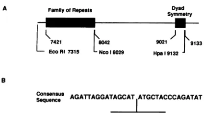
Connecticut 06510-8064 Received20July 1990/Accepted22October 1990The Epstein-Barr virus (EBV) latent origin of plasmid replication (oriP) contains two essential regions, a family of repeats with 20 imperfect copies of a 30-bp sequence and a dyad symmetry element with foursimilar 30-bp repeats. Eachof the repeats has an internal palindromic sequence and can bind EBNA 1, a protein that together withoriP constitutes the only viral element necessary for EBV maintenance and replication. Using single-strand-specific nucleases, we have probed plasmids containing oriP-derived sequences for the presence of secondary structural elements. Multiple single-stranded structures were detected within the oriP region. Of thetwoessential elements of oriP, the family of repeats seemed to extrude these structures at a much higher frequency than did sequences within the dyadsymmetry region. Though negative supercoiling was found to stabilize the single-stranded structures, they showed significant stability even after linearization of the oriP plasmids. Two majorsingle-stranded structures detected involved approximately 12 bp of DNA. These loci could betransiently unwound regions that form because of negativesupercoiling and the high A+T content of thisregion of DNA, or they could be cruciform structures extruded within thepalindromic sequences oforiP that may beimportant sites forprotein-DNA interactions in the EBV oriP.
Epstein-Barrvirus(EBV),ahumanherpesvirus, infectsB
lymphocytesandtransforms them into continuously growing cell lines. In most of these cells, the virus is latent. The 172-kb double-stranded DNA genome is carried as a
multi-copy, supercoiled plasmid, andonlyasmall numberof viral
genes are transcribed (reviewed in references 21 and 30).
Replication of the EBV genome during latency occurs in
synchronywith host cell DNAsynthesis(1, 15)andrequires
only two viral elements. One of these is a cis-acting se-quencecalled oriP(for origin of plasmid replication), which isfound on a 1.8-kb subfragment of the EBV genome (51)
(Fig.1A).Sequenceanalysisof oriP revealed the presenceof twounusualregions. One region, containing approximately 20 tandem, albeit imperfect copies ofa30-bp sequence, is called thefamilyofrepeats(FR). Locatedabout 1 kb away
isthesecondregion, whichcontainsa65-bpdyad symmetry
(DS) area within four additional copies ofthe 30-bp repeat
(2). Deletion analysis indicated that bothregionswere nec-essaryfororiPfunction (27,38),thoughneithertheir relative orientations nor the distances separating them seemed to
significantly affect their activity (38). The actual origin of replicationseems tobewithin the DS element, while the FR
region contains the termination siteforreplication (12). The secondviral elementrequired forEBVreplication in addition to oriP is EBNA 1, which acts in trans (27, 52). This
protein isencoded in theBamHI K fragment of the genome, far from the oriP region (9, 48). Studies using a bacterial
fusion protein containing the
carboxy-terminal
one-third ofEBNA 1 and partially purified EBNA 1 from lymphocytes
showed that this protein could bind to each of the two essential loci of oriP (18, 37). DNase footprinting demon-strated that binding of EBNA 1 to linearized, labeled sub-strate DNA protected 24 to 25 bp of each of the 30-bp
elementswithin the FR element aswellaseach of the four repeatelements in the DSregion.Aconsensus sequenceof
*Correspondingauthor.
the EBNA 1 binding sites reveals an internal 12-bp
palin-drome, overwhichtheDNase-protected regioncenters(37) (Fig. 1B). In addition to rolesin replication, EBNA 1 and
oriP seem to be involved in transcription during latent infection. Presenceof the FR elementincisand EBNA 1 in transhas been shown to enhance transcription from heter-ologous promoters (38). Furthermore, an EBV promoter downstreamoforiPwhichis thestartingpoint for transcrip-tion of at least two of the known latent gene products, including EBNA 1 itself, canbetransactivated by EBNA 1 (46). Thus, by virtue ofits interaction with EBNA 1, oriP influences the biologyof EBV inmany ways.
The EBV origin of replication used inlatent infection is
particularly interesting with respect to potential secondary
structures. Inverted repeat sequencesareknowntobe able
toformcruciformstructuresby intrastrand basepairing (14,
36). Palindromes can also bind with identical palindromes located nearby, and because the FR region contains 20
tandemrepeatsofanessentially 30-bp
palindromic
sequence(Fig. 1B), a very large number of alternative secondary
structures is possible (20). The DS element too may form
severalalternative secondary structures (20, 37), and all of thesewould occur within sequences thatareknowntohave the capacity to bind EBNA 1 (18, 37). In
addition
to the formation of such relatively stable structures, the oriPregion,because of itshighA+T content,mightbeexpected
toformtransiently unwound, lessstablestructuresunderthe
influence of
negative supercoiling.
These too could occur withinsequences thatbind EBNA-1. Adetailedunderstand-ing of theinteraction between oriP and EBNA 1andofthe molecular basis for the functions that thisinteraction
influ-ences mustthereforeinclude aknowledge ofthe structures
assumedbysequenceswithinthe oriP
region. Although
such structures werepredicted
(20, 37), noexperimental
dataastowhetheror notthey exist have beenavailable. Weshow here thatincubation of
supercoiled
plasmids
containing
oriP
sequenceswith
single-strand-specific
nucleasesresults inthe appearance ofmultiple nuclease cleavage sites. Restriction677
on November 10, 2019 by guest
http://jvi.asm.org/
A FamilyofRepeats Dyad Symmetry
L421 8042 9021 J9133
EcoRI 7315 -Nco 18029 Hpa19132
B
Consensus AGATTAGGATAGCATATGCTACCCAGATAT
SequenceI
FIG. 1. EBV oriPregion andconsensussequence of the 30-bp
repeats. Thetwoessential elementsof oriParethe FR and the DS elements (A), shown darkly stippled. Several restriction sites as
referencepointsareindicated,asarethe boundaries of the FR and
the DS regions, all numbered according to theirpositions on the B95-8 EBV genome sequence(2). (B) Consensus sequence of the
30-bp repeatsin oriP. Theinternal12-bp palindromeis indicated.
mappingshowed thatmanyof these sitesarelocalizedwithin
regions containingelements of oriP. The dataareconsistent
with the presence ofsingle-stranded structures within the
EBVoriP invitro.
MATERIALSANDMETHODS
Enzymes. Endonuclease T7 and nuclease P1 were from Pharmacia-LKB Biotechnology (Piscataway, N.J.);
nucle-ase S1was fromBoehringerMannheimBiochemicals (Indi-anapolis, Ind.). Restriction enzymes were purchased from
New England BioLabs (Beverly, Mass.) or Boehringer
Mannheim and were used according to the manufacturer's
specifications.
Plasmids. The EBV origin-containing plasmid pHEBO
was a giftform B. Sugden (University ofWisconsin) (45).
Other recombinant clones were constructed in our
labora-tory and have been described previously except for
pSV2neo: C60 (14a) and pSV2CAT: CHET (gift of J. L.
Kolman).Plasmidswerepropagatedinappropriatebacterial hosts and purifiedfrom Escherichia coliby standard tech-niques (28).
Probing single-stranded regions in plasmid DNAs. The presence of single-stranded regions in covalently closed, supercoiled plasmid DNAswas studied by using T7 endo-nuclease and endo-nuclease P1.Typically,200ngofplasmidDNA
was incubated with 6 U of T7or0.01 U of P1 at370C for60
min.Endonuclease T7 reaction buffercontained 50 mM Tris
hydrochloride (pH 8.0), 20 mMNaCl, 6 mM MgCl2,5 mM
dithiothreitol, and 100 ,ugof bovine serumalbumin perml;
nucleaseP1 buffer contained 10 mM Trishydrochloride (pH
7.5), 40 mM NaCl, and 0.4 mM ZnCl2. Reactions were
stopped by phenol-chloroform extraction, and products
were concentratedbyethanolprecipitation. Nuclease
cleav-age sites were mapped with respect to certain restriction
enzyme sites by cutting the resuspended DNA with the enzyme of choice. After removal of protein, the products
were analyzed by agarose gel electrophoresis and subse-quent Southern blotting usingradiolabeledprobes.
Theeffect ofnegative supercoiling onthe stabilityof the
single-stranded structures was studied by comparing
plas-midscleaved withendonuclease T7 andsubsequentlywitha
restriction enzyme with plasmids cleaved first with the
restriction enzymeandthen with theendonuclease. Boththe
endonuclease andrestrictionenzymereactionswerecarried
out at37°C for 60 min. DNAwasethanolprecipitatedondry ice for 15 min between the two reactions, and proteinwas removed before agarose gelelectrophoresis.
Estimates of the sizes ofsingle-stranded structures were obtainedby first digesting plasmid DNAs with endonuclease
T7;afterphenol-chloroform extraction,aliquots of the prod-ucts werefurtherdigested with 3 U of nuclease S1at37°C for 30 min. Mapping was again done with respect to known restriction enzymecleavagesites. Toaccuratelyseparatethe small fragments obtained from this procedure, reaction products were subjected to electrophoresis in 3.0% NuSieve GTG agarosegels (FMC Bioproducts; Rockland, Maine) and detectedby Southernblotting.
Agarose gel electrophoresis and Southern blotting. The
products of experiments studying plasmid DNA structure weresubjectedtoelectrophoresisin 0.8to1.2% agarose gels under standard conditions (28). HindlIl-digested lambda
bacteriophage DNA(NewEngland BioLabs)wassubjected toelectrophoresis alongwiththe samplestoprovide molec-ularsize markers.Agarose gelsweretransferredto nitrocel-lulose(BA85;Schleicher &Schuell,Keene,N.H.)orNytran
(Schleicher & Schuell) nylon filters and hybridized by a
modification of the method of Southern (44) in the presence of deionized formamide (28). Radiolabeled DNA probes were prepared by nick translation (40) and purified from
unincorporated radioactive dCTP on Pharmacia NICK-T
Sephadex G-50 columns. Specific activities of greater than 108cpm/,ugof substrate DNAwere regularlyobtained.
RESULTS
Plasmidscontaining oriP sequences extrude multiple single-stranded structures. T7 endonuclease, the product of the
bacteriophageT7 gene 3,isasingle-strand-specificnuclease that also hasa very high affinity for branchedstructuresin DNA suchascruciforms (8, 34).These elements are recog-nized by the enzymeand subsequently cleaved, generating
linear DNAmoleculesfrom thesupercoiled substrate. Posi-tions of thecuts canthen bemappedwith respectto known
restriction enzyme cleavage sites (35). This approach was
applied
tostudy theplasmids
pSV2neo: Bam C, pSV2neo:C6.0,
andpHEBO,
whichcontain oriP sequences, and,asacontrol, pSV2neo, whichcontains onlyvectorsequences.
pSV2neo DNA, aswellasthe otherplasmid preparations,
is a mixture ofcovalently closed supercoiled form I DNA with nicked form II and some linear form III species (Fig. 2A, lanepSV2neo /). Cleavage of this plasmidwith endonu-clease T7 slightly increased the amount of linear DNA present (lane pSV2neo T7), but subsequent digestion with therestrictionenzymeHindIIIrevealstwonovel bands(lane
pSV2neo
T7/H3, openarrowheads).
One distinct bandmi-grated
inaposition corresponding
toabout4,370 bp, while theother,aweak bandclearlyvisibleontheautoradiographsbutpoorly reproduced in the prints, migrated at aposition
corresponding
to about1,470bp.The sumof the base pairsof thesetwobandsisaboutequaltothe numberofbase pairs in the intact 5,729-bp plasmid, thus suggesting a single cleavagesite. This
finding,
alongwith data obtainedbyusing
otherrestriction enzymes, allowed mapping of thecleavage
site to an area between the origin ofreplication and the
ampicillin
resistance gene (see Fig. 2B for adiagrammatic
summary of the cleavage data). This part of pSV2neo is derived from the
plasmid
pBR322, which contains ase-quenceknown tobe abletoform acruciform(35) and thus serves as a good positive control for the activity of the endonuclease.
on November 10, 2019 by guest
http://jvi.asm.org/
[image:2.612.76.284.80.198.2]pSV 2Ei.C.: pS'V'? NEtC
pSV2NECI HAM f ½C,;
i._-.
AI1~ a*
pFiBE-W
I r- r- r
.- so, W- F I
_A
*F-B
47,- _1 7.
LHilnd3 Hind 3-J
1.140-
5.840-1.460 6.090
Hind 3 hyg amp
3.480 7.490
'4Z-rJ I.' 6.900 4.21
Hind 3 7,410
pSV2neo
5.729 bp
pHEBO
7.068bp
pSv2neo: C6.0
10.880 bp ?B0 Hind 3
7,040
}1
-a
".ps dyad
pSV2neo: Bam C
10,600 4,280 14952bp
Hind3
Cleavageof pSV2neo: Bam C DNA with T7 and then with HindIlI again identified the 1,470-bp band. Therefore, this nuclease-sensitive structure is also present in a plasmid
containing inserted EBV DNA. Many new cleavage prod-ucts, however, were detected. There were two major bands of about 7,410 and 7,040 bp which had approximately equal intensities and were therefore of about equal molarity (Fig. 2A, lane pSV2neo: Bam CT7/H3, large filled arrowheads). The sizes of these two fragments add up to approximately the size of the pSV2neo: Bam C plasmid itself, which suggests that a single cleavage was responsible for generat-ing these fragments; this site could be located within a region
corresponding to the EBV oriP, whose FR element is located 6,800 to 7,400 bp away from theHindIIIsite on the plasmid (Fig. 2B). In addition to these major bands, there were four weaker bands of10,600, 5,900, 4,280, and 3,740 bp. Two of these bands, those of 5,900 and 3,740 bp, were alsopresentinpSV2neo: Bam CdigestedonlywithT7 (lane
pSV2neo:Bam C T7, double open arrowheads). Since these
fragments are bounded by T7 cleavage sites, they are not useful in mapping. They do, however, suggest that the
endonucleasecancleave theplasmid DNA at a minimum of twositesin addition to cleavage within the region containing thepBR322-derivedfragmentand that the T7 cleavages that
generated them occurred in the cloned EBV DNA. The bands of 10,600 and 4,280 bp (small filled arrowheads),
which were approximately equimolar and added up to the size of the entire pSV2neo: Bam C plasmid, seemed to be
generated by a singleendonuclease T7 cleavage (Fig. 2B).
This recognitionsiteforT7wouldbelocated downstream of the oriP sequences in a region that contains an EBV pro-moterknown to functionduring latentinfection(5).
pSV2neo:
C6.0
contains a6.0-kb EcoRI-BamHIsubfrag-mentof theregionfoundin pSV2neo:BamC (14a).Cleavage
with T7 followed by HindIII resulted in the appearance of two major bands of about equal intensity with migration
rates corresponding to about 7,490 and 3,480 bp, respec-tively (Fig. 2A, lane pSV2neo:
C6.0
T7/H3, large filledarrowheads), suggestingthat they are present in equimolar
amounts.Thesizes ofthese twofragments addedup approx-imately to the size of the intact plasmid DNA, which suggests thatcleavage by endonucleaseT7 at onesitewithin the plasmid was responsible for generating both fragments (Fig.2B). Severalminorcomponents werealsovisible,with those corresponding to 6,900 and 4,280 bpbeing the most
1 3 5 7 9
l _ I
k oba sepair s
FIG. 2. Mapping of endonuclease T7-sensiti DNAs. (A) The oriP-containing plasmids p
pSV2neo: C6.0, and pHEBO were studied, ale which contains no EBV sequences. A 200-ng DNA consisting ofa mixture ofsupercoiled
nicked(N) formswasused in each reaction. P incubated either in the absence ofenzymes(/)
endonuclease T7 (T7), cleaved only with Hindl
withendonuclease T7and then with HindIIl (T7, T7digestions used 6 U ofenzymeandwere ca
37°C. Hindlll restriction endonuclease cleaves single site. Products of the reactionswere reso
electrophoresis anddetectedby Southern blottii fragments generatedweredeterminedby linear using HindlIl-digestedkDNAfragmentsasmol (indicated on the left in kilobases). DNA fral
T7/H3 lanearemarkedaccordingtothepositior
11 13 15
l_X Al site. Bands marked withanopenarrowhead result from T7cleavage
withinvectorsequences,those marked withalarge filledarrowhead are major cleavage sites within the EBV oriP-derived sequences,
ive sites in plasmid small filled arrowheads indicate minorcleavage sites withinother
)SV2neo: Bam C, regions of the EBV-derived DNA, and double open arrowheads
cng with pSV2neo, indicatefragmentsboundedatboth endsbyT7cleavage sites. The
sample of plasmid 1,570-bp bands in pSV2neo, pSV2neo: Bam C, and pSV2neo: C6.0
(S), linear (L), and T7/H3digestswerevisibleonautoradiographsbut did notreproduce
lasmid DNAswere well in the prints. (B) Major endonuclease T7 cleavage sites on ,cleaved onlywith plasmidsarepresentedindiagrammaticform andareoriented with III(H3),orcleaved respect to the singleHindIII site oneach of the plasmids, which
/H3).Endonuclease have beendrawn asiftheyhad beenlinearized atthis point. The
irriedoutfor 1 hat sizes of the fragments that resulted after digestion with T7 and
each plasmidata HindIll areindicated in basepairs.Bold numbers and lines indicate
lvedbyagarosegel cleavage within EBV-derived sequences, light numbers and lines ng(44). Sizesof the indicate cleavage within vector-derived sequences. Blackened
regression analysis, boxes represent the EBV oriP-derived sequences,darkly stippled lecular sizemarkers regions indicate cloned EBVsequencesthat flank theoriP, lightly gments within each stippled boxes represent antibiotic resistance genes, and blank
aoftheT7cleavage boxesrepresent thepBR322-derived procaryotic replication origin.
A
147.- 3.4850
ori amp H1ind 3
4a--m !':.
..Oft-Aalk.
514W
on November 10, 2019 by guest
http://jvi.asm.org/
[image:3.612.60.297.90.578.2]pSV2NEO pSV2NEO: C6.0
/ Hjnd3 Hind3 EroRI BOmHlIECORV KpnI R E
- -F O- - -+4-- + -- --'-- t - i- + iEBNA
44- d
23- A
2.o-PROBE: PSV2NEO: C60
2.360-2.900bp
Kpn 1
( ~~~~~~~~~~~~~~~~~~j-Kpnl,)
900 1.440bp
EcoRV 5,120-6.080bp
BamHi
IS50- 850 bp 550 850bp
Eco Ri 3,160
Hind 3
ori amp trps dyad
Bam Hi
EcoRI )
Hind3
neo
pSV2neo:C6.0
10.880 bp
1 3 5 7 9 11
I I
k oba s epa rs
FIG. 3. Mapping of nuclease P1-sensitive sites in pSV2neo and pSV2neo:
C6.0
in the presence and absence of EBNA 1. (A)Plasmids pSV2neoand pSV2neo:C6.0inthepresence(+EBNA 1) or absence (-EBNA 1) of 100 ng ofbacterially expressed (33),
immunoaffinity-purified EBNA 1wereexposedto0.01U of
nucle-aseP1.Productsweredeproteinized, either electrophoresed without
furthermodification (/)ordigested with various restrictionenzymes
(R.E.)asindicated,and analyzed bySouthern blotting(44).Sizesof
thedetected fragments were calculatedbylinear regression
analy-sis, using the HindIII-digested X DNA fragments as standards
(indicatedattheleft in kilobases).Becauseofalower abundanceof
cleavage products, in ordertovisualize small fragmentsthe
South-ern blot of this experiment had tobe overexposed, and only the
lowerhalfisshown for clarity. Open arrowheads indicate fragments resulting fromcleavage withinvectorsequences;closedarrowheads
indicate fragments resulting from cleavage within oriP-derived
se-quences.Thepurification of EBNA1,aswellasfurther studieson
its effect on plasmids bearing the EBV oriP, will be presented
elsewhere. (B)Major nuclease P1 cleavage sitesonpSV2neo: C6.0
evident (small closed arrowheads). The sizes ofthese
frag-mentsalso added up toapproximately the size of the entire
plasmid, which implies cleavage at asingle site. This
posi-tion maps to the sameregion identified above in pSV2neo:
Bam Cnearthelatentpromoter 3' tooriP.
Theplasmid pHEBO contains less of the oriP region than does eitherpSV2neo: BamCorpSV2neo:
C6.0
andbecause of its smaller size is a better target for mapping studies. Treatment with T7 and thenHindlIl generated a very strong band at5,840to 6,090bp and aladder of bands from about1,140 to 1,460 bp (Fig. 2A, lane pHEBO T7/H3, closed arrowheads). Thesetwosetsoffragments were boundedby
T7 andHindlIl sites, and the total of their sizes agrees well with the complete length of pHEBO, 7,068 bp. Thesedata, along with those presented subsequently, allowed mapping ofthecleavage sites resulting in these fragments to anarea clockwisetothe HindIII site. This area, from about 880to 1,480 bp, contains the FR element of oriP. Thus, these repeats, in the context of a supercoiled plasmid environ-ment,canextrude single-stranded structures. Furthermore,
several of the repeats have thiscapacity,since several bands areseenin this region, and it is likely that endonuclease T7 cuts atseveral structuresbeing extruded by different repeats on different plasmids. This would explain the width of the band at about 6,470 bp, since it is probably made up of several DNA fragments varying inlength by up to 600bp. Thesefragments would be generatedby endonuclease cleav-age at different repeats within the FR element of oriP, followed by linearization with HindIIl.
In additionto several of the repeats having theabilityto extrude single-stranded structures, more than one such structure seemed to be extruded in at least some of the oriP-containing plasmids. Examination of each of the T7 digestion lanes showed that though linear DNA was the major product, several fragments appeared that were the products of T7cleavage at morethanonesiteon aplasmid. This could occur either by simultaneous attack of endonu-cleaseat twopositionsorbyattackatonepositionfollowed by attack at a second before reabsorption of the second
regionduetolossofsupercoiling. pSV2neo,by comparison,
did not seem to yield any such products of double T7 digestion.
Finemapping of single-strand nuclease cleavage sites. Use of endonuclease T7 had demonstrated thatoriP-containing
plasmids contained regions of DNAsusceptibleto cleavage withthissingle-strandednuclease. Itwasof interesttoverify these results with a second nuclease that is often used to probe forsingle-stranded regionswithin DNA, nuclease P1 (7,22). Cleavage ofpSV2neowithnucleaseP1 andHindIlI
generated a fragment of about 4,480 bp and three closely spaced fragments at 1,280 to 1,460 bp (Fig. 3A, lane
pSV2neo-EBNA1, openarrowheads).Thecombination of the sizesofthelarger fragment with that of any ofthe smaller ones equaled approximately that of the pSV2neo plasmid
and allowed mapping of this site into the area identified earlier with T7 that contains a cruciform derived from
identified in panel Aare indicated in diagrammatic form and are
oriented with respecttothecleavagesiteof each ofthe restriction
enzymesused. Theplasmid is drawnasif thatrestrictionsitewere
at the left of the diagram. Blackened boxes represent the EBV
oriP-derived sequences, darkly stippled regions represent cloned EBVsequencesthat flank theoriP,lightly stippled boxesrepresent
antibioticresistancegenes, andblankboxesrepresentthe pBR322-derivedprocaryotic replication origin.
A
B
A Z=~~~~~~~~~~~~~
i
on November 10, 2019 by guest
http://jvi.asm.org/
[image:4.612.68.303.83.553.2]pBR322 sequences. Endonuclease T7 was also seen to generate three lower-molecular-weight products when it digested plasmids containing this region of DNA (see Fig. 5, lane pSV2CAT T7/H3). Cleavage of pSV2neo: C60 with nuclease P1 and then one of five different restriction en-zymesresulted in the generation of a set of intense bands at differentpositions, depending on the enzyme used (Fig. 3A, lane pSV2neo:
C6.0
-EBNA 1, closed arrowheads). Linear regression analysis indicated that the nuclease P1-sensitive sites were located 3,160 to 3,830 bp from the Hindlll site, 550 to 850bp from the EcoRI site, 5,120 to 6,080 bp from the BamHIsite, 900 to 1,440 bp from the EcoRV site, and 2,360 to 2,900 bp from the KpnI site (see Fig. 3B for a diagram-matic summary of these data). All of these sites map into a region of DNA that contains the FR element of oriP and confirm the data presented above for the T7 single-strandspecific endonuclease. Figure 3 also shows the effect of preincubating the plasmids with bacterially expressed (33), purified EBNA 1. The presence of EBNA 1 seemed to enhance cleavage by nucleaseP1within the same sequences that were recognized in unbound pSV2neo: C60 plasmid DNA, but it seemed to have little effect on pSV2neo, which does not contain high-affinity EBNA 1 binding sites. Further studies to determine the effect of binding of EBNA 1 to
plasmidscontaining the EBV oriP are under way and will be presented elsewhere.
Tomore closely map the endonucleaseT7-sensitive sites,
pSV2neo:
C6.0
was examined with additional restriction enzymes (Fig. 4A). Plasmid DNA was cleaved with T7 andthen with acombination of two restriction enzymes. Cleav-age with EcoRIand EcoRV selects against visualization of products formed by T7 cutting at the dyad because the EcoRV digestion would give fragments of 160 bp or less that would not be detected in this gel system. Three major
fragments are clearly visible. Two of these (open arrow-heads), those of about 9,300 and 1,350 bp, were derived from
cleavage ofpSV2neo:
C6.0
only withEcoRI and EcoRV, as seen in anadjacent control lane (pSV2neo:C6.0
R1/RV) inwhich products of a similar reaction with the same
restric-tion enzymes but in the absence of T7 were analyzed. The thirdmajor component of the reaction detected was a set of
bands ranging in size from about 1,020 to 1,150 bp (filled
arrowhead). Since these were seen only after treatment of the T7 digest with EcoRI and EcoRVnucleases, the
cleav-agesproducing these bandsoccur1,020to1,150 bp fromone of thesetwo enzymesites.
To determine more precisely the locations of the sites cleaved by endonuclease T7 to generate this last set of fragments, a parallel set of reactions was cleaved with EcoRV and KpnI. These once again revealed a set of fragments ranging in size from 1,020to 1,150 bp (Fig. 4A, lanepSV2neo:
C6.0
RV/K1, filled arrowhead). Were these T7 cleavage sites located about 1,100 bp from the EcoRI site, cleavage with EcoRV and KpnI would have increased their sizesby about 1,700 bp, but nosuchfragments of approxi-mately 2,800 bp are visible. The T7 cleavages, therefore, occurred 1,020 to 1,150 bp away fromthe EcoRV site and mapinto theright end of the FR element of oriP (see Fig. 4B for a diagrammatic summary of the data). This element is presentabout 960 to 1,540 bp away from the EcoRV site in pSV2neo:C6.0
A pSV2NEO:-.
NL Y
-
-I NN- NN<
0
4*40FIG. 4. Fine mapping of endonuclease T7-sensitive sites in pSV2neo:
C6.0.
(A) pSV2neo: C6.0 plasmidDNA was either incu-bated in the absence ofenzyme(/), cleavedonlywithendonucleaseT7 (T7), cleaved with endonuclease T7 and then with restriction
enzymesEcoRIandEcoRV(T7/R1/RV),cleaved with endonuclease
T7and then with restrictionenzymesEcoRVandKpnI (T7/RV/K1), cleavedonly with restrictionenzymesEcoRI and EcoRV(R1/RV),
orcleavedonlywithrestrictionenzymes EcoRV andKpnI (RV/K1). OpenarrowheadsindicateDNAfragmentsgenerated by restriction
enzymecleavage of pSV2neo:
C6.0;
filled arrowheads indicate bands resulting from17cleavage withincloned EBV sequences.Radiola-beledpSV2neo:BamCwasusedastheprobe. (B)Endonuclease T7
cleavage sites andthe fragments generated by restrictionenzyme
cleavageare shownindiagrammatic form.The topdiagram repre-sentsthe results obtained with theEcoRV andKpnIenzymes; the
bottom diagram represents results from the EcoRI-EcoRV trials.
Numbers in light type indicate the sizes of fragments generated
when pSV2neo: C60 was digested with the restriction enzymes
alone; numbers in boldtypeindicatethesizes offragments
gener-ated by endonuclease T7 cleavage followed by the respective restrictionenzymes.
B 930- 1.050 1.240 9,300 9.500
I.240 9.50-0
I
Dzzzi
X
EcOR
Ipnl
Eco RV Kpn EcoRI
930-1.050 1.660 9,300 1..660 .30wo
7II
_U-rps adye
EcoRI Eco RV Kpn
1 3 5
l
nee Orl amp
pSV2neo: C6.0
10.880bp
7 9 11
k oba s epa rs
-4,
..f.
7
on November 10, 2019 by guest
http://jvi.asm.org/
[image:5.612.340.521.284.736.2]Single-strand nuclease cleavage ofplasmids lacking the DS element. To determine whether the FR elements present alone on a plasmid without the DS area could extrude single-stranded regions of DNA, the plasmid pSV2CAT: CHET was examined. This construct contains a cloned 7,196-bp fragment derived fromaheterogeneous, rearranged strain ofEBV and contains part of the repeat elements butis missing the dyad (21a). Cleavage of the parent plasmid, pSV2CAT, with endonuclease T7 followed by
HindIll
yielded a novel fragment of about 4,120 bp that was visible between the bands representing linear pSV2CAT plasmid DNA and residual uncleaved supercoiled plasmid (Fig.
5A,
lane pSV2CAT T7/H3). Three additional fragments ofabout
1,420 bp were also generated which were visible on the original autoradiograph but poorly reproduced in the
photo-graphs. Thefact that the sizes of these two sets of fragments
(open arrowheads), withinthelimitsofresolutionofagarose
gel electrophoresis, added up to approximately the size of the pSV2CAT plasmid itself implies that a single cleavage event by the T7 enzyme created them. This event most
probablyoccurredat a site between the ampand
ori
genesofpSV2CAT(Fig.SB)which, asforpSV2neo,is derivedfrom pBR322 andcontains a sequenceknown tobe able to form a cruciform(35).
When pSV2CAT: CHET was exposedto endonuclease T7 followed by HindIll,twomajorfragments of approximately equal intensitywere formed(Fig. 5A,lane pSV2CAT: CHET
T7/H3, closed arrowheads), suggesting a single cleavage site. The sizes of these fragments are 7,000 and 5,660 bp,
respectively, and the cleavage site maps approximately to the middle of the CHET fragment where the 30-bp repeats derived fromoriP are located(Fig.SB).Againfaintlyevident on the original autoradiograph is a band ofabout 1,420 bp, along with a band of about 11,000 bp (open arrowheads), suggesting that cleavage at the pBR322-derived cruciform canalso occur. Cleavage within the sequences derivedfrom the FR element of oriP, however, seemed much more
pronounced.
Supercoiling stabilizes the single-stranded structures but relaxation is not instantaneous. The extrusion of double-stranded DNA intotransient or stable single-stranded struc-tures is often driven by negative supercoiling (17, 31). To determine whether this is the case for the oriP structures,
pSV2neo:
C6.0
either was cut with T7 and then withHindlIl
orBamHI orwas first cut with the restriction enzymes and then cut with the endonuclease (Fig. 6). Cleavage with T7 and then HindIII led to the appearance of three major bands having lengths of 7,480, 3,480, and 3,090 bp and a minor band of 4,370 bp with respect to theHindIll site. When the
plasmid was first linearized with
HindlIl,
however, subse-quent use of T7 produced bands of the same size but of reduced intensity (pSV2neo:C6.0
T7/H3 versusH3/T7).
Similarly, cleavage of the DNA with BamHI before addition of endonuclease reduced subsequent T7 cleavage of cruci-formstructures (T7/B1versusB1/T7). Calculations based on laser densitometric scanning of the original autoradiograph
(data not shown) indicated that precleavage with
BamHI
decreased subsequentT7 digestion by 1.8-fold, while
predi-gestion with HindIll decreased T7 sensitivity by about 2.7-fold. Completion oflinearization is demonstrated in the
last two lanes of Fig. 6, in which pSV2neo:
C6.0
cut either with BamHI orHindlIl was subjected to electrophoresis. Aliquots of these reaction mixtures were used for further analyses with endonuclease T7. Thus, loss of negativesupercoilingled to the relaxation of many of the structures
A f)
SE,
V C,A5pSV2
CAT:CItE`
Nl-,r- N
r' I"N-N- ( r -
i-~
r-_ iltosIs,,,
0 -*..
rn-0
-11'
':1
B
1,420 4.120
K
2
Hirnd3 Hind 3 1.420 11,000
6-fl--El-KCC amp
Hind3
pSV2CAT 5,018 bp
7,000 5,660
pSV2CAT: Chet
12,214bp
Hind 3
11 13 1
. . I ,, ,,
k o b a sepa rs
FIG. 5. Sensitivity oforiP plasmids lacking the DS symmetry element to cleavage by endonuclease T7. (A) Plasmid pSV2CAT: CHET, which contains only the FR element of oriP, and its control pSV2CAT, which contains no EBV DNA, were examined for endonuclease T7 sensitivity. As for Fig. 3, plasmids were incubated in the absence of enzyme
(/),
cleaved only with endonuclease T7 (T7), cleaved only withHindIll
(H3), or cleaved with endonuclease T7 and then withHindIII
(T7/H3). Supercoiled, linear, and nicked forms of pSV2CAT are indicated by S, L, and N. Filled arrowheads indicate fragments generated by endonuclease T7 cleavage in oriP-derived sequences; open arrowheads indicate fragments resulting from T7 cleavage within vector sequences. Radiolabeled pSV2neo: Bam C was used as the probe. (B) Major endonuclease T7 cleavage sites within pSV2CAT and pSV2CAT: CHET with respect to the singleHindIll
site in each plasmid are presented in diagrammatic form. Plasmids are drawn as described in the legend to Fig. 2B. Light numbers and lines indicate cleavage within vector sequences; bold lines and numbers indicate cleavage within EBV oriP-derived sequences.I
on November 10, 2019 by guest
http://jvi.asm.org/
[image:6.612.329.568.110.537.2]r4-) I,.- - r.
- r- N) r- - n
-N- N- I N m _ {X;
U')
- U)
-J I
- -:Z *cz
-a)i Cf En =
- t- r- r-
s-BP _
1353.-- _
1078 N _
872-622
-527
-404 --- 23
[image:7.612.92.245.76.284.2]2.0
FIG. 6. Effect of supercoilingonstability of the single-stranded
structures. Plasmid pSV2neo: C60 was incubated in the absence
ofenzymes(/),cleaved only with endonuclease T7 (T7), cleaved first with endonuclease T7 and then with HindIll (T7/H3), cleaved
first with HindIIl and then with endonuclease T7 (H3/T7), cleavedfirst with endonuclease T7 and then with BamHI (T7IBI), cleavedfirst with BamHI and then with endonuclease T7 (BIMT7),
or cleaved only with Hindlll (H3) orBamHI (BI). Reactions in which plasmid DNA was first linearized and then exposed to
endonuclease T7are aliquots of these cleaved with the restriction enzyme only. Filled arrowheads indicate fragments resulting from cleavage withinoriP-derived sequences.
that endonuclease T7 recognized, butthis process was not
instantaneous.
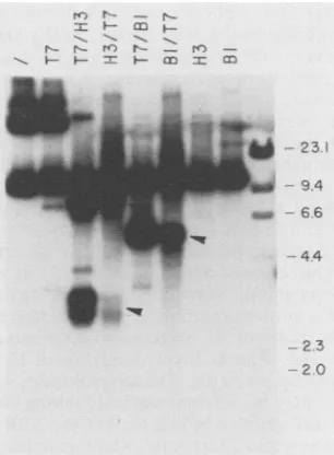
Twosingle-strandedstructuresextruded withinpHEBOare
approximately 12 bp long. Endonuclease T7 cutscruciform
structures at their bases, leaving one hairpin end on each
sideofthecleavage product. Since this hairpin issusceptible
to Si nuclease, use ofthis enzymeafter T7 allows calcula-tionof the number of bases involvedinacruciform (25).This
approach was applied to plasmidpHEBO, alarge
prepara-tion ofwhich wasdigested with T7 and then withBamHI, Sall,orHpaI. AliquotsofthedigestionswithSallandHpaI
were then treated with Si nuclease, and theproducts were
analyzed on a high-percentage agarose gel to resolve the smaller digestion products. Cleavage with T7 and BamHI revealed three major endonuclease-sensitive sites located 1,460, 1,070, and 780 bp from the Bam site (Fig. 7, lane T7/B1, one open and two filled arrowheads, respectively). Cleavage with T7 and SalI showed that the 1,460 site is locatedcounterclockwisefromthe Bamsiteandisprobably the structureidentified inpBR322 from which this region in pHEBO is derived. The sites at 1,070 and 780 bp from BamHI, however, are in the FR elements, since cleavage
withSall reduced their size by approximately the predicted
amount, which is 276 bp (compare lanes T7/B1 and T7/ SAL1). Probing of similar blots with anoriP-specific
radio-labeled DNA fragment identified only these last two sites (datanotshown),furthershowingthatthesecleavagesoccur
within oriP.Use of nuclease Si afterSall restriction accel-eratedthe apparentmigrationof bothfragments byabout 12 bp (lane T7/SAL1/S1 versus lane T7/SAL1). This same
[image:7.612.375.481.77.242.2]estimatewasobtainedby comparingthe sizes ofpHEBOcut
FIG. 7. Determination of the approximate sizes of the single-stranded structures within oriPby nuclease S1 analysis. pHEBO plasmid DNA was digested with endonuclease T7 and then with
restriction enzymeBamHI (Bi), Sall (SALl), orHpaI (Hi).
Ali-quots of the T7-SalI and T7-HpaI reaction mixtures were then digested with the single-strand-specific nuclease S1 (S1). HaelII-digested 4X174 and MspI-digested pBR322 DNAs were electro-phoresed in the gel to serve as molecular size standards. Filled arrowheadsindicate fragmentsgenerated by cleavage of
endonucle-aseT7 withinoriP-derivedsequencesfollowed by BamHI cleavage; theopenarrowhead indicates the fragment generated by T7 cleavage
within thepBR322-derived sequences.
withT7andHpaIwithpHEBOcutwithT7, HpaI,and then S1. Similar calculations estimate that the cruciform in the pBR322-derived DNA, cleavage at which generated the 1,460-bp fragment in thepHEBO T7/B1 lane (open
arrow-head), is about 27 bp long, which corresponds to the size predicted by sequencedata(35).
DISCUSSION
Palindromic sequences are often found near functionally
interesting regions ofDNA, such as in replication origins
thathave beenidentified in manyprocaryotes, including E. coli and Vibrioharveyi (29, 54),andalso inmanymammalian viruses, including JC and herpes simplex viruses (10, 50). Because inverted repeatscanformcruciforms(14, 36),these
structures have long been thought to be possible loci for protein-DNA interactions important in replication. That cruciforms exist in vivo has been shown for E. coli with the
use of an inducible T7 endonuclease expression system.
Both bacterial chromosomal DNA and exogenous plasmid DNAwerecleavedbythisenzymeinvivo,and theplasmids
weredigestedatthesamesites invivoasinvitro(34). Also, stabilization of cruciform structures withspecific monoclo-nalantibodieshas beenshowntostimulateDNAsynthesisin vivo(53). Thus, it seems likelythat cruciforms detected in
vitro will prove to have important functions in vivo. The
presence ofmany repeated palindromic regions in the oriP regionof the EBVgenome suggested that cruciforms could be extruded by these sequences. Furthermore, these se-quences areknown tobe able to bind the EBNA 1protein (18, 37),which isnecessary,along with oriP, for viral DNA
replication during latency (27, 52). An understandingofthe
interaction of EBNA 1 with oriP and of how thisinteraction
is important in EBV replication must therefore include a
knowledge ofany secondary structurepresent in the origin
region.
44
..z 3
Om
lqw
Wm
to
23.110 9.4
ow 6.6
on November 10, 2019 by guest
http://jvi.asm.org/
Studies
using
endonuclease T7 and nucleaseP1presented
here demonstrated that
oriP-containing
plasmids
containsingle-stranded
structures in vitro at severalplaces
within theorigin region.
Onepossible interpretation
of these results is that thesingle-stranded regions
detected representtran-siently
unwoundregions
ofDNA that form under theinflu-ence of
negative supercoiling.
Because the sequencescom-prising
oriP are 60%A+T,
whereas the rest of the EBV genome isonly
40% A+T(2),
thisregion
ofDNA wouldhave a
particularly high tendency
to form suchtransiently
melted areas. Such localizedunwinding, however,
may bemorethan
just
aby-product
ofthe unusual sequence of thisregion
and mayindicatethe presenceof elementsimportant
in the function of oriP.
Replication
of the E. coli chromo-somal DNA isinitiatedby
thebinding
ofthe DnaAprotein
tothe bacterial
origin,
oriC,
whichleadstoalocalizedunwind-ing
of DNA necessary for theassembly
of aprepriming
complex (6, 11, 19).
Thisunwinding
occurs within threerepeated 13-bp
sequences that areA+Trich(6).
However,
these sequencescanform
single-stranded regions
whenthey
are present on
supercoiled plasmids
even without thepres-ence ofboundDnaA
protein,
which has led to theirbeing
named
DNA-unwinding
elements(23).
Forplasmids
contain-ing
oriPtoreplicate, they
mustbesimilarly
unwound attheorigin
for theassembly
ofacomplex capable
ofinitiating
replication
ofthe twosingle
strands of DNA. Thatsuper-coiled
plasmids containing
oriP extrudesingle-stranded
structureswithin the sequences that would be
expected
tobe unwound firstinreplication
could indicate the presenceofDNA-unwinding
elementsanalogous
to those identified in the E.coli
oriC within the EBV oriP.A second
possible interpretation
of thefinding
thatsuper-coiled
plasmids containing
oriParesensitiveatseveral sitesto
cleavage by single-stranded
nucleases is that these sites represent cruciforms extruded within thepalindromic
se-quences of oriP. This
interpretation
issupported by
thekinetic data
presented
inFig.
6. If thesingle-stranded
structuresdetected weretransiently
formed meltedregions
of
DNA,
linearization oftheplasmids
would tendtoleadtoanalmost
complete
lossoftheseregions.
This isnotthecasewith the oriP structures,
however,
sincealthough
precleav-age ofthe
supercoiled plasmids
with a restriction enzyme does reduce thedegree
ofsubsequent susceptibility
toen-donuclease
T7,
it does not abolish it.Thus,
the structuresbeing
detectedhaveasignificant
half-lifeevenin the absenceof
negative supercoiling, arguing
for the presence ofamorestable structure such as acruciform. Kinetic studies have shown that both cruciform extrusion and
reabsorption
areslow processes
(13) having
half-livesofseveralhourseven at50°C
withcruciformsofabout700bp.
oriP sequences have been
predicted
to be able to form many structures(20, 37).
The DSregion,
forexample,
has beenpredicted
to have twopossible
alternative structures,one
involving
64bp
in ahairpin
structure with threemis-matches anda
second, larger
structureinvolving
116bp
ina stemplus loop
with more extensive mismatches(37).
Our data indicate that endonucleasecleavage
within the DSregion
is minimal and that at least in vitro themajor
secondary
structuralfeaturessensitive tocleavage
occurin the FRelement.Only
after exposure oforiPplasmids
to250 mMKCI,
which stimulates extrusion of certain types ofcruciforms
(47),
can anycleavage
within thedyad
bede-tected,
at whichpoint
four sensitive sites occur, probablycorresponding
to the four30-bp-like
repeats thatarefound there(data
notshown).
Itispossible that,
inanalogywith theinterpretation
of recent results obtained with the lambdabacteriophage(43), replication of EBV drives the extrusion of cruciforms within the dyad region. In the plasmidDNA itself, however, such extrusionseems toberare.
The FR element oforiP, because of its highlyrepetitive
andpalindromic nature, has beenpredictedto havea
large
number of possible alternative structures. Mathematical analysis of the EBV sequence localized two exact
palin-dromes within thisregion,both 18bpinlength(20).Our data areconsistent with the possibility that there are two major
single-stranded structures. Most of the cleavage patterns visible inFig. 2to4, 6, and 7 show twomajorbands, along
with several minor bands representative of endonuclease cleavage. This is most apparent in Fig. 7, in which three major T7 recognition sites are identified, two within the repeatsand a thirdrepresenting a pBR322-derivedstructure (35). On the basis of S1 nuclease experiments, these two major structures seem to involve only about 12 bp of DNA and not the 18 bp predicted. Though molecules withhairpins
at their endsmay migrateanomalouslyduring
electrophore-sis, similar calculations indicated that the pBR322-derived
cruciform was about 27 bp long, which is indeed thelength
predictedby sequence data(35). Interestingly, several30-bp
repeats within the 20-copy family contain internal 12-bp
perfect palindromes. It seems likely that these are the sequencesthatareextrudedinto cruciforms and that within the plasmid DNA itself there is little if any interaction betweenadjacent repeats.
The in vitro studies on oriP reported here are the firstto examine the secondary structure of this region of DNA. EBVplasmids in vivo, in latently infected cells, are present asnegatively supercoiled plasmids (26, 32), and this would tend to stabilize any single-stranded structures present. However, theseplasmids arethought to be packaged intoa nucleosomal structurevery similar to that of the host chro-mosomal material (42), and such packaging might be ex-pectedtointerfere with or even prevent cruciformextrusion. Nonetheless, it is interesting that the 3' DS region of oriP was recently shown to exclude nucleosomes in reconstitu-tionexperiments with linear DNA fragments containing the EBV oriP(41). The structures detected in oriP may consti-tute sites for interactionsimportant in the replication of the EBV genome during latency. Notably, the FR element has beenrecentlyfoundtocontain thetermination site for EBV
replication, and two major stop sites were in fact detected (12). It istemptingtospeculate that these two sites may have somerelationshipwith thetwomajor single-stranded regions thatwehave detected inthis study. Perhaps the presence of acruciform in the FR element provides a termination signal for replication of the origin-bearing DNA. EBNA 1, the
origin-bindingprotein, may have some role toplay in
con-junctionwithcruciforms. Formation ofacruciform structure at the simian virus 40 replication origin in vitro abolishes
binding oflarge T antigen (49), and if such is the case for EBNA 1, its inability to bind the cruciforms may in some way disrupt the progress of a replication complex passing
throughoriP. Indeed,wefindin related studies that binding of EBNA 1 adjacent to a single-stranded structure may stabilize thisconformation. Alternatively,since EBNA1has been reportedtohavesingle-stranded DNA-binding capacity
(16),it may beabletobind cruciforms, and such binding may causetheterminations ofreplicationwithin the repeats. Just such a cruciform-binding protein, which may have roles in both DNA transcription and replication, has recently been isolated from rat liver extracts (4). Further studies will be necessary to distinguish between these possibilities or to establish an alternative theory for the importance of the
on November 10, 2019 by guest
http://jvi.asm.org/
single-strandedoriP structuresin the functions of thisregion
of DNA.
ACKNOWLEDGMENTS
This workwas supported by grantsA122959 and CA16038 from theNational InstitutesofHealth.R.O. issupported byMSTPgrant
T32 GM07205.
We thank Don Crothers and Nigel Grindley for many helpful discussions.
REFERENCES
1. Adams, A. 1987. Replication of latent Epstein-Barr virus ge-nomesinRaji cells. J.Virol. 61:1743-1746.
2. Baer, R., A. T. Bankier, M. D. Biggin, P. L. Deininger,P. J. Farrell,T.J.Gibson, G.Hatfull,G.S.Hudson,S. C.Satchwell,
C.Seguin,P.S.Tufnell,andB.G. Barrell. 1984.DNA sequence
andexpression of the B95-8Epstein-Barrvirusgenome. Nature
(London) 310:207-211.
3. Benham, C. J.1981. Theoreticalanalysisofcompetitive confor-mational transitions intorsionallystressedDNA. J. Mol. Biol. 150:43-68.
4. Bianchi, M. E., M. Beltrame, and G. Paonessa. 1989. Specific recognition of cruciform DNAby nuclearprotein HMG1.
Sci-ence243:1056-1059.
5. Bodescot, M., M. Perricaudet, and P. J. Farrell. 1987. A promoter for the highly spliced EBNA family of RNAs of Epstein-Barr virus. J. Virol. 61:3424-3430.
6. Bramhill, D.,and A. Kornberg. 1988. Duplexopening bydnaA
protein at novel sequences in initiation of replication at the
origin of theE. coli chromosome. Cell 52:743-755.
7. CamilHoni, G.,F. DellaSeta,A.G.Gicca,and E. DiMauro.1986.
Purified Saccharomyces cerevisiae RNA polymerase II
inter-actshomologously withtwodifferentpromotersasrevealedby P1endonucleaseanalysis. Mol. Gen. Genet. 204:249-257.
8. deMassy, B.,R. A.Weisberg, and F.W.Studier. 1987. Gene3
endonuclease of bacteriophage T7 resolves conformationally branched structures in double-stranded DNA. J. Mol. Biol. 193:359-376.
9. Fischer,D. K., M. F.Robert,D.Shedd,W. P. Summers, J.E.
Robinson, J. Wolak, J.E.Stefano,andG.Miller. 1984. Identi-fication ofEpstein-Barr nuclearantigenpolypeptide in mouse
andmonkey cells aftergenetransfer withacloned 2.9-kilobase-pair subfragment of thegenome. Proc. Natl. Acad. Sci. USA
81:43-47.
10. Frisque, R. J. 1983. Nucleotidesequenceof theregion
encom-passing the JC virus origin of DNA replication. J. Virol. 46:170-176.
11. Fuller, R.S., B. E.Funnell,and A. Kornberg. 1984. The dnaA
protein complex with theE.coli chromosomalreplication origin (ori C) and otherDNAsites. Cell 38:889-900.
12. Gahn, T. A., and C. L. Schildkraut. 1989. The Epstein-Barr virus origin of plasmid replication, ori P, contains both the initiation andtermination sites ofDNAreplication.Cell 58:527-535.
13. Gellert,M., M. H. O'Dea, and K.Mizuuchi. 1983. Slow cruci-form transitions in palindromic DNA. Proc. Natl. Acad. Sci.
USA 80:5545-5549.
14. Gierer, A. 1966. Model forDNA andproteininteractions and thefunction ofthe operator. Nature(London) 212:1480-1481. 14a.Gradovifle, L., E. Grogan, N. Taylor, and G. Miller. 1990.
Differences in the extent of activation ofEpstein-Barr virus replicative geneexpressionamongfournonproducercelllines
stably transformed by OriP/BZLF 1 plasmids. Virology 178: 345-354.
15. Hampar, B., A. Tanaka, M. Nonoyama, and J. Derge. 1974.
Replication of the resident repressed Epstein-Barr virus
ge-nomes during the early S-phase (S-1 period) ofnon-producer
Raji cells. Proc. Natl. Acad.Sci. USA71:631-633.
16. Hearing, J., and A. J. Levine. 1985. The Epstein-Barr virus
nuclearantigen (BamHI Kantigen)isasingle-stranded DNA
bindingphosphoprotein.Virology 145:105-116.
17. Hsieh,T.-S.,andJ.C.Wang.1975.Thermodynamicproperties
ofsuperhelicalDNAs.Biochemistry 14:527-535.
18. Jones,C.H.,S.D.Hayward,and D. R. Rawlins.1989.
Interac-tionofthelymphocyte-derived Epstein-Barrvirus nuclear
anti-gen EBNA 1with itsDNA-bindingsites.J. Virol.63:101-110. 19. Kaguni,J. M.,and A.Kornberg. 1984. Replicationinitiatedat
theorigin (ori C)ofthe E. colichromosomereconstitutedwith
purifiedenzymes.Cell 38:183-190.
20. Karlin,S.1986.Significant potential secondarystructuresinthe
Epstein-Barr virus genome. Proc. Natl. Acad. Sci. USA 83:
6915-6919.
21. Knutson, J.C.,and B.Sugden.1989.Immortalization of
B-lym-phocytesbyEpstein-Barrvirus:whatdoes theviruscontribute
tothecell? In G. Klein(ed.),Advances in viraloncology.Raven
Press, New York.
21a.Kolman,J. Personal communication.
22. Kowalski,D. 1984. Changesinsitespecificityof single-strand-specificendonucleasesonsupercoiledPM2 DNAwith
temper-atureandionic environment. Nucleic AcidsRes.12:7071-7086. 23. Kowalski, D., and M. J. Eddy. 1989. The DNA
unwinding
element:anovel,cis-acting
component that facilitatesopening
of the Escherichiacolireplicationorigin.
EMBO J. 8:4335-4344.24. Lilley, D. M. J. 1985. The kinetic
properties
of cruciform extrusion are determined by DNA base sequence. Nucleic AcidsRes. 13:1443-1465.25. Lilley, D. M. J., and B. Kemper. 1984. Cruciform-resolvase interactionsinsupercoiledDNA. Cell 36:413-422.
26. Lindahl, T., A. Adams, B. Bjursell, G. W. Bornkamm, C.
Kaschka-Dierich,and U.Jehn. 1976.
Covalently
closed circular duplex DNA ofEpstein-Barr virus in a humanlymphoid
cell line. J.Mol. Biol. 102:511-530.27. Lupton,S.,and A.J.Levine.1985.Mapping
genetic
elementsofEpstein-Barrvirus thatfacilitate extrachromosomal
persistence
of Epstein-Barr virus-derivedplasmids
in human cells. Mol. Cell. Biol. 5:2533-2542.28. Maniatis, T., E. F. Fritsch, andJ. Sambrook. 1982.Molecular
cloning: alaboratorymanual. Cold
Spring
HarborLaboratory,
ColdSpring Harbor, N.Y.29.
Meier,
M., E. Beck, F. G. Hansen, H. E. N. Bergmans, W.Messer, K. von Meyenburg, andH. SchaHler. 1979. Nucleotide sequence oftheoriginof
replication
of theEscherichiacoliK-12 chromosome. Proc. Natl. Acad.Sci. USA 76:580-584.30. Miller,G. 1990.Epstein-Barrvirus:biology,
pathogenesis,
and medicalaspects,p.1921-1958.In B. N.Fields andD. M.Knipe
(ed.), Virology,2nd ed.RavenPress,NewYork.31. Mizuuchi, K., M. Mizuuchi, and M. Gellert. 1982. Cruciform
structuresinpalindromicDNAarefavored
by
DNA supercoil-ing.J. Mol. Biol. 156:229-243.32. Nonoyama,M., andJ. S.Pagano. 1972.
Separation
ofEpstein-Barr virus DNA from large chromosomal DNA in non-virus
producingcells.Nature(London)NewBiol. 238:169-171.
33. Orlowski, R., M.
Polvino-Bodnar,
J.Hearing,
and G. Miller. 1990. Inhibition of specific binding of EBNA 1 to DNAby
murine monoclonal and certain humanpolyclonal
antibodies.Virology 176:638-642.
34. Panayotatos, N., and A. Fontaine. 1987. A native cruciform
DNA structureprobedin bacteriabyrecombinantT7 endonu-clease. J. Biol. Chem. 262:11364-11368.
35. Panayotatos,N.,and R. D.Wells. 1981.Cruciformstructuresin
supercoiledDNA. Nature(London)289:466-470.
36. Platt,J.R.1955.Possible
separation
of intertwinednucleic acid chains by transfer-twist. Proc. Natl. Acad. Sci. USA 41:181-183.37. Rawlins,D.R.,G.Milman,S. D.Hayward,andG. S.Hayward.
1985.
Sequence-specific
DNAbinding
of theEpstein-Barr
virus nuclear antigen (EBNA 1) to clustered sites in theplasmid
maintenanceregion. Cell42:859-868.38. Reisman, D., and B. Sugden. 1986. trans activation of an
Epstein-Barrviral
transcriptional
enhancerby
theEpstein-Barr
viral nuclearantigen 1. Mol.Cell. Biol. 6:3838-3846.
39. Reisman, D.,J.Yates,and B.
Sugden.
1985. Aputative origin
ofreplication of
plasmids
derived fromEpstein-Barr
virus iscomposed of two
cis-acting
components. Mol. Cell. Biol. 5:1822-1832.on November 10, 2019 by guest
http://jvi.asm.org/
40. Rigby, P., M. Diekmann, C. Rhodes, and P. Berg. 1977.
Label-ling deoxyribonucleic acidto high specific activityin vitro by
nicktranslation with DNA polymerase. J. Mol. Biol.
113:237-251.
41. Sexton, C.J., andJ. S.Pagano. 1989. Analysisofthe
Epstein-Barr virusoriginofplasmid replication(oriP) reveals an areaof nucleosome sparing that spans the 3' dyad. J. Virol.
63:5505-5508.
42. Shaw,J. E., L. F. Levinger, and C. W. Carter. 1979. Nucleo-somalstructureof Epstein-Barr virusDNA in transformed cell
lines. J.Virol.29:657-665.
43. Shurvinton, C. E., M. M.Stahl, and F. W. Stahl. 1987. Large
palindromes in theAphagegenome arepreserved ina rec+ host
by inhibiting X DNA replication. Proc. Natl. Acad. Sci. USA 84:1624-1628.
44. Southern,E. M. 1975. Detection ofspecific sequences among DNAfragments separated by gel electrophoresis. J. Mol.Biol. 98:503-518.
45. Sugden, B., K. Marsh, and J. Yates. 1985. A vector that replicates as a plasmid and can be efficiently selected in B
lymphoblasts transformed by Epstein-Barr virus. Mol. Cell.
Biol.5:410-413.
46. Sugden, B., and N. Warren. 1989. A promoter ofEpstein-Barr virusthat canfunctionduring latent infection can be
transacti-vated by EBNA 1, a viral protein required for viral DNA
replication duringlatentinfection. J.Virol. 63:2644-2649. 47. Sullivan, K.M., and D. M. J. Lilley. 1987. Influence ofcation
size and chargeontheextrusion ofasalt-dependent cruciform.
J.Mol. Biol. 193:397-404.
48. Summers, W. P., E. A. Grogan, D. Shedd, M. Robert,C.-R. Liu,
and G. Miller. 1982.Stable expressionin mouse cellsof nuclear neoantigenaftertransferof a3.4-megadaltonclonedfragmentof
Epstein-Barr virusDNA. Proc.Natl. Acad. Sci. USA
79:5688-5692.
49. Tenen, D. G., L. L. Haines, U. M. Hansen, R. G. Martin, and D. M.Livingston. 1985. Formation of a cruciform structure at thesimian virus 40 replication origin abolishes T-antigen binding tothe origin in vitro. J. Virol. 56:293-297.
50. Weller, S. K., A. Spadaro, J. E. Schaffer, A. Murray, A. W. Maxam, and P. A. Schaffer. 1985. Cloning, sequencing, and
functionalanalysis of oriL, a herpes simplex virus type 1 origin of DNAsynthesis. Mol. Cell. Biol. 5:930-942.
51. Yates, J. L., N. Warren, D. Reisman, and B. Sugden. 1984. A cis-acting element from the Epstein-Barr viral genome that
permits stable replication of recombinant plasmidsin latently
infected cells. Proc. Natl. Acad. Sci. USA 81:3806-3810. 52. Yates, J. L., N. Warren, and B.Sugden. 1985. Stable replication
ofplasmids derived from Epstein-Barr virus in various mamma-lian cells. Nature(London) 313:812-815.
53. Zannis-Hadjopoulos, M., L. Frappier, M. Khoury, and G. B. Price. 1988.Effectof anti-cruciform DNAmonoclonal
antibod-ieson DNAreplication. EMBOJ. 7;1837-1844.
54. Zyskind, J. W., J. M. Cleary, W. S. A.Brusilow,N. E.Harding, and D. W.Smith. 1983. Chromosomal replication originfrom
the marine bacterium Vibrio harveyi functions in Escherichia coli: ori C consensus sequence. Proc. Natl. Acad. Sci. USA
80:1164-1168.