0022-538X/89/114697-06$02.00/0
Copyright© 1989, American Society for Microbiology
Use
of Ar+ Plasma Etching
To
Localize
Structural Proteins
in
the
Capsid of
Herpes
Simplex Virus
Type
1
WILLIAM W. NEWCOMB ANDJAY C. BROWN*
Departmentof Microbiology andCancer Center, University ofVirginiaHealthSciences Center, Charlottesville, Virginia 22908
Received16 May1989/Accepted 31July 1989
Partially cored herpes simplex virus type 1 (HSV-1) capsids (B capsids) were eroded in a low-energy (0.5-keV) Ar+ ion plasma underconditions in which the outermoststructural proteins wereexpected to be degraded before more internal ones. After various periods of etching, the proteins remaining intact were
separated by sodium dodecyl sulfate-polyacrylamide gel electrophoresis and determined quantitatively by densitometric scanning of the stainedgels. The results showed that the major capsid polypeptide (VP5) andtwo
other capsid proteins, VP19 andVP23,weredegraded rapidly beginningas soonascapsidswereexposedtothe ion plasma. Incontrast,significantlagswereobserved for erosion ofVP21,VP22a, and VP24, suggestingthat
theseproteinswereavailabletoacceleratedions onlyafter other,moreexternalstructureshad been damaged
orerodedaway. The resultssuggestthatVP5, VPl9, andVP23areexposedonthesurface ofthe capsid, while
VP21, VP22a, and VP24arefound insidethecapsidcavity. The experimentsareconsistentwith the view that VP5 constitutes themajorstructural componentof the hexavalent capsomers. Itis proposed that VPl9 and
VP23 may form other surface structures such as the pentavalent capsomers, the capsid floor, or the
intercapsomeric fibers.
Capsid architecture is among the mostcharacteristic fea-tures ofthe herpesvirus family. In all herpesvirus species
examined the capsid is icosahedral with a wall
approxi-mately14 nmthick anda"diameter" of105 to 116 nm (4, 8).
Capsids
consist of exactly 162 capsomers (triangulation number, 16)arranged insuch a waythat 150 capsomers, thehexavalent capsomers, form the edges and faces of the
icosahedron and have six nearest neighbors (25). The
re-maining 12 capsomers, the pentavalent capsomers, are
lo-catedatthecapsid verticesand havefivenearest neighbors.
Capsomers are connected by Y-shaped fibers linking the capsomersingroupsofthree and centered atpoints
equidis-tantfromthreecapsomers (21). In the intact virus thecapsid encloses the viral DNAandis surrounded bythe tegument and by aglycoprotein-containing membrane. Recent
cryo-electron microscopic studies by Schrag et al. (21) have
suggested that there is an icosahedral shell (triangulation number,4)betweenthe capsid andthe DNA mass.
Capsids can be isolated in high yield from the nuclei of herpesvirus-infected cells. Like several other herpesvirus species, herpes simplex virus type 1
(HSV-1)-infected
cells yield capsids ofthree distincttypes,calledA,B,and C(7). These havethe
same basic architecture but differ in sedi-mentationrate, amountof materialin thecapsidcavity, and ability to mature into infectious virions. Forexample,
whereasAcapsidsdo notcontainDNA, B(partially cored)
capsids
containasignificant
amountof internalmaterialand,
at least in equine herpesvirus type 1 (EHV-1), can mature
into intact virions (17). C (filled) capsids contain the entire DNAgenomeand arealso maturable.
Biochemical analyses have demonstrated that HSV-1 B
capsids consist of seven distinct
protein species.
One of these, called themajorcapsid protein
orVP5(Mr,
149,075;
12), constitutes approximately 60%of the total
capsid
mass*Correspondingauthor.
and is thought to be the basic structural component ofthe hexavalent capsomers (24). Three other proteins, VP19,
VP22a, and VP23, together account forapproximately 35% ofthecapsidmass,but theirlocationsin thenativestructure are lesscertain. One ormore could constitutethe pentava-lent capsomers, theprotein fibers connectingadjacent cap-somers, the "floor" materialinwhich capsomers appear to be embedded,ortheinternalcontents ofthecapsidcavity.
Thestudies described here wereundertakentoclarifythe
locationofB-capsidstructuralproteins by application ofthe
Ax+ plasma etching technique recently developed in our
laboratory (13). Intactcapsids wereexposedtolow energy
Ar+ plasmas(glow discharges)underconditions in which the outermost
polypeptides
were expected to bedegraded
be-foreinternalones. Irradiated
capsids
werethenanalyzedby
sodium dodecyl sulfate
(SDS)-polyacrylamide
gelelectro-phoresis to measure the amount ofeach
polypeptide
dam-aged by ion bombardment. Success ofthe method
depends
on the fact that Ar+ ions in the energy range
employed
(approximately
0.5 keV) are able tobreak chemical bonds and erode material from theparticle surface,
although
theirrange ofpenetration is small (2.2to4.4nm)
compared
with the capsid diameter (15). Analysis ofexperimentally
deter-mined protein decay curves has allowed us toclassify
sixcapsid polypeptides as exposed on the surface or as pro-tected inside thecapsidwall.
MATERIALS AND METHODS
Virusgrowth andcapsidpurification.All
experiments
werecarriedoutwith HSV-1 strain 17MP
(obtained
in 1986fromGlenn Gentry,
University
ofMississippi)
whichwasgrown at 37°C on monolayer cultures ofBHK-21 cells. The cellswere propagated in Dulbecco modified minimal essential
medium (DMEM)
containing
8% newborn calf serum andantibiotics.
Capsids
wereprepared
beginning
withlog-phase
(75% confluent)
cell cultures which were grown in 850-cm24697
on November 10, 2019 by guest
http://jvi.asm.org/
4698 NEWCOMB AND BROWN
roller bottles and infected with 1.5 ml ofvirus stock (at a
multiplicity of infection of 1 to 4 PFU per cell). The virus wasfirst allowed to attach tocells for 30 min; infection was thencontinued for15h at37°C in50 ml of DMEM containing
2% calfserumand 10%tryptose phosphate broth. Bcapsids wereisolated fromthe nuclei ofinfected cellsessentiallyby the method ofPerdue et al. (16). This procedure involved
disruption of cell nuclei by sonication and separation ofA,
B, and C capsids by centrifugation on a Renografin-76
gradient.
After centrifugation, the band of B capsids wasremoved from thegradientwith a Pasteurpipette, dilutedin TE buffer (10 mM Tris hydrochloride [pH 7.4], 1 mM
EDTA),andpelleted bycentrifugation for 1 h at24,000 rpm
inan SW28 rotor. Typical preparations beginning with five rollerbottles of infected cellsyielded 1 to 2mgofpurified B
capsids
which werefound, by electronmicroscopic analysisof
negatively
stainedpreparations
(23), tocontain less than10%Acapsids and5%or less Ccapsids.
Ar+ etching. B-capsid samples were prepared for Ar+
plasma etching by being freeze-dried on plastic cover slips
(2.2by2.2cm)aspreviouslydescribed(13). Each coverslip
contained 5 to 10 ,ug of purified B capsids, or enough to cover10to20% ofthe coversliparea.Coverslipscontaining
freeze-dried capsids were etched capsid side up on the
cathode of a modified Polaron E5100 sputter coater (13)
operated
at 5 mA(approximately
500 V) in 100% Arat 100 mtorr(13.3 Pa). Undertheseconditionsthepenetration depth ofAr+ ionsisexpectedtobe 2.2 to 4.4nm(15). Etching timeswere determined empirically. After etching was complete,
capsid proteins
wereremoved fromcoverslips by incubationat 60°C in
SDS-polyacrylamide
gel sample buffer(2% SDS,1.5%dithiothreitol, 20% glycerol,25 mM Trishydrochloride
[pH
8.8]) aspreviously described (13).Gelelectrophoresis, proteinquantitation, and measurement ofdecayrates. SDS-polyacrylamide gel electrophoresis was carried outwithproteins removed from fourto eight cover
slips.
These samples(40 to 50RIu)
wereboiled for2min andanalyzed by electrophoresis on 10% polyacrylamide slab
gels (10
cm by 13 cm by 1.5 mm), using the Laemmli (10)buffer system. Previously described procedures (13) were
employed for staining gels with 0.4% Coomassie brilliant
blue,
destaining, and measuring the amount of protein instained bands by densitometric scanning with the LKB
UltroScan laser densitometer. For each protein analyzed, theamountpresent inetchedspecimenswas expressed as a
fraction ofthe amount found in control, unetched capsids, which was taken to be 1.00. Gel analyses of proteins from
freeze-dried and undried capsids were found to be closely
similar.Forexample,incomparableanalyses of the two, no
protein
was found to differ in amount by more than 10%.Thus,
there was no evidence for systematic resistance ofparticular proteins
to solubilizationafter beingfreeze-dried. Protein decayrates weremeasured from the initial,loga-rithmicportions ofthe overalldecay curves. The observed
first-order decay rates were corrected for the effect of
protein
targetsize,assuming that allproteins were spherical and that target size wasproportional to surface area. Protein molecular weights used were those given by Gibson andRoizman(7), except for themolecular weight ofVP5,which was taken from McGeoch et al. (12).
Electronmicroscopy. Electron microscopy was performed
with
capsid
samplesthat wereadsorbed to carbon-Formvar-coatedcopperelectron microscopegrids (400 mesh), criticalpoint
driedwithoutfixationin a Tousimissamdri 780 criticalpoint dryer,
and etched(ifnecessary) as described above on aglass
support. Dried capsids were then rotary shadowedwith Pt-C at a 700 angle in a Balzers BAE 080 vacuum evaporator and photographed at 26,OOOx in a JEOL 100cx
transmission electron
microscope.
RESULTS
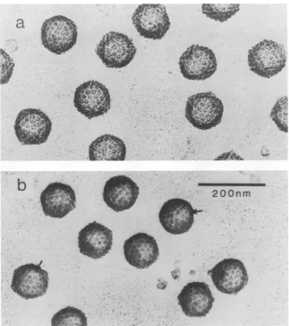
The B capsids employed in this study werecharacterized
by electron microscopy of shadowed preparations and
by
SDS-polyacrylamide gel electrophoresis. Electron micro-scopic analysis(Fig. la) revealedthatthe
capsids
werequite
homogeneous in morphology and relatively free from con-taminating noncapsid material. In most cases theiricosahe-dral shape could be discerned and individual capsomers
couldbe seen to protrudeabove afloororbase
layer.
Afteretching for10 s(themaximum time employedinthis
study),
there wasclearevidence thatindividual capsomershadbeen
eroded or smoothed (Fig. lb, arrows). The overall icosahe-dral capsid morphology was still clearly evident, however,
and reduction in the capsid "diameter" was less than 10%.
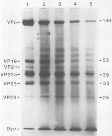
SDS-polyacrylamide gelanalyses of unetched capsids
dem-onstrated the presence of the six expected polypeptides
(Table 1; Fig. 2, lane 1), which together accounted for90%
or more of the total protein present. Their relative
abun-dance (Table 1) was in the range of values
previously
reported for B capsids (7). The 12-kilodalton polypeptide
(VP26) reported by Cohen et al.(3) either was not presentin
ourpreparations or migrated at the bromophenol blue
dye
front.
Figure 2 shows the results of SDS-polyacrylamide gel
analyses performed with capsids that had been etched for
various times in a 0.5-keV Ar+ plasma. Yields of the six major proteins are shown quantitatively as a function of
etching time in Fig. 3 and in the form of first-order rate (decay) constants in Table 2. From an examination of the stained gels (Fig. 2) one could seethat VP5 and VP19were lost rapidly as a function of etching time, while erosion of VP22a and VP24 was much slower. In all etched specimens (Fig. 2, lanes 2 through 5) there was a markedaccumulation of stained material between the major protein bands. Much of this material was present as a smear, but occasionally distinct bands were evident. Two of these bands are indi-cated by asterisks in Fig. 2, lane 2. Such bands most probably correspond to polypeptides produced by preferen-tial cleavage of larger proteins at defined sites. (Thus, the bands indicated by asterisks in Fig. 2, lane 2, are expected to be derived from VP5, since this is the only larger protein.) Erosion of the major protein species was also accompanied by an increase in the amount of stainable material, most probably small degradation products, migrating at the bro-mophenol blue dye front.
Quantitative analysis of the stained gels revealed that degradation of capsid proteins followed one of two quite different kinetic courses (Fig. 3). Loss of VP5, VP19, and VP23 began as soon as capsids were exposed to accelerated ions and proceeded more or less logarithmically until less than 20% of the original protein remained (approximately 6 s of etching). In contrast, biphasic kinetics were observed for degradation of VP21, VP22a, and VP24. Their loss was slow
duringtheinitial1 to 3 s of etching and more rapid thereafter. First-order rate constants (Table 2) calculated from the initial slopes (in two experiments) showed that proteins lost with monophasic kinetics (VP5, VP19, and VP23) had rates of 0.31 to 0.80s-1, while those for VP21, VP22a, and VP24 were 0.02 to 0.14 s-1. After correction for protein target size, thecorrespondingvalues were 0.69 to 1.31s-1and 0.07 to 0.37 s-1, respectively. In both experiments, corrected J. VIROL.
on November 10, 2019 by guest
http://jvi.asm.org/
HSV-1 CAPSID PROTEIN LOCALIZATION 4699
4ok~ ""
a
k .1
-WS...
i--.n..
..
-_
jcw
-2-wb
_
eEl
__
_r
Ir
_.t~~~~~~~~~~~~~~~~~~~~~~~~~~~~~~~~~~
__
200nm
'I:s
FIG. 1. Electron micrographsofHSV-1 B capsids before (a) and after (b) Ar+ etching. B capsids werepurified, criticalpoint dried, etched (for10s),andshadowed withPt-CasdescribedinMaterialsandMethods.Arrowsin panel bindicateregionswhere capsomers can be seen tobeeroded by the etchingprocess.
rateconstants were higher for VP19 and VP23than for VP5.
Thelargest inconsistencybetween the two experiments was
observed in the case of VP24, which had corrected rate constants of0.07
s-'
and 0.37s-'.
In allother cases, thecorrected rate constants differed by 50% or less in two
experiments.
TABLE 1. Proteincomposition of HSV-1Bcapsidsa
o%of totalprotein'
Protein Mr,103
Expt1 Expt 2 Avg
5 149 51.7 72.1 61.9
19 53 10.6 8.2 9.4
21 44 2.2 1.9 2.1
22a 39 23.6 7.2 15.4
23 33 10.8 9.6 10.2
24 25 1.1 1.0 1.0
aTheprocedures employedforB-capsidpreparation, protein
solubiliza-tion, gel electrophoresis, andproteinquantitationaredescribedinMaterials
andMethods.
ICapsids wereconsideredtoconsist onlyof the six indicatedproteins. Materialmigratingatthedye front and minorpolypeptides(whichaccounted
for less than1% of thestainedprotein)were notincluded intheanalysis.
DISCUSSION
The most
significant
feature ofthe resultsreported
hereis the time at which erosion of individualcapsid
proteins
began. VP5, VP19, and VP23 were degraded
beginning
as soonascapsidswereexposedtoacceleratedions,
suggesting
that all or part ofthese polypeptides was either exposed directly on the capsid surface or lay close enough to the
surface to be initially within the range (2.2 to 4.4 nm) of
accelerated ions. In contrast, there was a distinct lag or
delay
(biphasic
kinetics)
in the loss ofVP21, VP22a, and VP24. Theseproteins
must, therefore, have been burieddeeplyenough thattheywere
initially beyond
the range ofAr+ ions. The delay observed for their loss most
probably
represents the time
required
formoreexternal structures to be damagedoreroded away(13).
It is oflesser
significance
that the correcteddecay
con-stantsforVP19and VP23werehigher
than that for VP5. Inprinciple,this indicates that theratio of
exposed
surfacearea toproteinmassis greaterforVP19and VP23 than for VP5. Infact, however,thesmallratedifferencesobservedmay be due tosecondary
factors suchasdeviations ofproteins
fromthe assumed spherical geometry,
partial
shielding
of one VOL.63, 1989NO
t
I
on November 10, 2019 by guest
http://jvi.asm.org/
[image:3.612.153.476.76.440.2] [image:3.612.66.307.590.679.2]4700 NEWCOMB AND BROWN
1 2 3 4 5
VP5
-t4
4N.II
_ _VP19-g
VP2 1-
VP22a- VP23-
VP24-U.s.
a _._
149
-53
-39
-25
[image:4.612.314.556.77.341.2]Dye- _41
4i_
FIG. 2. SDS-polyacrylamide gel analysis of HSV-1 B capsid proteinsderived from unetched capsids (lane 1) and from capsids etched for 1s(lane 2), 3s(lane 3), 6s(lane4),and10s(lane 5).Gels
were runand stained with Coomassie blueasdescribed inMaterials
and Methods. Molecularweights(MW;inthousands)of themajor polypeptide speciesareshowntotheright.The asterisks in lane 2 indicatepolypeptides thoughttobe derived fromVP5by the action ofaccelerated ions.
surfaceprotein by another,oralignmentofproteinsrelative
to the direction of incident ions. The results, therefore, probably donotallowonetoorder theexposed polypeptides accordingtotheir relativedegreeofavailabilityonthecapsid surface.
In formulating the above interpretation it was assumed that erosion of capsids in the ionplasma was more orless geometrically uniform. Studies with model substrates such
asyeast cells andpolystyrene spheres have suggested that theassumptionisjustified. Thesematerialswerefoundtobe eroded inanisotropic oromnidirectional mannerwhenthey were irradiated on nonconducting supports at voltages of less than 5 to10keV (W. Newcomb andJ. Brown, unpub-lishedobservations). Both conditions applied inthepresent study. Moreover, direct electron microscopic analysis of etchedcapsids (Fig.lb) supportedtheview thaterosion had takenplacefrom alldirections.
Our conclusion that VP5 lies at or very near the capsid
surface is consistent with the idea that itforms the major structural component of the hexavalent (and also possibly pentavalent)capsomers(24).It ismostlikely,therefore, that the otherexposedpolypeptides, VP19andVP23, constitute the remaining structural features of the capsid surface. These could include the pentavalentcapsomers (ifthey are
notcomposed ofVP5), the trigonal, fiberlike material found to connect adjacent capsomers (21), and the floor or base
material in which capsomers appear to be embedded. The properties ofVP19 make it an attractive candidate for the
floor material. Its ability to bind DNA (1) and its disulfide bond(s) to the major capsid protein (26) would both be accommodatedifitweretoanchorcapsomers ontheouter side of the capsid wall while making contact with DNA inside.Althoughless isknown about thepropertiesofVP23,
4-)
0)0.50-C~~~~~~~~~~~~
E
a) 2
c-
0.20--60
0 9123
0.10
0 0
h0.05-ILL
0 3 6 9 12
Etching
Time
(seconds)
FIG. 3. Decay kinetics of the major proteins during Ar+ etching of HSV-1 Bcapsids. Individualproteinsweredetermined quantita-tively by densitometric scanning (as described in Materials and Methods) of the stainedgelshownin Fig. 2.
it could form the trigonal, intercapsomeric materialas
sug-gestedby Schragetal. (21).Thenumber of VP23 molecules
percapsid expectedonthebasis of this model(960)isclose to the range of experimental values (9, 14) reported for EHV-1 (approximately 700 copies) and HSV-1 (1,400 to 1,700 copies) capsids.
From the total amount of VP19 and VP23 found to be present in B capsids, one can argue that neither could
constitute only thepentavalentcapsomers. Forexample, if thetotal massof HSV-1 B capsids iscomparableto thatof EHV-1 intermediate capsids (which are closely similar in
otherrespects), thenonecan calculate that B capsids must contain approximately 21 megadaltons of VP19 and 23
TABLE 2. First-orderrate constantsfordegradationofHSV-1
capsid proteins duringAr+ plasma etchingof intactcapsidsa
Rate constant(s-1)
Rate constant(s-1) correctedforprotein
Protein targetsizeb
Expt1 Expt2 Expt1 Expt2
VP5 0.69 0.80 0.69 0.80
VP19 0.40 0.64 0.82 1.31
VP21 0.12 0.06 0.28 0.14
VP22a 0.08 0.14 0.20 0.35
VP23 0.36 0.31 1.00 0.86
VP24 0.02 0.11 0.07 0.37
aAllAr+plasmaetchingwascarried out at5mA asdescribedinMaterials
and Methods. Rate constants were calculated fromthe initial portionsof
logarithmic decay curves such as those shown in Fig. 3. Data in Fig. 3
correspondtoexperiment1.
bObservedrate constantswerecorrectedfor the effects ofproteintarget
sizebyassuming that allproteins were sphericaland that target size was
proportionaltoproteinsurfacearea(i.e.,proportionaltomolecularweightx
2/3).Corrections were made relative to the value forVP5 (Mr, 149,075).
J.VIROL.
l
on November 10, 2019 by guest
http://jvi.asm.org/
[image:4.612.82.273.87.319.2] [image:4.612.314.555.571.674.2]megadaltons of VP23 (14). Thiscontrastswithatotalmassof 9.6 megadaltons measured by Schrag etal. (21) for the 12 pentavalentcapsomers. Thus, if eitherVP19 or VP23 forms the pentavalent capsomers, then it must also be present
elsewhere in the capsid structure. Gibson's studies with
cytomegalovirus provide further evidence that VP19 is
un-likely
to constitute the pentamers. Cytomegalovirus Bcap-sidswerefoundtohavemorphologically normal pentamers
despite the fact that they lack apolypeptide analogous to
HSV-1 VP19 (5, 6).
The internal localization of VP22a as indicated by the evidence presented here is in conflict with the surface-labeling study of Braun et al. (2). These investigators ob-served that VP22a was iodinated when B capsids were
reacted in vitro with particle-bound lactoperoxidase and
concluded that VP22awas exposed on the capsid surface. Thereasonsfor the discrepancy with thepresent study are not yet clear. It is possible that the VP22a molecules iodinated intheearlier study (2)werefree in solutionor were presentin brokenordamaged capsids.Theviewthat VP22a
is located insideB capsids, however, is supported by elec-tron
microscopic
analyses of rotary-shadowedHSV-1 A and Bcapsids.
No detectable differences wereobserved in thesurface architecture ofthese structures (bothresembled the
images
shown inFig. la) despite the fact thatAcapsids lack VP22a(W. Newcomb andJ. Brown, unpublishedobserva-tions).
Similar results were obtained with shadowedprepa-rations of EHV-1 light and intermediate capsids, which resemble HSV-1AandBcapsids, respectively (14). Finally,
no evidence for surface VP22a was obtained when
cryo-electron
microscopic images
ofEHV-1 light andintermedi-ate
capsids
were employed to compute three-dimensional reconstructions ofthe two capsid types at high resolution (T. S. Baker, W. W. Newcomb, F. P. Booy, J. C. Brown,and A. C. Steven,inProceedingsofthe 47thMeetingofthe
Electron Microscopy SocietyofAmerica,abstr.,p. 822-823,
1989).
These latter results (together with the presention-etching
study)aremostcompatible with the viewthat VP22ais found insidethe
capsid
cavity. There it could constitute thevariously
shaped structures observed near the capsidcenterin thinsectionsof
purified
Bcapsids and ofBcapsids
in infected-cell nuclei (7, 20). The internal localization of VP22aas
suggested
here isinaccord with itsproposed
role inDNApackaging
(18, 19, 22)oras ascaffolding
orassemblyprotein
involved incapsid morphogenesis
(11).Thetwo
remaining
internalpolypeptides,
VP21andVP24,
are
quite
minorcomponents of the Bcapsid. Together they
accountfor less than5% of the total
capsid
mass(Table 1; 7),
and littleis known aboutthefunction ofVP24.The
proposed
location ofVP21inside thecapsidwalliscompatiblewith the
idea that itmaybea
partially processed
form ofVP22a(1)
orthat it may form the
proteinaceous
core around which the DNA appearstobewrapped
inthe maturevirion(7).
ACKNOWLEDGMENTS
Wethank WadeGibson,JohnBoring,AlasdairSteven,and Jace Houglandforproductivediscussionsaboutthisproject.
This work was supported by Public Health Service grant GM34036 from the National Institutes of Health.
LITERATURECITED
1. Braun,D. K.,W. Batterson,and B. Roizman. 1984. Identifica-tion and genetic mapping of a herpes simplex virus capsid
proteinthatbinds DNA. J. Virol. 50:645-648.
2. Braun,D.K., B. Roizman, andL.Pereira. 1984. Characteriza-tionofpost-translational productsofherpessimplexvirus gene 35proteins bindingtothe surfaces of fullcapsids butnotempty capsids.J.Virol. 49:142-153.
3. Cohen, G.,M. Ponce de Leon, H. Diggelmann, W. Lawrence, S. K.Vernon,and R.Eisenberg. 1980. Structuralanalysisof the capsid polypeptidesofherpes simplexviruses types1 and2.J. Virol. 34:521-531.
4. Dargan, D.J- 1986. The structure andassembly of herpesvi-ruses, p. 359-437. In J. Harris and R. Home (ed.), Electron microscopyofproteins,vol. 5.AcademicPress, Inc.(London), Ltd.,London.
5. Gibson,W.1981.Structural and nonstructuralproteinsof strain Colburncytomegalovirus. Virology111:516-537.
6. Gibson, W. 1983. Protein counterparts of human and simian cytomegaloviruses. Virology128:391-406.
7. Gibson, W.,and B. Roizman.1972.Proteinsspecified by herpes simplexvirus. VIII. Characterization andcompositionof mul-tiple capsid forms of subtypes 1 and 2. J. Virol. 10:1044-1052.
8. Hay, J.,C. R.Roberts,W. T.Ruyechan,and A. C. Steven.1987. Herpesviridae,p. 391-405.In M. NermutandA. Steven(ed.), Animal virusstructure. Elsevier,Amsterdam.
9. Heine, J. W.,R. W.Honess,E. Cassai,and B.Roizman. 1974. Proteins specified by herpes simplex virus. XII. The virion polypeptidesof type 1 strains. J. Virol. 14:640-651.
10. Laemmli,U. K.1970.Cleavageof structuralproteins duringthe assembly of the head ofbacteriophage T4. Nature (London) 227:680-685.
11. Lee, J. Y.,A.Irmiere,and W.Gibson. 1988. Primate cytomeg-alovirusassembly:evidence thatDNApackagingoccurs subse-quenttoBcapsid assembly. Virology167:87-96.
12. McGeoch, D. J., M. A. Dalrymple, A.J. Davidson, A.Dolan, M. C.Frame,D.McNab,L.J. Perry, J.A.Scott,and P.Taylor. 1988. ThecompleteDNAsequenceof thelong uniqueregionin the genome of herpes simplex virus type 1. J. Gen. Virol. 69:1531-1574.
13. Newcomb,W.W., andJ. C. Brown. 1988. UseofAr+plasma etching tolocalize structural proteins inviruses: studies with adenovirus2. Anal.Biochem. 169:279-286.
14. Newcomb,W.W., J. C.Brown,F.P. Booy, and A. C.Steven. 1989.Nucleocapsidmassandcapsomerprotein stoichiometryin equine herpesvirus 1: scanning transmission electron micro-scopic study.J. Virol.63:3777-3783.
15. Newcomb,W.W.,T. A.Johnston,andJ. C. Brown. 1987.Ar+ plasma etchingofpalmiticacidmultilayers: differential erosion
rates of exposed and protected layers. Langmuir
3:1000-1004.
16. Perdue, J., J. Cohen, M. Kemp, C. Randall, and D. O'Cal-laghan. 1975. Characterization of threespeciesofnucleocapsids
ofequine herpesvirus type 1. Virology 64:187-205.
17. Perdue,M. L., J. C. Cohen,C. C. Randall, and D.J. O'Cal-laghan. 1976. Biochemical studiesonthematurationof
herpes-virusnucleocapsid species.Virology74:194-208.
18. Preston,V.G., J.A. V.Coates,and F.J.Rixon.1983. Identifi-cation and characterization of a herpes simplex virus gene product required for encapsidation of virus DNA. J. Virol. 45:1056-1064.
19. Rixon,F.J.,A. M.Cross,C.Addison,and V.G. Preston. 1988. Theproductsofherpessimplexvirustype1geneUL26which are involved in DNA packaging are strongly associated with emptybutnotwithfullcapsids. J.Gen. Virol.69:2879-2891. 20. Roizman,B., andD.Furlong. 1974. Thereplication of
herpes-viruses,p. 229-403. InH. Fraenkel-Conratand R. R. Wagner
(ed.), Comprehensive virology, vol. 3. Plenum Publishing Corp., NewYork.
21. Schrag,J. D., B. V.Prasad, F. J. Rixon, and W. Chiu. 1989. Three-dimensional structure of the HSV-1
nucleocapsid.
Cell 56:651-660.22. Sherman,G.,andS. L.Bachenheimer.1988. Characterization of intranuclearcapsidsmadebyts
morphogenic
mutantsof HSV-1. Virology163:471-480.on November 10, 2019 by guest
http://jvi.asm.org/
4702 NEWCOMB AND BROWN
23. Thomas, D., W. W. Newcomb, J. C. Brown, J. S.Wall,J. F. Hainfeld, B. L. Trus,andA. C.Steven. 1985. Massand molec-ularcomposition of vesicular stomatitis virus:ascanning
trans-missionelectron microscopy analysis. J. Virol. 54:598-607. 24. Vernon, S.,M. Ponce de Leon,G. Cohen,R. Eisenberg, andB.
Rubin. 1981. Morphological components of herpesvirus. III.
Localization of herpes simplex virustype 1 nucleocapsid
poly-peptides by immune electron microscopy. J. Gen. Virol. 54: 39-46.
25. Wildy, P., W. Russell, and R. Horne. 1960. The morphology of herpes virus. Virology 12:204-222.
26. Zweig, M., C. Heilman, Jr., and B. Hampar. 1979. Identification of disulfide-linked protein complexes in the nucleocapsids of herpes simplex virustype2. Virology 94:442-450.
J. VIROL.