0022-538X/89/031162-12$02.00/0
Copyright©) 1989,American Society forMicrobiology
Structural Defect Linked
to
Nonrandom
Mutations in the Matrix
Gene of Biken
Strain Subacute
Sclerosing
Panencephalitis Virus
Defined by cDNA Cloning
and
Expression of Chimeric Genes
MINORU AYATA, AKIKO HIRANO, ANDTIMOTHY C. WONG*
Department ofMicrobiology, University of Washington Schoolof Medicine, Seattle, Washington 98195
Received 17 August 1988/Accepted 16 November 1988
Bikenstrain, anonproductive measles viruslike agent isolated froma subacutesclerosing panencephalitis
(SSPE) patient, containsaposttranscriptional defectaffecting matrix(M) protein. AputativeMprotein was
translated invitro with RNA from Biken strain-infected cells. A similarprotein wasdetected in vivo byan
antiserumagainstapeptide synthesizedfrom the cloned Mgeneof Edmonston strain measles virus.Byusing
a novelmethod, full-length cDNAs of the Biken Mgene were selectively cloned. The cloned Biken M gene
containedanopen readingframe which encoded 8extracarboxy-terminal amino acid residues and 20amino acid substitutionspredictedtoaffect both thehydrophobicityandsecondarystructureofthegeneproduct.The clonedgene wasexpressed in vitro and in vivo intoa37,500
Mr
proteinelectrophoretically andantigenicallydistinctfromthe M proteinofEdmonston strain but identicaltothe M proteininBiken strain-infectedcells.
ChimericMproteins synthesizedinvitroand in vivo showed that the mutations in thecarboxy-proximal region
altered the local antigenicity and those in the amino region affected the overall protein conformation. The protein expressedfromtheBikenMgenewasunstable invivo.Instabilitywasattributedtomultiplemutations inboth the amino andcarboxy regions.Asurprisingnumber of mutations in both thecodingand noncoding
regions of the BikenMgene wereidenticaltothoseinanindependentlyisolated SSPE virus strain withasimilar
defect. Theseresultsofferinsightsinto thebasis of the defect in Biken strain andposeintriguing questionsabout
theevolutionary originsof SSPE viruses in general.
Measlesvirustypicallycausesacuteandhighly contagious
infections in children andyoungadults. Onrareoccasions,a
few individuals might develop a chronic infection in the central nerous system (CNS) years after exposure in a
condition known as subacute sclerosing panencephalitis
(SSPE) (41). Unlikeacutemeasles virus infections, SSPE is
always fatal but noncontagious. Typically, measles
virus-related antigens and RNA can be detected in the CNS of
SSPE patients, but infectious virus can rarely be isolated.
Successfulisolation has occasionallybeenaccomplished by
cocultivating the infected CNS materials with permissive
primate cells. The virus thus isolated may continue to
replicate ina cell-associated mannerandproduce no
extra-cellular virions, or it may become productive after in vitro
passages (45). However, only the nonproductive strains retain neurovirulence in experimental animals (33, 42).
Therefore, the basisof nonproductive infection, a hallmark
ofSSPE, isofgreat interest.
Studies have linked nonproductive infections by SSPE strainstoabnormal expression of several viralproteins. The most common defects affect the matrix (M) protein, which
formstheintermediary layerbetween theribonucleoprotein
core and the outer lipid envelope of the virion (12, 24). Serumsamples from SSPEpatients frequently lack antibody
activities against M protein despite having high activities
against other measles virus proteins, and brain materials
from SSPEpatients often containnodetectable M protein(1,
11, 17, 18, 46). Biochemical analyseshave revealed possible
defects affecting transcription,translation, orstabilityof the
M protein in different cases (2, 7, 10, 40). More recent
studiesindicate that insome casesof SSPE, both M protein
andantibodiesspecific forMprotein canbe detected (2, 14,
*Correspondingauthor.
27, 31, 32). Furthermore, abnormalities in other viral
pro-teins, especially theenvelope-associated hemagglutinin (H)
and fusion (F) proteins, have also been observed (2, 27).
Current evidence suggests thatanabnormalityinanyoneof
several viralproteinscouldaffect virusmaturation andmight
contributeto anonproductiveinfection. A clearknowledge
of the putative viral defects is important forunderstanding
the basis of thistype of infections.
Biken strain is ameasles viruslikeagent thatwasisolated
from an SSPE patient by cocultivating the CNS materials
with human embryonic lung (HEL) cells (43). The agentis
nonproductive and spreads from cell to cell by fusion.
Biochemicalanalysis of the Bikenagenthas thus faryielded
conflictingdata. In anearly study, nonproductive infection
was attributed to abnormality in H function (5). Lin and
Thormarlatersuggested that the defectwasduetolack ofM
protein (28). In contrast, by immunofluorescence
tech-niques, Johnson et al. detected low levels ofM protein in
Biken strain-infected cells (23). However, this observation
wasnotconfirmedbyalaterstudy involving 14independent
monoclonal antisera(39).
In this study, we used biochemical, recombinant DNA,
and immunological approaches to identify and define a
defect affecting the M protein of Biken strain. The results show that Biken strain indeedencodesanM-relatedprotein.
However, the Biken M gene is riddled with mutations
predicted to affect both the hydrophobicity and secondary
structure of the gene product. By DNA-mediated gene
transfer, the cloned Biken M gene produces a structurally
altered Mproteinwithahalf-life 16 times shorterthan that of
the M protein ofEdmonston strain measles virus. By
ex-pressing chimeric M proteins containing different Biken
mutations, the structural alterations and instability of the
protein were attributed to multiple mutations in the amino
1162
on November 10, 2019 by guest
http://jvi.asm.org/
SSPE VIRUS BIKEN STRAIN STRUCTURAL DEFECT
and carboxy regions. The mutations in the Biken M gene
were surprisingly similar to those in an independently iso-lated SSPE virus strain with a similar defect. These results
shed light on the possible mechanism of the defect in the
Biken M protein and pose interesting questions about the origins of SSPE virus strains.
MATERIALS AND METHODS
Cellsandviruses. Biken strain isolated in Japan by
cocul-tivating infectedbrain materials from an SSPE patient with HEL cells (43) was a kind gift from S. Ueda, Osaka
University,
Osaka, Japan. The chronically infectedBiken-HELcells produced no extracellular virions and were
prop-agated by occasionally being replenished with fresh HEL
cellsin Eagleminimal essential medium supplemented with
10% fetal calfserum. Edmonstron strain measles virus was propagated in Vero cells or African green monkey kidney (CV-1) cells in the same medium. Simian virus SV40-trans-formed CV-1 (COS) cells have been described previously
(15).
Antisera. The GM antiserum against total measles virus
proteinswasalso a kind gift from S. Ueda and was originally prepared by immunizing a female African green monkey
withCV-1 cells infected with Nagahata strain measles virus
(44). The GMserumrecognizedall the structuralproteins of both Nagahata and Edmonston strain measles virus.
Mouse monoclonal antibodies were prepared against
af-finity
column-purified Nagahata strain measles virusanti-gensby usingapreviously described procedure (21). Hybrid cell lines
producing
measlesvirus-specific
antibodies were identified byanindirect immunofluorescence assay, and the antibody specificities weredeterminedby radioimmunepre-cipitation. Specific
antibodieswereharvested eitherfromthecellculturefluidorfromascites fluid from
syngeneic
animalswhich received thehybridomacells.
Region-specific polyclonal antiserawere prepared against peptides expressed from clonedDNAin bacteria. To
gener-ate the M-BC antiserum against the entire Edmonston M protein, we inserted the
BglII-ClaI
fragment from cDNAclone pcD-M2i (48) into a procaryotic
expression
vectorcontaining
an inducibletrpEpromoter(36) (seeFig.
6). Thehybrid protein containing trpEand M sequencesproducedin E. coli HB101 was
purified
by sodiumdodecyl
sulfate-10%polyacrylamide gel
electrophoresis
(SDS-PAGE)
andin-jected subcutaneously into a female New Zealand White rabbit in two 200- to 300-,ug doses administered 2 weeks
apart. Sera collected at 1-week intervals after the second
injection
were tested by radioimmuneprecipitation.
TheM-BE and M-EE antisera
against
the amino andcarboxy
regions
ofthe Edmonston Mprotein
wereprepared
by
thesame method with DNA
fragments
spanning
theBglIH
andthe
5'-proximal
EcoRI sites or the two EcoRI sites inpcD-M2i, respectively (48) (see
Fig. 6).
Similarly,
the N-EEantiserum was prepared
against
apeptide
encodedby
thesequencebetweentwoEcoRIsites in the N geneof
Edmon-ston strain
(S.
Castaneda and T.Wong,
submitted for publication).Protein and RNAanalyses. Cellular
proteins
werelabeled,
immunoprecipitated,
andanalyzed by
SDS-PAGE asprevi-ously
described(48),
except that CL-4Bprotein
A-Sepha-rose
(Pharmacia)
instead of fixedStaphylococcus
aureus(Calbiochem-Behring)
was used to recover the immunecomplexes
in someexperiments.
DNAprobes
specific
fortheN, P, M,
F,
and Hgenes ofmeasles virus were
prepared
fromfull-length
cDNA clones(47, 48; T. Wong, G. Wipf, M. Ayata, and A. Hirano, manuscriptinpreparation).RNA was prepared for Northern (RNA)blotanalysisasdescribed previously (48), except that electrophoresiswas performed in a 1% agarose gel contain-ing formaldehydeinstead of methylmercuric hydroxide (29).
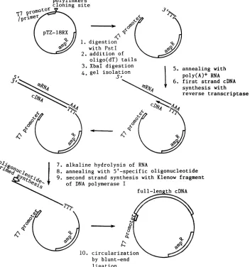
Novel approach for selective cloning of specific full-length
cDNA. A novel simple method was devised to selectively
obtainfull-lengthcDNArepresentingany geneforwhichthe
5' sequence is knownor canbe deducedfromother sources
suchasgenomicsequence.The vector pTZ18RX, generated by insertingaXhoI siteattheoriginal SmaI site in pTZ18R (Pharmacia, Inc.),waslinearizedatthePstI site, tailed with
oligo(dT),
and recut with XbaI (Fig. 1). The gel-purifiedoligo(dT)-tailed vector (2.8 ,ug or 1.4 pmol) was annealed
with 5
pLg
ofpoly(A)+ RNA,isolatedfrom Biken-HEL cellswhencytopathic effectswere maximal, to prime the
synthe-sis of the first cDNA strand in a fashion similar to the
Okayama-Berg
procedure (34). The firstcDNA strand wassynthesized at 37°C for 1 h in 50 ,ulofa reaction mixture
containing
50mMTrishydrochloride (pH 8.3), 75 mMKCI,3 mM MgCl2, 10 mM dithiothreitol, 1 mM each dATP, dGTP, dCTP,andTTP, 50 Uof RNasin (PromegaBiotec), and 1,000U ofreverse transcriptasefrom Moloneymurine
leukemia virus(BethesdaResearch Laboratories, Inc.).
Re-action products were extracted with phenol-chloroform,
precipitated
withethanol,andsuspendedin Trishydrochlo-ride(pH8.0)
containing
1 mMEDTA. Aportion
(10%)
of thereactionproductswastreated with 50mMNaOHat65°Cfor 1 h to hydrolyze RNA. After neutralizing with Tris hydro-chloride(pH8.0) and
HCI,
1pmolofasynthetic
oligonucle-otide(5'-AGGAGCAAAGTGATTGCCTC-3')
correspond-ing
to the 5' mRNA terminus ofthe M gene ofEdmonstonstrain was added and annealed to
full-length M-specific
first-strand cDNA
by being
heated to65°C
for 2 min andslowly
cooledto roomtemperature(Fig. 1).
The mixturewasadjusted
to100,ul,containing
50 mMTrishydrochloride (pH
7.4); 5 mM
MgCl2;
5 mMdithiothreitol;
50 ,ug of bovineserumalbumin per
ml;
50,uM
eachdATP, dGTP, dCTP,
andTTP;
and 10 U of the Klenowfragment
ofEscherichia coliDNApolymeraseI.Second-strand
synthesis
wascarriedoutat15°C
overnight.
The vector-cDNAwascircularizedby
T4DNA
ligase
andtransformed into competentE. coli x1776orDH5(29). Clones
containing
the Biken M genewereidenti-fied
by colony
hybridization
with a DNAprobe
prepared
fromthe Edmonston M gene.
Full-length
clones wereiden-tified
by
regeneration
of an XbaI site at the 5' end of thecDNA insert and confirmed
by
sequenceanalysis.
Structural
analysis
ofthecloned gene.Sequence analysis
was carried out
by
adideoxynucleotide-induced
chainter-mination method in both directions
(38),
withoverlapping
deletion clones
generated
by
digestion
with either ExolIlnuclease
(19)
orspecific
restrictionendonucleases.Hydropathy
andsecondary
protein
structure werede-duced fromsequenceinformation
by using
criteriadescribed by Kyte and Doolittle(26)
and Chou and Fasman(13),
respectively,
and a GenePro computer program(Riverside
Scientific
Enterprises, Seattle, Wash.).
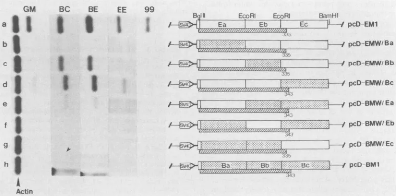
Constructionof chimeric Mgenes.Togenerate chimericM
genes for in vitro
expression,
theEcoRI sites in the vectorsequences of
parental
clonespTZ-EM1
andpTZ-BM1,
which containedthe
full-length
Mgenes of Edmonston andBiken
strains,
respectively,
werefirstdestroyed.
DNAfrag-ments from the
BglII
site to the5'-proximal
EcoRIsite,
between the two EcoRI
sites,
and from the3'-proximal
EcoRI sitetoaHindlllsite in thevectorwereisolated from
the
parental
clonesby gel
electrophoresis
andligated
withVOL. 63, 1989 1163
on November 10, 2019 by guest
http://jvi.asm.org/
annealing with poly(A)+ RNA first strand cDNA synthesis with
reverse transcriptase
7. alkaline hydrolysis of RNA
8. annealing with 5'-specific oligonucleotide
et°^tQd ' 9. second strand synthesis with Klenow fragment
es>SNP,
of DNA polymerase Ifull-len th cDNA
A$,
10. circularization by blunt-end ligation
FIG. 1. Simple method for selective cloningofexpressible full-length cDNAs fromspecificgenes (seeMaterialsandMethods).
the appropriate recipient clone to generate a series of
chi-meric genes containing different Edmonston and Biken se-quences(see Fig. 8). For in vivoexpression,the entire series ofchimeric Mgeneswerereleased from thepTZvectorsby
digestion with BamHI and HindlIl and inserted into the
BamHI site downstream of the SV40 early promoterin the
pcDvector (34) by usingaBamHI linker(see Fig. 9).
Invivoexpression ofclonedgenes. Culturescontaining 1.5
x 106 COS cells (15) weretransfected with 20 ,ugof cloned
DNA as previously described (48). Cellular proteins were
labeled for 6 h with 25 to 30 ,uCi of[35S]methionine (3,000
Ci/mmol; Du Pont; NENResearch Products) at 45 h
post-transfection andanalyzed asdescribed above.
Invitroexpressionof clonedgenes.Aftertheplasmidswere
linearized at the Hindlll site downstream of the cDNA
inserts, RNAwastranscribed in vitro byT7RNA
polymer-ase fromthe cDNA clones which contained an integral T7
promoter(30) (Fig. 1). Equal amounts (1 ,ug) ofRNA were
translatedin rabbit reticulocyte lysates(Promega Biotec)in
the presence of [35S]methionine (25). Equal amounts of
proteins standardizedbyscintillationcountsof
trichloroace-ticacid-precipitated radioactivity were immunoprecipitated
withantisera and analyzed by SDS-PAGE.
RESULTS
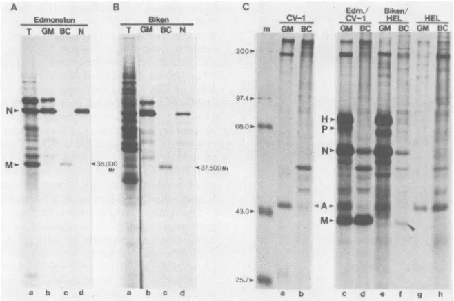
Posttranscriptional defect affecting M protein of Biken
strain. We first compared the overall gene expression of Biken strain with wild-type (Edmonston strain) measles
virus at the protein and RNA levels. Edmonston
strain-infectedCV-1 cellsproducedthe N(nucleoprotein),P
(phos-phoprotein), M, F1 (cleavage product ofF), and H measles
virus proteins detectable by the appropriate monoclonal antisera(Fig. 2A,lanesN, P, M,F, andH, respectively).All
thecorrespondingproteinsexceptMweredetected in Biken
strain-infected human embryonic lung cells (Biken-HEL)
(Fig. 2B, lanes N, P, M, F, and H, arrowheads). (The
autoradiogram for Biken-HEL cells was overexposed to show the lack of detectable Mprotein.The minor bandsnear
theexpected positionof Mproteinindicated byasterisksare
of cellular origin.) Similarly, an antiserum against total measlesviralproteins (GMserum[44])whichrecognizedthe
H, P, N, F, and M proteins in Edmonston strain-infected
cells (Fig. 2A, lane GM) also failed to detect Mprotein in Biken-HEL cells (Fig. 2B, lane GM). To rule out possible
host cell differences,we alsoexamined the viralproteins in polylinkers
cloning site
7r
'P
1.
1.
2. 3. 4.
on November 10, 2019 by guest
http://jvi.asm.org/
[image:3.612.142.494.77.453.2]SSPE VIRUS BIKEN STRAIN STRUCTURAL DEFECT 1165
B
GM N P M F H
_4p_. _ _b'1;
-0-N-0, 40.-Actin T
Al1 - ---.
-F, .a
-~~.-
MW
25.7.-0
Edmonston
c
N P M F orgin N
D
P M F H
-genome- _
-length n
9.49-7.46
-
4.40-
multi-W"
cistronic a64 4640
mono-2.37-7_ __cistronic-|cistronic | 1.351
Ednionston Biken
FIG. 2. Viral proteins andRNAs in Biken strain-infected cells. Proteins in Edmonston strain-infectedCV-1 cells (A)orBiken-HEL cells (B) were labeled with [35S]methionine for 12 h and immuno-precipitated with the GM antiserum against total measles viral
proteins (lane GM), orwith monoclonal antibodies against the N
(laneN), P(laneP), M
(Cl.99,
laneM),F(lane F), andH(lane H)proteins of measles virus. Immune precipitates were analyzed by
SDS-PAGE(10%o polyacrylamide). Thefirstlanerepresents molec-ular weight standards (shown in thousands). Total RNA purified from Edmonston strain-infected CV-1 cells (C)andBiken-HELcells (D)wasresolved by electrophoresis ina 1%agarose gelcontaining
formaldehyde, transferred ontonitrocellulose filterpaper, and
hy-bridized successively with 32P-labeled DNA probes specific forthe N, P, M, F,orHgeneof Edmonston strain (shownin lanes with the sameletters). Size markersareshown inkilobases.
Biken strain-infected CV-1 or Vero cells. Again, Mprotein was not detectedby these antisera (datanot shown).
The apparent absence of M protein in Biken-HEL cells
was not due to a transcriptional defect, since the
accumu-lated levels of monocistronic RNAs, multicistronic read-through RNAs, and genome-length RNAs were comparable
in both Biken-HEL and Edmonston strain-infected cells, as
shown by Northern blot analysis with five measles virus gene-specific DNA probes, including M (Fig. 2C and D, lanes N, P, M, F, and H). Although we did not test for expression from the L gene, which encodes the presumed
RNA polymerase (4), active transcription from the other viral genes implied afunctional polymerase activity. Thus,
theapparentdefectin M protein of Biken strain occurs ata
posttranscriptional level.
TranslationofanM-relatedprotein from Biken-HEL RNA
in vitro.Thefailuretodetect M proteindespite high levelsof
M-specific RNA in Biken-HEL cells could be due to a translational defect. Alternatively, it was equally possible that the Biken M protein assumed an antigenically altered conformation which was not recognized by the antisera. To distinguish between these two possibilities, polyadenylated RNA was prepared from Biken-HEL and Edmonston strain-infected CV-1 cells and translated in vitro in rabbit
reticulo-cytelysates. To maximize the chance of detecting a possible
Mprotein, a peptide containing nearly the entire M protein
of Edmonston strain was overproduced in bacteria from
cloned DNA spanning theBglII to ClaI sites of the
Edmon-ston M gene and used to generate a polyclonal antiserum
called M-BC (see Fig. 6 and Materials and Methods).
An-otherantiserum similarly prepared against the N protein of
Edmonston strain served as a control (N-EE serum).
Three major proteins were immunoprecipitated by theGM
antiserum from the translational products of RNA from Edmonston strain-infected cells (Fig. 3A, lanes a and b). Two of these proteins were identified as the M and N
proteins by the M-BC and N-EE antisera, respectively (Fig.
3A, lanes c and d, respectively). In contrast, the M protein
was not immunoprecipitated by the GMantiserum from the
in vitro-translated products of Biken-HEL RNA (Fig. 3B, lanes a and b). To our surprise, the M-BC antiserum
immu-noprecipitated a 37,500 Mr protein from the same
transla-tional products (Fig. 3B, lane c, arrowhead).
Immunoprecip-itation of this protein was specific, since theN-EEantiserum
recognized the N protein but not the 37,500 Mrprotein (Fig.
3B, lane d). These results provided the first hint that Biken
strain might produce an M-related protein.
Biken-HEL cells produce an altered M protein in vivo. To
see whether theputative M protein was actuallyproduced in
vivo, we immunoprecipitated the intracellular proteins in
Biken-HEL cells with the M-BC antiserum. This serum
clearly precipitated a low but detectable amount of a 37,500
Mr protein in Biken-HEL cells (Fig. 3C, lane f)which was
not present in uninfected CV-1 or HEL cells(Fig. 3C, lanes
a, b, g, and h). Since this protein was notrecognizedby the
GM antiserum (Fig. 3C, lane e), it might represent an
antigenically altered M protein. The apparent low level of
this protein could be due to inefficient recognition by the
antiserum. Alternatively, additional defects mightaffectthe
expression or stability of the protein. Furthermore, it
re-mained possible that the 37,500 Mr protein was a
cross-reacting cellular product induced by Biken virus infection.
Selective cloning of the Biken M gene by anovelapproach.
To allow definitiveidentification and detailed analysis ofthe
putative Biken M protein, we developed a simple general
method to obtain full-length cDNAs from specific genes.
This method involved the useof anoligo(dT)-tailedvectorto
synthesize the first cDNA strand from poly(A)+ RNA from
Biken-HEL cells. The second DNA strand was selectively
primed by a synthetic oligonucleotide specific for the 5'
terminus of the Edmonston M gene (Fig. 1) (see Materials
and Methods). The vector and primer were designed to
regenerate anXbaI recognition site only if thesecondstrand
was correctly primed on a full-length first-strand cDNA.
These features simplied theproduction and identification of
the desired full-length cDNA clones. All the M-specific
clones initiallyidentified by hybridization withaDNAprobe
prepared from theEdmonston M gene werefoundtocontain
full-length cDNAs of the Biken M gene.
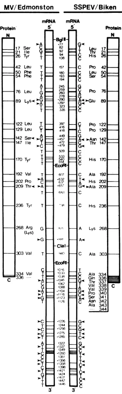
Structure of the Biken M gene. The sequence of a
full-length clone (pTZ-BM1) showed that the Biken M gene is
1,462 nucleotides (nt) long excluding the poly(A) tract,
identical in length to the M gene of the Edmonston strain
A
m GM N P M F H
200.0-- 4
97.4- _
68.0.- _
__m
43.0-
IS
VOL.63, 1989
** "
on November 10, 2019 by guest
http://jvi.asm.org/
[image:4.612.67.281.71.403.2]B
Edmonston Bil
T GM BC N T GM
I _I
a b c d
C Edm. Biken
ken CV-1 Cv-1 HEL HEL
BC N m GM BC GM BCGMBC GM 8C
9'.4'-<'' .g
H'- ^
l.i5....}
pN'-a b c d a b c d e t g h
FIG. 3. PutativeBikenMprotein detected in vitroand invivo. One microgramofpoly(A)+RNAfrom Edmonston strain-infectedCV-1 cells(A)orBiken-HEL cells (B)was translated invitro in rabbitreticulocyte lysates.Anequalamountof the translationalproductswas
immunoprecipitated with the GM, M-BC, or N-EE antisera (lanes b, c, and d, respectively), and analyzed by SDS-PAGE (10%
polyacrylamide) along with the total translational products (lane a).(C) ProteinsinCV-1 (lanesaandb),Edmonston strain-infectedCV-1 (lanes candd),Biken-HEL(lanes e andf),and HEL(lanesgandh)cellswerelabeledwith[35S]methioninefor4handimmunoprecipitated with GM (lanes a, c, e, and g) or M-BC (lanes b, d, f, and h) antiserum. Immune precipitates were analyzed by SDS-PAGE (10%
polyacrylamide).(The50,000to60,000Mr bands abovethe Mproteininlanes d and farenonspecific.)Molecularweightstandardsareshown in thousands.
previously determined from full-length cDNA clone
pcD-M2i (48). TheBikenM genecontains an openreadingframe
beginningatthe same translationalstartATG codon (nt 33) asin theEdmonston M gene (3). However, a T-to-C
transi-tion at position 1038 abolishes the normal translational
termination codon and lengthens the protein-coding region by 8 triplet codons (Fig. 4). Translation is predicted to terminate at position 1062, where anotherC-to-Ttransition
has generated aTGA codon (Fig. 4). As a result, the Biken
M gene potentially encodes a protein which contains 343
insteadof 335aminoacid residues,with a calculated molec-ular weight of38,249. Downstream ofthe new termination
codon is a long 3' untranslated region with two potential
overlappingopenreading frames preceded by ATGcodons. However, asin theEdmonstonMgene,these open reading framesare notfollowedby translational termination codons.
The M genesofBikenandEdmonston strains differ in 65
positions,ofwhich 40 lie in the protein-coding sequence and
25 lie in the normally untranslated 3' region (Fig. 4). Thus,
the overall sequences differ by about 4.4%, and the 3'
untranslated regions are more diverged, differing by about 6.1%. Some mutations are not found in all the Biken M
cDNAclones analyzed and probably represent genetic
vari-ation characteristic of SSPE virus strains (see below). A surprising number of mutations are shared with another SSPE virusstrain recently analyzed (Fig. 4, arrowhead) (10).
Thesignificance of this is discussed below (see Discussion).
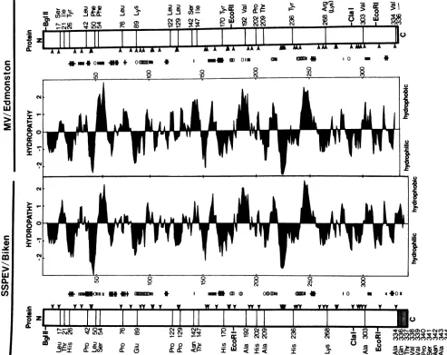
Predicted changes in the Biken M protein. On the basis of
parametersdescribed by Chou and Fasman (13), the amino
halfoftheEdmonston M protein encoded between theBglII
and the 5'-proximal EcoRI sites is predicted to contain
several beta-sheet structures (Fig. 5, parallel bars) and a
potential alpha helix (overlapping circles) interrupted by multiplepotential beta turns (crosses; see reference 3 for the
complete sequence of theEdmonston M protein). Thirteen
ofthe 20 amino acidsubstitutions in the predicted Biken M
protein occur in this amino region. With the exception of Leu-17, all the amino-proximal substitutions decrease the
hydropathy values according to a scale proposed by Kyte
and Doolittle (26), resulting in a noticeable shift toward
hydrophilicity in the hydropathy profile of this region. For instance, the inverted hydrophilic peak between Pro-42 and
Leu-50 is exaggerated, and the hydrophobic peaks around
Ser-54, Pro-76, and especially Asn-142 and Thr-147 are all
compromised (Fig. 5). Leu-17, the only substitution in the
aminoregion with increased hydropathy, occurs only intwo
offourfull-length cDNA clones analyzed and mightsimply
reflectgenetic variationcommoninSSPE virus strains(10).
In additiontotheireffectonhydrophobicity, the
substitu-tions by Pro-42 and Pro-76near twopotentialbeta turns are
predicted to extend the regions involved in the turns as a
result of the turn-promoting character of proline (Fig. 5,
crosses) (37). Furthermore, the Lys-89-to-Glu-89
substitu-tion might extend a potentialalpha helix as a result ofthe
helix-forming potential of glutamic acid (Fig. 5, overlapping
circles). These amino-proximal mutations might alter the
secondary protein structure and, with the reduced
hydro-phobicity described above, coulddrastically affect the
fold-ing andtertiary structure of the protein.
By contrast, most of the substitutions in the carboxy
region encoded between the two EcoRI sites do not
mark-edly affect the hydrophobicity of the protein. Perhaps the
most nonconservative mutations in thatregion are Ala-192
andAla-209, which might affectapotential beta-sheet
struc-tureand apotential alpha helix,respectively(Fig. 5, parallel
bars and overlapping circles, respectively). However,none
of thecarboxy-proximalmutationsaffects thepredictedbeta
turn norgenerates newpotential turns. Theextra
carboxy-A
N>3
on November 10, 2019 by guest
http://jvi.asm.org/
[image:5.612.151.480.73.291.2]SSPE VIRUS BIKEN STRAIN STRUCTURAL DEFECT
SSPEV/Biken
mRNA mRNA
5- 55
17 Ser
8
21 Ile26 Tyr T 42 Leu T
50 Phe T 54 Pe T
A
76 Leu GT
89 LyS(<'G
T
T
122 Leu 129 Leu
142 Ser<
147 1le 170 Tyr 192 Val 202 Pro 209 Thr< 236 Tyr T A T T C T T
268 Arg G
(Lys)
v-G
-1303 Val T
C
334 Vat T
336 T G
wT
T N-C T C >T A T 3 -BgIU-.77 82 92 94 108 157 180 193 194 245 259 260 278 .290 *297 323 335 397 416 418 449 *457 472 .479 509 533 540 554 607 *632 637 *657 R8-, .881 Clal-840 1016 10.33 103 .1046 1062 1088 1104 .1116 .1123 1126 .1226 1244 .1256 .1275 *1285 1322 .1337 1349 *1350 1351 *1356 *1358 1398 .1421 1424 .1431 1442 1446 3' G'KT Leu 17
T Thr 21 His 26
C Pro 42
CLeu 50
CSer
5456Pro 76 b' D-Glu 89
Ppro 122
C Pro 129
L
>Agn 142&<
Thr 147C
C His 170
C
C Ala 192
G His 202 G' Ala 209
C C A C His 23E Lys 261 Ala 30^ T Ala 334
8
Gin 336G' Thr 337
T Val 338
AG Val 339
CPro 340
A Ser 341
C Asn 342
Ala 343 344 Protei N C Gr- G-T<9 C< T4r T
C'9
FIG. 4. Sequence comparison between the Biken and
Edmon-ston M genes. The complete sequence of the Biken M gene in
full-length clone pTZ-BM1 was compared with pcD-M2i (48). The
terminal residues in the predicted Biken M protein are either
- hydrophobic (Val-338, Val-339, Pro-340, and Ala-343) or polar (Gln-336, Thr-337, Ser-341, and Asn-342). As
dis-n cussed below, these residues also affect the overall nature of
theprotein.
Invitro and invivoexpression of an antigenically altered M
protein from the cloned Biken M gene. The predicted Biken
M protein has a molecular weight higher than that of the
Edmonston M protein. However, the putative M protein in
Biken-HEL cells migrated faster than the Edmonston M
protein (Fig. 3). Since mRNAs of some SSPE virus strains have been found to be heterogeneous (10), it was important
to ascertain whether the cloned gene in fact encoded the
Biken Mprotein. RNA was transcribed in vitro from
pTZ-BM1 and pTZ-EM1 containing the M genes of Biken and
Edmonston strains, respectively (see Materials and
Meth-ods), and the proteins translated from these RNAs in rabbit
reticulocytelysates were analyzed by SDS-PAGE.
Thecloned M genes of Edmonston and Biken strains both
produced a single proteinin vitro (Fig. 6A, lanes a and h).
The Biken M gene product indeed migrated slightly faster
than the Edmonston M gene product. Furthermore, the
Edmonston M gene product wasimmunoprecipitatedbythe
GM,M-BC, and monoclonalCl.99antisera (Fig. 6A, lanes b,
c, andf, respectively), but the Biken M gene product was
recognized only by the M-BC antiserum but not by the GM andCl.99antisera(Fig. 6A, lanes j, i, and m, respectively), just as observed in Biken-HEL cells. As expected, neither
product wasrecognized by the anti-N serum (Fig. 6A, lanes
g and n,respectively).
To testwhether theBikenMgenealsoproduced the same
protein in vivo, the cloned gene was placed under the control
of the early promoter of SV40 (pcD-BM1) and transfected
into SV40-transformed CV-1 (COS) cells (15). Expression
wascompared with that of an equivalent plasmidcontaining
theEdmonston Mgene(pcD-PM-M). Cells transfected with
the Edmonston M gene produced a 38,000 Mr protein
immunoprecipitated by the GM, M-BC, and Cl.99 antisera (Fig. 6B, lanes a, b, and e,respectively). In contrast, cells
transfected with the Biken M gene expressed a low but
detectable levelof 37,500Mrprotein, whichwasrecognized
only bythe M-BC antiserum (Fig. 6B,lanes f, g, andj).
These experiments positively identify both the cloned
Biken M gene and the 37,500 Mr M protein in Biken-HEL
cellsand show that theantigenicalterations in the Biken M
protein arevirus encoded.
To localize the antigenic alterations, we used two addi-tionalantisera,M-BEandM-EE, directedagainsttheamino and carboxy halves of the Edmonston M protein,
respec-tively (see Materials and Methods). Both of these antisera
immunoprecipitated the Edmonston M protein synthesized
invitro(Fig. 6A, lanes dand e) and in vivo(Fig. 6B,lanesc
andd).However,neither antiserumrecognizedtheBikenM
proteinsynthesizedinvitro(Fig. 6A,lanes k and1)orinvivo (Fig. 6B, lanes h and i). Therefore, both the amino and
sequence shows the mRNA strand with the first adenine residue
corresponding to the 5' terminus of the mRNA as nucleotide 1.
Nucleotide differences are identified by their nucleotide numbers.
The protein-coding regions are shownas open boxes next to the
cDNAs. Predicted amino acid substitutions are identified bytheir
amino acid residue numbers. The apparent mutation at Lys-268 reflects variationamongdifferentvirusstocks,since the Edmonston
Mgene sequenced by Bellini et al. and Cattaneo et al.contains a
lysineat thisposition (3, 8). Mutations shared between Biken and
IP-3 strainsareindicatedbyarrowheads (see Discussion).
MV/
Edmonston
Protein N I I I I I 5 8 3 I 5 3 3 I I II
VOL.63, 1989 1167
r=09 M"
I'm
OMM" rm.
on November 10, 2019 by guest
http://jvi.asm.org/
[image:6.612.76.271.65.694.2]L-
,1
0)I.:a) D C'va
c-*?
I -i.£t
I
2C O
IL A AkAA
4*F..*Om4*+ 0o
i0.-0a
lrO
0
N
00 a
z. vi
I.AA0A11^nA 0AA ~~ANA A A MA A
lj* ZZolla),OIN , m o
I ItA11 - fI
.N111. NiUili11 Mt *+ mimi * mN U ( a oonii a * I *IO10
c t Y y v y y vws w y WY tYy ytt y y
=
0)A
gjgS
E
8I
SN
_D
RR
±|±
9
c2 |
E
t
i
FIG. 5. Predicted hydropathy and secondary structure of the Biken M protein. Open boxes representtheproteins predicted fromthe Edmonston and Biken Mgenes showing the identities and positions of amino acid substitutions. Thehydropathyprofileswerededucedby
aGenePro computerprogram basedon ascale developed by Kyte and Doolittle (26). Potential beta-sheet structures (11111), alpha helices (COMM),andbetaturns( )werededucedby usingparameters describedbyChouandFasman(13).Locationsof the conservedlysine andarginine residuesareindicatedbyarrows.
carboxy halves of the Biken M protein are antigenically altered.
Instability of the Biken M protein in vivo. The M-BC antiserum,whichefficientlyrecognized the Biken M protein
translated in vitro, could barely detect the same protein in
vivo (compare Fig. 3B, lane c, with Fig. 3C, lane f, and
compare Fig. 6A, lanej, with Fig. 6B, lane g). Since the
Biken M RNA seemed to be translated efficiently, we
compared the half-lives of the M proteins expressed in vivo
fromthecloned Edmonston and Biken M genes in a
pulse-chase experiment.
COS cells transfected with pcD-PM-M or pcD-BM1 (Fig.
6) were pulse-labeled with [35S]methionine for 30 min and
chased withunlabeled medium. The intracellular half-life of
theEdmonstonMprotein was about 8 h (Fig. 7, lanes a to d).
Incontrast,the Biken M protein had a half-life of less than 30
min (Fig. 7, lanes e to h). This explains the discrepancy betweenthe levels of the Biken M protein detected by the M-BCantiserum in vitro and in vivo.
Differential effects of the amino andcarboxy mutations on
the structure of the M protein. To directly examine the
mutationaleffects on protein structure rather thanstability,
we constructed chimeric M genes expressible in vitro into
chimericproteins which contained different Edmonston and
Biken sequences (Fig. 8). Clone pTZ-EMW/Ba contained
the 5'-proximal substitutions predicted to markedly affect
both hydrophobicity and secondary structure of the protein
(Fig. 5 legend). Clone pTZ-EMW/Bb contained the less
disruptive 3'-proximal substitutions (Fig. 8). Clone
pTZ-EMW/Bc encodedaprotein with theextracarboxy-terminal
residues. Clones pTZ-BMW/Ea, pTZ-BMW/Eb, and
pTZ-BMW/Ecweredesignedto testthe combined effects ofmore
thanoneof these mutatedregions (Fig. 8). RNAs transcribed
from these chimeric genes were translated in vitro, and
identicalamounts oftheradioactively labeled proteinswere
tested with the different antisera (see Materials and
Meth-ods).
The different mutations markedly affected the
electropho-retic mobility of the M protein. The amino mutations
re-tarded theelectrophoretic mobility (Fig. 8, column T,rowsb
c
0
-W
cn
Co 0
E
10 w
0.
-V
cn
-Tir
Pk PR Pk mm_ _
A A A A A
w
on November 10, 2019 by guest
http://jvi.asm.org/
[image:7.612.69.562.74.465.2]SSPE VIRUS BIKEN STRAIN STRUCTURAL DEFECT
A
Edmonstorn
m T GM BC BE EE 99 N
B
Biken
T GM BC BE EE 99 N
__-1
- _Edmons
m
M 3__
ston Biken
EE 99 C;M BC: BE EE 99
Wt.
mm
s B. &-~N
a b c d e f 9
pTZ-EM1
h i k m n
pTZ-BM1
a b c d e
pcD-PM-M
C
M gene 1Bgl EcoRI Cla EcoRi
MEgenetonl ,____
lEdmonstoni _
e.a. i
Antisera BE E}E
FIG. 6. Invitro andin vivo expression of thecloned Biken Mgene. (A)Plasmids pTZ-EM1 and pTZ-BM1 containing the full-length M
genesof Edmonston(lanesatog) and Biken strain,respectively (lanes hton),werelinearizedattheHindIll site downstreamof theMgene
andtranscribed into RNA in vitro by using the integral T7promoter. Onemicrogram of the invitro-generated RNAwastranslated in rabbit
reticulocyte lysate in thepresenceof[35S]methionine. Equalamountsof the translatedproteinswereimmunoprecipitatedwiththe GM(lanes b andi), M-BC (lanescandj), M-BE (lanes d and k), M-EE (laneseand 1), Cl.99 (lanesfandm),orN-EEantiserum(lanesgandn).Immune precipitateswerecomparedwiththe total translational products (lanesaandh) by SDS-PAGE (10%polyacrylamide). (B) Amounts of 20 fig ofpcD-PM-MorpcD-BM1 plasmid containingtheEdmonston(lanesatoe)orBiken Mgene(lanesftoj),respectively,drivenby theSV40 earlypromoterweretransfected into 1.5x 10⁶ COS cells.Transfected cellswerelabeled with30,uCi of[35S]methionine for 6 h beginning from
45 hposttransfection. Equalamountsof the labeled celllysateswereimmunoprecipitated with the GM (lanesaandf),M-BC(lanes b and g), M-BE(lanes c andh), M-EE (lanes d and i), orCl.99 antiserum (lanes e andj) and analyzed by SDS-PAGE (10% polyacrylamide). (C)
Polyclonalantiserum M-BCwasprepared againstapeptideencodedbyalmost theentireprotein-codingsequence( )between theBgIIl
andClaI sites inpcD-M2i. Amino- and carboxy-specific polyclonal antisera M-BE and M-EEweredirectedagainstpeptides encoded between
theBglIIandEcoRl sites,orbetween thetwoEcoRI sites,respectively (see Materials and Methods).
andg;migration from lefttoright).Unexpectedly,theadded
carboxy-terminal residues increased the mobility (Fig. 8,
columnT,rowsdande). Chimericproteinswhichcontained both the amino- and carboxy-terminal mutations, including
the Biken M protein itself, migrated with an intermediate
mobility slightly higher than that of the Edmonston M
protein (Fig. 8,columnT,rowsf andh). Onlythe mutations
in the carboxy-proximal region (excludingtheterminus) did notaffect mobility (Fig. 8, columnT, rows aandc). These results suggest that both the amino- and carboxy-terminal
mutations affect themobilityof the Biken Mprotein,
possi-bly by altering theprotein conformation.
Definitive evidencewasprovided by immunoprecipitating
equal amounts of these in vitro-synthesized proteins. The
Edmonston M protein from the parental clone was recog-nized by all the antisera(Fig. 8, row a, columns GM, BC,
BE, EE, and 99). Replacing the carboxy half of the M
proteinwith Biken sequence resulted in achimeric protein
undetectable by the carboxy-specific M-EE antiserum but still detectable bythe amino-specific M-BE antiserum(Fig. 8, row c, columns EE and BE, respectively). In contrast,
replacing the amino half of the M protein with Biken
sequence rendered the protein unrecognizable not only by
the amino-specific M-BE antiserum but also by the M-EE
antiserum(Fig; 8,rowb, columns BE andEE,respectively).
Since all the chimeric proteinsarerecognized by the M-BC
antiserum (Fig. 8, column BC), the mutations affected the
antigenicity and not the protein synthesis. Thus, the
car-boxy-proximal mutations, excluding the extra
carboxy-ter-minal residues, affected mainly the local antigenicity, and those in the amino half affectedantigenicitybothlocallyand in adistal region. Furthermore, achimeric proteinwith the extracarboxy-terminalresidueswasalsoinefficiently recog-nized bythe M-EE antiserum (Fig. 8, row d, column EE).
These results provide strong evidence that the amino- and
carboxy-terminalmutations altertheoverallprotein
confor-mation, as predicted from the nature of the amino acid
substitutions (Fig. 5 legend).
Instability of the Biken M protein in vivo is attributed to multiple mutationsinboth the aminoandcarboxyregions. To furtherinvestigatetheeffectsofthe Biken mutations invivo,
the entire series of chimeric Mgenes wereplacedunderthe control oftheSV40 earlypromoterand transfectedinto COS cells (see Materials and Methods). Northern blot analysis
showed that all the transfectants produced equivalent
amounts of M-specific RNA (data not shown). Proteins
expressedfrom the chimericgenes in parallelcultures were
analyzed by immunoprecipitation and SDS-PAGE. Each
-Actin
f g h i
pcD-BM1
VOL.63, 1989 1169
I
..:.
on November 10, 2019 by guest
http://jvi.asm.org/
[image:8.612.98.512.72.354.2]1170 AYATA ET AL.
Edmonston pcD-PM-M
m 0 2 4 8
--.4)l
I'. ...
Biken
pcD-BM1
0 2 4 8
a b c d e f g h
FIG. 7. Instability of the Biken M protein in vivo. Parallel culturescontaining1.5x 106 COS cellsweretransfected with20 ,ug of pcD-PM-M (lanesa tod)orpcD-BM1 (lanes e to h),containing theEdmonstonorBikenMgeneunder the controloftheSV40early promoter. At 42 h posttransfection, cells were pulse-labeled with
[35S]methioninefor30min(lanesaand e)and chased with medium
containingexcessunlabeledmethionine for2(lanesbandf),4(lanes candg), or 8(lanesd andh) h. Labeled proteinswere immunopre-cipitated with the M-BC antiserum and analyzed by SDS-PAGE (10% polyacrylamide). Molecular weight standards are shown in thousands.
column in Fig. 9 shows the different chimeric proteins
immunoprecipitated with the same antiserum as compared
withaparalleltransfection with the Edmonston M gene as a
T GM BC BE EE 99 N
al I
b3
c
1.t
d
e
9h
h3
I
II
S
S
'S
standard (Fig. 9, row a). The control for total cellular
proteins wasprovided by actin, which was
nonspecifically
precipitated by the GM antiserum (Fig. 9, column GM).
Theimmunoreactivity of thechimericproteins expressed invivoessentiallyparalleledthat invitro. Thus,the Edmon-stonMprotein expressed invivowasalsorecognizedbyall the antisera (Fig. 9, row a), and replacing the carboxy-proximal region with Biken sequence caused the chimeric protein undetectablebythe M-EEantiserumbut still
detect-ableby the M-BE and M-BC antisera, just as observed in
vitro (Fig. 9, row c,columns EE, BE, andBC,respectively).
Inaddition, effectsonprotein stabilitynotobservedinvitro
now became apparent. The M-BC antiserum which
recog-nized the Biken M protein in vitro could hardly detect the
sameproteininvivo(Fig.9,rowh,columnBC,
arrowhead).
Moreover, replacingthe aminoregion ofthe M
protein
withBiken sequenceaffecteddetectionbyall theantisera,
includ-ingthe M-BC antiserum(Fig.9,rowb). Similarly, replacing
both the
carboxy-proximal
andcarboxy-terminal regions
with Biken sequences also affecteddetection by the M-BC
antiserum (Fig. 9, row e), even though chimeric proteins
containingeitheroneof thesemutatedregionsweredetected (Fig. 9, rows cand d,column BC).
These resultsshow that instability ofthe Biken Mprotein
is due to both theamino-proximal mutationsaswellasthose
in thecarboxy-proximalandcarboxy-terminal
regions
actingin concert.
DISCUSSION
The present study shows that Biken strain-infected cells
produce a structurally altered M protein as a result of
multiplemutations in the M gene. Asdemonstrated withthe chimericproteinssynthesized fromthecloned genes invitro,
the amino- and carboxy-terminal mutations markedly alter
the overall protein conformation, while the carboxy-prox-imalmutations affectmainlythelocalantigenicity. Actingin
Aj II E: PI L ] pZ
pTZ EMW
-19-1'... pTZ EMW/Ba
ST-->.I W -IlrpTZ EMW Bb
p B
T --- ---- -W4 PTZ--EMW;BC
.... ... 4
'-2v-S&
-
...
...---iS pTZ_LBMW/EaLg.;,,.z/-JSazo
- p TMW/Eb
t~~~~~T *zzz~~~~~~~~~Az2 ~~~~~~~- -, ~~~~~~pTZ BMWEb
... -7" z2 -5- #PTZ BMWf:Ec
--^L.i>4-
... pTZ MW, .B.>a .N :B pTZ BMW
FIG. 8. Antigenicity of chimeric M proteins synthesized in vitro. Chimeric M genes containing the Biken mutations affecting the amino
(BgIIItoEcoRI), carboxy-proximal(EcoRIto EcoRI), orcarboxy-terminal(EcoRItoHindIll)region were constructed by swapping different
regionsbetween pTZ-EMW and pTZ-BMW, which were identical topTZ-EM1and pTZ-BM1, respectively (Fig. 6), except that theEcoRI site in the vector has been destroyed (see Materials and Methods). Symbols: X ii, Edmonston strain-derived DNA sequences; E:3, sequencesderived fromthe Biken strain; , regions encoding the chimeric proteins. The number of amino acid residues is shownfor each protein.These chimeric genes were linearized at theHindIll site and transcribed into RNA in vitro by using the integral T7promoter. One microgramof the invitro-synthesized RNA was translated in reticulocytelysatesin the presence of[35S]methionine,and equaltrichloroacetic acid-precipitablecountsof the translational products were immunoprecipitated with the GM, M-BC, M-BE, M-EE,Cl.99,and N-EE antisera andanalyzedbySDS-PAGE(10% polyacrylamide). Each column shows the proteins precipitated by the same antiserum resolved in the same gel (migration fromleft toright). Total translational products before immunoprecipitation are shown in column T.
J. VIROL.
on November 10, 2019 by guest
http://jvi.asm.org/
[image:9.612.100.266.72.283.2]SSPE VIRUS BIKEN STRAIN STRUCTURAL DEFECT
GM BC BE EE
l
I
I
I
99
I
I
BjC EcoRl EcoRl RiBumHI
,,>_Xa,EaJ ,;; ,,
,~~~~~~~~..
Eb...r, ~. ...____EC _ - pcD EMi7-i--
pcD EMW/BaL_-}
....
-I pcD-EMW:3Bb
j...pcD EMWi Bc
.:
i...
pcD BMW/Ea-pcD
BMWIEb_ -- -| } -- pcD BMW!Ec
A-4-J:Ba-IBp=D-IpcDBM1
FIG. 9. Effects of Biken mutationsonchimericMproteinsexpressed in vivo. The entire series of chimericMgenesshownin Fig.8was
placed under the control of theSV40 earlypromoterin thepcDvector(seethelegendtoFig. 8for description of the chimeric constructs). Equalamounts(20 ,ug) of the chimericMgenes weretransfected into cultures containing 1.5x 10⁶COS cells. Proteinswerelabeled with30
,uCi of[35S]methionine for6hstartingat45hposttransfection. Equalamountsof the labeledlysateswereimmunoprecipitated with the GM,
M-BC, M-BE, M-EE, and Cl.99 antisera andanalyzed by SDS-PAGE(10% polyacrylamide). Each verticalcolumn shows thechimericM proteinsprecipitated by thesameantiserum resolved inthesamegel(migration fromlefttoright).Cellular actin nonspecifically precipitated by the GM antiserumservedas aninternal control for totallabeledproteins.
concert, thesemutations render the Biken Mprotein highly
unstableinvivo. These results providestrongevidence that
thedefects in the BikenMproteinarevirus encoded andare
notdue tohost cell factors. Theseobservations explain the
apparentpleomorphicnatureof the defectinBikenstrain.In retrospect,detection of the Biken M protein insomebutnot otherstudiescould beduetoacombination of the instability
andalteredimmunoreactivity of the BikenMprotein (23, 28, 39).
Comparisonbetweenthe Mproteinsof Biken and
Edmon-ston strainsprovides significant insights into possible
mech-anisms of the defect in Biken strain. As pointed out by
Bellini et al. (3), the Edmonston M protein contains a
numberofcloselyspaced arginineandlysine clusters which
areassociated withhydrophilic regions likelytobeexposed
ontheexternal surfaces of theprotein (e.g.,Arg-45-Lys-46,
Arg-154-X-Arg-156, Arg-175-Arg-176,
Arg-225-Arg-226-Lys-227-Lys-228, Lys-265-X-X-Lys-268, and
Arg-293-X-X-X-Arg-297-X-Arg-299 [Fig. 5, arrowheads]).
Inter-spersed among these charged residues are a number of
hydrophobic regions,manyof whichassociated with
poten-tialbeta-sheetstructures(e.g., Fig. 5,parallelbarsspanning
amino acid residues 33to38,51to57, 100to103,156to163,
191 to 195, 218 to 220, 246 to 247, and 303 to 308). The
charged arginine and lysine pairs, the hydrophobic
beta-sheet structures, and the beta-turn-promoting proline and
glycine residues are the most highly conserved features
among the M proteins ofparamyxoviruses (3). Perhaps a
specific M-proteinconformation isrequiredforproper
inter-action with othercharged componentsduringvirus matura-tion. Although most of the arginine and lysine pairs are
conserved in the Biken Mprotein, the hydrophobicity and secondary structureoftheproteinarealtered. We
hypothe-size that these alterations affect thefolding ofthe Biken M
proteinintoafunctionaltertiary form. Indeed, such
confor-mationalchangesaredemonstrable bothin vitroand in vivo in thechimeric proteins (Fig. 8 and 9).
It is possible that some of the sequence alterations are
unrelatedtothenonproductive phenotype and simply reflect
genetic variations or mutations acquired during in vitro
passages of Edmonston and Biken strains. However, the
Edmonston M gene differs by less than 0.5% from the M
genes of other lytic strains of measles virus analyzed,
including a recent street isolate (9), suggesting that the M
genes oflytic strains do not diverge quickly even in vitro.
Furthermore, Cattaneo et al. recently characterized an
SSPE virus strain, IP-3, originally isolated in the United States, which also contains a posttranslational defect
af-fectingthestability ofMprotein (6, 8, 40). TheIP-3 Mgene
contains 45 mutations, representing an overall mutational
frequency of about 3%. However, 22 (50%) of those
muta-tions areexactlyidenticaltotheonesaffectingtheBikenM
gene (nt 77, 290, 297, 457, 479, 632, 657, 881, 1046, 1104, 1116, 1123, 1226, 1256, 1275, 1285, 1337, 1350, 1356, 1358,
1421, and 1431 [Fig. 4, arrowheads]). Eightof these shared
mutationsoccurin theprotein-coding region; fivearesilent,
and three leadto amino acid substitutions. Remarkably, all three common amino acid substitutions occur at positions
predicted to greatly alter the hydrophobicity or secondary
structure of the Biken M protein (Glu-89, Asn-142, and Ala-209[Fig. 4and 5 legends]). Infact, Glu-89 and Ala-209
are found in multiple SSPE virus strainsanalyzed to date, including those cloned directly from brain tissues (9; M.
Ayata,unpublished observation). Equally significant,23%of
the silent mutationsintheBikenMgene(nt 77, 245, 278,and 290) and27% ofthesilent mutations in the IP-3 Mgene(8)
occuratnucleotidesencodingeitherlysineorarginine, even
though these residues constitute only 13% of the total
peptide.Infact,twoofthese silent mutations (nt77 and290,
corresponding to Lys-15 and Lys-86, respectively) are
shared between the Biken and IP-3 M genes (Fig. 4). The
similarity betweenthe mutations in the Mproteinsof these
two SSPE virus strains supports thepostulated significance a|
bi
c
d|
e|
I
hi|
A
Actin
VOL.63, 1989 1171
on November 10, 2019 by guest
http://jvi.asm.org/
[image:10.612.98.499.73.272.2]ofthe affected residues and points to a parallel underlining mechanismfor the defects in these viruses.
Even more surprising, 14 mutations representing 56 and
61% of the mutations in the 3' untranslated regions of the
Bikenand IP-3 M genes, respectively, are exactly identical
(Fig. 4, arrowheads). Considering thehigh degreeofgenetic variationin negative-strand RNA viruses in general (20) and
in SSPE virus strains in particular (10), the similarity
be-tween the mutationsaffectingtheM genesoftwoSSPEvirus
strainsisolated in separate continentsis highly significant. It
not only implies that both the protein-coding and the 3'
untranslated regions of the M gene are functionally
impor-tant,but also suggests several intriguing possibilities
regard-ingtheevolution of SSPE virus strains.
First, Biken and IP-3 strains could have evolved from a
preexistingneurotropic strain of measles virus distinct from
all the lytic strains so far analyzed. Sequence comparison of
othergenesbesides M between Biken and IP-3 strains might
shed lightonthis possibility. Alternatively, generation of at
leastsome SSPE virus strains might not be a totally random
degenerative process as currently viewed. Instead, certain combinations of mutations might actually predispose the
virusfor chronic CNS infections and are positively selected
for.Such ahypothesiscould explain the rarity of SSPE more
easily than one invoking a simple loss of viral function. It
maybenoteworthy that two of the three amino acid
substi-tutions shared between the Biken and IP-3 M proteins
(Glu-89 and Asn-142) are also found in the M protein of caninedistempervirus (3), a particularly neurotropic relative ofmeasles virus. Third, the conservation between the Biken
and IP-3 M genes could also be explained by invoking a
second selectable function unrelated to virus maturation in either the M protein or the primary nucleotide sequence. In the rhabdovirus vesicular stomatitis virus, the M protein apparently actsas a suppressor for viral RNA transcription besides serving a maturation function (35). Whether the measles virus M protein serves a similar function is not known.
While a causal relationship between abnormality in M proteinandSSPE remainsto be established, the defect in the Biken M protein appears to be tightly associated with the nonproductive phenotype.We have recently isolated a panel ofclonal cell lines from Biken strain-infected CV-1 cells (a
kind gift from S. Ueda). Some of those cell lines began to
produce lowlevels ofextracellular virions. Concomitantly, theMproteinintheproductiveclones became detectable by antiserapreviouslyunable to detect the Biken M protein (A. Hirano, unpublished results). A similar phenomenon has also beenobservedintwo phenotypic revertants of the IP-3 strain (8). These observations mirror those in previous reports that chronic CNS infection by measles virus in hamsters was accompanied by disappearance of detectable M protein (22) and that progression of SSPE in a patient correlated with a drastic reduction in the M protein level (16). The present results do not exclude the possibility that additional oralternative changes in other viral components
could also contribute to chronic infections in some SSPE
cases (2, 27). However, the accumulated evidence strongly
indicates arole for the M protein in at least some cases, as originally proposedby Halland Choppin (11, 17).
ACKNOWLEDGMENTS
WethankShigeharuUeda for providing the Biken-HEL cells and GMantiserum; GregoryWipf for preparing the M-BC, M-BE, and M-EE antisera; David Powell for assistance in DNA sequencing; and Sharon Castanedafor critical reading of the manuscript.
This work was supported by grant MV-238 from the American Cancer Society and Public Health Service grantA123732 from the National Institutes of Health.
LITERATURE CITED
1. Baczko, K., M. J. Carter, M. Billeter,and V. ter
Meulen.
1984.Measles virus gene expression in subacute sclerosing panen-cephalitis. VirusRes. 1:585-595.
2. Baczko, K., U. G. Liebert, M. Billeter, R. Cattaneo, H. Budka, and V.terMeulen.1986. Expression of defective measles virus genes in brain tissues of patients with subacute sclerosing panencephalitis. J. Virol. 59:472-478.
3. Bellini, W. J.,G. Englund,C. D.Richardson,S.Rozenblatt,and R. A. Lazzarini. 1986.Matrixgenesof measles virus and canine distemper virus: cloning, nucleotide sequences, and deduced amino acid sequences.
J.
Virol. 58:408-416.4. Blumberg, B. M., J. C. Crowley, J. I. Silverman, J. Menonna, S. D. Cook, and P. C. Dowling. 1988. Measles virus L protein evidences elements of ancestral RNA polymerase. Virology 164:487-497.
5. Breschkin, A. M., E. M. Morgan, J. McKimm, and F. Rapp. 1979. SSPE-Biken: a naturally arising hemagglutination-defec-tive mutantof measles virus. J. Med. Virol. 4:67-80.
6. Burnstein, T., L. B. Jacobsen, W. Zeman, and T. T.Chen. 1974. Persistent infection ofBSC-1 cells by defective measles virus derived from subacute sclerosing panencephalitis. Infect. Im-mun. 10:1378-1382.
7. Carter, M. J., M. M.
Willcocks,
and V. terMeulen.
1983. Defective translation of measles virus matrix protein in a subacute sclerosing panencephalitis cell line. Nature (London) 305:153-155.8. Cattaneo, R., A. Schmid, M. Billeter, R. D. Sheppard, and S. A. Udem. 1988. Multiple viral mutations rather than host factors cause defective measles virus gene expression in a subacute sclerosing panencephalitis cell line. J. Virol. 62:1388-1397. 9. Cattaneo, R., A. Schmid, D. Eschle, K. Baczko, V. ter Meulen,
and M. A. Billeter. 1988. Biased hypermutation and other genetic changes in defective measles viruses in human brain infections. Cell 55:255-265.
10. Cattaneo, R., A. Schmid, G. Rebmann, K. Baczko, V. ter Meulen, W. J. Bellini, S. Rozenblatt, and M. A. Billeter. 1986. Accumulated measles virus mutations in a case of subacute sclerosing panencephalitis: interrupted matrix protein reading frame and transcription alteration. Virology 154:97-107. 11. Choppin, P. W. 1981. Measles virus and chronic neurological
diseases. Ann. Neurol. 9:17-20.
12. Choppin, P. W., and R. W. Compans. 1975. Reproduction of paramyxoviruses, p. 95-187. In H. Fraenkel-Conrat and R. R. Wagner (ed.), Comprehensive virology, vol. 4. Plenum Publish-ing Corp., New York.
13. Chou, P. Y., and G. D. Fasman. 1974. Conformational parame-ters for amino acids in beta sheet, and random coil regions calculated from proteins. Biochemistry 13:211-222.
14. Dhib-Jalbut, S., H. F. McFarland, E. S. Mingioli, J. L. Sever,
and D. E. McFarlin. 1988. Humoral and cellular immune re-sponses to matrix protein of measles virus in subacute
scleros-ing panencephalitis virus. J. Virol. 62:2483-2489.15. Gluzman, Y. 1981. SV40-transformed simian cells support the replication of earlySV40mutants. Cell 23:175-182.
16. Haase, A. T., D. Gantz, B. Eble, D. Walker, L. Stowring, P. Ventura, H. Blum, S. Wietgrefe, M. Zupancic, W. Tourtelotte, C. J. Gibbs, Jr., E. Norrby, and S. Rozenblatt. 1985. Natural history of restricted synthesis and expression of measles virus genes in subacute sclerosing panencephalitis. Proc.
Natl.
Acad. Sci. USA 82:3020-3024.17. Hall, W. W., and P. W. Choppin. 1979. Evidence for lack of synthesis of the M polypeptide of measles virus in brain cells in subacute sclerosing panencephalitis. Virology 99:443-447. 18. Hall, W. W., R. A. Lamb, and P. W. Choppin. 1979. Measles
and subacute sclerosing panencephalitis virus proteins: lack of antibodies to the M protein in patients with subacute sclerosing panencephalitis. Proc.Natl.Acad. Sci. USA 76:2047-2051. 19. Henikoff,S.1984. Unidirectional digestion with exonuclease III
on November 10, 2019 by guest
http://jvi.asm.org/
SSPE VIRUS BIKEN STRAIN STRUCTURAL DEFECT 1173 creates targeted breakpoints for DNA sequencing. Gene 28:
351-359.
20. Holland, J., K. Spindler, F. Horodyski, E. Grabau, S. Nichol, and S. VandePol. 1982. Rapid evolution of RNA genomes. Science 215:1577-1585.
21. Ikuta, K., H. Homma, K. Maotani, S. Ueda, S. Kato, and K. Hirai. 1982. Monoclonal antibodies specific to and cross-reac-tive with Marek's disease virus and herpesvirus of turkeys. Biken J. 25:171-175.
22. Johnson, K. P., E. Norrby, P. Swoveland, and D. R. Carrigan. 1981. Experimental subacutesclerosingpanencephalitis: selec-tive disappearance of measles virus matrix protein from the centralnervoussystem. J.Infect. Dis. 144:161-169.
23. Johnson, K. P., E. Norrby, P. Swoveland, and D. R. Carrigan. 1982. Expression offiveantigens incells infected with wild-type and SSPE strains of measles virus:correlationwith cytopathic effectsandproductivity of infections. Arch. Virol. 73:255-262. 24. Kingsbury,D.W.1985. Orthomyxo- andparamyxoviruses and theirreplication,p. 1157-1178. In B. N. Fields (ed.),Virology. RavenPress, NewYork.
25. Krieg, P. A., and D. A. Melton. 1984. Functional messenger RNAs are produced by SP6 in vitro transcription of cloned cDNAs. Nucleic AcidsRes. 12:7057-7070.
26. Kyte, J., and R. F. Doolittle. 1982. A simple method for displaying the hydropathic character of a protein. J. Mol. Biol. 157:105-132.
27. Liebert, U. G., K. Baczko, H. Budka, and V. terMeulen. 1986. Restricted expression ofmeaslesvirus proteins in brains from cases of subacute sclerosing panencephalitis. J. Gen. Virol. 67:2435-2444.
28. Lin, F. H., and H. Thormar. 1980. Absence ofMprotein in a cell-associated subacute sclerosing panencephalitis virus. Na-ture(London) 285:490-492.
29. Maniatis, T., E. F. Fritsch, and J. Sambrook. 1982. Molecular cloning: alaboratory manual. ColdSpringHarborLaboratory, ColdSpring Harbor, N.Y.
30. Melton, D. A., P. A. Krieg, M. R Rebagliati, R. Maniatis, K. Zinn, and M. R. Green. 1984. Efficient in vitro synthesis of biologically active RNA and RNA hybridization probes from plasmids containing a bacteriophage SP6 promoter. Nucleic AcidsRes. 12:7035-7056.
31. Norrby, E., K. Kristensson, W. J. Brzosko, and J. G. Kapsen-berg. 1985. Measles virus matrix protein detected by immune fluorescence with monoclonal antibodies inthebrain ofpatients withsubacute sclerosing panencephalitis.J.Virol. 56:337-340. 32. Ohara, Y., M. Tashiro, S. Takase, and M. Homma. 1985. Detection ofantibodytoMprotein of measles virusinpatients with subacute sclerosing panencephalitis: acomparative study onimmunoprecipitation. Microbiol. Immunol. 29:709-723. 33. Ohuchi, M., R.Ohuchi,and M. Homma. 1981. Modeof
suba-cutesclerosingpanencephalitis (SSPE) virus infectionin tissue culture cells. III. Neurovirulence of cell-free SSPE viruses of Niigata-1, Kitaken-1, and Biken strains. Microbiol. Immunol.
25:887-893.
34. Okayama,H., and P. Berg. 1983. A cDNAcloning vector that permits expression ofcDNA insertsin mammalian cells. Mol. Cell. Biol. 3:280-289.
35. Pal, R., B. W. Grinnell, R. M.Snyder, and R. R. Wagner. 1985. Regulation ofviral transcription by the matrixprotein of vesic-ular stomatitis virus probed by monoclonal antibodies and temperature-sensitivemutants. J. Virol.56:386-394.
36. Pape, L. K., T. J. Koerner, and A. Tzagoloff. 1985. Character-ization ofayeastnucleargene(MST1)coding for the mitochon-drial
threonyl-tRNA,
synthetase. J. Biol. Chem. 260:15362-15370.37. Richardson, J. S. 1981. The anatomyand taxonomyof protein structure. Adv. Protein Chem. 34:167-339.
38. Sanger, F., S. Nicklen, and A. R. Coulson. 1977. DNA sequenc-ing with chain-terminating inhibitors. Proc. Natl. Acad. Sci. USA 74:5463-5467.
39. Sato, T. A., A. Fukuda, and A.Sugiura. 1985. Characterization ofmajor structural proteins of measles virus with monoclonal antibodies.J. Gen. Virol. 66:1397-1409.
40. Sheppard, R. D., C. S. Raine, M. B. Bornstein, and S. A. Udem. 1986. Rapiddegradation restricts measles virus matrixprotein expression in a subacute sclerosing panencephalitis cell line. Proc.Natl. Acad. Sci. USA 83:7913-7917.
41. ter Meulen, V., J. R. Stephenson, and H. W. Kreth. 1983. Subacute sclerosing panencephalitis, p. 105-142. In H. Fraen-kel-Conratand R. R. Wagner(ed.), Comprehensive virology, vol. 18.Plenum Publishing Corp., New York.
42. Thormar, H., P. D. Mehta, and H. R. Brown. 1978.Comparison ofwild-type and subacutesclerosing panencephalitis strains of measles virus. Neurovirulence inferrets andbiological proper-ties incellcultures.J. Exp. Med. 148:674-691.
43. Ueda, S., Y. Okuno, Y. Okuno,Y. Hamamoto, and H. Ohya. 1975.Subacute sclerosing panencephalitis(SSPE): isolation ofa defective variant of measles virus from brain obtained at au-topsy. BikenJ. 18:113-122.
44. Ueda,S.,M.Takahashi,T.Kurimura, Y.Minekawa,N.Suzuki, K. Yamanishi, K. Baba, Y. Okuno, T. Sasada, K. Onishi, T. Konobe, K. Takaku, Y. Yamada, T. Tanami, F. Kusano, Y. Hayakawa, and T. Kurose. 1972. Development of extremely attenuated live measles virus vaccine (CAM-EX). Biken J. 15:173-177.
45. Wechsler, S. L., and H. C. Meissner. 1982.Measles andSSPE viruses: similarities anddifferences.Prog.Med. Virol. 28:65-95. 46. Wechsler, S. L., H. L. Weiner, and B. N. Fields. 1979. Immune responseinsubacutesclerosingpanencephalitis: Reduced anti-bodyresponsetothematrixprotein of measles virus.J. Immu-nol. 123:884-889.
47. Wong, T. C., and A. Hirano.1986.FunctionalcDNAlibrary for efficientexpression ofmeasles virus-specific gene products in primate cells. J.Virol. 57:343-348.
48. Wong, T.C.,G.Wipf,and A. Hirano. 1987. Themeasles virus matrixgene and gene product defined byin vitro and in vivo expression. Virology 157:497-508.
VOL.63,1989


![FIG. 7.theculturesofpromoter.[35S]methioninecontainingccipitated(10%thousands. and pcD-PM-M Instability of the Biken M protein in vivo.Parallel containing 1.5 x 10⁶ COS cells were transfected with 20 ,ug (lanes a to d) or pcD-BM1 (lanes e to h), containing](https://thumb-us.123doks.com/thumbv2/123dok_us/1326634.86541/9.612.100.266.72.283/theculturesofpromoter-methioninecontainingccipitated-thousands-instability-parallel-containing-transfected-containing.webp)