Vol. 66, No. 1 JOURNALOFVIROLOGY,Jan. 1992,p.458-468
0022-538X/92/010458-11$02.00/0
CopyrightC)1992, AmericanSociety forMicrobiology
The
UL5 Gene of
Herpes
Simplex
Virus
Type
1: Isolation of
a
lacZ
Insertion
Mutant
and
Association
of the UL5 Gene Product with
Other Members of the Helicase-Primase
Complex
LIANG ZHUt ANDSANDRA K. WELLER*
Department ofMicrobiology, The University ofConnecticut HealthCenter, Farmington, Connecticut 06030
Received 19 June 1991/Accepted 26September 1991
The UL5 gene product is required continuously during viral DNA synthesis (L. Zhu and S. K. Weller,
Virology 166:366-378, 1988) and has been shown to be a component of a three
protein
helicase-primase complex encoded byherpessimplex virustype 1(J. J. Crute,T.Tsurumi,
L.Zhu,
S. K.Weller,
P. D.Olivo, M. D.Challberg, E. S.Mocarski, andI. R. Lehman,Proc. Natl. Acad. Sci. USA86:2186-2189,
1989). The other membersof thecomplexareviralproteinsencoded bygenes UL8 and UL52. In thisstudy, weisolated apermissive cell line (L2-5) which containsthewild-typeUL5gene under the control of thestrongandinducible promoter for thelarge subunit of herpes simplexvirus type 1ribonucleotide reductase(ICP6).
Aninsertion mutantcontaining a mutation in the UL5 gene(hr99)wasisolatedbyusingtheinsertionalmutagenICP6::lacZ,inwhich theEscherichiacoli lacZgene is expressed under control oftheviral ICP6 promoter. When grown on Verocells, hr99 does not form plaques or synthesize viral DNA, although both defects are complemented efficiently on the L2-5 cells. These results confirm that the UL5geneproductisessential for viralgrowthand DNAreplication.Furthermore, since no detectable UL5proteinissynthesizedinhr99-infectedcells,these cells provide a valuable control not only for the detection ofthe UL5 protein itself but also for the detection of
protein-proteininteractions with UL8 and UL52bycoimmunoprecipitation. Inaddition, thelacZ insertion in hr99provides a convenientscreeningsystem for the introduction ofsite-specificmutations into the viralgenome (L. Zhu and S. K.Weller, J.Virol.66:469479, 1992). Thus,hr99 isavaluable tool in the structure-function analysis of the UL5 gene.
The 152-kb genome of herpes simplex virus type 1
(HSV-1) contains three cis-acting origins ofDNAreplication (45, 46, 54) and at least 72 open reading frames (31). Two
complementary approaches have beenused toidentify
trans-acting viral factors required for viral DNA synthesis. By
usingageneticapproach, HSV-1 mutantsrepresentingseven
distinct complementationgroups havebeen identifiedwhich
fail to induce viral DNA synthesis under nonpermissive growthconditions(2-4, 19,27, 30, 40, 51, 52, 57;reviewedin
reference 50). These complementation groups represent seven viral genes which have been shown to be necessary
andsufficient fororigin-dependent plasmid amplification ina
transient transfection assay (32, 55). Biochemical activities
have now been assigned to the products of these genes:
UL30and UL42 encode the viral DNApolymerase and its
accessory protein (4, 5, 17, 37, 39, 48); UL29 encodes the
major DNA-binding protein, ICP8 (7, 52); UL9 encodes the
HSV origin-specific DNA-binding protein (35); and UL5, UL8,and UL52encodethreemembers of a protein complex
with helicase-primase activities (9).
The initial indication that UL5 is essential for DNA
replication camethroughthephysical mapping of the
muta-tions in two temperature-sensitive (ts) mutants of HSV-1
(57). The existence ofa consensus ATP binding site in the UL5 gene raised thepossibility that the UL5 gene product may be an ATPase or a helicase (32, 57). We previously
reported theexpression ofthe UL5geneproduct in
Esche-richia coliandthe generation of specificanti-UL5 polyclonal antisera
(a-UL5)
(57); however, direct attempts to demon-stratethattheUL5 proteinexpressedin E. colihas intrinsic*Correspondingauthor.
tPresent address: MGHCancerCenter,Charlestown,MA 02129.
helicase activity were unsuccessful (60). Helicase
activity
was,however,detected in extractsof HSV-infectedcells
(8).
In that report,apartiallypurified protein complexconsisting
ofthreemajor polypeptides with molecularweights of
120,
97, and 70 kDa was shown to exhibit helicase and
DNA-dependent ATPase activities. Subsequently, a.-UL5 was
used to demonstrate that the UL5 protein is a component of this complex which exhibits not only helicase but also primase activity (9). The other two components of this
complexwereidentifiedby specific antisera as theproducts
of theUL8 and UL52genes(9), both of which are essential for viral DNA replication (3, 19, 55). These three gene products coexpressed in insect cells form afunctional
com-plexexhibitingDNA-dependentATPase, DNAhelicase, and DNAprimase activities (12). To date, UL5 expressedalone,
eitherin E. coli (60) or in insect cells (12), fails to exhibit
helicase activity; however, when UL5 is coexpressed with UL52, the two proteins together exhibithelicase activity(1). Subsequently, this two-protein complex was shown to ex-hibitboth helicase and primase activities (13). The elucida-tion of the functional roles of each protein in helicase-primase activity and the significance of the interaction
between the members of the complex await further
biochem-ical and genetic characterization. This problem can be
approached genetically through structure-functionanalysis, which will also facilitate the definition of protein domains that contribute to the enzymatic activities ofhelicase and
primase and to the interactions between members of the complex.
The studiesreported in this and theaccompanyingreport
(59)represent initial attempts todefinedomains required for UL5 function. ts mutants were useful in the initial identifi-cationof the UL5 gene as anessential DNA replication gene.
458
on November 10, 2019 by guest
http://jvi.asm.org/
ULS GENE OF HSV-1 459
In particular, temperature shift experiments have indicated that the UL5 gene product is required continuously during DNA replication (57). On the other hand, the analysis of
ts
mutants can be hindered by leaky ts mutations and possible interference caused byts gene products. These problems can be circumvented by the isolation of host range mutations in DNA replication genes which can be propagated in cell lines capable of providing the wild-type gene products in trans. For the isolation of host range mutants, we previously
reported an insertional mutagen consisting of the lacZ gene of E. coli expressed under the control of an inducible HSV promoter, permitting the screening of insertion mutants on the basis of plaque color (3, 19, 53). We report herein the establishment of a UL5 permissive cell line and theisolation
and characterization of an ICP6: :lacZinsertionmutant in the UL5 gene. The availability of this host range mutant, hr99, facilitates the analysis of functional domains of the UL5 protein in several ways. First, hr99-infected cells, inwhich no UL5 protein is expressed, provide avaluablecontrol not only for the detection of the UL5 proteinitselfbutalso for the detection of protein-protein interactions by
coimmuno-precipitation (this report). In addition, hr99-infected cells provide an ideal environmentinwhich totest the function of cloned copies of UL5 genes containing site-specific muta-tions in the absence of potentially interfering mutant pep-tides (59). Finally, the availability ofamutantthatproduces
blue plaques provides a convenientsystem forscreeningfor the introduction of site-specific mutations into the viral genome (20, 59).
MATERIALS ANDMETHODS
Cells and viruses. African green monkey kidney cells (Vero; American Type Culture Collection, Rockville, Md.) were propagated and maintained as described previously
(52). HSV-1 strain KOS wasused asthewild-type virus and the parental virus for the isolation of the UL5 host range mutant. Viruses were propagatedand assayed as described
previously (43). tsmutants tsK13andtsM19ofstrain KOS, isolated bynitrosoguanidine andUVradiation,respectively,
were obtained from P. A. Schaffer (Dana-Farber Cancer Institute, Boston, Mass.)(42).
Plasmids. Plasmid pDG-2 wasconstructed by inserting a 549-bp
Narl-to-BalI
fragment containing the HSV-1 strainKOS ICP6 gene upstream regulatory region into the vector Bluescribe (Stratagene, San Diego Calif.) which had been digested withAccIand SmaI (20a). pDG-2 contains 315 bp
upstream from the ICP6transcriptional startsite and228bp
of the ICP6 5' untranslated sequence (Fig. 1). pCW8 con-taining an XbaI-to-KpnI fragment from HSV-1 strain KOS
(sequence coordinates 10636 to 16269) encompassing the entire UL5 gene(sequencecoordinates 12487 to 15133; Fig.
2) inserted intothe vector Bluescribe was provided by M. Challberg (National Institutes of Health, Bethesda,
Md.).
The expression clonep6UL5 was constructed by digesting
pCW8 with MluI, releasing a 3,044-bp fragment (12127 to
15162) containingtheentireUL5 codingregion (Fig. 2).The
MluI
fragmentwas treated with Klenowfragment,
ligated
toXhoI linkers (New England Biolabs), and digested with XhoI. Thisfragment was inserted at the singleXhoI site of pDG-2 such that the entire UL5 open reading frame is
downstream from the ICP6 promoter and 5' untranslated
region(Fig. 1).TocreatepUL5Lac, inwhichthe UL5gene is disrupted, the 4,276-bp BamHI fragment from pD6p
(19)
containing the lacZ gene under the control of the ICP6 promoter (the ICP6::lacZ cassette) was inserted into the
-315
N/A
+195
x +231
B/S
0
TATABOX
f OVERLAPPING OCTAMER
ANDTAATGARAT
M
SP-1BNDINGSTE
* AP BINDING NME1
FIG. 1. Diagramofplasmid
pDG-2
showing
theICP6promoter region. A 549-bpNarl-to-BalIfragment
from the ICP6 gene pro-moterregionwasinserted into thevectorBluescribe which had beendigestedwithAccI andSmaI.Thestartsite of the ICP6
transcript
is marked by an arrow and numberedas +1 (33); the315-bp region
located upstream from +1 contains consensus sequence elements knowntoberesponsivetovarioustranscription
factors,illustrated with boxes as shown. The ICP6 gene translation start codon isat position +231. Restriction enzymerecognition
sites: X,XhoI; N, Narl; A,AccI;
B,BalI;
S, SmaI. For reference, the XhoI site markedisatposition86409 in the sequence ofHSV-1 strain17(31).BglII
site inpCW8
(at
position
14589,
544bp
downstream ofthestartcodonof the
UL5
gene;Fig.
2).
pSV2neo containing
the
neomycin
resistancegeneunder the controlofthesimianvirus 40 promoter wasdescribed
previously
(44).
Generation of UL5
permissive
cell lines. Togenerate
UL5
permissive
celllines,
Vero cells were cotransfected withplasmid
pSV2neo
and eitherpCW8
orp6UL5
as describedpreviously
(18).
G418-resistant colonies werepicked
andscreened for their
ability
tosupport
thegrowth
oftsM19
at39°C.
Cell lines whichsupported
thegrowth
oftsM19
atthenonpermissive
temperaturewereanalyzed
forcopy number of the UL5 geneby
Southern blotanalysis
as described below.Preparation
andanalysis
of cellular and viralDNA. Intactinfectious viral
genomic
DNArequired
in marker transferexperiments
wasprepared
as describedpreviously
(57).
Viral DNA used for Southernanalysis
was isolated asdescribed for infectious DNA
except
that the DNA wasdeproteinized by
extraction withphenol-chloroform-isoamyl
alcohol(25:24:1)
andprecipitated
with ethanol.Alterna-tively,
viral DNA was isolated from infected cells asde-scribed
by
Coen et al.(6).
Infected cells werescraped
into medium and thensubjected
to threecycles
offreezing
andthawing.
Thesample
wassonicated for1minandcentrifuged
at
2,500
rpm for 10 min topellet
the cell debris. Thesupernatantwastransferred intoafreshtube and
centrifuged
at
13,000
x gfor 1h topellet
the crude virions. The virionpellet
wasresuspended
in 400 ,ul ofTris-EDTA,
deprotein-ized
by
extraction withphenol-chloroform-isoamyl
alcohol(25:24:1),
andprecipitated
with ethanol.High-molecular-weight
cellularDNAfor Southern blotanalysis
was isolatedas described
previously
(54).
Southern blotanalysis
wasperformed
onGeneScreenPlus membranesaccording
tothemanufacturer's instructions. DNA
probes
wereprepared
by
randompriming
ofgel-isolated
restrictionfragments
with[a-32P]dATP
(14).
Marker transfer and marker rescue
experiments.
Marker VOL.66, 1992+1
on November 10, 2019 by guest
http://jvi.asm.org/
[image:2.612.319.561.55.235.2]460 ZHU AND WELLER
ab
UL
bEa,c,UusCa0.0 0.1 02 0.3 OA 0.5 0.6 0.7 0.8 0.9 1.0
-~~~~~~
X V P P GP P M P K
l l I I I lIl l I
16,261
_
_UL
Xba I to Kpn Ifragment InpCW8
M U
-1
N N
[-u P P GP P VP
I I I I II
Mlu IfragmentInp6UL5
N-N fragment probe
K
ICP6::Iac Z Insertion InpUL5Lac
[image:3.612.137.485.73.313.2]B B
FIG. 2. Maplocation of the HSV-1 UL5gene.The HSV-1genomeandmapcoordinatesareshown in the first two lines.Below,theinsert inpCW8,theXbaI-to-KpnIfragment (sequence positions10636to16261) spanningtheUL5openreadingframe,has beenexpandedtoshow internalcleavage sites: X,XbaI; M,MluI; P, PstI; G, BglII; K, KpnI. Thenextthree linescorrespondtothe MluIfragmentinp6UL5,an NaeIfragmentusedas aprobefor Southern blothybridization, andadiagramof the ICP6::IacZ insertinpUL5Lac.N refers toNaeI sites and B referstoBamHI sites.
transfer and marker rescueexperiments were performedas described previously (20).
Analysis of viralDNAsynthesis. Viral DNAsynthesiswas determinedaccordingtoamethod describedby Gadler (15). Specifically, cells in 35-mm plateswereinfected with virusat amultiplicity of infection of 2to5for 16 hat37°C. The plates were washed with phosphate-buffered saline (PBS) twice, and thecellswerescraped into 15-ml tubes. The cells were precipitated by centrifugationat4°C for 10 minat2,000rpm, andthepelletswere resuspended in 100 p.l of fresh PBS. A series of fivefold dilutions was spotted onto a GeneScreen Plus membrane, using a Microsample Filtration Manifold (Schleicher & Schuell). The membrane was treated twice with0.4 NNaOH and twice with 1 M Tris-HCl (pH 7.5). The membranewasdriedatroomtemperatureandprobed witha 32P-labeled HSV-specific probe (EcoRI F fragment from plasmid pSG18;mapcoordinates 0.315to0.422)asdescribed above.
Detection of UL5 proteins in viral infection and transient transfection by immunoprecipitation and immunoblotting. Cells in 100-mmplates wereinfected with virus stocks ata multiplicity of infection of 20 PFU per cell at 34°C. L-[355]methionine was addedat5h; cellswere harvestedat 17hpostinfection and processed for immunoprecipitationas described below. To detect UL5 protein in cellstransiently transfected with theexpression clones pCW8andp6UL5, 3 x 10⁶ Vero cells were transfected with 12 p.gof pCW8 or
p6UL5asdescribed previously (20) and plated ina 100-mm
tissueculture dish. Induction of expressionwasachieved by
superinfectionat30 h posttransfection with hr99.
L-[35S]me-thioninewas addedat 32 h posttransfection,and cells were
harvested at48 h. Priorto labeling, cellswere washedwith
prewarmed methionine-free minimal essential medium
(GIBCO) and incubated in this medium for 30 min. The
medium was removed,
L-[35S]methionine
(716 Ci/mmol;NEN ResearchProducts, Wilmington, Del.)wasaddedtoa concentration of 400 ,uCi/ml in 0.5 ml ofmedium, and the plates were rocked constantly at 34°C. At the end ofthe labeling period, each platewaswashedthree times withcold PBS and cellswerelysedontheplate by the addition of 1 ml of RIPA buffer(50 mM Tris-HCl [pH 8.0], 150 mM NaCl, 0.5%deoxycholate, 1.0% Nonidet P-40,0.1% sodium dode-cyl sulfate [SDS]) containing 1 mMEDTA, 0.5 mM phenyl-methylsulfonyl fluoride, 2 ,ug of aprotinin per ml, 200 ,uM tolylsulfonyl phenylalanyl chloromethyl ketone, (TPCK), and 200 F.M tolylsulfonyl leucylalanyl chloromethyl ketone (TLCK) at 4°C for 30 min with occasional rocking. The lysate was scraped into 1.5-ml microcentrifuge tubes, soni-cated for1 min, and cleared bya 10-min centrifugation ina microcentrifuge at4°C. The resulting lysatewasprecleared twiceby the addition of nonimmune rabbit serum(50 ,ul) at 4°C for 30 min. Then 40 p.l of a 50% solution of protein A-Sepharose CL-4B beads (Pharmacia, Uppsala, Sweden) in RIPA bufferplus 2% bovine serum albumin (BSA) was added, and the incubationwascontinued for another 30min withrocking. The immune complexeswerecentrifuged for 2 min in themicrocentrifuge. Thesupernatantwastransferred to a fresh tube to which 2.5 to 10 p.l (depending on the experiment) of a-UL5 (57) was added; the mixture was incubated for 1 h in ice, and the immune complexes were pelleted after the addition of 40 pI of protein A beads as described above. The beads were washed once with RIPA bufferplus 2%BSA, oncewith RIPA bufferplus1 MNaCl, twice with RIPAbuffer,oncewith RIPAbuffer minusSDS, oncewith RIPAbufferplus 0.2 M LiCl and 1 Murea, once with RIPA bufferminus SDS, and once with RIPA buffer. The pelletcontaining the immune complexeswas boiled for 5 min in protein SDS-polyacrylamide gel electrophoresis (PAGE) sample buffer (0.0625 M Tris [pH 8.6], 2% SDS, 5% 2-mercaptoethanol, 0.001% bromophenol blue, 10% glyc-10,636
x
J. VIROL.
on November 10, 2019 by guest
http://jvi.asm.org/
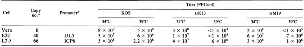
UL5 GENE OF HSV-1 461 TABLE 1. Plaquing efficiencies of HSV ts mutants on Vero, E22, and L2-5 cells
Titer (PFU/ml)
Cell
Copy
Promoterb KOS tsK13 tsM19no.' rmtr O sl sl
34°C 39°C 34°C 39°C 34°C 39°C
Vero 0 8 x 10⁸ 5 x 10⁸ 3 x 10⁸ <1 x 10³ 2 x 10⁸ <1x 10³
E22 40 UL5 3 x 10⁷ 6 x 10⁶ 1 X 10⁷ <1 X 10³ 6 x 10⁷ 7 x 10⁶
L2-5 66 ICP6 5 x 10⁸ 2.2 x10⁸ 4 x 10⁷ 6 x 10⁶ 3x 10⁸ 1 x 10⁸
aSee Fig. 3 for determination of UL5 copy number in L2-5 cells. b Refers to promoter used for UL5 expression.
erol)(25). The proteins were separated by SDS-PAGE (9%
acrylamide). The gelwas fixed inH20-methanol-acetic acid
(5:5:1) for 1 h, rinsed in water for 15 min, and soaked in
Autofluorfor 2 h. The dried gel was fluorographed at -70°C.
14C-labeled rainbow protein molecular weight markers were
purchased fromAmersham (Arlington Heights, Ill.). Insomeexperiments, acombination of immunoprecipita-tion and immunoblottingwas used to detect theUL5protein. The infected cells were not labeled, and the immune
com-plexes were washed only three times with RIPA buffer. Immune complexes were subjected to SDS-PAGE as
de-scribed above, and proteins were electrophoretically
trans-ferredtofiltersessentially as described by Towbin et al. (47).
Immobilon membrane filters (Millipore, Bedford, Mass.) were used as recommended by the supplier. Filters were
incubated with a 1:500 dilution of serum, and the
immuno-reactive proteins were detected by reaction with a goat
anti-rabbit immunoglobulin G (IgG) (Fc) antibody
conju-gated to alkaline phosphatase (Promega, Madison, Wis.).
Alkalinephosphatase color development was carried out as
describedpreviously (57).
Detection of protein complexes by coimmunoprecipitation. Coimmunoprecipitation was performed ina manner similar toimmunoprecipitationexceptthat cellswere lysedin TNE
buffer (50mMTris-HCI [pH 8.0], 1%NonidetP-40, 150 mM
NaCl,
2 mM EDTA) and the immune complexes werewashedthreetimes with TNE buffer. Following SDS-PAGE,
individual proteins in the complex were detected by
immu-noblotting with their corresponding antibodies as described above. ot-UL8 wasraised ina rabbit against a UL8 protein expressed in E. coli (3a); oa-UL52 was provided by M.
Challberg (36).
RESULTS
Isolation of UL5 permissive cell lines. To isolate and propagate a UL5 insertion mutant, a cell line that can
provide awild-typecopyofthe UL5 gene productin trans was necessary. Initially, Verocellswerecotransfected with plasmid pCW8 containing the UL5 gene under its
endoge-nouspromoter andpSV2neo. Stably transfected G418-resis-tant cell lines were isolated and tested for their ability to
supportthegrowth ofUL5ts mutant, tsM19. One cell
line,
E22, which could support tsM19atthe
nonpermissive
tem-perature, was chosen forfurther
study.
Surprisingly,
how-ever, this cell line was unable to support tsK13 at thenonpermissive
temperature(Table
1). Thisallele-specific
difference wasalso observed in another cell line
containing
pCW8.
Although thisphenomenon
is notunderstood,
thesimplestexplanation is thatthe endogenous UL5gene pro-moter is not very active and that levels ofthe UL5 gene
product are insufficient to
complement certain
ts mutants.This explanation is consistentwith the
finding
that the UL5protein is not abundant in infected cells (36, 57). To express
higherlevelsof the UL5 protein in infected cells, we decided toexpress the UL5 gene under the control of the promoter
forICP6, the large subunitof ribonucleotide reductase. The reasonfor choosing the ICP6 promoter is threefold: (i) the
ICP6protein is an abundant protein in HSV-infected cells;
(ii)theICP6 promoter is inducible by viral infection (18, 58);
and(iii) in previous experiments, areporter gene (the lacZ gene) was expressed successfully at relatively high levels under the control of the ICP6 regulatory elements (18, 19).
Plasmid p6UL5, in which the UL5 gene is undercontrol of the ICP6 regulatory region (Fig. 1), was used to transform
Vero cells as described in Materials and Methods. G418-resistant clones were tested fortheirability to support tsM19 and tsK13 at the nonpermissive temperature. Of 16 clones which were able to support tsM19, 13 were also able to
support the growth oftsK13 at the nonpermissive tempera-ture (data not shown). One ofthe 13 cell lines, L2-5, was chosenfor furtherstudy,itsabilitytosupport the ts mutants
isshownin Table 1.
To confirm the presence of the UL5 construct and to
determine itscopy number inthe L2-5 cell genome,
South-ernblotanalysis ofhigh-molecular-weightcellular DNA was
performed (Fig. 3). Cellular DNA and p6UL5 DNA were
digested, subjectedtoelectrophoresis, transferredtoanylon membrane, and probed with a
32P-labeled
MIuI-MluIfrag-ment covering the UL5 gene (Fig. 2). Various dilutions of p6UL5 provided standards for the determination of copy number (Fig. 3, lanes 1 to 5). Digestion of p6UL5 and
cellularDNA with
BglII
andEcoRIgeneratesthree internal fragments, 523bp, 2.0 kb, and4.3 kb. Thediffering intensi-ties of each band are due to theposition
of eachfragment
relativeto theprobe. Cellline L2-5 DNAexhibits thesame
pattern asp6UL5
(Fig.
3, lane7),confirming
thepresenceof p6UL5in thecellgenome. Bycomparing
theintensity
oftheinternal 2.0-kb fragment in lane 7 with the
standards,
it appears that the L2-5 genome contains about 60copies
ofp6UL5 DNA per
haploid
genome. L2-5 DNA was alsodigested withHindIII,
generating
twoexpected
fragments
of3.0and 3.8kb
(Fig.
3,lane9).VerocellDNA,
whendigested
andprobedin
parallel,
exhibitednospecific
hybridization
to the probe(Fig.
3, lanes 6 and8).
The E22 cell line and 15otherL-celllineswere
subjected
toSouthernblotanalysis
inparallel(datanotshown).The E22cell linecontainsabout 40
copies oftheUL5 gene per
haploid
genome. The UL5gene copy numbers of those L-cell lines thatsupported
bothtsK13and tsM19rangedfromaslittleas2
copies
toabout 60copies
perhaploid
genome. The three L-cell lines thatsupported only tsM19 contain
approximately
onecopy of the UL5 gene perhaploid
genome. These results support thehypothesis
that theallele-specific
differences inability
tocomplement mutants tsK13 and tsM19 are dueto levels of
VOL. 66, 1992
on November 10, 2019 by guest
http://jvi.asm.org/
462 ZHU AND WELLER
1 2 3 4 5 6 7 8 9
4.3-kb - m
-A KOS hr99
-3.8-kb
_- 3.0-kb 5.4-kb
2.0-kb -O
_i -1.1-kb
523-bp
FIG. 3. Southern analysis of cell line L2-5 DNA. DNA from plasmid p6UL5, Vero cells, or L2-5 cells was digested with the appropriate restriction enzyme, subjected to electrophoresis, and
transferred toGeneScreenPlus membranes. In lanes 1to7,DNAs
were digested with BglII and EcoRI;in lanes 8 and9,DNAswere
digested with HindIII. Lanes 1 to 5 contain various amounts of digested p6UL5 DNA: lane 1, 3.125 ng; lane 2,0.625 ng;lane 3, 0.125ng;lane4,0.025ng;lane5,0.005ng.Lanes 6to9contain 7jig ofcellular DNA from Vero(lanes6 and8)orL2-5 (lanes7 and9). The blotwasprobedwith the 32P-labeled MluIfragment containing the entire UL5gene(seeFig. 2). Fragmentsizesareindicated.
expression of the wild-type version of the UL5 genein the cell lines.This hypothesis was testeddirectly by lookingat the ability of the E- and L-cell lines to express detectable UL5protein (see below).
Isolation ofaUL5insertionmutant. Itwasanticipated that the availability of a null mutant in the UL5 gene would facilitate the analysis of functional domains of the UL5 protein and extendourunderstandingof its role in viral DNA replication. We previously demonstrated that the E. coli lacZ gene can be introduced into the HSV genome by marker transfer, permitting the screening of insertion mu-tants on the basis ofplaque color (3, 19, 53). We initially attempted to construct a UL5 disruption mutant with an in-frame insertion of the structuralgene of E. coli ,-galac-tosidaseattheBglII site such thataUL5-lacZfusion would be synthesized under the control of the endogenous UL5 regulatory signals. It was expected that following marker transfer, recombinant viruses would easily be detected by screening for 1-galactosidase activity in the presence of
5-bromo-4-choro-3-indolyl-i-D-galactopyranoside (X-Gal); however, noblue plaquesweredetectedinseveralseparate marker transfer experiments. Our inability to detect blue plaque formation led us to postulate that
P-galactosidase
synthesisfrom these recombinants was below the limits of detection, consistent withourbelief thatthe UL5promoter isweak. Wepreviously reported the constructionanduseofaplasmid, pD6p, bearing aninsertional mutagenconsisting
ofthe lacZgeneexpressedunder the control of theupstream regulatory signals of the ICP6gene (3, 19, 53). Restriction digestion of pD6p with BamHI results in the excision ofa
4.2-kbICP6: :lacZcassette.Thiscassettewasinsertedintoa
UL5 clone at a BglII site located at a position 544 bp downstream from the 5' end of the initiation AUG of the UL5 gene (Fig. 2). The resulting construct, pUL5lacZ,
* * - 409bp
ON*
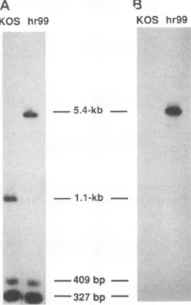
327bpFIG. 4. Southern blotanalysis of viral DNA. Viral DNAs from wild-type KOS and hr99 were digested with PstI, subjected to electrophoresis, and transferred toaGeneScreen Plus membrane. Membraneswereprobed either with the HSV-1 NaeI fragment (see Fig. 2) (A)orlacZ DNA(B). Fragmentsizesareindicated.
would be expectedtoencodeanN-terminallytruncatedUL5
peptidecontainingthe first 181 amino acid residues ofUL5
which would terminate within the upstream regulatory se-quences of the ICP6gene togeneratea protein of approxi-mately20 kDa.
AnHSVmutantcontainingalacZ insertionwasrecovered
by cotransfecting L2-5 cells with infectious wild-type DNA andpUL5LacZ. Homologous recombination between KOS genome DNA and pUL5LacZ resulted in the generation of recombinants which formed dark blueplaques in thepresence of X-Gal. Mutant viruseswere picked, purified, and propa-gated in L2-5 cells. One recombinantvirus, hr99,wasusedin further studies. To confirm that hr99 contains theICP6::lacZ
cassette at the appropriate position, DNAs from KOS and hr99were purified, digested withPstl, and subjected togel electrophoresis and Southern blot hybridization in duplicate (Fig. 4). Filterswereprobed witha32P-labeled NaeIfragment from inside theUL5gene(seeFig. 2 for position of probe)or with a 32P-labeled LacZ fragment probe. KOS contained a 1.1-kb fragmentas expected, and hr99 contained the 5.4-kb version expected if the ICP6::lacZ fragment were inserted (Fig. 4A); furthermore, the 5.4-kb fragment from hr99 was also recognizedby the LacZ probe asanticipated (Fig. 4B). The 409- and 327-bp fragmentsseenin KOS and hr99 DNA probed with the NaeIfragment correspondtoPstIfragments from within the UL5gene(Fig. 2).
Phenotypic properties of hr99.Themutanthr99wastested for itsabilitytoformplaques in untransformed Vero cells. A
stockof hr99waspropagatedonL2-5 cells, and the titerson L2-5 andonVero cellsweredetermined. Table 2 shows that hr99 failstoformplaquesonVero cells and that this growth defect can be complemented by the L2-5 cell line. These
results confirm that the ULS gene is essential for virus growth.The ability of hr99toinduceDNA synthesis in L2-5
and Vero cells was assessed by dot blot hybridization.
Previously wemeasured viral DNA synthesisby the
incor-KOS hr99
0..
J. VIROL.
on November 10, 2019 by guest
http://jvi.asm.org/
[image:5.612.82.289.77.264.2] [image:5.612.376.511.78.293.2]UL5 GENE OF HSV-1 463
TABLE 2. Plaquing efficiencies of KOS andhr99 on Vero and L2-5 cells
virus Cell Titer
line (PFU/ml)
KOS Vero 6 x 10⁸
L2-5 6x 10⁸
hr99 Vero <1 x 10³
L2-5 1.6 x 10⁹
porationof[methyl-3H]thymidine into viral DNA in mutant andwild-type virus-infected cells; by this method, we
dem-onstrated that tsK13 and tsM19 were unable to synthesize viral DNA at the nonpermissive temperature (39.6°C) (57). In this study, we measured viral DNA synthesis by dot
hybridization of total DNA from infected cells to a
virus-specific probe (see Materials andMethods). Theresults for
tsK13- and tsM19-infected cells areentirely consistent with ourprevious results (data not shown), thereby establishing the validity of this method. The results in Fig. 5 clearly demonstratethat hr99 was notableto synthesize detectable
viral DNA inVero cells. The figure shown was exposed to
filmfor S h, but no viral DNA inhr99-infectedVero cells was
detectedevenafter1weekofexposure(data not shown). In contrast, hr99 was able tosynthesizewild-type levels of viral DNAinL2-5cells (Fig. 5). These results clearly demonstrate that the UL5 gene is essential for HSV DNA synthesis, consistent with results obtained with the UL5 tsmutants.
Theability of hr99toform plaques in L2-5 cells and not in Vero cells clearlydemonstratesthat the lesion in hr99 is in the UL5 gene.Thiswas furtherverifiedbymarker rescue
exper-iments: transfections which includedhr99 DNA and p6UL5
generated approximately 4,000-fold more recombinants that grew on Vero cells than did transfections
with
hr99 DNAalone (data not shown). These recombinants form white plaques in the presence of X-Gal, since wild-type UL5 sequences have replaced the UL5 gene containing a
lacZ
insertion.Theabilitytopick whiteplaques from abackground of blueplaques formsthebasis fortheselection of additional
UL5 mutations introduced into theviralgenome (59). Detection of UL5protein in KOS- and hr99-infected Vero and L2-5 cells. In a previous report, polyclonal antibodies (ot-UL5) raised against an E. coli-expressed UL5 protein
were described (57). Immunoblot analysis of infected cell
extracts witha-UL5demonstrated that a
protein
of99 kDaMock L2-5
Vero
L2-5
0
KOS-Vero 9 L2-5
hr99
--Vero
FIG. 5. Analysisof viralDNAsynthesis.VerocellsorL2-5 cells (asmarked)wereinfected with the indicated virusat370C for 18h. Foreach sample, aseries of fivefold dilutions ofacellsuspension wasspottedontoGeneScreen Plusmembranes, andthecellswere
lysedonthemembrane.32P-labeled EcoRIFfragment(coordinates
0.315 to 0.422 on the HSV genome) was used as a hybridization
probe.
c-UL5 nonimmune
e 4 4 mock KOS
200-kDa
4
97.4-kDa
-69-kDa
46-kDa
-30-kDa 21.5-kDa
-14.3-kDa
1 2 3 4 5 6 7
FIG. 6. Detection of UL5 protein in KOS- andhr99-infected cells by immunoprecipitation. Verocellswere mockinfected or infected with KOS or hr99 and labeled with [35S]methionineas described in Materialsand Methods.Cell extracts were prepared in RIPA buffer andprecleared twice by precipitation with nonimmune rabbit serum and subsequently with a-UL5 (lanes 1 to 3). Lanes 4 and 6 are samples from the firstpreclearing step; lanes5and7aresamples from the second preclearing step. Only preclearing steps of mock- and KOS-infected cell extracts are shown, although extracts from hr99-infected cells were treated identically prior to precipitation with a-UL5. Positions of molecular weight markers are indicated.
was barelyvisiblebut that a majority of theproteins recog-nizedbytheantibodymigratedatpositionscorrespondingto
proteins of approximately45 to50 kDa(57).Theseproteins
were notrecognized by preimmune rabbit serum. The sig-nificance ofthesefaster-migrating specieswas notknownat thetime.Toimprovethe sensitivity of detection oftheUL5 proteinin infectedcells andtodetermine whether the 45- to
50-kDa
proteins
correspondtoanalternateform oftheUL5 protein, infected cellswerelabeledwith[35S]methionine
and subjected toimmunoprecipitation
as describedin Materialsand Methods. In the
experiment
shown in Fig. 6, extractsfrom mock-, hr99-, or KOS-infected cells were
precleared
twice by
precipitation
with nonimmune rabbit serum andthen specifically
precipitated
with a-UL5(lanes
1 to3).
InKOS-infected cell extracts, aband of99kDacan
readily
be detected afterimmunoprecipitation
withspecific antisera,
although no suchband wasobserved in hr99-infected cells (Fig. 6, lanes 2 and 3,
respectively).
Thesensitivity
of detection wasgreatly
improved
over that of the initialimmunoblots, andthe 45-to50-kDa bandsare not
observed,
indicating that these smaller bands were
probably
nonspe-cific reactionproducts. A
protein
ofapproximately
155 kDa was also precipitated by aL-UL5 from KOS- and hr99-infected cells. Thisprotein
was not seen in immunecom-plexes from mock-infected cell extracts
(Fig.
6,
lane1),
indicatingthat it was induced
by
HSV infection. To deter-mine whether thisprotein
reactedspecifically
witha-UL5,
mock- orKOS-infected cellextractswere
precipitated
withnonimmune rabbit serum and
subjected
toelectrophoresis.
The presence ofthe 155-kDaprotein
in both the first andsecond
preclearing
stepsinKOS-infectedcells(Fig.
6,
lanesVOL. 66,1992
on November 10, 2019 by guest
http://jvi.asm.org/
[image:6.612.61.302.97.163.2] [image:6.612.119.249.559.661.2]464 ZHU AND WELLER
L2-5 Vero
C ) U) iu
0 i0 L.
U,
E
cn
CL
CD e
C. C.
200 kd
0i
_IgG
--FIG. 7. Detection of UL5 gene product in KOS- and
hr99-infected cells by combination immunoprecipitationandimmunoblot analysis. Cellextractsfrommock-,KOS-,orhr99-infected Veroor L2-5 cells were immunoprecipitated with a-UL5. The immune complexes were subjected to SDS-PAGE, and UL5 protein was detected by immunoblotting with a-UL5. Positions of the UL5 protein and IgGareindicated.
6 and7, respectively) indicatesnotonlythat it isprecipitated nonspecifically but also that it is afairly abundant protein, since preclearing twice failed to deplete it. The absenceof
the 155-kDa band in the first and second preclearing steps
from mock-infected cells (Fig. 6, lanes4 and5,respectively) confirms that it isvirally induced.
To confirm the results obtained by immunoprecipitation andtofurthercharacterize UL5 expression in Vero andL2-5
cells, an alternative method for detecting immunoreactive material in infectedcellswas employed. Unlabeledextracts from mock-, hr99-, and KOS-infected cells were immuno-precipitated with oa-UL5 and immune complexes were sub-jected to SDS-PAGE and immunoblot analysis with a-UL5 (Fig. 7). The only bands detected were the 99-kDa UL5 protein andabandcorrespondingtoimmunoglobulin protein present in the immune complexes (marked IgG). hr99 is predicted to encode a peptide of approximately 20 kDa; however, a band of this size was not observed either by
immunoprecipitation orby the combined immunoprecipita-tion-immunoblot method (Fig. 6, lane 3; Fig. 7), indicating that this truncatedproteinmaynotbestable. Theinability of hr99 to induce the expression of either full-length or a 20-kDatruncated version of the UL5 protein indicates that it canbeconsideredtobeanullmutant.Theability of the cell line L2-5 to synthesize the UL5 protein was investigated. The results ofa combined immunoprecipitation and immu-noblot analysis are presented in Fig. 7. In mock-infected L2-5cells,noUL5protein could be detected, indicating that UL5 expressed from p6UL5 isnot constitutively expressed inthese cells. On the other hand, in hr99-infected L2-5 cells, significant amounts of the UL5 gene product were readily detected (Fig. 7). These results demonstrate that although hr99 isincapable of synthesizing full-length UL5 protein in
infected Vero cells, it is capable of providingthenecessary
factors in transfor the inductionof the the ICP6 regulatory
element, allowing expression of the UL5 gene in infected
L2-5 cells (see Discussion). In addition, this experiment
confirms that the abilityof L2-5 cells to supportviralDNA replication andgrowthof hr99 is duetoits abilitytoprovide the missing UL5 protein.
Detection of UL5 in transient and stable transfections. To
demonstrate directly that the ICP6 regulatory region is
significantly more efficient than the UL5 endogenous
pro-moterregion in directing the synthesis ofUL5 protein, levels
of UL5 protein expression level from the two promoters
97.4kd d
69 kd
46 kd
30 kd
21.5 kd
-FIG. 8. Detectionof UL5 protein expressed in Vero cells tran-sientlytransfected withplasmid pCW8orp6UL5. Vero cellswere
transfected with 12 jig of pCW8 or p6UL5 as indicated. In the leftmost lane, Vero cells were transfected with 12 ,ugof salmon
spermDNA. After transfection, cellsweresuperinfected with hr99 andlabeledwith35S]methionine, andextractswere immunoprecip-itated with a-UL5 as described in Materials and Methods.
14C-labeled rainbow protein molecular weight markers (Amersham)
were electrophoresed in parallel. Positions of molecular weight markersareindicated.
were compared in a transient transfection experiment. As shown above, hr99 is unableto synthesize UL5 protein but cantransactivatethe ICP6promoterin L2-5cells,leadingto
the expression of the UL5 protein; therefore, it seemed logical that hr99 would also be able to induce p6UL5 in transient transfection assays. In pCW8, the UL5 gene is undercontrol of itsownpromoterand is alsoexpectedtobe inducedby superinfection with hr99, since the expression of immediate-early genes is predicted to be normal in this mutant. Vero cells were transfected with either pCW8 or p6UL5 and superinfected with hr99. Infected cells were labeled with [35S]methionine, and cell extracts were pre-pared and immunoprecipitated with a-UL5 as described in Materials and Methods. UL5protein expression(Fig. 8)was induced in cellstransiently transfected with either pCW8 or p6UL5 and superinfected with hr99. These results confirm that hr99caninduce the synthesis of UL5 fromeither p6UL5
orpCW8. Inaddition, the level of UL5 protein synthesized by p6UL5 isatleast 10timeshigher than in cells transfected with pCW8, indicating that the ICP6promoter is consider-ably strongerthantheendogenous UL5genepromoter. The relative strength of the ICP6 promoter is also indicated by analysis of cell lines transfected with pCW8 (E-cell lines)or withp6UL5 (L-celllines). As mentioned above, several such
cell lines were isolated, some capable of supporting the growth of both tsM19 and tsK13 and others capable of UL5
-_-J. VIROL.
on November 10, 2019 by guest
http://jvi.asm.org/
[image:7.612.389.480.76.357.2] [image:7.612.109.252.78.196.2]UL5 GENE OF HSV-1 465
C
° o (n E Y. 0'
-180-kDa -180-kDa
-116-kDa UL52 _ -116-kDa
-84-kDa -B4-kDa
IgG-_- '44 5-58-kDa
-36-kDa
[image:8.612.155.470.76.204.2]-58-kDa -36-kDa
FIG. 9. Detectionof UL5 protein interaction with UL8and UL52 proteins bycoimmunoprecipitation. Extractsfrom mock-, KOS-, or
hr99-infected Vero cells were immunoprecipitated with a-UL5. Immune complexes were divided into three aliquots and subjected to electrophoresis in parallel. Each blotwasprobed with either at-UL5(A),a-UL8 (B),ora-UL52(C). Positionsof the UL5 and IgG proteins
aremarked in panelA;positions ofthe UL8 and UL52 proteinsaremarkedin panels B andC,respectively. Positions of molecular weight markersareindicated.
supporting the growth of tsM19 only.We suggestedthatthe
abilityto support both ts mutants wasdue to the levels of expression of the UL5gene.This hypothesiswasconfirmed byimmunoprecipitation of[35S]methionine-labeled extracts from representative cell lines. When E4 cells, containing approximately 50 copies of pCW8,were infected with hr99, nodetectable UL5 proteinwasobserved; however,in
hr99-infected L-cell lines, containing between 1and 60copies of
p6UL5, the UL5 protein could be detected (datanotshown). In these cells, the amount of UL5 protein correlates well
with the copy numberof the UL5 gene. Barely detectable
UL5 protein was observed in one cell line (L1-18) which contains approximately one copy of p6UL5 and is not supportive of tsK13; onthe other hand, considerably more protein was seen in two permissive cell lines, L2-13 and L2-5, containing 2.5 and 60 copies of the plasmid, respec-tively (data not shown). The fact that E4 and L2-5 cells which contain approximately thesamenumbers of copies of the UL5 gene vary so greatly in levels of UL5 synthesis confirms thatthe ICP6 promoter is much strongerthan the endogenousUL5promoter. Inaddition, theobservation that the cell lines which support both tsmutants exhibit higher levelsof UL5protein than do cell lines which donotsupport tsK13 confirms our hypothesis that the ability to support both tsmutants is dueto levelsof UL5 expression.
Detection of associations among UL5, UL8, and UL52. UL5, UL8, and UL52 proteinswerecopurifiedasan active helicase-primase complex, indicating that physical associa-tions among the proteins exist (9). We wished to provide additional evidence for the specific interaction between these threeproteins. We reasoned that since UL5 could be immunoprecipitated with a-UL5, it might be possible to coimmunoprecipitate UL8 and UL52 with the same sera. Immunoprecipitation experiments described above (Fig. 6) inwhich[35S]methionine-labeled KOS-infected cellextracts
were precipitated with oa-UL5 did not reveal any other proteins specific to KOS-infected cells; however, in those
experiments, cells were lysed in RIPA buffer containing deoxycholate, Nonidet P-40, and SDS and the immune
complexes were washed very stringently to decrease non-specific background. To detect interactions between pro-teins, the conditionsweremodifiedasdescribedinMaterials
and Methods. In addition, instead oflooking for UL8 and UL52 directly in immune complexes after SDS-PAGE, the
presence ofUL8 and/or UL52 in the a-UL5 immune com-plexeswasdeterminedbyimmunoblotanalysis usinga-UL8
or a-UL52 (see Materials and Methods). To establish the specificity ofcoimmunoprecipitationin KOS-infected cells, hr99, whichis unabletosynthesize UL5 protein,wasusedto infect cells in parallel. In the experiment shown in Fig. 9, mock-, hr99- or KOS-infected Vero cell extracts were
im-munoprecipitatedwith a-UL5. The immune complexeswere washed three times with TNE buffer and resolved by SDS-PAGE. Three identical protein gels were blotted onto Im-mobilon membranes and probed with a-UL5, a-UL8, or a-UL52to detect thepresenceof each protein in each blot. ot-UL52 raised againstaC-terminal oligopeptide was previ-ouslyreportedtoimmunoprecipitatea115-kDaprotein from KOS-infected cell extracts (36). a-UL8 reacts specifically with aprotein of approximately 85 kDa from KOS-infected cells which is absent in cellsinfected withaUL8 nullmutant (3, 43a). In the gel system thatwe used, UL8 consistently migratesatapproximately 85 kDa whether it is expressed in KOS-infected cells or in cells transiently transfected with three different UL8expression clones, slightly higher than the previously reported size of 80 kDa or smaller (9, 36). Figure 9A demonstrates that as shown above, if KOS-infected cell extracts are initially precipitated with a-UL5, UL5 can be detected by immunoblot analysis and no UL5 protein is detected in hr99-ormock-infected cells. InFig. 9B and C, UL8 and UL52 (marked by arrows) are detected in
immune complexes from KOS-infected cells precipitated with a-UL5. The bands marked UL8 and UL52comigrated with UL8 and UL52 proteins detected by the combined
immunoprecipitation-immunoblot method using antisera to UL8 and UL52, respectively (datanot shown). That these
interactionsspecifically require UL5issuggested bythefact
that they are not observed in hr99- or mock-infected cells
(Fig. 9, laneshr99). InFig. 9B,aprotein justabove theIgG band is also visible. The appearance of this band was not reproducible, and it was not observed inextracts
immuno-precipitated and immunoblottedwitha-UL8. Therefore,we believethatitisnonspecific. The resultspresentedinFig.9
indicate that we can detect specific physical interactions
between members of the helicase-primase complex. These resultsareconsistent with the factthatthese threeproteins can be copurified as an helicase-primase complex from infected cells(8, 9).To further demonstratethespecificityof thisinteraction, the UL5 immune complexes were assayed forthe presenceof several otherviral proteins by
immuno-blot. We tested andwereunabletodetect thepresenceof the UL9, DNA polymerase, alkaline nuclease, and ICP8
pro-A
°o
E
-B
C. (1)Co
OO2!
E
U2
mUL5-_
-1 80-kDa -116-kDa
-84-kDa UL8 _
-58-kDa
-36-kDa
VOL.66, 1992
on November 10, 2019 by guest
http://jvi.asm.org/
466 ZHU AND WELLER
teins
(data
notshown).
The absence of theseproteins
in immunecomplexes
indicates that(i)
the interactions ob-served betweenUL5, UL8,
andUL52
arelikely
to bespecific
and(ii)
ifprotein-protein
interactions betweenUL5 andotherreplication proteins
exist,
they
cannotbedetectedby
coimmunoprecipitation using
the conditionsdescribedinthis
report.
It isanticipated
that theability
to detect UL5association with UL8 and UL52
by coimmunoprecipitation
should
provide
aconvenientassaytolocalize residuesonthe UL5protein
whicharerequired
for interaction(59).
DISCUSSION
Inthis
study,
weisolatedapermissive
cellline(L2-5)
which contains thewild-type
UL5geneand iscapable
ofsupporting
the
growth
of ts mutants in the gene at thenonpermissive
temperature.
AmutantwithalacZ
insertion in theUL5
gene,hr99,
wasisolated andpropagated
inL2-5cells. Theexpres-sion of UL5
protein
in mutant-and KOS-infected Veroand L2-5 cells was monitoredby
immunoprecipitation
andby
immunoblotting
with aUL5-specific
antiserum. Inaddition,
coimmunoprecipitation
wasusedtodetect thespecific
inter-action between
UL5,
UL8,
and UL52.Isolation ofa
complementing
cell line.Inourfirstattempt
toisolate a
permissive
cell line for UL5 mutants, Vero cells were cotransfected withpSV2neo
andpCW8,
aplasmid
containing
theUL5
geneunder the control of itsendogenous
promoter.
Theresulting
cell lines(the
Eseries)
wereabletosupport
thegrowth
ofamutantwithatsmutation intheUL5gene,
tsM19;
however,
these cell lines were not able tosupport
anotherUL5
ts mutant,tsK13.
Wehypothesized
that the level of UL5
expression
from itsendogenous
pro-moter in the E-series cells is too low to allow efficientcomplementation
ofcertainmutants.ThattheUL5promoter
itself is weak is
suggested by
two observations:(i)
the UL5protein
isnot abundant in the infected cells(36, 57)
and(ii)
initial
attempts
to isolateanin-frame lacZinsertion mutantin which the
lacZ
gene isexpressed
from theendogenous
UL5
promoter
failed,
presumably
because,-galactosidase
levels wereinsufficient for blue color formation.
To isolate a cell line
capable
of increased levels of UL5protein expression,
we wished to use a promoter which isboth
strong
andinducible,
since itwaspossible
thatoverex-pression
of UL5 in aconstitutive mannermight
be toxictocells. The ICP6
promoter
was chosen for many reasons: ICP6isanabundantprotein
ininfectedcells,
andits unusualregulation
hasgenerated
considerable interest.Although
in infected cells the kinetics of induction of ribonucleotide reductaseactivity
is similar to that of other HSVearly
proteins
such asthymidine
kinase(26),
thelarge subunit,
ICP6,
isregulated
in somerespects
likeanimmediate-early
gene.The ICP6
transcript
andprotein
areexpressed
within 2 hpostinfection
(21-23),
and small amountsof thetranscript
are
synthesized
in the presence ofcycloheximide
(49).
Furthermore,
the ICP6transcript
andprotein
areexpressed
in ICP4mutantsunder
nonpermissive
growth
conditions(10,
11, 28, 33).
Wehavepreviously
reported
theisolation of cell lines(D14)
containing
theentire ICP6gene(18).
In D14cells,
ICP6 is not
constitutively
expressed
and itssynthesis
is inducedby
ICPO
but notICP4,
unliketypical early
(,B)promoters.
The existence of animmediate-early-type
cisresponseelement
(TAATGARAT)
inthe promoterforICP6(Fig.
1)
provides
apartial explanation
for itsunusualregu-lation. Thissequence foundupstream fromseveral
immedi-ate-early
genes is thetarget
for the viraltranscriptional
activator VP16
(also
knownas Vmw65 anda-TIF)
(16,
24,
34, 38). It is now clear that the promoters for the
large
subunit of ribonucleotide reductase in both HSV-1 and HSV-2 contain a TAATGARAT-related sequence and that both promoters are indeed responsiveto VP16
(56,
58).
In cells transiently transfected withpD6p
in which the lacZ gene is expressedfrom the ICP6promoter(19),,B-galactosi-dase activityis induced 30- to 40-fold
by
VP16 and atleast10-fold byICPO (58). In thisreport, weshow that the ICP6
promotercandirect the synthesisofatleast 10 times more UL5proteinthan can theendogenousUL5gene promoterin transient transfection experiments(Fig.
8). Thus,
the ICP6promoter appearstobe astronginduciblepromoter,andwe have used the ICP6 construct, p6UL5, to transform Vero cells. Of 16 cell lines (L series)
isolated,
13 were found tosupport thegrowth of bothtsM19andtsK13. We
speculate
that this is due to increased expression of UL5 in these
L-cell lines. The three L-cell lines which were unable to
supportthegrowth of tsK13containedonlyonecopyof the UL5 gene per haploid genome, and one such cell line
synthesized barely detectable levels of UL5 when induced
byinfection withhr99. The 13complementingcelllines, on
the other hand, contained between 2 and 60 copies per haploid genome, and two such cell lines synthesized
easily
detectable UL5following hr99 infection. These results
sup-port ourhypothesis that abilitytocomplement tsK13
corre-lates with levels of UL5 expression. Taken together,these
results indicate that the ICP6 promoter is both strongand
inducible and hasgeneral applicationin the overexpression
ofpoorly expressedHSV proteins.
Isolation of an ICP6::1acZ insertion mutant. To isolate a mutant which would be incapable ofexpressingfunctional
UL5, the gene was disrupted by the insertion of an
ICP6::lacZ mutageniccassetteatthe unique BglIIsite within
thegene.hr99, arecombinantviruscapable offormingblue
plaques, was isolated and propagated in L2-5 cells. This mutantfailed to produce plaques or synthesize viralDNA
when grownin Vero cells, consistent withprevious results
with ts mutants indicating that UL5 is essential for viral
DNA replication (57). This insertion mutant has several
advantages
overpreviously identifiedts mutantsin the UL5gene for the analysis of functional aspects of the UL5
protein. Since no UL5 gene product wasdetected in hr99-infected
cells,
it will now bepossible tocharacterizefunc-tional activities ofmutantUL5proteins in complementation
assayswithoutinterference fromtsgene products (59). The
fact that hr99 does not synthesize detectable UL5 protein also makes it ideal as a control in coimmunoprecipitation
experiments
designed to study UL5 interaction with otherHSV
proteins
(seebelow).
In addition, hr99 is able toprovide
the necessarytrans-acting factors fortheinductionof the ICP6promoterin p6UL5 notonly in stable cell lines but also in transiently transfected cells. Furthermore, the
availability
of the blue plaque mutant, hr99, provides aconvenient system for screening for the introduction of
site-specific
mutations into the viralgenome (59).UL5 interaction with other HSV proteins. The specific
interaction ofUL5 withotherHSVreplication proteinswas
originally
suggested bythe factthat partiallypurifiedprep-arations of theHSV-1helicase containedthreepolypeptides
(8). By using specific
antisera, the three proteins weresubsequently
identifiedastheproductsof theUL5,UL8,andUL52genes(9). In thisreport, weprovideadditionalevidence for the
specific
interaction between these threeproteins. We showthatthe UL52 andUL8 gene products are specificallycoimmunoprecipitated
with UL5protein by a-UL5(Fig. 9).This interaction was observed by immunoprecipitation of
J. VIROL.
on November 10, 2019 by guest
http://jvi.asm.org/
ULS GENE OF HSV-1 467 infected cell extractswith a-UL5 followed by SDS-PAGE of
the immunocomplexes; UL8 and UL52 were detected by
immunoblot with appropriate antibodies. This method was developed to circumvent high levels of background observed
indirect immunoprecipitation of[35S]methionine-labeled
pro-teins. The specificity of this method was demonstrated by
includinghr99 as a control; no UL8 or UL52 is precipitated by
a-UL5in hr99-infected cells (Fig. 9). This assay will provide a system to map functional domains required for specific protein-protein interactions. In the accompanying report (59), three mutants which abolish the replication ability of the UL5
protein are shown to maintain their ability to form specific
interactions with UL8 and UL52. This result strongly sug-geststhat the sites for interaction are separable from regions
of the UL5 protein required forreplication activity. Further
site-directed mutagenesis will be required to extend these initial findings and demonstrate whether theseinteractionsare essential for enzymatic function.
Function of UL5 inhelicase-primase
activity.
UL5, UL8, and UL52 canbecoexpressedin insect cells to form a functional complex exhibiting helicase and primase activities (12). A subcomplex made up of only UL5 and UL52 was shown toexhibit helicase (1) and subsequently helicase and primase (13) activities. The question of whichactivities are associated with each protein has not been addressed directly because neither UL5 nor UL52 appears to be functional when ex-pressed alone (1, 12, 13); however, the presence of six conserved helicase motifs in the UL5 gene (59) strongly suggests that the UL5 protein will turn out to be associated with helicase activity. It is possible however, that correct
association between the UL5 and UL52 is required for both helicase and primase activity. The UL5-UL52 complex may be
analogous to the primeosome ofT4 bacteriophage, which is
believed to consist of two proteins, ahelicase (gene 41) anda
primase (gene 61). The T4 primase-helicase requires boththe 41 and 61 proteins for optimalprimase andhelicase activities
(29, 41). In the HSV system, the question ofwhich proteins
have intrinsic helicase and primase activities awaits further
genetic and biochemicalcharacterization. It isanticipated that structure-function analysis of the relevant geneswill not only
aidin the assignment of function to theindividual members of
the complex butalso define protein domainsthatcontributeto the enzymatic activities ofhelicase andprimase (59).
ACKNOWLEDGMENTS
We are grateful to M. Challberg for providing plasmids and
a-UL52and to P. Shaffer for supplyingts mutants tsK13 and tsM19. We also thank D. Goldstein for constructing plasmid pDG-2 and members of our laboratory for helpful comments on the manuscript. This investigation was supported by Public Health Service grant
A121747.S.K.W. is the recipient of an American Heart Association-Genentech Established Investigator Award.
REFERENCES
1. Calder, J. M., and N. D. Stow. 1990. Herpes simplex virus helicase-primase: the UL8 protein is not required for DNA-dependent ATPase and DNA helicase activities. Nucleic Acids Res. 18:3573-3578.
2. Carmichael, E. P., M. J. Kosovsky, and S. K. Weller. 1988. Isolation and characterization of herpes simplex virus type 1 host range mutants defective in viral DNA synthesis. J. Virol. 62:91-99.
3. Carmichael, E. P., and S. K. Weller. 1989. Herpessimplexvirus type 1 DNA synthesis requires the product of the UL8 gene: isolation andcharacterization of anICP6::IacZ insertion muta-tion. J. Virol. 63:591-599.
3a.Carmichael, E. P., and S. K. Weller. Unpublished data.
4. Chartrand, P., C. S. Crumpacker, P. A. Schaffer, and N. M. Wilkie. 1980. Physical and genetic analysis of the herpes sim-plex virus DNA polymerase locus. Virology 103:311-326. 5. Coen, D. M., D. P. Aschman, P. T. Gelep, M. J. Retondo, S. K.
Weller, and P. A. Schaffer. 1984. Fine mapping and molecular cloning of mutations in the herpes simplex virus DNA polymer-aselocus. J. Virol. 49:236-247.
6. Coen, D. M., S. Weinheimer, and S. L. McKnight. 1986. A genetic approach to promoter recognition during trans induction of viralgene expression. Science 234:53-59.
7. Conley, A. J., D. M. Knipe, P. C. Jones, and B. Roizman. 1981. Molecular genetics of herpes simplex virus. VII. Characteriza-tion of a temperature-sensitive mutant produced by in vitro mutagenesis and defective in DNA synthesis and accumulation of -y polypeptides. J. Virol. 37:191-206.
8. Crute, J. J., E. S. Mocarski, and I. R. Lehman. 1988. ADNA helicaseinduced by herpes simplex virus type 1. Nucleic Acids Res. 16:6585-6596.
9. Crute, J. J., T. Tsurumi, L. Zhu, S. K. Weller, P. D. Olivo, M. D. Challberg, E. S. Mocarski, and I. R. Lehman. 1989. Herpes simplex virus 1 helicase-primase: a complex of three herpes-encoded gene products. Proc. Natl. Acad. Sci. USA 86:2186-2189.
10. DeLuca, N. A., M. A. Courtney, and P. A. Schaffer. 1984. Temperature-sensitive mutants in herpes simplex virus type 1 ICP4 permissive for early gene expression. J. Virol. 52:767-776. 11. DeLuca, N. A., A. M. McCarthy, and P. A. Schaffer. 1985. Isolation and characterization of deletion mutants of herpes simplex virus type 1 in the gene encoding immediate-early regulatory protein ICP4. J. Virol. 56:558-70.
12. Dodson, M. S., J. J. Crute, R. C. Bruckner, and I. R.Lehman. 1989. Overexpression and assembly of the herpes simplex virus type 1 helicase-primase in insect cells. J. Biol. Chem. 264: 20835-20838.
13. Dodson, M. S., and I. R. Lehman. 1991. Association ofDNA helicase and primase activities withasubassemblyof theherpes simplex virus 1 helicase-primase composed of the UL5 and UL52 geneproducts. Proc. Natl. Acad. Sci. USA88:1105-1109. 14. Feinberg, A. P., and B. Vogelstein. 1983. A technique for radiolabeling DNA restriction fragments tohigh specific activ-ity. Anal. Biochem. 132:6-13.
15. Gadler,H. 1983. Nucleic acid hybridization formeasurement of effects of antiviral compounds on human cytomegalovirus DNA replication. Antimicrob. Agents Chemother. 15:758-762. 16. Gaffney, D. F., J. McLauchlan, J. L. Whitton, and J. B.
Clements.1985. Amodular system for the assayoftranscription regulatory signals: the sequence TAATGARAT is required for herpes simplex virus immediate early gene activation. Nucleic Acids Res. 13:7847-7863.
17. Gallo, M. L., D. I. Dorsky, C. S.Crumpacker,and D. S.Parris. 1989. The essential 65-kilodalton DNA-binding protein of her-pes simplex virus stimulates the virus-encoded DNA polymer-ase. J. Virol. 63:5023-9.
18. Goldstein, D. J., and S. K.Weller. 1988. Herpes simplex virus type 1-induced ribonucleotide reductaseactivity is dispensable for virus growth and DNA synthesis: isolation and characteri-zation of an ICP6 lacZ insertion mutant. J. Virol. 62:196-205. 19. Goldstein, D. J., and S. K. Weller. 1988. An ICP6::lacZ
inser-tional mutagen is used to demonstrate that the UL52 gene of herpes simplex virus type 1 is required for virus growth and DNA synthesis. J. Virol. 62:2970-2977.
20. Goldstein, D. J., and S. K. Weller. 1988. Factor(s) present in herpes simplex virus type 1-infected cells can compensate for the loss of the large subunit of the viral ribonucleotide reduc-tase: characterization of an ICP6 deletion mutant. Virology 166:41-51.
20a.Goldstein, D. J., and S. K. Weller. Unpublished data. 21. Holland, L. E., K. P. Anderson, J. R. Stringer, and E. K.
Wagner. 1979. Isolationandlocalizationofherpessimplexvirus type 1 mRNA abundant before viral DNA synthesis. J. Virol. 31:447-462.
22. Honess, R. W., and B. Roizman. 1973. Proteins specified by herpessimplexvirus. XI. Identification and relative molar rates VOL.66, 1992
on November 10, 2019 by guest
http://jvi.asm.org/
468 ZHU AND WELLER
of synthesis ofstructural and nonstructural herpesvirus poly-peptidesin infected cells. J. Virol. 12:1346-1365.
23. Honess, R. W., and B. Roizman. 1974. Regulation of herpes virus macromolecular synthesis. I. Cascade regulation of the
synthesis ofthreegroupsof viralproteins.J. Virol. 14:8-19. 24. Kristie, T. M.,and B. Roizman. 1984. Separationof sequences
definingbasalexpressionfrom thoseconferringagene recogni-tion within theregulatorydomains ofherpes simplex1agenes.
Proc. Natl. Acad. Sci. USA 81:4065-4069.
25. Laemmli, U. K. 1970. Cleavageof structuralproteins duringthe
assembly of the head of bacteriophage T4. Nature (London) 227:680-685.
26. Langelier, Y., and G. Buttin. 1981. Characterization of
ribonu-cleotidereductase induction in BHK-21/C13Syrianhamster cell
line upon infection by herpes simplex virus (HSV). J. Gen.
Virol. 57:21-31.
27. Marchetti, M. E., C. A. Smith, and P. A. Schaffer. 1988. A
temperature-sensitive mutation inaherpes simplexvirustype1
gene required for viral DNA synthesis maps to coordinates 0.609through0.614 in UL. J. Virol. 62:715-721.
28. Marsden, H. S., I. K. Crombie, andJ.H. Subak-Sharpe. 1976.
Control ofprotein synthesisinherpesvirus-infected cells:
anal-ysis of the polypeptides induced by wild-type and sixteen
temperature-sensitive mutants of strain 17. J. Gen. Virol. 31: 347-372.
29. Matson, S., and K. A. Kaiser-Rogers. 1990. DNA helicases. Annu. Rev. Biochem. 59:289-329.
30. Matz, B., J. H.Subak-Sharpe,and V.G. Preston. 1983.Physical mapping of temperature-sensitive mutations ofherpes simplex virustype1using cloned restriction endonuclease fragments.J. Gen. Virol. 64:2261-2270.
31. McGeoch, D. J., M. A. Dalrymple, A. J. Davison, A. Dolan, M. C.Frame, D. McNab,L.J. Perry, J.E.Scott,and P.Taylor. 1988. Thecomplete DNAsequenceof thelong unique regionin the genome of herpes simplex virus type 1. J. Gen. Virol. 74:1531-1574.
32. McGeoch, D.J., M. A. Dalrymple, A. Dolan, D. McNab, L.J. Perry, P. Taylor, and M. D. Challberg. 1988. Structures of herpes simplex virus type 1 genes required for replication of virus DNA. J. Virol. 62:444-453.
33. McLauchlan, H.,andJ.B. Clements. 1983. Organizationof the herpes simplex type 1 transcription unit encoding two early proteins with molecular weights of 140,000and40,000. J. Gen. Virol. 64:997-1006.
34. O'Hare, P., and G. S. Hayward. 1987. Comparisonof upstream
sequencerequirementsforpositiveandnegative regulationofa
herpes simplex virus immediate-early gene by three virus-encodedtrans-acting factors. J. Virol. 61:190-199.
35. Olivo,P. D., N. J. Nelson, and M. D.Challberg. 1988. Herpes simplex virus DNA replication: the UL9 gene encodes an
origin-binding protein. Proc. Natl. Acad. Sci. USA 85:5414-5418.
36. Olivo,P. D., N. J. Nelson, and M. D. Challberg. 1989. Herpes simplex virus type 1gene products required forDNA replica-tion: identification andoverexpression. J. Virol. 63:196-204. 37. Parris, D. S., A. Cross, L. Haarr, A. Orr, M. C. Frame, M.
Murphy, D. J. McGeoch, and H. S. Marsden. 1988. Identifica-tion of thegeneencoding the 65-kilodalton DNA-binding protein
ofherpes simplex virustype1. J. Virol. 62:818-825.
38. Preston, C. M., M. G. Cordingley, and N. D. Stow. 1984. Analysis of DNA sequenceswhich regulate the transcription ofaherpes simplexvirusimmediateearlygene.J. Virol. 50:708-716. 39. Purifoy, D. J. M., R. B. Lewis, and K. L. Powell. 1977.
Identification of the herpes simplex virus DNA polymerase gene. Nature(London) 269:621-623.
40. Purifoy, D. J. M., and K. L. Powell. 1981. Temperature-sensitive mutants in two distinct complementation groups of herpes simplex virustype1 specify thermolabile DNA
polymer-ase.J. Gen. Virol. 54:219-222.
41. Richardson, R. W., and N. G. Nossal. 1989. Characterization of
the bacteriophage T4 gene 41 DNA helicase. J. Biol. Chem. 264:4725-4731.
42. Schaffer, P. A., G. M. Aron, N. Biswal, and M.
Benyesh-Melnick. 1973.
Temperature-sensitive
mutants ofherpessim-plex virustype 1: isolation,
complementation
andpartial char-acterization.Virology52:57-71.43. Schaffer, P. A., V. C. Carter, and M. C. Timbury. 1978. Collaborative
complementation study
oftemperature-sensitive
mutants of herpes
simplex
virus types 1 and 2. J. Virol. 27:490-504.43a.Shao, L.,andS. K. Weller.
Unpublished
data.44. Southern,P.J.,and P.Berg. 1982. Transformation of
mamma-lian cells to antibiotic resistance with a bacterial gene under controlof the SV40
early
region
promoter.J.Mol.Appl.Genet. 1:327-341.45. Spaete, R. R.,and N. Frenkel. 1982.The
herpes
simplexvirusamplicon: a neweucaryoticdefective-virus cloning-amplifying vector. Cell 30:295-304.
46. Stow,N. D. 1982.Localization ofan
origin
of DNAreplication
withintheTRS/IRSrepeatedregionof theherpessimples
virus type 1 genome. EMBO J. 1:863-867.47. Towbin, H.,T.Staehelin,andJ.Gordon. 1979.
Electrophoretic
transfer ofproteins frompolyacrylamide gels
tonitrocellulose sheets:procedureandsomeapplications.
Proc. Natl. Acad. Sci. USA 76:4350-4354.48. Vaughan, P.J., D. J. Purifoy, andK. L. Powell. 1985.
DNA-binding protein associated with
herpes
simplex
virus DNApolymerase. J. Virol. 53:501-8.
49. Watson, R. J., C. M. Preston, and J. B. Clements. 1979.
Separationad characterization of
herpes
simplex
virus type 1immediate-earlymRNAs. J. Virol. 31:42-52.
50. Weller,S.K.1990.Genetic
Analysis
of HSVgenesrequired
for genomereplication,p. 105-135.In E.Wagner(ed.),Herpesvirus
transcriptionand itsregulation.CRCPress, BocaRaton, Fla. 51. Weller,S.K.,E.P.Carmichael,D. P.Aschman,D.J.Goldstein,
and P. A. Schaffer. 1987. Genetic and
phenotypic
characteriza-tion ofmutantsin four essentialgenesthatmaptothe lefthalf of HSV-1 ULDNA.Virology 161:198-210.52. Weller, S. K., K. J. Lee, D. J. Sabourin, and P. A. Schaffer. 1983. Geneticanalysis of
temperature-sensitive
mutantswhich define the gene for the major herpessimplex
virus type 1DNA-bindingprotein.J. Virol. 45:354-366.
53. Weller, S. K., R. M. Seghatoleslami, L. Shao, D. Rowse, and E.P.Carmichael. 1990.Theherpessimplexvirustype1alkaline nuclease isnotessentialfor viralDNAsynthesis:isolation and characterization of a lacZ insertion mutant. J. Gen. Virol. 71:2941-2952.
54. Weller,S.K.,A.Spadaro,J.E.Schaffer,A. W.Murray,A. M. Maxam, and P. A. Schaffer. 1985. Cloning, sequencing, and functionalanalysisoforiL,aherpessimplexvirus type1
origin
ofDNAsynthesis. Mol. Cell. Biol. 5:930-942.55. Wu,C.A.,N.J.Nelson,D.J.McGeoch,and M.D. Challberg.
1988. Identification ofherpes simplex virus type 1 genes re-quired fororigin-dependent DNA synthesis. J. Virol. 62:435-443.
56. Wymer,J. P.,T. D.Cheung,C. Y.-N.,G.S.Hayward,and L. Aurelian. 1989. Identification of immediate-early-type cis-re-sponse elements in the promoterfor the ribonucleotide
reduc-tase large subunit from herpes simplex virus type 2. J. Virol. 63:2773-2784.
57. Zhu, L., andS. K. Weller. 1988. UL5, aprotein requiredfor HSVDNAsynthesis: genetic analysis, overexpressionin Esch-erichiacoli, and generationofpolyclonal antibodies.Virology
166:366-378.
58. Zhu, L., R.Martinez, and S. K. Weller.Unpublisheddata. 59. Zhu, L., and S. K. Weller. 1992. The six conserved helicase
motifs of the UL5 gene product, a component ofthe herpes
simplex virus type 1 helicase-primase, are essential for its function.J. Virol.66:469-479.
60. Zhu, L.,andS. K. Weller. Unpublisheddata.
J. VIROL.