0022-538X/93/031734-05$02.00/0
Copyright ©1993, AmericanSocietyforMicrobiology
Efficient Analysis of Nonviable Poliovirus Capsid Mutants
JOHNSIMONS,' ANDREW ROGOVE,2t NICOLA MOSCUFO 2* CAROLREYNOLDS,1§ANDMARIECHOW2*
Departments ofApplied Biological Sciences' and Biology,2 Massachusetts Institute
of
Technology,
Cambridge,
Massachusetts 02139Received 28September1992/Accepted 10 December 1992
Nonviablepolioviruscapsid mutantswerestudied byanefficient infection-transfection system.
Phenotypi-cally, nonviable poliovirus capsid mutants appear to segregate into three classes: those that form only
protomers, those thatcanformpentamers, andonethatcanformcompleted virions.
The ability to construct site-specific mutations within genomic cDNA clones ofRNAviruses hasyieldedawealth of information on viral gene function. Through the use of standard recombinant techniques,construction of viable and conditional lethal poliovirus mutants has enabled the effects on viral infection, genome replication, immune avoidance, and host pathology of specific amino acid or nucleotide substitutions to be examined. However, site-specific muta-genesis studies have generated additionalnonviable poliovi-rus clones (7, 8, 12, 16, 18, 19). Although the mutations leading to these nonviable viruses demonstrate the impor-tance ofspecific protein and/or nucleic acidresidues, it has been difficult to identify the replication stages affected in these mutants. Thesedifficulties arein parttechnical, stem-ming from the low efficiencies typically observed upon transfection of plasmids containing the mutant cDNAs or RNAtranscripts and/or the asynchrony of viral gene expres-sion observed after transfection. Here we describe an effi-cient transient transfection system that allows thestudy of nonviable poliovirus capsid mutants.
Transfection ofpoliovirus cDNAs. Transfection of poliovi-ruscDNAsutilized the high transfectionefficiency afforded by electroporation (4); the plasmid pPVM-1, which con-tained an infectious cDNA copy of the poliovirus genome
(serotype 1,Mahoneystrain) downstream ofabacteriophage T7 RNA promoter (17, 20); and the expression of T7 RNA
polymerasefroma recombinant vacciniavirus, vTF7-3 (5).
HeLa cells, maintained in suspension, were pelleted and resuspended at a concentration of 5 x
106
cells per ml inJoklik-minimumessentialmedium(GIBCO)-20mMHEPES
(N-2-hydroxyethylpiperazine-N'-2-ethanesulfonic
acid) (pH7.2)-0.1% bovine serum albumin. Cells were infected with sucrose-purified vTF7-3 at a multiplicity of infection of 20. Viruswasallowed to adsorb for 60 min at 37°C with gentle
shakingevery10to15 min. PlasmidpPVM-1(20,ug/ml with 60 ,ug of salmon sperm DNA per ml as a carrier) was transfected into vTF7-3-infected HeLa cells (2 x
10'
cells per0.4ml)withanelectroporation device (BTX Electro CellManipulator 600) set at 170 to 175 V (delivering a field strength of 0.68 to 0.7 kV/cm), 13 fl, and 1,650 ,uF, using
2-mm-gap electroporation cuvettes (Bio-Rad). (The
electro-*Corresponding author.
tPresent address: School ofMedicine,StateUniversityof New YorkatStonyBrook, Stony Brook, NY 11794.
tPresent address: DepartmentofDermatology, TuftsUniversity MedicalSchool, Boston, MA 02111.
§Present address: Department of Pathology, Brigham and Wom-en'sHospital, Boston, MA02115.
poration conditionswere determinedto beyieldingoptimal
transfectionefficiencies with approximately 30% cell viabil-ity [4]. For unknown reasons, if poliovirus infection is initiated after 4 h post-vaccinia virus infection, poliovirus replication is inhibited [as detected by plaqueproduction]. If poliovirus infection is initiated within 2 h post-vaccinia virus infection,noinhibition ofpoliovirus replication isobserved;
rather, vaccinia virus replication is inhibited[1Sa]. Thus, the timing of the transfection is likely to be important for the overallefficiency of poliovirusexpression.) The transfected cells were incubated at room temperature for 10 min, sub-sequently plated into tissue culture dishes in Dulbecco's modifiedEagle's medium(GIBCO) without methionine sup-plemented with 5% fetal bovine serum, and incubated at 37°C, 5%CO2. At 4 h posttransfection, medium and nonad-herent cells were aspirated from the plates and fresh medium containing [35S]methionine (50 ,uCi/ml; specific activity, 1,170Ci/mmol;NEN)wasadded. Cellswereharvestedat7 hposttransfection and lysed in RIPA buffer (10 mM Tris [pH 7.5], 150 mM NaCl, 1% deoxycholate, 1% Triton X-100, 0.1% sodium dodecyl sulfate [SDS]), and total cellular protein wasmeasured (Pierceprotein assay). Cell lysates (10 ,ug each) were subjected to electrophoresis through an SDS-10%polyacrylamide gel and transferred onto nitrocel-lulosefilters. Thesewereanalyzed byautoradiography (Fig. 1).
Cells infected with vaccinia virus vTF7-3 and transfected withplasmid pPVM-1 (vTF7-3-infected/pPVM-1-transfected cells) produce thesameprotein profileas that observed for wild-type poliovirus-infected cells, with a few additional vacciniavirus-specificproteins (Fig. 1, lanes2 and4). The identities ofpoliovirus-specific bands seen in the autoradio-graphwereconfirmedby Western blot(immunoblot) analy-sis (data not shown). The inclusion of dactinomycin (5
,ug/ml)at 2 hposttransfection stronglyreduced both polio-virusprotein and background protein bands. Consistent with poliovirus transcription being driven initially from the T7 promoter and with the kinetics of T7 RNA polymerase expression from the vaccinia virus vTF7-3vector(5), DNA-dependent RNA synthesis is necessary for more than 2 h posttransfection to provide sufficient templates for poliovi-rus RNAandproteinsynthesis. The relatively low amount of background cellular protein synthesis observedmost likely resultsfrom the shutoff of host protein synthesis that occurs inbothvaccinia virus- andpoliovirus-infectedcells. The low
backgroundof cellularprotein synthesis allowed the produc-tion ofpoliovirus-specificproteinstobedirectly assessed in labeled celllysates without additional analyses by
immuno-precipitations or Western blots. The efficiency of the cou-1734
on November 9, 2019 by guest
http://jvi.asm.org/
NOTES 1735
1
2
3
4
5
6
--i 06
-'i- 80
--49.5
-*32.5
-m27.5
P1
-vPO
2C\
vP1
-VP2
-VP3
--"-i- 8.5
FIG. 1. HeLa cells infected with vTF7-3 and transfected with pPVM-1 expresspoliovirus proteins. Lysatesfrom
infected-trans-fected cells (10 ,ugofprotein perwell) were separated ona 10%
polyacrylamide gel and electroblotted onto a nitrocellulose
mem-brane. The membrane was then subjected to autoradiography (2 days at -70'C). Lanes: 1, prestained standards; 2,
poliovirus-infected HeLa cell lysates (labeledfrom 3 to5 hpostinfection); 3, vTF7-3-infected/pPVM-1-transfected cells treated with dactinomy-cin at2 hposttransfection; 4, vTF7-3-infected/pPVM-1-transfected cells; 5, cells infectedonlywith vTF7-3; 6, cells thatwere mock infected andtransfected withpPVM-1.Thepositionsofthecapsid protein precursor, P1; capsid proteins; and prestained molecular
massmarkers (in kilodaltons)are indicated.
pledvaccinia virus infection-cDNA transfection system is also reflectedbythehighpercentageof cellsproducingvirus
upon transfection of the infectiouswild-type pPVM-1 plas-mid; approximately30to35% of the viable cells(i.e.,10% of theelectroporated cells)release infectious virusasmeasured byinfectiouscenterassays (datanot shown).
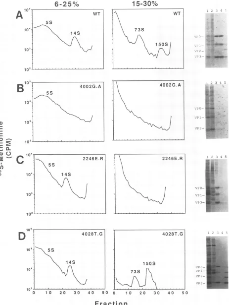
The low background of labeled host and vaccinia virus proteins allowed the poliovirus capsid assembly intermedi-ates formed in transfected cells to be examineddirectlyon sucrose gradients (15). The major assembly products (5S protomers, 14S pentamers, 73S empty capsids, and 150S
mature virions) are present in labeled lysates from vTF7-3-infected/pPVM-1-transfectedcells(Fig. 2A). SDS-polyacryl-amide gel analysis of the peakfractions from the gradients displaystheexpectedVPO-VP1-VP3protein compositionfor the various assembly intermediates. In addition, the 150S particles are infectious and are indistinguishable from
su-crose-purified, mature virions obtained from wild-type-in-fected HeLa cells. Thus, the poliovirus assembly pathway observed in the coupled poliovirus plasmid transfection-vTF7-3 infection systemrecapitulatesthat whichisobserved
during poliovirus infection and enables the assembly of nonviable poliovirus capsid mutants to be analyzed with an ease comparable to that for viable viral mutants.
Analyses
ofnonviable poliovirus capsid mutants. Analyses of nonviable poliovirus capsid mutants using the described infection-transfection technique were undertaken to deter-mine at which stage in the assembly pathway nonviable mutants wereblocked.Previous site-specific substitutions in residues that form the myristoylation signal sequence (18), form neutralization site 3B (16), or interact with the myristate moiety (12) have produced poliovirus mutants which upon transfection of the mutant cDNA plasmids themselvesorgenomic RNA transcripts of the cDNA plas-midsproducednoplaques. Characterization of these nonvi-able mutants has not been achievable by previously pub-lished methods (6, 10). Study of viable mutants obtained from thesemutagenesis studies suggested that critical points in the poliovirus replicative cyclewere affected. Mutations in the myristate signal sequence reduced the levels ofmyristate modification on capsid protein VP4 precursors. This, in turn, affected proper assembly of pentamers and emptycapsids (14). Mutations of highly conserved, solvent-exposed surface residues inantigenicsite 3B(which spansa pentamer-pentamerboundary)disturbed P1 (uncleaved
pro-tomer) folding (15). Finally,mutations which affect interpro-tomermyristate-protein interactions,incomparisonto wild-typevirus,resulted in anaccumulation of 14S pentamers in infected cells and reduced levels ofinfectivity and
thermo-stability (12). But because these "viable" substitutions do not inhibit completely the formation ofinfectious virus, it has beendifficulttoidentify thesite(s)atwhich the mutation affects the viral replicative cycle. In contrast, characteriza-tion of nonviable mutants allows the rapid identification of anysevereblocks present in theassembly pathway of these viruses.
Wild-typeandmutantpPVM-1 plasmidsweretransfected into vaccinia virus vTF7-3-infected HeLa cells(1 x 10⁷to2 X 107 cells). Lysates from
[35S]methionine-labeled
cells wereprepared (14) and analyzed bySDS-polyacrylamidegelelectrophoresis. Lysates from cells transfected with nonvi-ablemutantscontainingsubstitutions that alter the
myristoy-lation signalsequence, neutralization site 3B,or myristate-protein interactions showed protein profiles comparable to that from the wild-type-transfected cells, indicating that
capsid protein processing in these mutants wasnot
signifi-cantly affected (Fig. 2B, C, and D and data not shown).
Thus, the lethal defect for each of these groups of nonviable
capsid mutants does not appear to be due to defects in
protein synthesis and/or processing.
Sucrosegradient analysisofassemblyintermediates found in the transfected cells demonstrates the existence of three different classes ofnonviablemutants (Table 1): those that canformonly5S protomers(e.g.,4002G.A
[Fig.
2B]), those that form only 5S protomers and 14S pentamers(e.g.,
2246E.R [Fig. 2C]), and one that forms fully assembled virions (4028T.G [Fig. 2D]). With the exceptionofmutants withsubstitutionsatThr-28 ofVP4(4028T),thephenotypes
of the nonviable mutants are generally consistent with pre-dictions basedonanalysisof the assembled virionstructure. The myristate moietyis locatedat the fivefold axis. Elimi-nation of thismodificationbydestroyingthe
myristoylation
signal sequence prevents the
assembly
of 14S pentamers. These results agree with studies byAnsardiet al.(2) using
recombinant vaccinia
virus-poliovirus
expression
vectors. Because unmodified protomers can beincorporated
into pentamerandempty-capsidintermediates in the presence of VOL.67, 1993on November 9, 2019 by guest
http://jvi.asm.org/
[image:2.612.96.272.71.368.2]6-25%
15-30%
10'WT WT
5S
104 1 73S
IX
S
I \
~~~15S
I
;°qI
03; j 102B
~~~~~~4002G.A
4002G.A124
5S
1 4 2 s
103
0
lo,
P~
10*
00
icr
12246E.R
2246E.R12345
i01
o4 14S'Os~~~~ ~ ~ ~ ~ ~ ~ ~ ~ ~ ~ ~ ~~VO
14S4X | 4 73S 1 50S vP3 E
10'~~ ~ ~ ~ ~ ~ ~ 10
[image:3.612.73.536.40.655.2]0 1 0 20 30 40 50 0 1 0 20 30 40 50
Fraction
FIG. 2. Analysis of assemblyintermediates present in and nonviable poliovirus mutant-transfected cells. Lysates from wild-type-(A)and from 4002G.A(B),2246E.R(C), and 4028T.G (D) poliovirus-transfected cells were analyzed by standard sucrose gradient techniques (15).Thepositionsof the 5S protomers, 14S pentamers, 73S empty capsids, and150S mature virions were identified in a gradient containing the wild-type-transfectedlysates which was run in parallel with the gradients containing the mutant lysate during each centrifugation. Autoradio-graphsofanunenhanced gel of the peak fractions are displayed on the right of the figure. Lanes 1, unfractionated lysate; lanes 2,5Sregion; lanes 3,14S region; lanes 4, 73S region; lanes 5,
i5OS
region.The 4028T.G profile was generated by fractionating the entire lysate on a 6 to25%sucrose gradient, afterwhich thepelletfrom this gradient was resuspended and separated on a 15 to30% sucrosegradient.1736
on November 9, 2019 by guest
http://jvi.asm.org/
NOTES 1737 TABLE 1. Analysis ofnonviable poliovirus capsid mutants
Mutanta mntermediateLargest Site ofmutationc 4002G.A(GCG) 5S Myristoyl signal sequence 4002G.V (GTG) 5S Myristoyl signal sequence 4002G.M (ATG) 5S Myristoyl signal sequence 4002G.Y (TAC) 5S Myristoyl signal sequence 4002G.E (GAG) 5S Myristoyl signal sequence 4002G.R(CGG) 55 Myristoyl signalsequence 2074E.K(AAA) 14S Neutralizationsite 3B 2243S.P (CCG) 14S Neutralization site 3B 2246E.R(CGC) 14S Neutralization site 3B 4028T.K(CGG) 14S Myristate-protein interactions 4028T.G(GGG) 150S Myristate-protein interactions
aMutantnomenclature: the number and lettersidentifythelocation and amino acid substitution withinthepoliovirusgenome. The first numberrefers
tothecapsid protein in which the mutation islocated; thefollowingthree numbers refer to the amino acidresidueposition;thefinaltwo letters refer to the wild-type and mutantresidues, respectively. In addition, the mutated codon is reported in parentheses. Therefore, 4002G.A, for example, is a substitution of an alanine for the wild-type glycine at thesecond residue
(counting from the N-terminalmethionine) ofVP4.Wild-type codonsare 4002
(GGT),2074(GAG),2243(TCG),2246(GAG),and 4028(ACC).
b Fastest-sedimenting (i.e.,mostcomplete) assemblyintermediate found by sucrosegradient analysisofinfected-transfectedcelllysates.
c Proposeddisturbance causedbythemutation.
myristoyl-modified protomers (14), the myristoyl moiety maycatalyze the nucleation of pentamers and stabilize this structure. Thus, the small amount of 14S and 73S assembly intermediates previously observed for these nonviable mu-tants (9, 10) may have resulted from the presence of a low level of myristoyl-modified revertants, as a single base transversion restores the wild-type codon. Neutralization site 3B is formed from residues that span pentamer-pentamer boundaries. The nonviablemutantscontaining substitutions attheseresidues appeartoassemble pentamerstructuresbut failto assemble pentamers into emptycapsids. Thus, muta-tions near the pentamer-pentamer boundary prevent the assembly of 14S pentamers into largerstructures. The phe-notypes of the nonviable 4028T mutants wereunexpected. The threonine 28 residue of VP4 forms an interprotomer hydrogen bond between its hydroxyl moiety and the car-bonyloxygenofaneighboring myristate (3). Thishydrogen
bond has been hypothesized to stabilize the pentamer sub-unitstructure.Surprisingly,4028T.Kcanform5S protomers and 14S pentamers even though the mutation drastically changes the chemical properties at this residue position.
Even more unexpected was thephenotype of the 4028T.G mutant. 4028T.G forms complete virions that are similarto wild-typevirions by several biochemical criteria (13). The exact nature of the lethal defect remains to be identified. However, the presence of a 150S particle containing VP2 indicates that theseparticles packageRNA.
Theabilitytostudynonviable virusmutantsallowsone to examinehowessential genes function inavirus'sreplicative
cycle. Three methods for examining nonviable poliovirus
clones have beenpublishedpreviously. Oneutilizes in vitro translation systems programmed with mutant poliovirus RNA (6, 8, 11). Another method relies on the DEAE-dextran-mediated transfection of in vitro-transcribed viral RNAintosusceptiblecells inanefforttoforceonecycleof replication(9, 10).Athird method allows characterization of theprocessingandassemblyof nonviable
capsid
mutantsbycoinfection of recombinantvaccinia virusvectorsexpressing poliovirus capsid proteins and 3CD protease (1, 2). The infection-transfection system described here has several advantages over itspredecessors: it isvery efficient (wild-type cDNAproduces approximately108PFU/107 HeLa cells
originally electroporated), the low level ofbackground cel-lularprotein synthesis allows the direct analysis of labeled viral proteins without additional immunoprecipitations or Western analyses, and nonviable clones can be analyzed
directly without havingto construct acomparable mutation inavaccinia virusvector.
Byusing this method tostudy nonviablecapsid mutants, three classes of mutants have been identified: those that cannotassemble14S andlarger structures, those thatcannot assemble73S andlargerstructures, andonethat assembles completed virions that are not infectious. Although only a small number ofmutantshave been examinedto date, it is likely that further studies will identify RNA packaging mutantswhich assemble5S, 14S, and 73Sstructuresbut do notform 150S virions.Inaddition, this generalmethodology
may beapplicabletothe characterization ofnonviable virus
arising from mutations in other regions of the poliovirus
genome.
We thank Rich Condit for initial help with the vaccinia virus technology, Dan Treiber and Jill Mello for assistance with the electroporations,andEd Niles forsupplyingvTF7-3.
This work was supported by Public Health Service grant A1122627 from NIH and by research grant MV-466 from the American CancerSociety.
REFERENCES
1. Ansardi, D. C.,D. C. Porter, and C. D. Morrow. 1991. Coin-fection with recombinant vaccinia virusesexpressing poliovirus P1and P3proteinsresults inpolyprotein processingand forma-tion of emptycapsidstructures.J. Virol. 65:2088-2092. 2. Ansardi, D. C.,D. C.Porter, andC. D. Morrow. 1992.
Myri-stylation of poliovirus capsid precursor P1 is required for assemblyof subviralparticles.J.Virol. 66:4556-4563. 3. Chow, M., J. F. E. Newman, D. Filman, J. M. Hogle, D. J.
Rowlands, and F. Brown. 1987. Myristylation ofpicornavirus capsidproteinVP4and its structuralsignificance.Nature (Lon-don)327:482-486.
4. Chu, G., H.Hayakawa,andP. Berg.1987. Electroporationfor the efficient transfection of mammalian cells with DNA. Nucleic Acids Res. 15:1311-1325.
5. Fuerst,T.R., E. G. Niles,F. W. Studier,and B. Moss. 1986. Eukaryotictransient-expression systembasedonrecombinant vaccinia virus that synthesizes bacteriophage T7 RNA
poly-merase.Proc. NatI.Acad. Sci. USA 83:8122-8126.
6. Krausslich, H.-G., C. Holscher, Q. Reuer, J. Harber, and E. Wimmer. 1990. Myristoylationof the poliovirus polyproteinis requiredfor proteolytic processingof the capsid and for viral infectivity. J. Virol.64:2433-2436.
7. Kuhn, R. J.,H. Tada, M.F. Ypma-Wong, J. J. Dunn, B. L. Semler,and E. Wimmer.1988.Constructionofa"mutagenesis cartridge" forpoliovirus genome-linked protein: isolation and characterization of viable and nonviable mutants. Proc. Natl. Acad. Sci. USA 85:519-523.
8. Marc, D.,G. Drugeon, A.Haenni, M.Girard, andS.van der
Werf. 1989. Role ofmyristoylation ofpoliovirus capsidprotein VP4asdeterminedbysite-directedmutagenesis of its N-termi-nalsequence.EMBO J. 8:2661-2668.
9. Marc, D.,M.Girard, and S.vander Werf. 1991. Aglycineto
ALA substitution in poliovirus capsid protein VPO blocks its myristoylation and prevents viral assembly. J. Gen. Virol. 72:1151-1157.
10. Marc, D., G. Masson,M.Girard, and S.van der Werf. 1990. Lack of myristoylation of poliovirus capsid polypeptide VPO preventsthe formation of virions orresults in theassemblyof VOL.67, 1993
on November 9, 2019 by guest
http://jvi.asm.org/
noninfectious virus particles.J.Virol. 64:4099-4107.
11. Molla, A., A.V.Paul, and E. Wimmer. 1991. Cell-free denovo
synthesis ofpoliovirus. Science 254:1647-1651.
12. Moscufo,N., andM.Chow.1992.Myristate-protein interactions inpoliovirus: interactions of VP4 threonine 28 contributetothe structuralconformation of assembly intermediates and the
sta-bility of assembled virions. J.Virol. 66:6849-6857. 13. Moscufo,N., andM. Chow.Unpublished observations. 14. Moscufo, N., J.Simons, and M. Chow. 1991. Myristoylationis
important atmultiple stages in poliovirus assembly. J. Virol. 65:2372-2380.
15. Reynolds, C., D. Birnby, and M. Chow. 1992. Folding and
processing of the capsid protein precursor P1 is kinetically retarded inneutralization site3Bmutantsofpoliovirus.J.Virol. 66:1641-1648.
15a.Reynolds, C., and M. Chow. Unpublished observations. 16. Reynolds, C.,G. Page,H. Zhou, and M. Chow. 1991.
Identifi-cation ofresidues in VP2 that contributetopoliovirus neutral-ization antigenic site 3B. Virology 184:391-396.
17. Sarnow, P. 1989. Role of 3'-end sequences in infectivity of poliovirus transcripts made in vitro. J. Virol. 63:467-470. 18. Simons, J.,C.Reynolds,N.Moscufo, L. Curry, and M. Chow.
1990.Myristylation ofpoliovirusVP4capsid proteins,p. 158-165. In M. A. Brintonand F. X. Heinz (ed.), Newaspectsof positive-strand RNA viruses. American Society for
Microbiol-ogy,Washington, D.C.
19. Trono, D., R. Andino, and D. Baltimore. 1988. An RNA
se-quenceofhundreds of nucleotides atthe 5' end ofpoliovirus RNA is involved in allowing viral protein synthesis. J. Virol. 62:2291-2299.
20. vanderWerf, S., J. Bradley, E. Wimmer, F. W. Studier, and
J. J. Dunn. 1986. Synthesis ofinfectious poliovirus RNAby purified T7 RNA polymerase. Proc. Natl. Acad. Sci. USA 83:2330-2334.