
0022-538X/90/031021-07$02.00/0
Copyright ©1990, American SocietyforMicrobiology
A
Specific Base Transition Occurs
on
Replicating Hepatitis
Delta Virus RNA
GUANGXIANG LUO,' MEICHAO,' SEN-YUNG HSIEH,' CAMILLESUREAU,2 KAZUKO NISHIKURA,3 AND JOHN
TAYLOR'*
Fox Chase CancerCenter, 7701 BurholmeAvenue, Philadelphia, Pennsylvania 191111;Departmentof Virology andImmunology, Southwest Foundation forBiomedicalResearch, San Antonio, Texas782842;
andThe WistarInstitute, Philadelphia, Pennsylvania 191043 Received 25September1989/Accepted 6 November 1989
Threeindependentlinesofevidence showed that whenaninfectiouscloneof hepatitisdeltavirusofknown sequence wasusedtoinitiategenomereplication, upto41% of the genomes werespecifically mutatedinthe
ambertermination codon(UAGtoUGG) fortheopenreading frameofthe deltaantigen, thereby increasing
the length ofthe predicted protein from195to214 amino acids. This changewas detected onlyonmolecules
thatparticipated inRNA-directedRNA synthesis.
Hepatitis delta virus (HDV) wasdiscovered inhumansin association with hepatitis B virus (HBV) (21). It has since beenproved that HDV isasubviral satellite of HBV (11); it
depends onHBV forpackaging ofthe HDVgenomic RNA
and thedelta antigen intovirusparticles (4, 5, 35). Within the
infected cell, the HDVgenome replicates independently of
HBV(11), usingarolling-circle mechanism involving
RNA-directed RNA synthesis of genomic RNA and its comple-ment, the so-called antigenomic RNA (8). It is the antige-nomicRNA that encodesthe delta antigen, the only known protein ofHDV (7, 19, 32).
In natural infections, the delta antigen appears as two species of similar electrophoretic mobility, which are
re-ferredtohere asthe 22- and 24-kilodalton (kDa) species (3,
4, 19, 32, 35). Three laboratories have published complete HDV sequenceswhichbetween thempredictdeltaantigens of 195 and 214amino acids(12, 14, 30, 31).Thesequencesof Makinoetal. (14) and Wangetal. (30) predicta protein of 214 amino acids. The prediction ofWang etal. (30), after correction for a single sequencing error (31), was actually morecomplicated; they sequenced seven partially
overlap-ping cDNA clones and thus found 16cases of
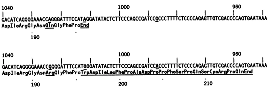
microhetero-geneityinthecompositesequence.The variationswereonly single-base changes, butas shown in Fig. 1, one wasofan
adenosine to a guanosine in the termination codon for a
predicted 195-amino-acid species, thereby increasing the predicted sizeto214amino acids. (Twomoreof the
single-basechangesareshowninFig. 1; theycausechangesonlyin thepredictedaminoacids.)The thirdpublishedsequence,by
Kuoetal.(12), predicts onlythe195-amino-acid species. As related elsewhere, thesequenceisotherwisevery similarto those described by Wang et al. (30, 31). This is not too surprising, because the infectious material used in the two cDNAcloningstudieswasderived, albeit bydifferent
exper-imental animal transmissions, from the same initial human
isolate.
Studies in several laboratories (7, 32) argue that the above-mentioned RNAsequencedifferences explain thetwo
electrophoreticformsof the delta antigen thatare normally
observed in infected livers. Thus, when we expressed the
cloned sequence of Kuoet al. (12) in the absence of RNA replication, we obtained only a single species, which we
* Correspondingauthor.
designated as the 22-kDa species and expected to be the predicted 195-amino-acid species. However,asexplainedin
this report, we made a puzzling observation when the full
HDV sequence was used to transfect cells and genome replication occurred (11). There appeared along with the 22-kDa speciesa smallbutsignificantamountofthe 24-kDa species. We found that this observation correlated with a
specific base change being made on the replicating RNA genome.
This finding led us to consider the source of this RNA replication-associated mutagenesis. We already knew from the above-mentioned study of Wang et al. (30) that 16 examples ofsingle-base substitutions had been found after the cDNA cloningof HDV from a single pool of virus. On
closerexamination,wenoted that 15 of the 16changeswere
basetransitions, thatis,between AandGorC and U. This could have been causedby transcriptionerrorsduringHDV
genome replication; viral RNA-directed RNA synthesis is frequentlycitedasbeingpronetoerrorsbymisincorporation (9, 24). However,recentstudiessuggestanothermechanism that specifically causes specific base transitions. Normal cells contain an activity, inappropriately named "RNA duplex unwindase," that acts in vitro on double-stranded
RNA substratesand deaminatesadenosine, thusconverting itto inosine (I) (1, 20, 27, 28). The in vivofunction ofthis activityisnotestablished, butif it actedon areplicatingviral RNA, the I would first directincorporation ofC, which in
turn would direct the incorporation of G. The net effect wouldbe the replacement ofA withGon onestrand and of U with Conthecomplementarystrand.There is circumstan-tial evidence that such unwindase action may have led to certainneurological variantsof measlesvirus(2, 6)and also
a variant of vesicular stomatitis virus (18). As presented
here, we have direct evidence thatduringthe replication of the HDV genome atleast one specific A is replaced by G, leadingtothetranslation of thelargerdeltaantigen. Also,we
evaluate the evidence that the change was via unwindase action.
MATERIALS AND METHODS
Plasmids and transfections. HDV sequences cloned into the RNAexpression vectorpGem4B (pG4B; Promega Bio-tec, Madison, Wis.) were synthesized by standard
proce-dures (23). Cloning into the eucaryotic expression vector
1021
on November 10, 2019 by guest
http://jvi.asm.org/
1040
II
I
1000 960I
I
I
I
I
I
I
GACATCAGGGGAAACCAGGGATMTCCATAGGATATACTCTTCCCAGCCGATCCGCCCIICTCCCCAGAGTGTCGACCCCAGTGMTAA
AspII
eArgG
IyAsn9j G I yPheProEnd190
1040 1000 960
GACATCAGGGGAACCIGGGAIMICCATIGGATATACTMCCCAGCCGATCCACCCIIIiCTCCCCAGAGTTGTCGACCCCAGTGAT
AspIIeArgGI
yAsnArG
IyPhProTrpAspIIoLouPhoProAIaAspProProPhoSerProGInSerCysAraProG
InEnd190 200 210
FIG. 1. Microheterogeneity on antigenomic HDV RNA in the region spanning the Cterminus ofthecoding region forthedelta antigen. The two sequences are from Wang et al. (30) and contain single-base differences at three places (underlined), one of which, at position 1012, changesanamber termination codon into a tryptophan codon. The upper sequence is alsoidenticaltothat of Kuo et al. (12).
pSVL (Pharmacia, Inc., Piscataway, N.J.) was as previously
described (11). This construct was used to transfect both
COS7 cells (11) and a chimpanzee (25). The animal was
alreadyacarrier of HBV. Six weeks after HDV transfection,
theanimal developed an apparently typical HDV infection. The serum and liver biopsy samples taken at this time
containedHDVRNA and were used for the present study.
For the unwindase studies, we used the transcription vector pG4B with subgenomic inserts of HDV, as summa-rized below according to designation and location on the HDV genome: pG4B(PX), PstI (1087) to XbaI (781); pG4B(XP), XbaI (781) to PstI (655); pG4B(PP),PstI (1087) toPstI (655). For RNAtranscription, the vectors were first
linearizedat arestriction enzyme site located downstream of
the HDV insert and then copied with the RNA polymerases
of phageT7 orSP6 (Bethesda ResearchLaboratories, Inc.,
Gaithersburg, Md.). When necessary, the RNAs were
la-beled with
[a-32P]ATP
(800 Ci/mmol; Dupont, NENRe-searchProducts, Boston, Mass.). The products were treated with DNase I (Promega) before gel purification and, as necessary, subjected tohybridization to make the
appropri-ateintermolecularRNA-RNA structures shownin Fig. 5A.
Theelectrophoretic mobility of the two intermolecular struc-tureswastested byelectrophoresis into polyacrylamide gels undernondenaturing conditions.
Oligonucleotide primers. The following HDV-specific
oli-gonucleotides were chemically synthesized and are listed
accordingtoadesignatedlettercode,positionon HDV(12),
and actual sequence, respectively: A, 894 to 919, CCGAC CCGAA GAGGAAAGAA GGACGC; B, 1152 to 1135,
TGGGGGGTGT GAACTCGAAG GTGGATCGA; C, 1004
to 1022, GTATATCCCA TGGAAATCC; D, 1082 to 1069,
GGAGTCCCGG AGTC; E, 999 to 1012, GAAGAGTATA
TCCT;F, 999 to 1012,GAAGAGTATATCCC.Notethat at
position1012, C andF aremutated relativetothe sequence
of Kuo et al. (12).
Reverse transcription and polymerase chain reactions. RNA samples were extracted by treatment with pronase in the presenceof sodium dodecyl sulfate. After one extraction with phenol and two with ether, the samples were precip-itated with ethanol in the presence of salt and carrier dextran (Sigma Chemical Co., St. Louis, Mo.) (29). Some samples wereadditionally treated with DNase I (Promega) and
repu-rified. Reverse transcription was done under standard
con-ditions(23)with the enzyme from avian myeloblastosis virus
(Life Sciences, Inc., St. Petersburg, Fla.). For polymerase chain reaction (PCR), the products were next treated with
alkali to destroy the RNA and then aliquots were used as
templatesfor PCR. PCR reaction mixtures of 50 ,ul with Taq polymerase (The Perkin-Elmer Corp., Norwalk, Conn.) were asdescribed previously (10), using 30 to 40 cycles on a TechneTempblok setfor 1 min at 94°C, 2 min at 55°C, and 3min at72°C. The primercombinations are described in the
textandfigure legends. With theprimersmismatched atthe
3' terminus, we followed the method of Wu etal. (34) and
empirically determined that at anannealing temperature of
55°C there was optimum discrimination in the ability of primer E versus F to replicate wild-type versus mutant HDV.
As explained in the text, we also used PCR as part of a
cDNA cloning strategy. We converted HDV RNA into
double-stranded DNAfragments spanningSall (962) to PstI (1087). Thesewereinserted intopG4B. Recombinants were
screened (15) withanRNAprobe [pG4B(PX)] as described
above or with the oligonucleotide C that had been end labeled. Special conditions were needed for hybridization
with the mutant oligonucleotide C to discriminate against the
detection of unmutated sequences. We found empirically
thatoptimum specificitywas obtained withawashing
solu-tionat45°C(17).
For nucleotide sequencing, recombinant plasmid DNAs wereused in adideoxy sequencing protocol (22) as modified tomake use of the Taqpolymerase (10).
In vitro modification of RNA and assay by thin-layer chromatography. RNA substrates labeled with [a-32P]ATP wereprepared as described above. HeLa cell extracts were
prepared by a modificationof the method ofManley et al.
(16)asdescribed previously (28). Approximately 10 fmol of various RNA samples was incubated with 50 ,ug of cell extractproteinin 20 ,ulof50mMTris(pH7.8)-0.15MKCl-5 mM EDTA-25% glycerol-1mMdithiothreitol.Thereaction mixture was incubated at 37°C for 2 h, deproteinized with
proteinase K, phenol extracted, and ethanol precipitated.
The RNAswerethendigested with nucleaseP1 (Sigma) and
subjected to thin-layer chromatography with solvent 2 of
Wagner et al. (28) to assay the conversion ofadenosine to inosine. Radioactivity was detected either by
autoradiogra-phy or with a Radioanalytic Imaging system (AMBIS, San
Diego, Calif.).
Proteinanalyses. HDVantigenwastranslated invitrowith a rabbit reticulocyte lysate (Bethesda Research Laborato-ries). Samples from DNA transfections along with the
West-ern(immunoblot) analysis were all aspreviously described
(11, 13).
on November 10, 2019 by guest
http://jvi.asm.org/
[image:2.612.97.529.72.213.2]1 2 3 4 5
kDa
44.2
29.2
- 24 -22
18.2
[image:3.612.57.298.74.291.2]14.4
FIG. 2. Westernanalysis ofdeltaantigen species. After
separa-tion on a 12% acrylamide gel by the method of Laemmli (13), a
Westernanalysis wasdone(11) withapatient antibody specific for
deltaantigen. The mobilities ofprestained markers (Bethesda Re-search Laboratories)areshownattheleft; lane 1 isa samplefrom
the liver ofaninfectedpatient; lane 2 isaninvitro rabbit
reticulo-cyte translationproductofantigenomicHDV RNAwhich encodes the deltaantigen;lanes 3 and 4arefromCOS7cells transfectedwith
pSVL(Ag)andpSVL(D3), respectively; lane 5 is aplasma sample
fromachimpanzeetransfected withpSVL(D3).
RESULTS
Electrophoreticforms of deltaantigen.Asexplained below,
wemadeanunexpected findingfrom whatwethoughtwould
bearoutine Westernanalysisof the deltaantigeninvarious samples. Liversamplesfromaninfectedpatient (Fig. 2,lane 1) showed two species, designated the 22- and 24-kDa species, just as others have described (3, 4, 19, 32, 35). (Other species, such as one at about 20 kDa, were also present.)Invitro translation of the HDVsequenceclonedby Kuo et al. (12) yielded only the 22-kDa species (lane 2), which was, as predictedfrom the nucleotidesequence, 195 amino acids. A band of the same electrophoretic mobility wasalsoseenwhenasubgenome-size fragmentof the HDV
sequence of Kuo et al. (12) was expressed in transfected COS7 cells from theeucaryotic expressionvectorpSVL(Ag) (Fig. 2, lane 3) (11). However, a surprising finding was
obtained when we studied the antigenmade in cells trans-fected with thevectorpSVL(D3), containingatrimer of the
HDVgenome(11).This constructallowedreplicationof the HDVgenome. We still obtained the 22-kDaspecies, butwe now obtained small amounts of additional protein species, includingonethat migrated thesame asthe 24-kDaspecies
observed ininfected liver(Fig. 2, lane4). From densitomet-ric analysis, the relative amount of this larger species was
5%. Whenwe studied the plasma proteins ofa chimpanzee
thathad beensuccessfullytransfected with thesame
recom-binant HDV construct (25), we again obtained the 24-kDa
band(Fig. 2, lane5),but the relativeamount now was20%. Onepossible explanationfor theappearanceofthenovel bandwasthattherewasreadthroughof the amber termina-tioncodonand, as aresult, synthesisofthe214-amino-acid species that contained at its C terminus an additional 19
aminoacids (see Introduction andFig. 1). Such readthrough
can occur by tRNA suppression of the termination codon;
Weineretal. (32)documentedthisfordelta RNAexpressed
inasuppressor-positivestrain of Escherichia coli. However,
inourstudies, suchamechanism ofsuppressionwas
appar-ently notinvolved because we found that in the absence of
HDV genome replication (Fig. 2, lane 3) the large protein was notmade. (There wasthe remotepossibility that HDV
replicationinducedasuppressortRNA.) We thus tested the
alternativehypothesis that HDV genome replication leadsto
single-base changes, some of which are in the termination
codon and thus leadtothesynthesisof thelongerform of the deltaantigen.
Sequence analysisof HDV RNAs from transfected cells. To
test the above-described hypothesis, we sequenced the
region of the genome that surrounds the termination codon
for the smallerantigen (Fig. 1). Also, sincetheproteindata indicated that onlya smallfraction (5 to 20%) ofthe RNAs would be changed, it was considered necessary to obtain
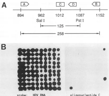
cDNA clones andscreenabout 100 recombinant clones. The strategy we usedis explained in Fig. 3. Antigenomic HDV RNA was reverse transcribed with an oligonucleotide
primer, designatedA in Fig. 3A, and thenamplified bythe
PCR with primers Aand B. The 258-base-pair productwas
gel purifiedand then digestedwithboth Sall and PstI. The
resultinginternal125-base-pair fragmentwasgelpurifiedand
ligatedinto themultiple cloningsiteofplasmid pG4B,which
had beensimilarly digestedandgelpurified.Theligationmix was used to transform E. coli HB101, and the colonies obtainedwere screened(15)witharadiolabeled RNAprobe
specificfor thisregionof theHDVgenome. Setsof 100 such
positive colonies were transferred and grown up on three
identicalagarplates;one waskeptas amasterplate,one was
rescreened with the RNAprobe toconfirm the presence of the HDV insert(for example, Fig. 3B,leftside),andonewas screened with end-labeled
oligonucleotide
C(for
example,Fig. 3B, right side). This
oligonucleotide
was designed todetect those recombinants in which the nucleotide at posi-tion 1012on the
antigenomic
strand waschanged fromAtoG; this convertsthe termination codon on the
antigenomic
RNA from UAG to UGG (Fig. 1). In the example shown,
three positive clones were found and subsequently
con-firmed by dideoxy sequencing (10, 22) with end-labeled
oligonucleotide
D astheprimer (Fig.
3C). Theinput
RNA used in theexample shown in Fig. 3 wasfromCOS7 cells 2 weeks after transfection with thereplication-inducing
clone pSVL(D3).Whenwe usedthis approachwith RNAfrom cells
trans-fected withpSVL(Ag)orwith
pSVL(D2M),
a mutantunableto replicate, and screened atotal of255 recombinant
colo-nies, wefound none thatcontained the
changed
base(data
notshown). Thisdemonstrated that the
previously
obtainedchanged sequences werenot some
quirk
ofusing
PCR(10).Italsowasconsistent with thepreviouslymentioned
hypoth-esis that the change depended on the
ability
ofthe HDV RNA sequences toundergo RNA-directed RNAsynthesis.
We also tested RNA samples from an HDV-transfected
chimpanzee. UsingliverRNA,wefound41of100
recombi-nantcoloniestobe mutant. Wealso wishedto testthe HDV
RNAthatwas packaged and released into the
blood;
how-ever,becausevirions contain
only genomic
RNA,wehadtomodify the strategy of Fig. 3A and carry out the reverse
transcription stepusing primerB.Theoutcome wasthatwe
obtained 73 recombinant colonies from 200 thatwere
posi-tive for thechange
(data
notshown).
Thus,
notonly
werethe serum and liver resultscomparable,
butapparently
weon November 10, 2019 by guest
http://jvi.asm.org/
A
X>
-
I-894 962
SalI
1012 1
p 1087 :StI
k----
125-l
lI---
--- 258 >B
.
0I l
_
..
probe: HDV RNA
C
T C A G G A C T1152 46. .._-W
s ^ 3=~~d
i =
w
7iw.i k*., :.fi..
..f:
oligonucleotide C
FIG. 3. Recombinant DNA cloning and sequencing of specific HDVRNA sequences. (A) The strategy for going from RNA to clonable double-stranded DNA spanning the region Sall (962) to PstI (1087) is explained in the text. (B) Recombinant clones were screened with an HDV RNAprobe and theneitherreconfirmed as such(left side)ortested forhybridization tooligonucleotideC(right side). (C)Tworecombinantclones from panel B were subjected to dideoxy sequencing. Oneclone was expected to be unchanged (wt) and the othertobe modified(mutant) atposition1012. The sequenc-ing confirmed this;the twoclones differed onlyin A toGat1012 (*).
_
-
_I
4F
- p
*i_ _
low
_w
:"m le
_
-_ t-4
AP#N.
du ;'t
'`4..,:. ?
iis.i
_-ak
: _
obtainedcomparable resultsfor both genomic RNA (serum)
andantigenomicRNA (liver).
Thus, the approach of examining the HDV RNA gave results roughly comparable to the earlier protein studies (Fig. 2) in which we assayed the relative abundance of the longer form of delta antigen in the transfected chimpanzee andcellcultures, in which genome replicationoccurred.
Independent PCR-based assayfordetecting HDV sequence changes. Theabove strategyforthe detection of nucleotide changes at position 1012 wasquantitative, but thesensitivity waslimited by the number of recombinant clones screened.
Inaddition, it depended oncloning and sequencing. Thus,
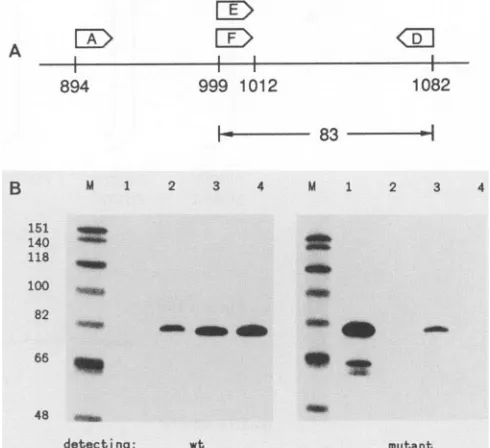
weexploited the alternative strategy of Wu et al. (34), which is able to specifically amplify only those molecules that contain the base change. This can beexplained with Fig. 4.
An RNA sample containing antigenomic HDV sequences
was reverse transcribed with primer A, and then aliquots weresubjected to PCR amplification with the primer pair E
plusD or Fplus D. The primers E and F were end labeled,
and so the PCR products were assayed by a denaturing
sequencing gel and screened for an 83-basespecies.Thefirst
primerpair detects the normalHDV sequenceof Kuo et al.
(12). Primer F, however, differs from E in its 3' nucleotide, which corresponds to position 1012. F will only pair
pre-cisely to antigenomic HDV RNA that has been modified
from AtoGatposition 1012. AsWuetal. (34) have shown, the annealing temperature of the PCR reaction required
optimization so that the first primer pair detected only
unmodified HDV(Fig. 4B, left panel, lane 2 versus lane 1),
while the second pair detected only modified HDV (Fig. 4B,
rightpanel, lane 1 versus lane 2).
This strategy was applied to various RNA samples
con-tainingHDVsequences. For the COS7 cells transfected with
pSVL(Ag), we detected only unmodified DNA (lane 4),
plasmid: wt mutant
whereas in those transfected with pSVL(D3), we detected
modifiedaswellasunmodified DNA(lane 3).
Areconstructiontodetermine thespecificityand
sensitiv-ityof this assayprocedure was performedwith unmodified DNA in the presence of decreasing amounts of modified DNA. Wecouldreadilydetect1partof modifiedDNAin the presenceof 100 partsof unmodified DNA(datanotshown). This result also made clear thespecificity ofthisPCR-based
assay.
Action of unwindase on HDV RNA. In the previous
sec-tions,we showed thatin theterminationcodonof the delta
antigen, someRNAmoleculesarechangedfrom AtoG. As
mentioned in theIntroduction,wewishedto testthe hypoth-esis that thischangewasinitiatedbyRNAduplexunwindase action converting A to I, followed by RNA-directed RNA
synthesis
perpetuating
the I asG. Wesoughtdirectexperi-mental evidence to support the role of unwindase. Also,
since unwindase is knownto have a specificityfor double-stranded rather than single-stranded RNA, we were inter-estedto know whether the rod structure of the deltaRNA, withanaverageof70%of basespaired, would alsoact as a substratefor unwindase.
FourdifferentHDV RNA constructs weremade(Fig.5A). These RNA specieswere initially transcribed via
bacterio-phage RNA polymerases from recombinant transcription
plasmidsandwerelabeled with[Ot-32P]ATP.Theywerethen
hybridized, as necessary, to make the structures shown,
beforebeingincubated for2hat37°C inan extractof HeLa cells that contained unwindase activity (16, 27, 28). After
this,the RNAs were extracted, digested with nuclease P1,
f
on November 10, 2019 by guest
http://jvi.asm.org/
[image:4.612.65.295.72.275.2]and analyzed by thin-layer chromatography, followed by radioimaging. Since nuclease
P1
releases 5' nucleotides, the conversion of A to I could be monitored simply as the transfer of label from pA topl.
This occurred to a significant extent only for the 100% double-stranded RNA (Fig. SB,lane 2), and the extent of conversion was 30%. With the three other RNA substrates, the extent of conversion was much lower. It could, however, be detected with a 5-day exposure of an autoradiogram and was about 1%. Such modifications were proved to depend on treatment of the RNA with the cell extract that contained unwindase activity. Also, a heat-inactivated extract failed to induce the changes (data not shown). Figure5B shows additional controls. Lane 5 showed that without
P1
digestion the label remained exclu-sively at the origin. Lane 6 showed that with P1 digestion followed by phosphatase treatment, the label was released and migrated as free phosphate. The migrations of pA andplwere confirmed by using unlabeled standards that were lo-cated by UV light. From these studies, we deduced that if unwindase was involved in vivo in the modification of HDV RNA, then a fully double-stranded RNA would be a preferred substrate. Yet it remains possible that a lower extent of modification, but with some specificity, could also occur on HDV RNA that was either single stranded or folded into the rod structure. Another possible substrate could be created by an intermolecular interaction between HDV RNA and an RNA in the cellular extract. If unwindase caused the A-to-G change at position 1012 in the termination codon, the A would have to be in the antigenomic RNA. The modified base would then have to replicate to be perpetuated as a G.
DISCUSSION
Three lines of evidence showed that during the replication of the HDV genome there was a nucleotide change in the amber termination codon of the delta antigen. In the three clones that were partially sequenced and did have the A-to-G change, we were unable to find any additional changes in the approximately 100 bases of flanking
sequence.
To search for other changes on the HDV genome, we modified the strategy used in Fig. 3 to obtain more sensitivity. We thus found eight changes at other sites, but the frequency was about 500-fold lower than in the termination codon. Also, the changes were independent of genome replication (data not shown).We found that the fraction of molecules changed at the termination codon was as much as41%for the HDV RNAs from the liver and serum of the infected chimpanzee. This relatively high level could be related to the fact that the infection had been established more than 6 weeks in the animal, in contrast to the transient transfection studies with the cultured cells. However, in a separate series of transient transfections, we observed that the extent of change (as judged by Western analysis) depended on the extent of genome replication and could be at least as high as in the infected chimpanzee (data not shown). Thus, the appearance of the change was independent of cell-to-cell spread.
The change only occurred when there was RNA-directed RNA replication of HDV. Presumably the change occurred either after or during rounds of RNA-directed RNA synthe-sis. What is the evidence that the change might have occurred via the action of the recently found cellular activ-ity, RNA duplex unwindase (1, 20, 27, 28)? (i) Examples exist with other RNA viruses (2, 6, 18). (ii) The change of A to G was consistent with the known properties of unwindase action (1, 28). (iii) We could reconstruct A-to-I changes on HDV RNA in vitro. (iv) The 15 of 16 examples from the
A ,
894 999 1012 1082
B-3_ 83
B M l 2 3 4 M 1 2 3 4
151 140 118
100 82
2
Em, 4f# - ..l
48 _
[image:5.612.317.562.72.296.2]detecting: wt mutant
FIG. 4. PCR-based approach to detect specific single-base changes in HDV RNAs. (A) Asexplainedin the text, the strategy was toreverse transcribe antigenomic HDV RNA sequences with primer A and then use samplesof thisproductas atemplatefor PCR witheither primersE and D or F and D.PrimerF, unlikeE, willgive perfect base pairingonly to antigenomic sequences that havebeen modifiedby anA-to-G changeatposition1012. (B)Primers EandF were endlabeled sothat the PCRproduct could beassayedbyusing a denaturing acrylamide gel and screening for an 83-base species. The left panel showsthe detection ofwild-type (wt) sequence; the right panel showsthe mutant sequence. Lanes 3 and 4 madeuse of RNAfromCOS7 cells transfected with pSVL(D3) and pSVL(Ag),
respectively. As controls for specificity ofamplification, we used PCR templates that were pure wild-type DNA (lane 2) or pure mutated DNA (lane 1). The nucleotide sequence of these two
controls was obtained in Fig. 3C. Lanes M and the associated numbers refer to a size marker of bacteriophage X174 DNA digestedwith Hinfl and end labeled.
HDVsequencing ofWangetal. (30)wereallconsistentwith unwindase action. At best, these four lines of evidence are
circumstantial. For item iv, the
directionality
of the base transitions is not known. For itemiii,
our reconstructionswithdouble-stranded RNA could be argued to be irrelevant
because they allow multiplechanges, whereas HDV
replica-tion has so far revealed only the
single
change
in the termination codon. (Of course, molecules withmultiple
changes could have been selected
against,
forexample,
by
means of competence for replication.) It remains
possible
that unwindase action was on a specific
region
of intermo-lecular or evenintramolecular double-stranded RNA. And it is also possible that unwindase was not involved atall.Ifunwindase actionwas definitely the cause of
change
onHDV and certain other RNA viruses
(2, 6,
18),
then there would be important consequences for ourunderstanding
of the replicative structures of these viruses. Sinceunwindase,
at least in vitro, has a clear preference for RNA substrates that aredoublestranded, wewould havetoconclude that the RNAspecies that aremodified must,atleast
transiently,
exist in a double-stranded conformation. In this respect, we have previously isolated double-stranded HDVspecies
from in-fected cells and tissues (8, 26). However,there is thedogma,
as recently restatedby Weissmann
(33),
thatdouble-strandedviral RNA structures are either the
nonproductive
on November 10, 2019 by guest
http://jvi.asm.org/
A
single
double
intermolecularintramolecular
strand
strand
rod
rod
B
single strand
double strand
intermolecular rod
intramolecular rod
undigested
digested
plus phosphatase
I I I
[image:6.612.140.464.66.560.2]p pI pA origin
FIG. 5. Ability of HDV RNAstructures to actassubstrates for RNA duplex unwindase. (A)Thefourconstructsshowncontaineddifferent
amountsofintermolecular and intramolecular base pairing. Theywereassembledby using RNA species synthesizedin vitroin thepresence
of[a-32P]ATP. (B) Theywerethensubjectedto treatment withunwindaseactivity,followedbyextractionanddigestionwith nuclease P1 beforechromatography with solvent 2 of Wagneretal.(28). Radioactivitywasquantitated in situ withaRadioanalytical Imagingsystem.The
indicatedpositions of pA and plwereestablished by cochromatography of unlabeledstandards (Sigma),aslocalized with UV light. Sample
5 was acontrol of undigested RNA, which remained at the origin. Sample 6 corresponds toadigestas in sample 1 but followed by an
additionaltreatmentwith bacterial alkaline phosphatasetoverify the releaseof free phosphate (p).
quences of"collapsed" transcription orartifacts of extrac- lished data), implyingthatthe small antigen is sufficient for
tion. genome replication. Also, recent experiments have shown
Whileourstudies offeranexplanation of thelong-standing that in the absenceof the small antigen, thelargeantigen is
puzzleofHDVencodingtwodifferentelectrophoretic forms notsufficient forgenomereplication(M. Chao, S.-Y.Hsieh,
of delta antigen, they also raise newquestions. Previously, andJ. Taylor, unpublished data). Experimentsareneededto
we have shown that the smaller antigen when provided in address whether the changes occur in every infected cell.
trans allows a mutant genome to replicate (11); in this Maybe the two forms have different functions or even a
experiment,nolarge antigenwasdetected(M. Chao, unpub- cooperative function, suchasinpackaging. Wearetempted
I/
on November 10, 2019 by guest
http://jvi.asm.org/
to speculate that the replicating HDV exploits the ability of its genome to be modified by the host, so as to allow the synthesis ofthe additional larger form of the delta antigen.
ACKNOWLEDGMENTS
J.T. was supported by grantfMV-7M from the American Cancer Society, by Public Health Service grants CA-06927, RR-05539, and AI-26522 from the National Institutes of Health, and by an appro-priation from the Commonwealth of Pennsylvania, and K.N. was supported by grant CA-46676 from the National Cancer Institute.
We thank William Mason, Wang-Shick Ryu, and Richard Katz for critical reading of the manuscript, Laura Coates for technical assistance, and Tony Yeung for chemical synthesis of the oligonu-cleotides used in the study. The sample of HDV-infected human tissue was provided by Eric Gowans, and the human antidelta serum was provided by John Gerin.
LITERATURE CITED
1. Bass, B. L., and H. Weintraub. 1988. An unwinding activity that covalently modifies its double-stranded RNA substrate. Cell 55:1089-1098.
2. Bass, B. L., H. Weintraub, R. Cattaneo, and M. A. Billeter. 1989. Biased hypermutation of viral RNA genomes could be due to unwinding/modification of double-stranded RNA. Cell 56:331. 3. Bergmann, K., and J. L.Gerin. 1986. Antigens of hepatitis delta virus in the liver and serum of humans andanimals. J. Infect. Dis. 154:702-706.
4. Bonino, F., K. H. Heermann, M. Rizzetto, and W. H. Gerlich. 1986. Hepatitis delta virus: protein composition of delta antigen and its hepatitis B virus-derived envelope. J. Virol.58:945-950. 5. Bonino, F., B. Hoyer, J. W.-K. Shih, M. Rizzetto,R. H. Purcell, and J. L. Gerin. 1984. Delta hepatitis agent: structural and antigenic properties of the delta-associated particles. Infect. Immun. 43:1000-1005.
6. Cattaneo, R., A. Schmid, D. Eschle, K. Baczko, V. ter Meulen, and M. A. Bilieter. 1988. Biased hypermutation and other ge-netic changes in defective measles viruses in human brain infections. Cell 55:255-265.
7. Chang, M.-F., S. C. Baker, L.H.Soe, T.Kamahora,J. G. Keck, S. Makino, S. Govindarajan, and M. M. C. Lai. 1988. Human hepatitis delta antigen is a nuclear phosphoprotein with RNA-binding activity. J. Virol. 62:2403-2410.
8. Chen, P.-J., G. Kalpana, J. Goldberg, W. Mason, B.Werner, J. Gerin, and J. Taylor. 1986. The structure and replication of the genome of the hepatitis delta virus. Proc. Natl.Acad. Sci. USA 83:8774-8778.
9. Domingo, E., E. Martinez-Salas, F. Sobrino, J. C. delaTorre, A. Portela, J. Ortin, C. L6pez-Galindez,P. Perez-Brena, N. Villa-nueva, R.
Naijera,
S. VandePol, D. Steinhauer, N.DePolo,and J. Holland. 1985. The quasispecies (extremely heterogeneous) nature of viral RNA genome populations:biological relevance-a review. Gene 40:1-8.10. Higuchi, R. 1989. Using PCR to engineer DNA, p. 61-70. In H. A. Ehrlich (ed.), PCR technology. Stockton Press, New York.
11. Kuo, M. Y.-P., M. Chao, and J. Taylor. 1989. Initiation of replication of the human hepatitis delta virus genome from cloned DNA: role of delta antigen. J. Virol. 63:1945-1950. 12. Kuo, M. Y.-P., J.Goldberg, L.Coates, W.Mason,J.Gerin,and
J. Taylor. 1988. Molecular cloningof hepatitis delta virus RNA from an infected woodchuck liver: sequence, structure, and applications. J. Virol. 62:1855-1861.
13. Laemmli, U. K. 1970. Cleavage ofstructural proteins duringthe assembly of the head ofbacteriophage T4. Nature (London) 227:680-685.
14. Makino, S., M.-F. Chang, C.-K. Shieh, T. Kamahora, D. M. Vannier, S. Govindarajan, and M. M. C. Lai. 1987. Molecular cloning and sequencing of a humanhepatitis deltavirus RNA. Nature (London) 329:343-346.
15. Maniatis, T., E. F. Fritsch, and J. Sambrook. 1982. Molecular cloning: a laboratory manual. Cold SpringHarborLaboratory, Cold Spring Harbor, N.Y.
16. Manley, J. L., A. Fire, A. Cano, P. A. Sharp, and M. L. Gefter. DNA-dependenttranscriptionofadenovirus genes in asoluble whole-cell extract. Proc. Natl. Acad. Sci. USA 77:3855-3859. 17. Mullis, K., F. Faloona, S. Scharf, R. Saiki, G. Horn, and H.
Erlich. 1986. Specificenzymatic amplification ofDNA in vitro: the polymerase chain reaction. Cold Spring Harbor Symp. Quant. Biol.51:263-273.
18. O'Hara, P. J., S. T. Nichol, F. M. Horodyski, and J. J. Holland. 1984. Vesicular stomatitis virus defective interfering particles can contain extensive genomic sequence rearrangements and base substitutions. Cell36:915-924.
19. Pohl, C., B. M. Baroudy, K. E. Bergmann, P. J. Cote, R. H. Purcell,J. Hoofnagle, andJ. L.Gerin. 1987. A human monoclo-nal antibody that recognizes viral polypeptides and in vitro translation products of the genome of hepatitis D virus. J. Infect. Dis. 156:622-629.
20. Rebagliatti, M. R., and D. A. Melton. 1987. Antisense RNA injections in fertilized frog eggs reveal an RNA duplex unwind-ing activity. Cell48:599-605.
21. Rizzetto, M., M. G. Canese, J. Arico, 0. Crivelli, F. Bonino, C. G. Trepo, and G. Verme. 1977. Immunofluorescence detec-tion of a new antigen-antibody system (delta-antidelta) associ-ated to the hepatitis B virus in the liver and in the serum of HBsAgcarriers. Gut 18:997-1003.
22. Sanger, F.,S. Nicklen, and R. A. Coulson. 1977. DNA sequenc-ing with chain-terminatsequenc-ing inhibitors. Proc. Natl. Acad. Sci. USA74:5463-5467.
23. Sharmeen, L., M. Y.-P. Kuo, G. Dinter-Gottlieb, and J. Taylor. 1988. The antigenomic RNAof human hepatitis delta viruscan
undergo self-cleavage. J. Virol. 62:2674-2679.
24. Steinhauer, D. A., and J. J. Holland. 1986. Rapid evolution of RNA viruses. Annu. Rev. Microbiol. 41:409-433.
25. Sureau, C., J. Taylor, M. Chao, J. E. Eichberg, and R. E. Lanford. 1989. Cloned hepatitis delta virus cDNAisinfectious in the chimpanzee. J. Virol. 63:4292-4297.
26. Taylor, J., M. Kuo, P.-J. Chen, G. Kalpana, J. Goldberg, C. Aldrich, L. Coates, W. Mason, and J. Summers. 1987. Replica-tion of hepatitis delta virus, p. 541-548. In W. Robinson, K. Koike, and H. Will (ed.), Hepadna viruses. Alan R. Liss, Inc., NewYork.
27. Wagner, R. W., and K. Nishikura. 1988. Cell cycleexpression of RNAduplex unwindaseactivity inmammaliancells. Mol. Cell. Biol. 8:770-777.
28. Wagner, R. W., J. E. Smith, B. R. Cooperman, and K. Nishikura. 1989. A double-strand RNA unwinding activity in-troducesstructural alterationsby means of adenosine to inosine conversions in mammalian cells and Xenopus oocytes. Proc. Natl. Acad. Sci. USA 86:2647-2651.
29. Wain-Hobson, W., P. Sonigo,0. Danos, S. Cole,and M. Alizon. 1985. Nucleotide sequence of the AIDSvirus LAV. Cell 40:9-17. 30. Wang, K.-S., Q.-L. Choo, A. J. Weiner, H.-J. Ou, R. C. Najarian, R. M. Thayer, G. T. Mullenbach, K. J. Denniston, J. L.Gerin, and M. Houghton. 1986. Structures, sequence and expression of the hepatitisdelta viralgenome.Nature(London) 323:508-513.
31. Wang, K.-S., Q.-L. Choo, A. J. Weiner, H.-J.Ou,R.C. Najarian, R. M. Thayer, G. T. Mullenbach, K. J. Denniston, J. L. Gerin, and M. Houghton. 1987. Structure, sequence and expression of the hepatitis delta viralgenome. Nature(London)328:456. 32. Weiner, A. J., Q.-L. Choo, K.-S.Wang, S.Govindarajan,A. G.
Redeker, J. L.Gerin, and M. Houghton. 1988.Asingleantigenic open reading frame of the hepatitis delta virus encodes the epitope(s) of both hepatitis delta antigen polypeptides p24'and p278. J. Virol. 62:594-599.
33. Weissmann, C. 1989. Single-stranded RNA. Nature (London) 337:415.
34. Wu, D. Y., L. Ugozzi, B. K. Pal, and R. B. Wallace. 1989. Allele-specific enzymatic amplification ofP-globin specific ge-nomic DNA for diagnosis of sickle cell anemia. Proc. Natl. Acad. Sci. USA 86:2757-2760.
35. Zyzik, E., A. Ponzetto, B. Forzani, C. Hele, K.-H. Heermann, and W. A. Gerlich. 1987. Proteins ofhepatitis delta virus in serum and liver. UCLA Symp. Mol. Cell. Biol. 70:565-577.

![FIG. 5.amounts5additionalofbeforeindicated was [a-32P]ATP. Ability of HDV RNA structures to act as substrates for RNA duplex unwindase](https://thumb-us.123doks.com/thumbv2/123dok_us/1321851.85889/6.612.140.464.66.560/fig-amounts-additionalofbeforeindicated-ability-structures-substrates-duplex-unwindase.webp)