A DATA MINING-BASED APPROACH FOR INVESTIGATING THE RELATIONSHIP BETWEEN DNA REPAIR GENES AND AGEING
Thesis submitted in accordance with the requirements of the University of Liverpool for the degree of Master in Philosophy
by
Alex Alves Freitas
ABSTRACT
There is a clear motivation for ageing research, since ageing is the greatest risk factor for many diseases, including most types of cancer. Arguably, another strong motivation for ageing research is that, despite the large progress in this area in the last two decades, ageing is still to a large extent a poorly understood process, especially in humans.
The vast majority of biogerontology research is still based on “wet lab” experiments done with simpler organisms, due to the problems associated with performing ageing-related experiments with humans. In contrast, this thesis proposes a data mining approach, based on classification algorithms, for analysing data about human DNA repair genes and their relationship to ageing. The classification algorithms – more precisely, decision tree induction and Naive Bayes algorithms – were applied to datasets prepared specifically for this research, by adapting and integrating data from several bioinformatics resources, namely: (a) the GenAge database of ageing-related genes; (b) a web site with a comprehensive list of human DNA repair genes; (c) Uniprot, a centralized repository of richly-annotated data about proteins; (d) the HPRD (Human Protein Reference Database); and (e) the Gene Ontology – a controlled vocabulary for describing gene or protein functions. Some experiments also used a separate dataset including gene expression data. Applying classification algorithms to such datasets aimed at producing classification models that identify which gene properties are most effective in discriminating ageing-related DNA repair genes from other types of genes – mainly non-ageing-ageing-related DNA repair genes, but in some experiments the other types of genes also included genes whose protein product interact with DNA repair genes. A related goal of this research was to analyse the automatically-built classification models from two perspectives, namely: (a) measuring the predictive accuracy (or “generalization ability”) of those models from a data mining perspective; and (b) interpreting the meaning of the main gene properties relevant for classification in those models, in the light of biological knowledge about DNA repair genes and the process of ageing.
In summary, the main gene properties that were found effective in discriminating ageing-related DNA repair genes from other types of genes (mainly non-ageing-ageing-related DNA repair genes) in the datasets created in this research are as follows: ageing-related DNA repair genes‟ protein products tend to interact with a considerably larger number of proteins; their protein products are much more likely to interact with WRN (a protein whose defect causes the Werner‟s progeroid syndrome) and XRCC5 (KU80, a key protein in the initiation of DNA double-strand repair by the error-prone non-homologous end joining DNA repair pathway); they are more likely to be involved in response to chemical stimulus and, to a lesser extent, in response to endogenous stimulus or oxidative stress; and they are more likely to have high expression in T lymphocytes.
CONTENTS
ABSTRACT ... II
CONTENTS ... III
LIST OF FIGURES ... VI
LIST OF TABLES ... VII
ACKNOWLEDGMENTS ... VIII
DECLARATION ... IX
CHAPTER 1 – INTRODUCTION ... 1
1.1WHAT IS AGEING? ... 1
1.1.1 Defining ageing ... 1
1.1.2 Ageing at the cellular and tissue levels ... 2
1.1.3 The motivation for ageing research ... 5
1.2 THEORIES OF AGEING ... 6
1.2.1 Evolutionary theories of ageing ... 6
1.2.2 DNA damage theory of ageing ... 8
1.3 PROGEROID SYNDROMES ... 12
1.3.1 An overview of progeroid syndromes ... 13
1.3.1.1 Werner syndrome (WS) ... 13
1.3.1.2 Hutchinson-Gilford progeroid syndrome (HGPS) ... 14
1.3.1.3 Trichothiodystrophy (TTD) ... 15
1.3.1.4 Cockayne syndrome (CS) ... 15
1.3.1.5 Ataxia telangiectasia (AT) ... 16
1.3.1.6 Rothmund-Thomsom (RT) syndrome ... 16
1.3.1.7 Xeroderma pigmentosum (XP) ... 17
1.3.2 On the relevance of progeroid syndromes to the study of human ageing ... 18
1.4DNADAMAGE ... 20
1.4.1 Two major sources of DNA damage ... 20
1.4.1.1 Oxidative damage ... 20
1.4.1.2 Damage induced by ultraviolet (UV) radiation ... 21
1.4.2 An overview of major types of DNA damage ... 22
1.4.2.1 Depurination and depyrimidination ... 22
1.4.2.2 Deamination ... 23
1.4.2.3 Abasic (AP) sites ... 25
1.5DNAREPAIR ... 27
1.5.1 Base excision repair (BER) ... 27
1.5.2 Nucleotide excision repair (NER) ... 30
1.5.3 Repair of double-strand breaks... 35
1.5.3.1 Homologous recombination (HR) ... 35
1.5.3.2 Non-homologous end joining (NHEJ) ... 36
1.5.4 Mismatch repair ... 38
1.6 OBJECTIVES ... 39
CHAPTER 2 – BIOINFORMATICS AND DATA MINING ... 41
2.1BIOLOGICAL DATABASES ... 41
2.1.1 GenAge ... 41
2.1.2 Other ageing-related databases ... 43
2.1.3 Uniprot ... 44
2.1.4 HPRD (Human Protein Reference Database) ... 45
2.2GENE ONTOLOGY (GO) ... 46
2.2.1 The motivation for the gene ontology ... 46
2.2.2 The basic structure of the gene ontology ... 47
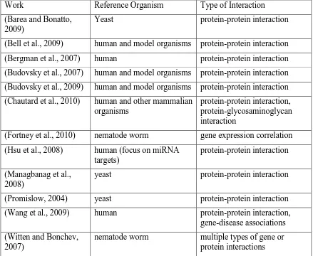
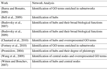
2.3ANALYSING AGEING-RELATED GENE OR PROTEIN NETWORKS ... 49
2.3.1 Types of interactions and reference organisms in ageing-related networks .... 49
2.3.2 Analysing ageing-related gene or protein networks ... 53
2.4CONCEPTS AND PRINCIPLES OF DATA MINING ... 57
2.4.1 Basic concepts of data mining ... 57
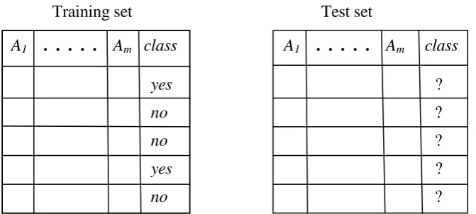
2.4.2 The classification task of data mining ... 58
2.4.2.1 Overfitting and underfitting ... 61
2.4.2.2 Classification versus clustering ... 61
2.5CLASSIFICATION METHODS USED IN THIS RESEARCH ... 63
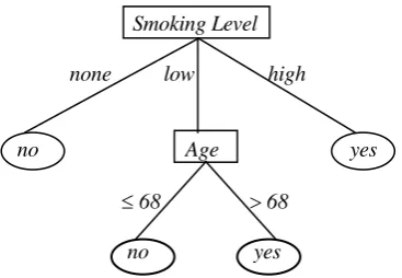
2.5.1 Decision tree induction ... 63
2.5.2 Naive Bayes ... 68
2.6RELATED WORK ON PREDICTING PROTEIN FUNCTION WITH CLASSIFICATION METHODS... 69
CHAPTER 3 – DATASET CREATION AND EXPERIMENTAL SET UP ... 75
3.1 CREATING DATASETS WITH TWO CLASSES AND MULTIPLE ATTRIBUTE TYPES ... 75
3.1.1 Creating two classes: ageing-related vs. non-ageing-related DNA repair ... 75
3.1.2 Creating the predictor attribute type of DNA repair ... 76
3.1.3 Creating a predictor attribute measuring the rate of evolutionary change (Ka/Ki ratio) ... 77
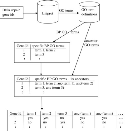
3.1.4 Creating a set of predictor attributes representing GO terms ... 78
3.1.5 Creating a set of attributes representing protein-protein interaction information... 81
3.1.6 Removing duplicate data instances ... 82
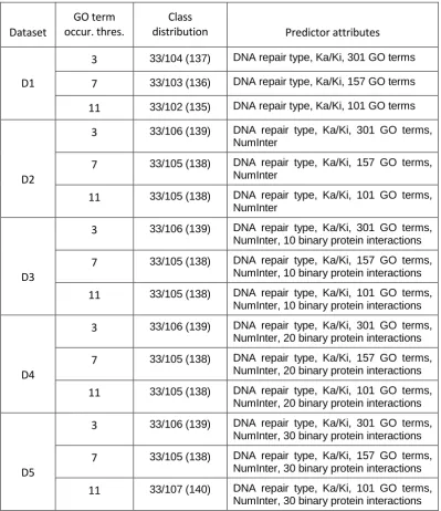
3.1.7 Dataset specifications... 83
3.2 CREATING A DATASET WITH TWO CLASSES AND GENE EXPRESSION ATTRIBUTES ... 86
3.3.1 Creating the four classes to be predicted ... 88
3.3.2 Creating the predictor attributes ... 89
3.3.3 Dataset specifications... 89
3.4 MEASURING PREDICTIVE ACCURACY ... 91
3.5 STATISTICAL SIGNIFICANCE ... 94
CHAPTER 4 – COMPUTATIONAL RESULTS AND DISCUSSION ... 96
4.1 RESULTS AND DISCUSSION FOR DATASETS WITH TWO CLASSES AND MULTIPLE ATTRIBUTE TYPES ... 96
4.1.1 Results for the J4.8 decision tree induction algorithm ... 97
4.1.2 Results for the CART decision tree induction algorithm... 100
4.1.3 Results for the Naive Bayes algorithm ... 103
4.1.4 Discussion on predictive patterns extracted from the decision trees ... 104
4.1.4.1 Discussion on attributes chosen as root nodes in the decision trees ... 105
4.1.4.2 Issues on selecting and interpreting rules extracted from decision trees . 108 4.1.4.3 Discussion on selected rules extracted from decision trees ... 111
4.2 RESULTS AND DISCUSSION FOR DATASETS WITH TWO CLASSES AND GENE EXPRESSION ATTRIBUTES ... 117
4.2.1 Predictive accuracies for J4.8, CART and Naive Bayes algorithms ... 118
4.2.2 Interpreting a rule extracted from the decision tree built by J4.8 ... 118
4.2.3 Integrating results for gene expression and other types of predictor attributes ... 120
4.3 RESULTS AND DISCUSSION FOR DATASETS WITH FOUR CLASSES AND MULTIPLE ATTRIBUTE TYPES ... 123
4.3.1 Results for the J4.8 decision tree induction algorithm ... 124
4.3.2 Results for the CART decision tree induction algorithm... 127
4.3.3 Results for the Naive Bayes algorithm ... 130
4.3.4 Discussion on predictive patterns extracted from the decision trees ... 131
4.3.4.1 Discussion on attributes chosen as root nodes in the decision trees ... 131
4.3.4.2 Discussion on selected rules extracted from decision trees ... 132
CHAPTER 5 – CONCLUSIONS ... 140
5.1 CONTRIBUTIONS ... 140
5.2 SUMMARY OF THE MAIN DISCOVERED PREDICTIVE PATTERNS ... 141
5.3 FUTURE RESEARCH DIRECTIONS ... 148
LIST OF FIGURES
LIST OF TABLES
ACKNOWLEDGMENTS
The author thanks his primary supervisor, Dr. Joao Pedro de Magalhaes, for his help in defining the topic of this research and for his valuable comments throughout this research project.
The author also thanks his secondary supervisor, Dr. Olga Vasieva, for her use of the Genevestigator® and Ingenuity® software tools and her help in the analysis of the results obtained by using those tools.
Thanks are also due to Dr. Fernando E.B. Otero, from the University of Kent, for his use of software to process Gene Ontology terms.
In addition, the author thanks all his colleagues in the Magalhaes‟ lab for creating a
DECLARATION
C h a p t e r 1 – I n t r o d u c t i o n
1.1 WHAT IS AGEING?
1.1.1 Defining ageing
Ageing is a natural process, occurring in almost all species (although at very different rates in different species), and ageing signs in humans are “obvious” to a lay person.
However, from a scientific point of view, ageing is still a mysterious process, whose fundamental causes are still strongly debated, and it is difficult to find a definition of ageing which is accepted as a “standard” in the literature.
Several candidate definitions are reviewed in (Arking, 2006). Two main conclusions can be drawn from that review. First, several definitions of ageing refer to changes that happen to an organism with the passage of chronological (physical) time. In principle it would be better to refer to a more relevant measure of “biological time”, in
terms of biomarkers of ageing involving physiological processes; but in practice it is not easy to define such biomarkers, due to our lack of knowledge of the fundamental causes of ageing.
Secondly, it seems useful to make a distinction between at least two kinds of changes affecting an organism in two different stages of its life, namely the developmental stage and the post-maturational stage. This distinction is important because organismal changes during development are not normally deleterious. In contrast, a major characteristic of the ageing process is that it produces deleterious changes (involving functional decline) in an organism, and such changes usually manifest themselves after the organism‟s maturation. Hence, gerontology usually focuses on
the study of the ageing process after an organism has completed its development
Taking the above points into account, ageing can be defined as: “...the
time-independent series of cumulative, progressive, intrinsic, and deleterious functional and structural changes that usually begin to manifest themselves at reproductive maturity and eventually culminate in death” (Arking, 2006).
It is worth discussing the relationship between the process of ageing and age-related diseases, which seems controversial. It has been argued that ageing and ageing-related
diseases are different types of biological processes. In this spirit, (Hayflick, 2000) points out several features of the process of ageing that are not features of any specific disease,
such as: ageing affects every animal that reaches adulthood, it occurs in all members of a species only after the age of reproduction, and it takes place in virtually all species. There is no specific disease with these features. On the other hand, ageing and ageing-related diseases are very related processes, and in general mutations that slow ageing also postpone age-related diseases (Kenyon, 2010). Hence, it has also been argued that the “aging is no more and no less than the collective early stages of the various age-related diseases” (Grey and Rae, 2007).
In any case, it is worth noting that the distinction between ageing and age-related diseases is made by several governmental agencies. For instance, the US Food and Drug Administration will not approve any drug whose purpose is “only” to slow down ageing,
rather than combating a disease (Vijg and Campisi, 2008).
1.1.2 Ageing at the cellular and tissue levels
In order to understand ageing in complex multicellular organisms, it is often useful to study ageing at the more basic cellular and tissue levels, as discussed next.
The term “senescence” derives from the Latin word senex, meaning old man or old age,
the behaviour of a cell when it gets senescent. Senescent cells also have altered morphology (in general an enlarged flattened shape) and altered functionality, and they are often resistant to apoptotic signals. Hence, cellular senescence is now understood as a stress response process, rather than as a process that mimics organismal ageing at the cellular level.
Functionality changes associated with cellular senescence have been extensively studied
in fibroblasts – the cell type that synthesizes the stroma, the structure underlying the cells of epithelial tissues. Senescent human fibroblasts exhibit, for instance, an increased
secretion of inflammatory cytokines and epithelial growth factors (Campisi, 2005).
Cellular senescence can be caused not only by the well-known shortening of telomeres (which typically occurs in humans, but not usually in mice (Lombard et al., 2005)), but also by several other factors such as DNA damage, perturbations to chromatin organization, and expression of certain oncogenes (Campisi, 2005), (Campisi and Fagagna, 2007), (Passos et al., 2009). In addition, in vitro culturing can be regarded, by itself, as a form of stress that induces premature cellular senescence (Pelicci, 2004). Cellular senescence induced by dysfunctional telomeres is often called telomere-initiated cellular senescence, whilst in general senescence induced by stress – independently of telomere dysfunction – is called stress-induced premature senescence (Zglinicki et al., 2005), (Debacq-Chainiaux et al., 2005).
There are two major ways in which cellular senescence can contribute to the ageing of an organism (Lombard et al., 2005). First, as the number of senescent progenitor or stem cells increases with age, tissue renewal is gradually impaired, leading to a loss of tissue homeostasis (Maslov and Vijg, 2009), (Pelicci, 2004). The issue of whether stem cell depletion per se is a major cause of ageing-related tissue or organ dysfunction, in the absence of high levels of genotoxic stress, is still an open question, since stem cells provide just one out of several types of homeostatic or defence mechanisms against
note that stem cell depletion seems not to have any significant contribution to the ageing of post-mitotic tissues such as the brain and the heart – where the vast majority of cells are not replaced during adult life. Furthermore, (Maslov and Vijg, 2009) point out that in general stem cell populations are not completely lost, and they suggest that a more important role of stem cells in ageing is that mutations accumulated in stem cells are transmitted to their daughter cells that become newly differentiated cells, and this in turn contributes to a decline of tissue functionality.
The second way in which cellular senescence can contribute to organismal ageing is that
the aforementioned secretory phenotype of senescent cells can modify the local tissue environment in a way that contributes to ageing-related diseases, including cancer (Campisi, 2005) – which seems ironic, considering that cellular senescence is believed to have evolved as a tumor suppressor mechanism (Campisi and Fagagna, 2007).
Turning to the tissue level, one should recall that complex multicellular organisms such as humans have a large number of different types of tissues, with very different functionality and patterns of gene expression, and therefore it is not surprising that the process of ageing manifests itself in different ways in different tissues (Arking, 2006).
Another example of differences between tissues – which is relevant for the DNA damage theory of ageing, which will be briefly discussed later – is that the number of endogenous abasic sites – i.e. sites in a DNA strand without a base, characterizing a type of DNA damage – has been observed to vary widely between tissues, but not within the same tissue (Nakamura and Swenberg, 1999). The greatest number of abasic sites was observed in the brain.
1.1.3 The motivation for ageing research
A major motivation for ageing research is that ageing is the greatest risk factor for many
diseases, including virtually all types of cancer.
It is important to recall here the distinction between ageing and ageing-related diseases that is often made by policy makers in government and research-funding agencies. For instance, it has been noted that more than half of the budget of the US National Institute on Ageing is spent on Alzheimer‟s disease, a single ageing-related disease; even though the likelihood of dying from Alzheimer‟s disease is just 0.7% and a complete elimination
or cure of this disease would add only about 19 days onto the average human life expectancy (Hayflick, 2000).
In contrast, if we could significantly slow down ageing in humans, we would be simultaneously postponing the onset of a large number of age-related diseases (Grey and Rae, 2007), (Kenyon, 2010), greatly increasing both life expectancy and quality of life. Therefore, there is a clear motivation to do research on the process of ageing, rather than just on individual age-related diseases.
One can also consider that there is a great need for ageing research because, despite the large progress in this area in the last two decades, ageing is still to a large extent a poorly understood process. To quote (Miller, 2004b):
“We now know as much about aging as scientists knew about infection after John Snow‟s
1.2 THEORIES OF AGEING
In this section we discuss just two types of theories of ageing, namely evolutionary theories – which at present provide the most accepted explanation about why we age from an evolutionary perspective – and the DNA damage theory of ageing – which is particularly relevant for this thesis. It should be noted, however, that there are many other theories of ageing, and for a discussion of other theories the reader is referred to (Arking, 2006), (Magalhaes, 2011).
1.2.1 Evolutionary theories of ageing
Explaining why ageing occurs in evolutionary terms is not easy, since at first glance it seems that natural selection would not favour the evolution of ageing, given its seriously harmful (eventually lethal) effects to an organism. However, the existence of ageing can be explained, in broad terms, by evolutionary theories of ageing (Arking, 2006), (Magalhaes, 2011), as follows.
The mutation accumulation theory is based on Peter Medawar‟s insight that the force of
natural selection fades with age (Medawar, 1952). That is, in the wild most animals have a very high rate of mortality – due, for instance, to predators. Hence, in general, if a
mutation produces a harmful or lethal effect only in very old age, the gene allele associated with that mutation will not be eliminated from the population by natural selection, because the vast majority of animals will die from other causes before the mutation‟s effect is triggered. Hence, mutations that are harmful at old age tend to
accumulate. The mutation accumulation theory of ageing was also influenced by Szilard, a physicist who proposed that somatic mutations are the elementary step in the ageing process based on an analogy with the known effects of radiation (Arking, 2006).
George Williams has proposed another evolutionary theory of ageing which is complementary to the mutation accumulation theory. Williams‟ theory, called the
different functions. Hence, if a gene allele has the opposite (antagonistic) effects of increasing the chances of an animal surviving until reproduction and being harmful at an older age, that gene allele will be favoured by natural selection despite its harmful effects later in life.
There is reasonable evidence for the existence of genes with antagonistic pleiotropic effects, with the caveat that most of the evidence is derived from animal models, and the
evidence for genes with such effects in the wild is much weaker (Leroi et al., 2005). Evidence for the mutation accumulation theory seems less convincing and more
controversial than the evidence for the antagonistic pleiotropy theory (Kirkwood, 2005), (Kirkwood and Austad, 2000).
To illustrate the difficulty of finding genes with antagonistic pleiotropy, let us consider the following case. A typical example of a gene which is often considered to have antagonistic pleiotropic effects is the well-known p53 tumor suppressor gene (Campisi and Fagagna, 2007), (Lombard et al., 2005), which contains mis-sense mutations in approximately 50% of the major forms of cancers (Ko and Prives, 1996). This gene has major roles in apoptosis and cellular senescence, and also has a role in DNA repair (Cao et al., 2006), (Ko and Prives, 1996), (Oren, 2003). Mutant mice with hyperactive p53 are, as expected, much more resistant to cancer than the wild type, but in some cases, surprisingly, they have a somewhat shorter life span (despite the lower incidence of cancer) and display multiple signs of premature ageing (Tyner et al., 2002), (Maier et al., 2004). Apparently, these results can be explained by the antagonistic pleiotrophy theory.
However, a more recent analysis of those experiments suggests that the aging phenotype of those mutant mice seems to be due to the fact they had a truncated form of the p53 protein expressed along with the wild type of the protein, and apparently it was that truncated form (with altered function) - rather than an overall increase in the normal form of the protein – that mainly contributed to the premature ageing phenotype (Mendrysa et
are resistant to tumor formation but do not show signs of premature ageing. Hence, it is not clear if p53 is really a gene with antagonistic pleiotropy after all.
In any case, the concept of antagonistic pleiotropy has been applied not only to genes, but to ageing-related biological processes or mechanisms. For instance, cellular senescence can be considered a tumor suppressor mechanism at relatively young ages, but senescent cells seem to contribute to an ageing phenotype in later ages – as discussed earlier. A
similar comment applies to apoptosis (Campisi, 2005).
Another evolutionary theory of ageing is the disposable soma theory, proposed by Kirkwood (Kirkwood, 1996). In essence, this theory postulates that there is a trade-off between the investment of metabolic resources in the maintenance of the soma and in reproduction. Hence, organisms tend to invest more metabolic resources in the maintenance of the soma as long as they are expected to survive, but the use of metabolic resources tends to shift from soma maintenance to reproduction as the chance of the organism dying in the wild becomes higher.
Although at a high level of abstraction the disposable soma theory seems somewhat similar to the antagonistic pleiotropy theory, the disposable soma theory is more specific about the mechanisms underlying ageing (Kirkwood and Austad, 2000), (Kirkwood, 2005). In particular, the disposable soma theory emphasises the role of somatic repair mechanisms (for instance, DNA repair) in ageing, predicting that a fundamental factor underlying ageing is the accumulation of unrepaired cellular damage due to imperfect repair mechanisms. In contrast, the antagonistic pleiotropy theory is formulated in terms of a more abstract general pattern of temporal differences in gene effects.
1.2.2 DNA damage theory of ageing
normal double-helix structure and consists of an uninterrupted sequence of bases. In contrast, DNA damage refers to physical or chemical alterations in the structure of a DNA molecule, which is no longer a normal double-helix. In other words, mutations change the informational content of a DNA molecule, but preserve its normal structure. Damage modifies the structure of a DNA molecule, producing an abnormal structure. Although damage to other kinds of molecules found in cells can also influence ageing, DNA damage seems a particularly important kind of damage because, unlike other cellular components which can be replaced, DNA must last the lifetime of the cell (Lombard et
al., 2005).
In essence, the DNA damage theory of ageing postulates that the main cause of the functional decline associated with ageing is the accumulation of DNA damage. Note that DNA damage can have multiple effects. In particular, DNA damage can impair transcription, cause an interruption of the cell cycle until the damage is repaired or (if the damage is too serious) lead to programmed cell death (apoptosis). DNA damage can also lead to mutations when the DNA is replicated, as will be discussed later. Hence, the DNA damage theory of ageing can be interpreted in different ways, depending on how one interprets the relative contribution of each of those effects to the ageing process.
Although this theory is clearly related to the mutation accumulation theory (both involve alterations to DNA), they are also quite different, given the aforementioned differences between mutations and damage in DNA. However, the theories would tend to converge if one believed that the main effect of DNA damage causing ageing were the DNA mutations resulting from that damage.
Within the variations of the DNA damage theory of ageing, one can also distinguish between variations emphasizing either the role of nuclear DNA damage or the role of mitochondrial DNA damage in ageing. It has been argued that one reason why nuclear DNA damage might be a more important cause of ageing is that normally there are only
thousand copies of the mitochondrial DNA in a cell (Lombard et al., 2005). In addition, nuclear DNA (nDNA) accounts for about 99% of the cellular DNA.
On the other hand, there are also possible reasons for the greater importance of mitochondrial DNA (mtDNA) in ageing, as follows (Graziewicz et al., 2006). First, mtDNA is much more prone to damage than nDNA (since mtDNA is not protected by histone proteins and it is very close to the site of ROS – reactive oxygen species –
generation in the mitochondrial membrane). In addition, overall the repair of mtDNA is less efficient than the repair of nDNA.
The relative importance of mtDNA damage and mutations for ageing is still controversial, though. In a recent review of several mouse models with increased levels of mtDNA, (Khrapko & Vijg 2008) noted that, although in general those mutant mice show multiple signs of premature ageing, the interpretation of the results requires great caution. For instance, in general the mutant mice start to have a high frequency of mutation in the developing embryo, whilst in a mice undergoing normal ageing the accumulation of mtDNA mutations would presumably be driven by ROS or other factors in a time- and tissue-dependent manner. Khrapko & Vijg conclude their review with the remark that
“Despite decades of research and recent advances in generating mouse models with
increased mutational loads, the study of mitochondrial DNA mutations in aging still has not reached a stage at which clear, definitive conclusions can be drawn regarding causal relationships.”
There is good evidence for (a), as follows. First, many human progeroid syndromes – diseases characterized by accelerated ageing, which will be reviewed later – are due to defective nDNA repair, and overall the ones with the most severe progeroid phenotype are the ones with most severe nDNA repair defects (Best, 2009), (Lombard et al., 2005). In addition, there are many DNA repair genes whose deletion leads to a premature ageing phenotype in mouse models (Hasty et al., 2003), (Magalhaes and Faragher, 2008).
In summary, as pointed out by (Arking, 2006): “Damage to nuclear DNA likely contributes to the aging process, at least in certain tissues. The data strongly suggests that
the absence of DNA repair ability probably has a causal relationship to the expression of a shortened life span and accelerated senescence.”
However, it is much harder to find evidence for (b), which increased DNA repair leads to increased life span – and even harder to find evidence that such increased life span is due to slower aging, since life span could be extended due only to a reduced risk of death from disease. One study showing an increase in life span with improved DNA repair in flies is presented in (Symphorien and Woodruff, 2003). In this work, D. melanogaster
with one or two extra copies of a DNA repair gene had a significantly extended life span, although the extension was not large. Also, in this study DNA repair was not directly measured, it was simply assumed to directly vary with gene dose (Arking, 2006).
This kind of experiment artificially increasing the number of copies of a gene cannot usually be done in humans (for ethical reasons, at least), but one can study the DNA repair systems of centenarians, to determine if they are more efficient than the DNA repair systems of most old individuals. (Chevanne et al., 2007) compared the efficiency with which cells from young, old and centenarian subjects repair DNA strand breaks caused by sublethal concentrations of the oxidant hydrogen peroxide (H2O2). They
observed that cells from centenarians are about as efficient in that kind of repair as the cells from young subjects, and both types of cell were considerably more efficient in that
significantly decreased in the cells of old subjects, but not in the cells of young and centenarian subjects. In addition, centenarians have significantly higher levels of the KU70 protein (a key factor in the repair of DNA double-strand breaks – as will be discussed later). These results support the hypothesis that improved DNA repair systems may lead to longer life span.
The second major prediction of the DNA damage theory of ageing is that a systemic,
age-related shift in DNA repair activity is a major factor underlying the functional decline associated with the process of ageing (Arking, 2006). There are many studies showing
evidence that many kinds of DNA repair activity decrease with age, and a number of such studies will be discussed later in this thesis, in the context of specific DNA repair pathways. Here we just mention that, in a recent review of work in this area, (Gorbunova et al., 2007) concluded that: “There is sufficient evidence that all pathways of DNA repair...become less efficient with age”.
There are, however, two caveats to this kind of conclusion. First, it should be noted that most studies that measure DNA repair tend to focus on just one type of DNA repair, but the decrease of one type of DNA repair may be compensated by an increase in another type (Engels et al., 2007). Also, DNA repair activity is very difficult to measure, especially in vivo (Maslov and Vijg, 2009).
1.3 PROGEROID SYNDROMES
This section is divided into two parts. First, we review some major progeroid syndromes caused by defects in DNA repair. Secondly, we discuss the relevance of such progeroid syndromes to the study of normal human ageing.
1.3.1 An overview of progeroid syndromes
1.3.1.1 Werner syndrome (WS)
This is usually considered the progeroid syndrome that shows more symptoms of normal
ageing and ageing-related diseases (Magalhaes and Faragher, 2008). WS is caused by a variety of loss-of-function mutations in a gene coding for a protein that is a member of the
RecQ DNA helicase family (WRN) (Kipling et al., 2004). The WRN protein is involved in several important biological processes, related to DNA replication, recombination, apoptosis and telomere metabolism, but its major function seems to be the re-initiation of stalled replication forks. In vitro, the absence of WRN leads to a mutator phenotype, characterized by an increased frequency of deletional mutations, resulting from the inability to re-initiate stalled replication forks.
WS patients usually seem to be normal during childhood, but stop growing during the teenage years (Best, 2009). WS patients usually show the following ageing symptoms (Davis and Kipling, 2006), (Martin and Oshima, 2000): premature graying of the hair and baldness, skin and muscular atrophy, hypogonadism, poor wound healing, atherosclerosis, osteoporosis, soft-tissue calcification, juvenile cataracts, a tendency toward diabetes, and an elevated cancer frequency (Arking, 2006), (Kipling et al., 2004). On the other hand, WS patients show no increased tendency for neurodegeneration or Alzheimer‟s Disease,
and the immune system remains normal. The median age at death is 47 years, and death is usually a result of cancer or arteriosclerosis.
The cells of WS patients show significant chromosomal abnormalities and accumulation of DNA double-strand breaks (Ariyoshi et al., 2007), (Best, 2009). WS fibroblasts reach the stage of replicative senescence considerably faster than normal fibroblasts, but both
1.3.1.2 Hutchinson-Gilford progeroid syndrome (HGPS)
This progeroid syndrome has an onset in childhood, much earlier than the onset of WS. For this reason, HGPS is sometimes called “child progeria”, whilst WS is sometimes called “adult progeria”. HGPS is caused by a point mutation in the gene for lamin A, a
type of protein that forms a network of filaments beneath the inner nuclear membrane (among other possible locations in the nucleus) (Bridger and Kill, 2004). A-type lamins are also believed to be involved in nDNA replication and RNA polymerase II-dependent transcription, and higher-order chromatin structure (they can directly bind to DNA and to
chromatin).
There are several types of HGPS, which can be divided into classical and non-classical HGPS (Hennekam, 2006). Here we focus on the classical HGPS, which is associated with a strong resemblance of symptoms among the affected patients and tends to be the more severe form of the disease.
HGPS patients show the following symptoms (Hennekam, 2006), (Arking, 2006): premature loss of hair and subcutaneous fat (starting in the first year), postnatal growth is severely disturbed, no pre-pubertal or pubertal growth spurt, osteolysis, decreased joint mobility from the second to third year, thinning of the skin, limited sexual development and severe vascular problems in the brain and elsewhere – strokes occur at the median age of nine years. The vast majority of patients have a normal cognitive development. No neurofibrillary tangles appear in the central nervous system. The median age at death is 12 years. The cause of death is usually of vascular origin, in particular myocardial infarctions.
The study of HGPS seems less relevant for the understanding of normal human ageing than the study of WS, because, although HGPS patients show symptoms that superficially resemble premature ageing, there seems to be no basic mechanism shared between HGPS symptoms and normal human ageing, as pointed out by (Arking, 2006). However, it
genomic instability and both lead to accelerated ageing in some tissues (Bridger and Kill, 2004).
1.3.1.3 Trichothiodystrophy (TTD)
TTD is caused by point mutations in the XPD gene, which encodes one of the two core transcription factor IIH (TFIIH) helicases (Hasty et al., 2003). Different mutations in this gene can give rise to TTD, XP (xeroderma pigmentosum) or CS (Cockayne syndrome).
The helicase encoded by the XPD gene is involved in both DNA repair and transcription initiation (Boer et al., 2002).
TTD patients show the following symptoms (Hasty et al., 2003), (Boer et al., 2002): neurodegeneration (including cerebellar ataxia), skeletal degeneration, impaired sexual development, cachexia (i.e, a patient‟s unintentional loss of body mass that cannot be reversed by nutrional means), osteoporosis, cataracts, brittle hair and nails. Patients have a mean life span of just about 10 years, and show no predisposition to cancer.
1.3.1.4 Cockayne syndrome (CS)
1.3.1.5Ataxia telangiectasia (AT)
AT is caused by a loss-of-function mutation in the ATM (ataxia-telangiectasia mutated) gene. The term ataxia refers to the shaky and unsteady limb movements, due to the brain's failure to regulate the body's posture and regulate the strength and direction of limb movements. The ATM gene‟s product is a protein kinase which is involved in several signal transduction pathways, which operate both under stress and in normal physiological conditions (Rotman and Shiloh, 1997). In particular, ATM is involved in cell cycle progression and checkpoint response to DNA damage.
AT patients show the following main symptoms (Pesce and Rothe, 1996), (Rotman and Shiloh, 1997), (Wong et al., 2003): progressive neurodegeneration – with cerebellar ataxia becoming apparent when the patient begins to walk, telangiectases (dilated blood vessels) – with onset typically between three and five years of age, immunodeficiency, genomic
instability, strong cancer predisposition (mainly of lymphoid origin) and sensitivity to radiation, accelerated telomere loss, and growth retardation in many patients – although those who reach puberty usually have normal height and weight. The average life span of AT patients is about 20 years (Hasty et al., 2003). ATM-deficient mice exhibit most of the symptoms of the human disease (Rotman and Shiloh, 1997), (Wong et al., 2003).
1.3.1.6 Rothmund-Thomsom (RT) syndrome
1.3.1.7Xeroderma pigmentosum (XP)
This is a disease due to a defect in one of seven genes (XPA – XPG) required for nucleotide excision repair (a form of DNA repair to be reviewed later). XP victims show dramatically accelerated aging only in areas of skin exposed to the sun and a skin cancer rate more than a thousand times greater than normal, and frequently exhibit neurodegeneration (Hasty et al., 2003), (Best, 2009). Overall, the symptoms of accelerated ageing in XP seem to be more focused than the broader symptoms associated with other progeroid syndromes such as Werner.
[image:26.595.109.544.447.710.2]To summarize the previous discussion on progeroid syndromes, Table 1.1 shows, for each of those syndromes: which genetic defect is associated with it and what are the main processes affected by that defect; what is the mean life span (in years) of patients with that syndrome; and whether or not the syndrome is associated with an increased incidence of cancer.
Table 1.1: Summary of major human progeroid syndromes (adapted from (Hasty et al., 2003), (Arking, 2006), (Lans and Hoeijmakers, 2006))
Syndrome Genetic defect Mean life
span (years)
Predisposition to cancer?
Werner RecQ-like DNA helicase and
exonuclease, involved in DNA repair
47 yes
Hutchinson-Guilford Lamin A, involved in nDNA replication, transcription, nuclear organisation
13 no
Trichothiodystrophy TFIIH helicase, involved in DNA repair and transcription
10 no
Cockayne CSA or CSB gene, involved in DNA repair and transcription
12-20 no
Ataxia telangiectasia ATM protein kinase, involved in DNA damage response
20 yes
Rothmund-Thomson RecQ-like DNA helicase Normal? yes
Xeroderma Pigmentosum
XPA – XPG genes, involved in DNA repair
Lower than normal?
1.3.2 On the relevance of progeroid syndromes to the study of human ageing
The relevance of the study of progeroid syndromes for the understanding of normal ageing is controversial, particularly when such progeroid syndromes are observed in mouse models of their human counterpart. (Miller, 2004a) has strongly criticized this relevance, arguing that progeroid syndromes and animal models of premature or accelerated ageing in general do not offer any significant insights into the normal human ageing process. A major point of his criticism is that patients or animal models of progeroid syndromes show just a subset of the symptoms of normal ageing, whilst normal
ageing is characterized by a much broader range of symptoms. Actually, progeroid syndromes are often called segmental progeroid syndromes, to emphasize the fact that their pathologies are limited to one or a few organs or tissue types, so that they resemble ageing only in part (Arking, 2006).
In addition, Miller points out that it is relatively easy to make an animal have a significantly shorter life span by introducing a defect in some crucial DNA repair gene, but it is much more difficult to show that the defect is really accelerating ageing – particularly considering that we still do not have a very reliable biomarker of ageing. Furthermore, although in several cases a mutation in a single DNA repair gene leads to signs of premature or accelerated ageing in mice, not all mice mutants with defective DNA-repair genes show signs of premature or accelerated ageing.
Counter-arguments to Miller‟s criticism have been provided in (Hasty and Vijg, 2004b) and (Kipling et al., 2004). (Hasty and Vijg, 2004b) point out that the “segmental” nature
of progeroid syndromes does not invalidate their relevance for the study of normal ageing,
because every individual who undergoes normal ageing exhibits a segmental ageing
phenotype, by comparison with all possible ageing phenotypes in the population. That is,
segmental ageing is natural and normal. A similar argument is made by (Best, 2009). The
extent to which progeroid syndromes patients have symptoms of normal ageing varies
significantly among those syndromes – as discussed earlier – but at least Werner‟s
syndromes for the understanding of normal human ageing, (Kipling et al., 2004) conclude that there is good evidence that the premature replicative senescence of Werner‟s
syndrome cells is a causal factor in the aspects of that syndrome that resemble premature
ageing, and that this evidence supports a causal role for replicative senescence in normal
human ageing.
Concerning Miller‟s argument that not all mice mutants with DNA-repair defects show
signs of accelerated ageing, used as an argument against the relevance of DNA repair in
ageing, (Hasty and Vijg, 2004b) point out that deletion of some crucial DNA repair genes
leads to embryonic death or cancer at an early age, so that there is simply no time for the
ageing phenotype to appear. This shows that DNA repair is crucial for survival, but this is
not incompatible with the fact that DNA repair is also important for ageing. Hence, some
DNA repair defects are so harmful that they lead to early death, whilst other defects allow
the organism to survive long enough to show signs of premature or accelerated ageing.
The authors conclude that progeroid syndromes in human and mouse mutants with
defects in some DNA repair genes offer strong support to the idea that a major causal
factor of ageing is the accumulation of DNA damage and mutations with time, which
eventually leads to cellular senescence or apoptosis (Hasty et al., 2003).
In any case, one should recall that, unlike normal ageing, in general progeroid syndromes
are subject to the force of natural selection (so that the existence of progeroid syndromes
cannot be explained by the evolutionary theory of ageing), and some phenotypic features
of those syndromes may result from gross abnormalities in development which are not
directly relevant for gerontology (Martin and Oshima, 2000) – recall that gerontology
1.4 DNA DAMAGE
This section is divided into two subsections. The first one discusses two major types of sources of DNA damage (arguably the two most studied ones), namely oxidative damage and damage induced by ultra-violet radiation. The second subsection discusses specific types of damages regardless of the cause of the damage. That subsection focuses just on describing the damage itself, not how to repair it, which will be the focus of section 1.5.
1.4.1 Two major sources of DNA damage
1.4.1.1 Oxidative damage
A common cause of DNA damage is exposure to reactive oxygen species (ROS). ROS include superoxide, hydrogen peroxide, hydroxyl radicals and singlet oxygen. Such ROS can oxidize DNA, which can produce several kinds of DNA damage, in particular oxidized bases, abasic sites and single- and double-strand breaks (Bont and Larebeke, 2004), (Jackson and Loeb, 2001).
Different types of ROS vary considerably in their degree of reactivity with cellular components. Superoxide has limited reactivity but it is converted to hydrogen peroxide by
superoxide dismutase. Hydrogen peroxide is reduced to water by catalase and glutathione peroxidase; but, in the presence of some metals such as iron, hydrogen peroxide is reduced to hydroxyl radicals. These radicals‟ reactivity is so great that they do not diffuse more than one or two molecular diameters before reacting with a cellular component (Friedberg et al., 2006), (Bont and Larebeke, 2004). Hence, a hydroxyl radical is able to oxidize DNA only if that radical is produced immediately adjacent to DNA. On the other hand, hydrogen peroxide can be seen as a diffusible, latent form of hydroxyl radical that reacts, for instance, with iron (Fe+2) adjacent to a DNA molecule to generate a hydroxyl radical.
that normally the nucleus is practically anoxic, helping to protect nuclear DNA from the harmful effects of ROS. Also, in mammals the level of oxygen concentration in tissues is only 3 to 4%, which is much smaller than the environmental level of about 20%. In addition, to reduce the use of iron available to be used in the production of hydroxyl radicals as mentioned earlier, most iron in cells is safely stored in ferritin and transferrin. Furthermore, organisms produce a number of antioxidant enzymes that eliminate ROS. For instance, catalase removes hydrogen peroxide, and superoxide dismutase (SOD) eliminates superoxide. There are also several redox-activated transcription factors,
indicating that mechanisms to cope with ROS are genetically regulated. Despite all these defence mechanisms against ROS-induced damage, sometimes the production of ROS is so overwhelming that those defence mechanisms are not enough, and in this case oxidative stress occurs.
ROS can produce many different kinds of damage and mutation in DNA. For instance, the cytosine base alone can undergo oxidative damage producing at least 40 different modified species (Jackson and Loeb, 2001). Some oxidatively modified bases block DNA replication, whilst others are mispaired and lead to base substitutions in the DNA. It is interesting to note that some of the progeroid syndromes caused by defective DNA repair discussed earlier – such as xeroderma pigmentosum and ataxia telangiectasia – are associated with a high amount of 8-oxo-dG (Bont and Larebeke, 2004).
In any case, in the last few years it has become clearer that ROS is not just a source of damage, but also relevant players in signalling pathways, involved in the regulation of gene expression, development, growth and apoptosis (Magalhaes and Church, 2006).
1.4.1.2 Damage induced by ultraviolet (UV) radiation
The UV radiation spectrum can be divided into several segments based on wavelength (Friedberg et al., 2006): UV-A (320-400nm), UV-B (295-320nm), UV-C (100 to 295nm). It should be noted that, in order to study DNA repair, most laboratories use UV-C light,
although (Friedberg et al., 2006) point out that many of the DNA lesions produced by UV-C are also produced at longer wavelengths, and UV-C light is normally used because it is more efficient in producing those lesions.
In any case, in the natural world UV-A and UV-B are the major cause of DNA damage (Yaar and Gilchrest, 2007). UV-B photons are on average 1,000 times more energetic than UV-A photons, and so UV-B radiation is believed to be the main type of UV
radiation responsible for direct DNA damage and photocarcinogenesis after sun exposure. UV-A radiation is still likely to be a relevant cause of photodamage, though, because it is
at least 10-fold more abundant in terrestrial sunlight and on average it has a greater depth of penetration into the dermis, by comparison with UV-B radiation. UV-A radiation also triggers mtDNA mutations.
The main types of DNA damage caused by UV radiation are single-strand breaks and numerous photoproducts (Friedberg et al., 2006) – the most frequent type of which is cyclobutane pyrimidine dimers (Yamada et al., 2006). (These types of DNA damage will be further discussed below.) UV radiation is also a major exogenous source of ROS (Yaar and Gilchrest, 2007). UV-B light crosses the epidermis and results in the generation of ROS and DNA damage, leading to the activation of signalling pathways related to stress response and ageing (Debacq-Chainiaux et al., 2005). UV radiation also has an effect in transcriptional down-regulation of a transforming growth factor-β (TGF- β) receptor, which leads to reduced production of collagen (Quan et al., 2004). In experiments with yeast, (Kozmin et al., 2005) concluded that UVA radiation can be strongly mutagenic due to the generation of oxidative DNA damage.
1.4.2 An overview of major types of DNA damage
1.4.2.1 Depurination and depyrimidination
abasic site. In general about 10,000 purine bases are lost every day from the DNA of each mammalian cell by spontaneous depurination reactions (Friedberg et al., 2006).
Depyrimidination involves the loss of pyrimidine bases (cytosine and thymine) from DNA. Depyrimidination is much less common than depurination, however, since the N-glycosyl bond between a pyrimidine base and the deoxyribose is more stable than the corresponding bond for purine bases. The rate of loss of depyrimidination has been
estimated as about only 5% of the rate of depurination (Lindahl, 1993).
1.4.2.2 Deamination
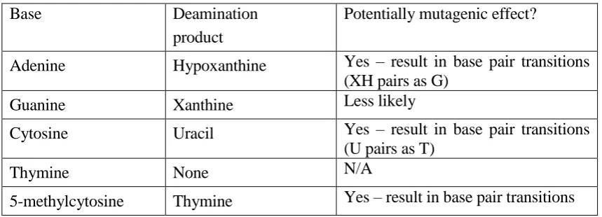
Deamination involves the loss of amino groups from DNA bases. Almost all DNA bases undergo deamination in spontaneous reactions, with the exception of thymine – which does not have an amino group – as summarized in Table 1.2. It should be noted that most types of deamination shown in the table produce a base that does not naturally occur in DNA (the only exception is the deamination of 5-methylcytosine), and this fact facilitates the identification and excision of the deaminated base by a DNA glycosylase enzyme, as will be discussed later.
Interestingly, the fact that DNA uses T as a base, rather than the corresponding U base in RNA, provides one possible reason why the genetic code, which is thought to have been carried in RNA bases (A, C, G, U) a long time ago, was replaced by the current code carried in DNA bases. In the current code, a deaminated C converted to a U can be easily recognized as damage and excised from DNA. However, if DNA used U, rather than T, as a natural base, a deanimated C converted into a U would not be so easily recognized as damage.
damage by themselves. In addition, cytosine can deaminate to uracil as a result of specific biological processes, such as the somatic hypermutation phase of antibody production.
Table 1.2: Types of deamination in DNA bases
Base Deamination
product
Potentially mutagenic effect?
Adenine Hypoxanthine Yes – result in base pair transitions (XH pairs as G)
Guanine Xanthine Less likely
Cytosine Uracil Yes – result in base pair transitions (U pairs as T)
Thymine None N/A
5-methylcytosine Thymine Yes – result in base pair transitions
DNA can also contain the base 5-methylcytosine, which base pairs with guanine and is involved in silencing gene expression at CpG sequences. The deamination of 5-methylcytosine into thymine leads to the formation of a G-T base pair. This is potentially mutagenic because, although such unnatural base pair can be corrected by the mismatch repair pathway, this process is relatively slow – by comparison with the rapid excision of a deaminated cytosine by a uracil-DNA glycosylase (which is normally abundantly present in cells). Interestingly, although only about 3% of the C bases in human DNA are methylated, GC → AT transitions at the sites of those methylated cytosines account for
about one-third of the single-base mutations in inherited human diseases (Cooper and Youssoufian, 1988), (Alberts et al., 2002).
It is interesting to note that, although the deamination of cytosine to uracil is normally a form of damage to DNA that has to be repaired, in some cases the deamination of cytosine can be beneficial to the organism – at least in the short term. For instance, as part of a cell‟s defence against retroviruses, a cytosine deaminase catalyzes the conversion of
The deamination of adenine and guanine normally occurs at a lower rate than the deamination of cytosine. More precisely, adenine is deaminated into hypoxanthine at only about 2-3% of the rate of cytosine deamination (Bont and Larebeke, 2004). The deamination of adenine into hypoxanthine is a potentially mutagenic event, because hypoxanthine can base pair with cytosine during DNA replication, which can generate A-T → G-C transitions. Hypoxanthine-DNA glycosylase is normally present at low levels in
a cell, and so the excision of hypoxanthine from DNA is less efficient than the excision of uracil (deaminated cytosine).
The deamination of guanine produces xanthine, which pairs with cytosine, and so this lesion is less mutagenic than the deamination of adenine. The rate of deamination of guanine is similar to the rate of deamination of adenine (about 2-3% of the rate of cytosine deamination) (Bont and Larebeke, 2004).
1.4.2.3 Abasic (AP) sites
An abasic site, also called an “apurinic or apyrimidinic” (AP) site, is formed when a base
is lost from the DNA by cleavage of a N-glycosyl bond, leaving the sugar-phosphate chain intact (Friedberg et al., 2006). At normal physiological conditions, it has been estimated that 50,000-200,000 AP site lesions persist at a steady-state level in mammalian cells (Nakamura and Swenberg, 1999). This number results from the balance between the continuous generation and repair of AP sites in cells.
Abasic sites are produced not only by spontaneous depurination reactions, but also by ROS (Nakamura and Swenberg, 1999), (Nakamura et al., 2000). Abasic sites are also produced in intermediate steps of the base excision repair pathway (to be discussed later). When that repair pathway is successfully completed, abasic sites are repaired, but inefficient or incomplete base excision repair might leave abasic sites in DNA. Abasic sites are potentially mutagenic, because, if they are not repaired, DNA polymerase would preferentially incorporate an adenine opposite the abasic site during DNA replication
1.4.2.4 DNA strand breaks
Some strand breaks are produced in intermediate steps of natural reactions and are not necessarily considered as a form of damage. An example involves DNA strand breaks occurring during lagging-strand DNA replication, which are protected by multi-protein complexes and therefore are not accessible to poly(ADP-ribose) polymerase (which modulates DNA repair) (Friedberg et al., 2006). In addition, the process of V(D)J recombination during lymphocyte development is initiated by a kind of programmed double-strand break between two recombining variable-region gene segments and their
flanking sequences (Taccioli et al., 1994), (Walker et al., 2001).
However, some strand breaks are clearly a serious form of DNA damage and inhibit DNA replication, leading to the activation of DNA repair mechanisms. DNA strand breaks can be caused by oxidative damage to DNA (Sharma, 2007) or by ionizing radiation (Friedberg et al., 2006). Single-strand breaks caused by ionizing radiation and free radicals usually have complex terminal structures due to the destruction of the deoxyribose residue at the 3‟ or 5‟ end of the break, and as a result such breaks cannot
usually be rejoined directly by DNA ligase. Double-strand breaks can also result from the blockage or pausing of DNA replication – which can lead to replication fork collapse and free double-stranded ends (Engels et al., 2007).
Misrepaired double-strand breaks lead to genomic rearrangements, a common and serious problem in aging organisms (Gorbunova et al., 2007). A considerably-increased frequency of DNA double strand breaks is observed in patients of some progeroid syndromes discussed earlier, such as Werner‟s syndrome and ataxia telangiectasia (Fishel
et al., 2007). The number of single- and double-strand breaks in the neurons of rat cerebral cortex has been shown to considerably increase with age (Rao, 2007).
1.4.2.5 Cyclobutane pyrimidine dimers (CPDs)
CPDs are characterized by covalent linkages between adjacent pyrimidines in the same
2006). The type of CPD most frequently found in DNA consists of a thymine dimer, which is known to be mutagenic in mammalian cells (Yamada et al., 2006). The formation of CPDs can also enhance the deamination of cytosine (Friedberg et al., 2006).
1.5 DNA REPAIR
1.5.1 Base excision repair (BER)
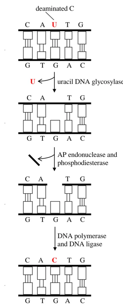
The BER pathway corrects small alterations in a DNA strand that do not distort the overall structure of the DNA helix, such as a base altered by deamination or a missing base due to a depurination reaction. The base alterations targeted by BER may or may not block transcription and normal replication, but they frequently lead to changes in DNA sequence, being therefore potentially mutagenic (Hoeijmakers, 2001). BER is the main pathway to repair oxidative damage. The BER pathway removes lesions affecting only one DNA strand, which permits the use of the information in the complementary strand to correct the lesion in the damaged strand.
The main steps of the BER pathway are shown, in a simplified form, in Figure 1.1 (adapted from (Alberts et al., 2002)), using as example the repair of DNA with a uracil
DNA polymerase and DNA ligase. At the end of this repair process, the U that was produced by an accidental deamination has been restored to a C.
deaminated C
C A U T G
G T G A C
U uracil DNA glycosylase
C A T G
G T G A C
AP endonuclease and phosphodiesterase
C A T G
G T G A C
DNA polymerase and DNA ligase
C A C T G
[image:37.595.239.448.156.676.2]G T G A C
The BER pathway can also be used to repair the result of a depurination event. In this case, since there is no need for a DNA glycosylase (the purine base was already removed by a spontaneous reaction), the BER pathway starts with the action of the AP endonuclease (Alberts et al., 2002). This is possible because the AP endonuclease recognizes any site that contains a deoxyribose sugar with a missing base, regardless of
which event produced that abasic site.
The BER pathway can be categorized into two sub-pathways, namely short-patch BER, where only one nucleotide is replaced (as illustrated in Figure 1.1); or long-patch BER, where 2-13 nucleotides are replaced (Gorbunova et al., 2007). The decision between performing a short-patch or long-patch repair is modulated by PARP-1 and PARP2 (poly(ADP-ribose) polymerases) (Beneke and Burkle, 2007), and the short-patch BER sub-pathway is used by cells in approximately 80-90% of the cases (Friedberg et al., 2006).
Some differences between the short-patch and long-patch pathway are as follows (Lee et al., 2005). First, in the short-patch pathway, DNA polymerase β (polβ) is the main gap-filling enzyme; whilst in the long-patch pathway this activity seems to be performed by polβ, pol δ, polε. In addition, in the long-patch pathway, the WRN protein (defective in
patients with Werner‟s syndrome) interacts physically and functionally with several other proteins such as PCNA and RPA, which is not the case in the short-patch pathway (Lee et al., 2005), (Rao, 2007).
(non-dividing) cells, and in principle other DNA repair pathways such as homologous recombination and mismatch repair (reviewed later) are not important in neurons.
There is good evidence that, overall, the level of BER activity is reduced with age. In particular, the activity of polβ – an important component of the BER pathway – has been
shown to be considerably reduced with age in mice in many investigations (Rao, 2007), (Cabelof et al., 2006), (Krishna et al., 2005), (Kaneko et al., 2002), (Cabelof et al., 2002). The activity of polγ – which performs the gap-filling step of BER in mitochondrial DNA
(Graziewicz et al., 2006) – has also been observed to decrease with age (Kaneko et al.,
2002). There are, however, studies reporting that some BER enzymes have an increased expression with age – see, for instance, (Lu et al., 2004). This seems likely to be a response to increased levels of oxidative DNA damage with age, although the response is presumably not effective due to the aforementioned decrease in polβ activity.
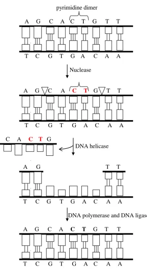
1.5.2 Nucleotide excision repair (NER)
The NER pathway copes with lesions in a DNA strand that distort the DNA double helix. This kind of lesion interferes with base pairing and usually blocks transcription and normal replication (Hoeijmakers, 2001). NER is considered the most versatile DNA repair pathway in terms of the variety of lesions that it can recognize – it recognizes several types of bulky lesions, produced, for instance, by ultraviolet light and carcinogens. Like the lesions targeted by BER, the lesions targeted by NER affect a single DNA strand, which allows the use of the information in the complementary strand to correct the lesion in the damaged strand.
illustrates a small gap in the strand for simplicity, but the actual gap in humans is considerably larger, more than 20 nucleotides (Alberts et al., 2002). Next, the gap is filled by DNA polymerase and DNA ligase.
pyrimidine dimer
A G C A C T G T T
T C G T G A C A A
Nuclease
A G C A C T G T T
T C G T G A C A A
C A C T G
DNA helicase
A G T T
T C G T G A C A A
DNA polymerase and DNA ligase
A G C A C T G T T
T C G T G A C A A
[image:40.595.186.480.168.704.2]The NER pathway is usually classified into two types, namely global genome NER (GG-NER), which occurs everywhere in the genome, and transcription-coupled NER (TC-NER), which occurs in the transcribed strand of active genes. The repair of DNA damage by TC-NER is faster than the repair by GG-NER (Moriwaki and Takahashi, 2008).
In GG-NER, the first step is the recognition of the DNA damage by the XPC-HR23B complex. In contrast, in TC-NER the repair process is believed to be triggered by a stalled RNA polymerase, and initiation of the repair requires the proteins CSB and CSA (whose mutations cause the Cockayone‟s progeroid syndrome) (Gorbunova et al., 2007),
(Hoeijmakers, 2001). After the initial stage, GG-NER and TC-NER seem to proceed in an identical way. The presence of damage is verified by XPA, and if damage is absent the repair process is aborted. The XPB (ERCC3) and XPD (ERCC2) helicases in complex with the TFIIH transcription factor open the DNA helix double helix around the damage.
RPA (Replication Protein A) stabilizes the open DNA by binding to the undamaged strand. The endonucleases XPF and XPG cleave the borders of the open segment in the damaged strand. The damaged segment is then removed, and the repair is completed by DNA polymerase and DNA ligase.
There has been many experiments investigating whether or not NER efficiency in repairing ultraviolet light-induced damage decreases with age, but the results of these experiments are sometimes conflicting (Best, 2009). For instance, NER efficiency was observed to decrease with age in (Yamada et al., 2006), (Hazane et al., 2006), (Takahashi et al., 2005), but observed not to decrease with age in (Merkle et al., 2004). It seems like these different results are due to the use of different experimental procedures and different types of damages being investigated.
in humans: Xeroderma Pigmentosum (XP), Cockayne Syndrome (CS), and Trichothiodystrophy (TTD).
These diseases were reviewed earlier, but a few additional remarks are relevant here, in the context of their association with defects in the NER pathway. The severity of the symptoms in XP varies significantly across the different types of XP – associated with defects in different complementation genes – and in general the more the mutation affects
the NER pathway, the more severe the symptoms are (Niedernhofer, 2008). Moreover, multiple mutations in NER genes have been observed to result in dramatically accelerated
aging phenotypes (Niedernhofer et al., 2006), (Pluijm et al., 2007), (Ven et al., 2006), (Gorbunova et al., 2007).
In addition, XPD-mediated NER has been observed to have a significantly role in maintaining the functional capacity of long-term reconstituting haematopoietic stem cells (LT-HSCs) with age, by helping to preserve the proliferative capacity and to prevent apoptosis under stress (Rossi et al., 2007).
It should also be noted that XP – which is associated with a dramatic increase in skin cancers – is mainly caused by a defect in GG-NER; whilst the progeroid syndromes CS and TTD – which show no evidence of increased risk cancer – are caused mainly by defects in TC-NER (Niedernhofer et al., 2006). This is because GG-NER is responsible mainly for repairing pre-mutagenic DNA lesions, preventing carcinogenesis; whilst TC-NER is responsible mainly for repairing DNA lesions that block transcription (Ljungman and Lane, 2004). Hence, in general defects in GG-NER (but not in TC-NER) tend to greatly increase predisposition to cancer.