Interleukin 6 (IL-6) is a multifunctional cytokine with important roles in the acute phase response,1,2
haematopoesis3and multiple aspects of immune
regu-lation.4IL-6 also has roles as an autocrine stimulator of
growth in a number of tumours (most notably plasma-cytomas and myelomas)5,6as well as for some normal
cell types; it is inducible by poly(I).poly(C),7and has a
number of functions in the endocrine and nervous sys-tems. It has been suggested that it acts as a long range alarm signal in the body. In line with these functions, it has previously been known as B-cell stimulatory factor 2 (BSF-2), interferon β2 (IFN-β2), hepatocyte stimula-tory factor (HSF)m 26 kD protein and hybridoma plas-macytoma growth factor (HPGF). Over-expression of IL-6 is known to be an important feature of the pathogenesis of a number of inflammatory diseases such as rheumatoid arthritis, glomerula nephritis and psoriasis, as well as the development of myeloma/ plasmacytoma development. It may also be important in the development of pathology in a number of infectious diseases such as mycobacterial disease and AIDS.
IL-6 in the human is a 184 amino acid protein which is processed from a 212 amino acid preprotein. It is vari-ably glycosylated and phosphorylated. The main
tran-script is 1.3 kb, although 2 further downstream polyadenylation sites have been mapped. Although the promoter regions of IL-6 genes are closely conserved between mouse and human, the structural sequences of the gene are less well conserved (42% at the amino acid level). Nonetheless, human IL-6 is able to support the growth of murine IL-6 dependent myeloma lines.
In order to study whether IL-6 mediates the patho-genesis of lentiviral disease in ruminants, we have cloned, sequenced and expressed in a yeast based sys-tem the structural regions of the ovine IL-6. Biological assays on the product show that there is a species bar-rier which prevents ovine IL-6 from supporting growth of murine IL-6 dependent cell lines, and similarly that human IL-6 is not recognized by ovine B-cells.
RESULTS
Cloning and sequence of ovine IL-6
To obtain the ovine IL-6 proprotein coding sequence, single stranded cDNA was made from total RNA of LPS stimulated alveolar macrophages and amplified using the polymerase chain reaction with primers derived from conserved regions of the 59and 39untranslated regions of IL-6 mRNA’s of human and mouse. The product of this reaction was cloned into pTZ18R and sequenced (Fig. 1B). The derived sequence contains an open reading frame of 624 bp, which encodes a protein of predicted Mr523 429. This protein is 53% identical to human IL-6 in sequence, and 43% identical to mouse IL-6. By analogy with the known maturation pattern of human IL-6 it is likely that the mature ovine protein is cleaved from the pro-pro-tein at the proline at position 29. A predicted N-linked
232 CYTOKINE, Vol. 7, No. 3 (April), 1995: pp 232–236
THE OVINE INTERLEUKIN 6 GENE
B. Ebrahimi, D. J. Roy, P. Bird, D. R. Sargan
Gene amplification by reverse transcriptase PCR with heterologous primers has been used to obtain a cDNA clone encoding the structural sequences of ovine interleukin 6 from alve-olar macrophages. This cDNA encodes a protein of Mr523 429, which is 53% homologous
in amino acid sequence to human IL 56. The clone hybridizes to an RNA of size 1260 nt in alveolar macrophages, expression of which is potentiated by LPS. The ovine IL-6 structural gene has been cloned into the yeast expression vector pOGS40, and used to produce a recom-binant protein. This protein is capable of causing increased immunoglobulin production in pokeweed mitogen stimulated ovine peripheral blood mononuclear cells at concentrations of 10–100 ng/ml, but it only causes very limited replication of B9 cells, a murine IL-6 dependent cell line. This is in contrast to recombinant human IL-6, which is capable of stimulating B9 cell proliferation, but not immunoglobulin production by ovine PBMC.
From the Department of Veterinary Pathology, University of Edinburgh, Edinburgh, UK
Correspondence to: D. R. Sargan, at present address, Department of Clinical Veterinary Medicine, University of Cambridge, Madingley Road, Cambridge CB3 0ES, UK
Received 18 May 1994; accepted for publication 28 October 1994 © 1995 Academic Press Limited
1043-4666/95/03023215 $08.00/0
glycosylation site at amino acid position 38 is conserved in the bovine sequence but not in other sequenced IL-6 proteins.
Expression of ovine IL-6 in yeast
The same CDNA population was used as a sub-strate to amplify an expression cassette containing this open reading frame. This cassette was cloned into pTZ18R, and subsequently into the galactose inducible yeast Ty expression vector pOGS40. After resequenc-ing, the resulting plasmid pOGIL6 was used to trans-form yeast strain BJ2168. After cloning, individual transformants were grown on in liquid culture, and lysed after growth in induction conditions. An inducible protein was produced by the transformed cells which
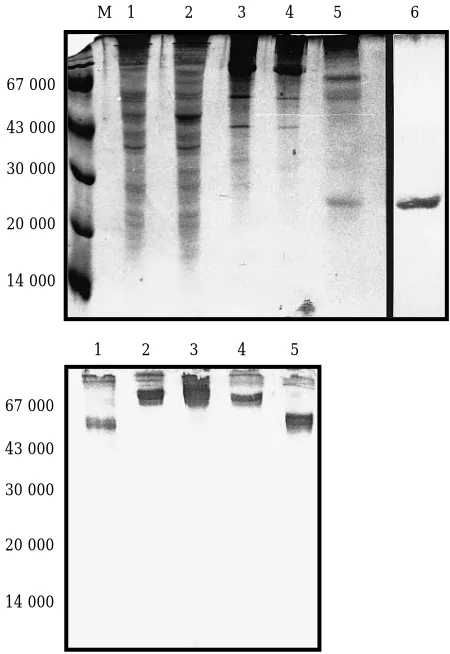
was of the predicted size for a TyP1-IL-6 fusion protein product, and blotted with an anti-P1 rabbit antiserum (Fig. 2). When purified by differential sucrose gradient centrifugation, this fusion protein could be digested with Factor Xa to yield a Mr524 000 protein,
puta-tively ovine IL-6, as well as a Mr550 000 band which
[image:2.595.314.539.62.389.2]corresponds to P1 protein present as vlps. Further cen-trifugation to remove remaining vlps allows the recov-ery of the released IL-6 with a yield of c100 µg/litre of culture, at a purity of about 90% as estimated by silver stain staining of PAGE gels.
Figure 1A. Primers used in cloning the ovine IL-6 cDNA. 054R and 056R were derived from regions of consensus between murine and human genes, whilst 053R contains IL-6 sequence derived from the bovine gene (B. Collins, personal communication) and shows two changes from the sequence of the ovine gene in Figure 1B. Non IL-6 sequences used in the expression construct are underlined.
Figure 1B. Sequence of the coding region of ovine IL-6 cDNA. The sequence of the PCR product of 054R and 056R was determined from both strands of three clones. The complete protein coding region is shown. Maturation of the protein leads to a probable amino-terminus at position 29 (underlined).
Figure 2. Expression of rovIL-6 protein in yeast.
(A) Proteins from various stages of IL-6 purification were analysed by electrophoresis on SDS-15% polyacrylamide gels, followed by staining with Coomassie blue R. Lane M: marker proteins. Lane 1: total proteins from a lysed control yeast expressing the TyP1 protein from the plasmid pOGS41 after 16 h induction with galactose. Lane 2: total proteins from a yeast strain transfected with pOGIL-6 after 16 h induction with galactose. Lane 3: partially purified virus-like particles of P1/IL-6 fusion protein from the 60% sucrose cushion after the first centrifugation step. Lane 4: virus-like particles of P1/IL-6 fusion protein with factor Xa added, prior to digestion. Lane 5: virus like particles after factor Xa cleavage. Lane 6: purified rov-IL-6 after removal of vlp’s by centrifugation. This lane is a silver stained preparation, from a different gel. (B) A western blot of a similar gel (lanes 1–5) was probed with a polyclonal antiserum raised against P1 protein.
M 1 2 3 4 5 6
1 2 3 4 5 67 000
43 000
30 000
20 000
14 000
67 000
43 000
30 000
20 000
[image:2.595.50.288.62.146.2]Biological activity of rov IL-6
The purified material was used in two assays of IL-6 activity. B9 cells offer a highly sensitive prolifera-tion assay for human and murine IL-6, although these cells will also proliferate in the presence of murine (but not human) IL-4. Proliferation assays were performed using these cells and the yeast expressed rovIL-6 (Fig. 3). This material was able to support proliferation of these cells, but only relatively poorly, and at concentrations of 1–10 ng/ml and above, some four orders of magnitude higher than the concentrations at which rhuIL-6 is active. (In the assay shown, rhuIL-6 is already super-optimal for stimulation at 20 pg/ml. In other assays, optimal stimulation by rhuIL-6 was achieved in the range 0.1–1 pg/ml, in good agreement with the findings of other authors for this cell line.8) Mixing rovIL-6 with rhuIL-6
did not affect proliferation to rhuIL-6, showing that there was no toxicity from rovIL-6 (data not shown). P1 pro-tein alone was unable to cause proliferation of B9 cells at any concentration measured.
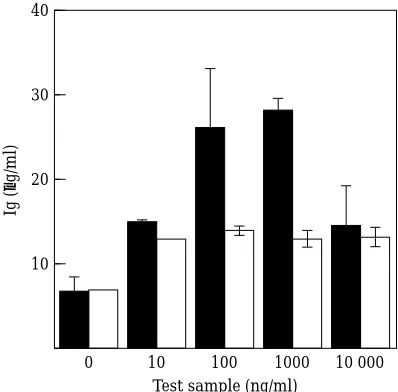
To measure the activity of rovIL-6 on ovine cells, IL-6 was used to drive immunoglobulin production by peripheral blood mononuclear cells9(Fig. 4). RovIL-6
at concentrations above 10 ng/ml was able to potenti-ate total immunoglobulin production by these cells in the presence of pokeweed mitogen (measured in an ELISA assay) by anything from 2–8 fold in different sheep. (Production of Ig in the absence of IL-6 varied between 2 and 7 µg/ml.) These concentrations are com-parable to those required to cause similar enhance-ments of Ig production by rhuIL-6 acting on human EBV transformed B-cells.16The same ovine cell
popu-lations were much less responsive to a control protein preparation consisting of the equivalent purification fractions from a factor Xa digested P1 expressing yeast. Nor were they sensitive to rhuIL-6 at these
concentra-tions (data not shown). This suggests that there is a par-tial barrier in IL-6 recognition between sheep IL-6 and mouse IL-6 receptors and between human IL-6 and sheep IL-6 receptors which does not exist between human IL-6 and mouse IL-6 receptors.
DISCUSSION
Ovine recombinant IL-6 has been cloned and expressed in a yeast based system. The sequence is iden-tical to that reported very recently by Andrews et al.,10
although these authors did not express protein from their gene. On Northern blotting the clone described in the current study hybridizes to a rare transcript in sheep alveolar macrophages of 1.26 kb. RT PCR experiments showed that this manuscript was induced in the macrophages by LPS stimulation in vitro. Induction of the transcript occurs by 1 h post treatment, and con-tinues for at least 3 h (data not shown).
The protein expressed in the current study is a pro-protein, but it retains functional activity on ovine cells. However, there is a partial species barrier between ovine IL-6 and murine cells which is not present between human IL-6 and murine cells, preventing extensive proliferation of the B9 myeloma line in response to the ovine molecule. Similarly, human IL-6 is not able to potentiate immunoglobulin synthesis by ovine peripheral blood mononuclear cells. This type of species barrier has been noted before in other species.
100 000 120 000
0 1
IL-6 (pg/ml)
cpm
100 000
80 000
60 000
40 000
20 000
[image:3.595.323.522.63.259.2]10 100 1000 10 000
Figure 3. Proliferative response of B9 cells to recombinant IL-6. Medium control (d), rhuIL-6 (h) or rovIL-6 (j) was used to stimu-late proliferation of the murine myeloma cell line B9 for 36 h. 3 H-Thymidine incorporation was measured over the next 5 h. Means and ranges of incorporated counts are shown. Where no error bars are shown, they are within the diameter of the symbol.
40
0
Test sample (ng/ml)
Ig (
µ
g/ml)
10 30
20
10
[image:3.595.52.283.536.666.2]100 1000 10 000
Figure 4. Immunoglobulin production by peripheral blood lym-phocytes in response to IL-6.
Although human IL-6 will bind to the murine IL-6 receptor and activate murine IL-6 dependent cell lines, murine IL-6 is unable to activate human cells in the same way.11
IL-6 sequences are not extensively conserved between species, but four cysteine residues forming disulphide bridges at positions 72–78 and 101–111 are conserved across the ovine protein as well as those of other species.12(These cysteines define a structural
fam-ily of cytokines including IL-2, IL-4, GM-CSF, G-CSF, to which LIF and erythropoietin are more distantly related.) The most closely conserved regions across species, within the IL-6 gene, are within the leader sequence and also between residues 68 and 126. These residues also form the best conserved region between the human and murine proteins and this region consti-tutes parts of two alpha helices, two turns defined by the cysteine bridges and a long loop, at least part of which is required for interaction between IL-6 and the IL-6 binding subunit of IL-6R.19
Most studies of IL-6 structure/activity relation-ships have used site directed mutagenesis of human IL-6 and have concentrated on the important role of the carboxyl end of the protein in receptor binding.13–16
Residues 176–183 of the human mature protein and two sets of periodically arranged leucines (positions 167, 174 and 181; and positions 151, 158, 165 of the human sequence) have both been suggested to be important in this regard.22 Of these leucines in
the human sequence, only those at positions 167 and 174 have equivalents in the ovine sequence (positions 191 and 198 of the sequence in Fig. 2). That at position 181 (human) is not present in most other IL-6 sequences, and can be substituted with several other residues without effect.23However, the absence of the
other spaced set of leucines may be important in the observed species barrier.
MATERIALS AND METHODS
Cloning and sequencing of ovine IL-6
Alveolar macrophages were obtained from gnotobiotic
Finn 3Blackface sheep by alveolar lavage and red blood cells
removed by hypoosmotic shock as before.172 3107cells were
plated into plastic tissue culture bottles in Iscove’s serum free
medium and allowed to adhere for 24 h. Adherent cells (.90%
macrophages by non-specific esterase staining) were then
stim-ulated with 10 µg/ml LPS from Salmonella abortus equi for 4 h.
Total RNA was purified by the acidified guanidine
thio-cyanate method18and used as a template for single stranded
cDNA synthesis using oligo dT primers and AMV reverse transcriptase. Primers 054R and 056R for amplification of the ovine IL-6 gene were selected from well conserved regions of
the 59and 39untranslated regions of the IL-6 gene in human
and mouse (Fig. 1A). The cDNA was amplified through 35 cycles using these primers and the buffer conditions of Ohara
and Gilbert.19The product of PCR was purified by agarose
gel electrophoresis and electroelution. This product was lig-ated directly into the phosphoryllig-ated cloning site of the plas-mid pCR2 according to the manufacturer’s instructions (Invitrogen, Ca., USA). Clones containing inserts were sequenced on both strands using the di-deoxy nucleotide chain termination method.
Construction of a ruminant IL-6 cassette
A primer was designed containing the 5922 bases of a
ruminant IL-6 sequence (derived from an unpublished bovine IL-6 sequence, B Collins, personal communication) attached to a coagulation Factor Xa recognition site, a BamHI site and a GC clamp (Fig. 1A). This primer (053R) was used in com-bination with a second primer (159R) containing a GC clamp and BamHI site attached to the antisense of the last 22 bases of the ovine IL-6 structural sequence (up to and including the stop codon) in a PCR using the same cDNA population as before. The resulting product, after gel purification, was digested with BamHI and cloned into the BamHI site of pTZ18R (Pharmacia). A clone containing an insert of the expected size was sequenced, and the insert cleaved out with BamHI and purified by agarose gel electrophoresis. The insert was ligated into the BamHI site of the yeast Ty expression plasmid pOGS40. Plasmids containing the correct size of insert were sequenced across the junctions between insert and vector to determine the direction of cloning and make sure that no frame shifts had occurred during cloning. A plasmid pOGIL6 was selected for further work.
Expression and purification of IL-6 from yeast
The method used was essentially that of Gilmour et al.20
with modifications as published previously.21Briefly, pOGIL6
was transfected into yeast BJ2168 by spheroplasting, plating in regeneration agar (0.67% yeast nitrogen base; 1M sorbitol; 1% glucose; 3.0% Difco agar) and incubating at 30°C. Colonies obtained, after re-streaking, were grown in liquid culture. For inducible expression they were grown at 30°C for 48 h in YNB (0.67% yeast nitrogen base) containing 1% glu-cose and 0.002% tryptophan, then induced for 16–24 h with
1% galactose 10.3% glucose and and tryptophan. Cells were
harvested, suspended in 10 mM Tris-HCl pH 7.4, 2 mM EDTA, 140 mM NaCl containing protease inhibitors and lysed by vortexing with acid washed glass beads. Supernatants
were spun at 10 000 3g for 5 mins, the yeast debris discarded,
and the vlp containing suspension partially purified and
con-centrated by spinning at 30 000 3g for 90 min onto a 60%
sucrose cushion. The interface and cushion material were
dial-ysed into 100 mM Tris/HCl pH 7.4, 10 mM CaCl2containing
0.1% DOC (Na deoxycholate). Expressed protein was released from the vlps by digestion with Factor Xa at a con-centration of 1:50 w/w for 4 h at 25°C and further purified by
centrifugation at 40 000 3g for 40 min to remove the P1, vlps
present post cleavage as well as any remaining yeast proteins.
Samples were dialysed into PBS and stored at 270°C.
Analysis of ovine IL-6 RNA and protein
Alveolar macrophages were plated in Iscoves medium
RNA extracted using RNAzol B (Cinna Biotix) was analysed by Northern blotting followed standard methods. For RT
PCR, cDNA was prepared from 1 µg total RNA, using the
method of Rolfs et al.22Primers 053R and 159R were then
used in a 40 cycle PCR (otherwise as before) with 1/10 of the cDNA sample. SDS polyacrylamide gel electrophoresis and
Western blots were as before.12
Cell proliferation assay
B9 cells, a murine IL-6-dependent hybridoma cell line, were used in proliferation assays to measure the biological activity of recombinant ovine IL-6 (rovIL-6). Proliferation
was measured according to the method of Aarden et al., 1987.23
4 3103B9 cells were incubated in duplicate wells with test
sample in RPMI1640, 10% foetal calf serum 100 units ml21
penicillin, 100 µg ml21streptomycin, in 200 µl total volume
for 3 days at 37°C. They were then pulsed with 1µCi
3H-thymidine for 5 h, prior to harvesting, washing and
scin-tillation counting.
Total immunoglobulin production assay
Washed peripheral blood mononuclear cells
(13105) were incubated in 96-well flat-bottomed ELISA
plates in 200 µl RPMI1640 containing 2 mM glutamine, 5 3
1025M 2-mercaptoethanol, 200 µg/ml gentamycin, 10% fetal
calf serum, supplemented with pokeweed mitogen (Gibco product 061-05360B, used at 1 in 10 000) and test samples as stated in figure legends. Assays were performed in triplicate. After 7 days in culture the supernatants were assayed in a cap-ture ELISA for total immunoglobulin. Total Ig was measured using a mouse anti-sheep Ig light chain monoclonal antibody,
VPM8,28to coat ELISA plates as capture antibody, and a
puri-fied rabbit anti-sheep Ig antibody directly conjugated to horse-radish peroxidase to develop (P. Bird, in preparation).
Acknowledgements
We thank Mr B. Collins for giving us access to his results prior to publication, Dr S. Pooles for rhuIL-6, Dr L. Aarden for the B9 cell line and Ms D. Allen for technical assistance. The work was supported by AFRC project grant no. AG15/602 and Wellcome Trust Programme grant no. 035157.
REFERENCES
1. Ritchie DG, Fuller GM (1983) Hepatocyte-stimulating fac-tor: a monocyte derived acute phase regulatory protein. Ann NY Acad Sci 408:490–502.
2. Gauldie J, Richards C, Harnish D, Lansdorp P, Naumann H (1987) Interferon β2 / B-cell stimulating factor type 2 shares identity with monocyte derived hepatocyte-stimulating factor and regulates the major acute phase protein response in liver cells. Proc Natl Acad Sci USA 84:7251–7255.
3. Ikebuchi K, Wong GG, Clark SC. Ihle JN, Hirai Y, Ogawa M (1987) Interleukin-6 enchancement of interleukin-3 -dependent proliferation of multipotential hemopoietic progenitors. Proc Natl Acad Sci USA 87:9035–9039.
4. Reviewed in Van Snick T (1990) Interleukin-6. An overview. Ann Rev Immunol 8:253–278
5. Nordan RP, Potter M (1986) A macrophage-derived factor required by plasmacytomas for survival and proliferation in vitro. Science 233:566–569.
6. Van Damme J, Opdenakker G, Simpson RJ, Rubira MR, Cayphas S, Vink A, Billiau A, Van Snick J (1987) Identification of the human 26kD protein interferon β2 (IFNβ2), as a B cell hybridoma/plasmacytoma growth factor induced by interleukin 1 and tumour necrosis factor. J Exp Med 165:914–919.
7. Weissenbach J, Chernakovsky Y, Zeevi M, Shulman L, Soreq H, Nir U, Wallach D, Perricaude M, Tiollais P, Revel M (1980) Two inter-feron mRNA’s in human fibroblasts: in vitro translation and Escherichia coli cloning studies. Proc Natl Acad Sci USA 77:7152–7156.
8. Helle M, Boeji L, Aarden LA (1988) Functional discrimi-nation between interleukin 6 and interleukin 1. Eur J Immunol 18: 1535–1540.
9. Muraguchi A, Hirano T, Tang B, Matsuda T, Horii Y, Nakajimi K, Kishimoto T (1988) The essential role of B cell stimula-tory factor 2 (BSF-2/IL-6) for the terminal differentiation of B cells. J Exp Med 167:332–344.
10. Andrews AE, Barcham GJ, Ahman K, Meeusen ENT, Brandon MR, Nash AD (1993) Molecular cloning and characteris-ation of ruminant interleukin-6 cDNA. Immunol and Cell Biol (1993) 71:341–348.
11. Fiorillo MT, Cabibbo A, Iacopetti P, Fattori E, Ciliberto G (1992) Analysis of human-mouse interleukin-6 hybrid proteins —both amino and carboxy termini of human interleukin-6 are .required for in vitro receptor binding. Eur J Immunol 22:2609–2615. 12. Simpson RJ, Moritz RL, Van Roost E, Van Snick J (1988) Characterisation of a recombinant murine IL-6: assignment of disul-phide bonds. Biochem Biophys Res Comm 157:364–72.
13. Lutticken C, Kruttgen A, Moller C, Heinrich PC, Rose-John S (1991) Evidence for the importance of a positive charge and an alpha-helical structure of the C-terminus for biological-activity of human-IL-6. FEBS Lett. 282:265–267.
14. Leebeck FW, Kariya K, Schwabe M, Fowlkes DM (1992) Identification of a receptor binding site in the carboxyl terminus of human interleukin-6. J Biol Chem 267:14382–14388.
15. Nishimura C, Ekida T, Nomura K, Sakamoto K, Suzuki H, Yasukawa K, Kishimoto T, Arata Y (1992) Role of leucine residues in the C-terminal region of human interleukin-6 in biological activity. FEBS Letters 311:271–275.
16. Savino R, Lahm A, Giorgio M, Cabibbo A, Tramontano A, Ciliberto G (1993) Saturation mutagenesis of human interleukin 6 receptor binding site: implications for its three dimensional structure. Proc Natl Acad Sci USA 90:4067–4071.
17. Green IR, Sargan DR (1991) Sequence of the cDNA encod-ing the ovine tumour necrosis factor-A: problems with clonencod-ing by inverse PCR. Gene 109:203–210.
18. Chomczynski P, Sacchi N (1987) Single step method of RNA isolation by acid guanidinium thiocyanate-phenol-chloroform extraction. Analytical Biochem 162:156–159.
19. Ohara O, Dorit RL, Gilbert W (1989) One sided polymerase chain reaction: the amplification of cDNA. Proc Natl Acad Sci USA 86:5673–5677.
20. Gilmour JEM, Senior JM, Burns NR, Esnouf MP, Gull K, Kingsman SM, Kingsman AJ, Adams SE (1989) A novel method for the publication of HIV-1 p24 protein from hybrid Ty virus-like-particles (Ty-VLP). AIDS 3:717–723.
21. Fiskerstrand CE, Roy DJ, Green IR, Sargan DR (1992) Cloning, Expression and characterisation of ovine interleukins 1α and β. Cytokine 4:418–428.
22. Rolfs A, Schuller I, Finckh U, Weber-Rolfs I (1992) PCR: Clinical Diagnosis and Research. Springer Verlag, Heidelberg.
23. Aarden LA, De Groot ER, Schaap OL, Lansdorp PM (1987) Production of hybridoma growth factor by human monocytes. Eur J Immunol 17:1411–1416.