
Minor groove binders: some recent research in drug development
11
0
0
Full text
(2) 54. Abedawn I. Khalaf. furan, oxazole and thiophene. On the other hand, some of the amide linkages were also modified; these can be replaced with an olefinic double bond or diazo groups.. and Table 1. Two compounds that were prepared and studied extensively alongside distamycin 1 and netropsin 2 are Thiazotropsin A 3 and Thiazotropsin B 4.. In 2007, Anthony et al. [10] reported the synthesis of a large number of lexitropsins with a variety of modifications to the head groups, tail groups and the heterocyclic moieties. In this review some of these modifications and the synthetic strategy for these syntheses have been reported: Scheme 1. These polyamides were synthesised using a variety of coupling methods such as HBTU, dicyclohexylcarbodiimide DCC, or acid chloride coupling reactions. The amidines were prepared from the methylation of thioamides and finally the displacement of methane thiol by an aminopyrrole. H O. H. NH NH. NH2. O. HN. H N. N. N NH. HN O. N N. O. Thiazotropsin A (3). Distamycin (1). N. NH. NH. O. H 2N. NH. HN NH. N. H N. HN. H N. N. NH2. N. O O. Thiazotropsin B (4). Netropsin (2). TG. i ArCOX. S. N. O. N. O. HN. HN. N. ArCO2 H. O. S. N. O. N. HN. O. O. O. HN. H N. N. O. +. N O. HN N. HN. NH. ii. O H N. TG. Ar. HN. O2 N. O. O. N. H N. N. O. HN. O. O. N. TG RAr. iii S. SMe +H. H2N. 2N. TG. HN. O2 N. RAr. H N. N O. HN N. RAr. ii. O. NH. N. HN. O. H N. O PO(OEt)2. +. CO 2Et. HN. O2 N. iv, v N. O. N. TG. Ar. H. N. HN. N O. Ar. 6. HN O. +. 5. N. N. CO 2H. HN TG. H N. + N O. N. ii. Ar. 7 O. O. H N. HN. N O. N. N. O. Scheme 1. Synthesis of minor groove binders [5, 6, 7]. Ar and R represent head groups; TG: tail group; N-methylpyrrole have been shown throughout as the linked components. Reagents: i: coupling agent: HBTU, dicyclohexylcarbodiimide DCC, or thionyl chloride; ii: H2/Pd-C then immediate reaction with the coupling partner after filtration of the catalyst; iii: MeI; iv: NaH; v: NaOH(aq)..
(3) Minor groove binders: Recent research in drug development. 55. Table 1. Activities of polyamides [8-13] against representative bacterial species. No. 8. Compound. Staphylococcus aureus 5. H O. NH. O N. H N. N. N. N H. HN S. O. 9. Staphylococcus faecalis 10. N. O. Cl O. Cl. 6. 50. 13. 6. 13. 6. 13. 25. 13. 13. N N H. N NH O. N. HN. H N N. O. N. O. 10. N O NH Cl. H N. N. HN H N. O. O N. N. O. 11. N O NH N Cl. H N. HN H N. O. S O. N. N. O. 12. N O. O. NH N. O H N. HN H N. O. S O. N. N. O. 13. HN NH2 O NH MeO. H N O. N. HN N. H N. S. O N. O. All data are minimium inhibitory concentrations, given as µg mL-1. Organisms used: Staphlococcus aureus NCTC 6571. Streptococcus faecalis NCTC 775..
(4) 56. Abedawn I. Khalaf. oligomer. The other class of compounds contained stilbenoid head groups. These were prepared either from the reaction of arylmethylphosphonates with aromatic aldehydes in the presence of sodium hydride [Horner-Wadsworth-Emmons conditions] or in the case of the pyridyl derivatives which was bearing an active methyl group; a condensation reaction was used in the presence of zinc chloride, as a catalyst, in acetic anhydride. The esters formed were hydrolysed to give the corresponding carboxylic acids, which were coupled to the amino pyrrole derivatives: Scheme 1.. in the thermal denaturation measurements. The authors developed a new set of compounds containing diaromatic monoguanidinium and mono-2-aminoimidazolinium derivatives and a series of diaromatic mono- and bis-isouroniums as well as a series of asymmetric diaromatic guanidinium/ 2-aminoimidazolinium dications Table 2: compounds 14-21. All the compounds studied by the authors contained a furamidine scaffold which had been modified by changing both the terminal cationic arms and the bridging furan part of the molecule. It was proposed that the cationic groups could bind to DNA in two ways: first through direct hydrogen bonds through the NH groups and second through water-mediated interactions with the bases. In this work the authors exchanged the amidinium cations with guanidinium, 2-aminoimidazolinium, and isouronium cations. In these cases they aimed to enhance hydrogen bond contacts in the minor groove by increasing the number of hydrogen bond donors (NH groups in the guanidinium and 2-aminoimidazolinium cations) or introducing an additional hydrogen bond acceptor (an O atom in the isouronium cation). Also, it was hoped that the optimum linker would allow for a better orientation of the positive charges within the groove and introduce the curvature or. 2. Sleeping sickness. Nagle et al. [11a] prepared and studied a large number of symmetric diaromatic bis-guanidinium and bis-2-aminoimidazolinium derivatives which are structurally related to furamidine and have the potential to treat sleeping sickness. Sleeping sickness is a chronic disease caused by Trypanosoma parasites, which affect the human central nervous system. Jackson et al. [11b] have generated a high-quality draft genome sequence for the strain of Trypanosoma brucei that is responsible for almost all reported cases of human African trypanosomiasis. Nagle’s compounds showed good affinity for DNA as noticed by an increase of the melting temperature (∆Tm) values observed. Table 2. Results obtained from the thermal melting experiments showing the change in melting temperature in the presence of the monoamidine-like cations [14-21] to Salmon Sperm DNA (∆Tm(SS), oC); melting temperature in phosphate buffer (10 mM) is 69oC. X. X H2N NH2 Y=. H2N. Y. N H. NH 2. No.. X. 14 15 16 17 18 19 20 21. CH2 CH2CH2 O S NH Piperazine CO NHCONH. Y NH2. HN Y=. N H. N H. (∆Tm(SS), oC) 3 -. Y=. NH 2. O. (∆Tm(SS), oC) 0 2 2 2 2 0 2. 3 -. (∆Tm(SS), oC) 0 0 0 2 1 4. 2 4 2 -.
(5) Minor groove binders: Recent research in drug development geometry best suited to fitting in the groove. However, Boykin et al. have shown that linear molecules are also capable of strongly binding to the minor-groove [12]. Therefore the curvature is not essential to fit into the minor groove. This is also supported by the work carried out by Nguyen et al. [13]. This showed that in the linear molecule CPG- 40215A, the N,N’-diaminoguanidine bridging group served as a “see-saw” hinge facilitating a variety of interactions with the bases through motions that position the terminal amidines cations in a range of binding modes [13]: Figures 1 and 2. Moreover, to investigate the optimal linking scaffold, we have exchanged the furan moiety of furamidine for a number of smaller functional groups (O, S, NH, CO, CH2, CH2CH2, or NHCONH). It was assumed that such a substitution should afford a 2-fold advantage by facilitating the interaction with DNA through Hydrogen bonds (as donors or acceptors), and as the cations are larger than in the furamidine series, making this central group smaller should ensure. optimization of the distance between the cationic moieties (guanidinium and 2-aminoimidazolinium) for DNA binding. In 2009 Vooturi et al. [14] focused their work on the role played by the three-dimensional structure of DNA on gene expression [15]. They concentrated on the development of minor groove DNA binding agents with the potential to “bend DNA”. The design hypothesis of these agents consisted of connecting the sequence recognition properties of N-methylpyrroles to sterically bulky, hydrophobic groups that could force open the minor groove of DNA, resulting in a bend. As part of this project, approximately a dozen compounds were prepared, and on the basis of previous reports of antimicrobial activity of DNA binding agents, they examined these compounds for antibacterial activity against both Gram-positive and Gramnegative bacteria. Most of the agents displayed no antibacterial activity up to a concentration of 32 mg/L. However, one compound, 22, gave MIC. H2N. NH 2. H2 N. X. NH 2. NH 2. O. H 2N. 57. N H. N H X. NH N H. NH 2 NH2. HN. N H. N H. N H. X=O, S, NH, CH 2, CO, SO2, CH2 CH 2, NHCONH, NHCSNH, NHCO, piperidine, piperazine. Figure 1. Structure of furamidine (left) and general structure of the related compounds previously reported by Rozas’ group [27] that have shown good affinity toward DNA.. X. HN. H2 N. NH. N H. n=2, nil Family I. NH 2 H 2N. X. (CH 2) n R. NH2. O NH 2 R=-NH2 , -O-C(NH 2) 2+ Family II. X. N H. HN N H. N H. Family III. Figure 2. Structures of three families studied..
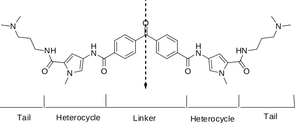
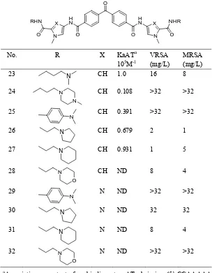
(6) 58. Abedawn I. Khalaf. values in the 8-16 mg/L range against VRSA, MRSA, and GISA. No activity was observed against Gram-negative bacteria. This result is similar to published examples of DNA binding antibiotic (DBA) [16-25]. Although 22 did display antibacterial activity, the MIC values were too high to be useful, thus they decided to prepare a series of analogues of the lead agent in an effort to identify more potent molecules and to elucidate some of the key structural features of this molecule that were necessary for activity.. imidazole was used. The last part was the linker. As well the benzophenone, the authors reduced this moiety to the corresponding alcohol. Also, they used an aliphatic linker of approximately the same length as the benzophenone linker: Table 4, compounds 33-38. 3. Studies of DNA displacement in bacteria. Several extensive studies showed that there was no correlation between the DNA binding affinity and antibacterial activity. This was explained on the basis of the differences in the transport of these compounds into the bacterium and this consequently will lead to a lack of activity [10, 14, 18, 22].. The authors [14] designed their compounds on the basis of the tail region, which is the portion of the molecule that contains cationic groups Figure 3. It is known that the positive charge of the tail should interact with the negative charge on the phosphate backbone of DNA. The authors were also interested in the hydrophobic character of this region, since the tail also interacts with the hydrophobic part of the sugar residue located in the minor groove of DNA. It has been reported before that enhancing the hydrophobicity of the cationic tail could increase the binding affinity of the molecule to DNA and help the transport of the drug [16-20, 25]. The authors chose the following tail groups for the synthesis of their compounds: methylpiperazine, dimethylaniline, morpholine, piperidine, and pyrolidine: Table 3, compounds: 23-32. The second part of the molecule they investigated was the heterocyclic region. It was clear from the literature that the heterocyclic moiety can modify the antibacterial activity of the molecule [17, 19, 22, 26]. N-methylimidazole was chosen based on the enhanced activity when. Vooturi et al. [14] examined the binding ability of their own compounds to DNA in vivo using a Hoescht dye competition assay. The result of the incubation of Hoescht dye with bacteria was the formation of fluorescent bacteria. Individual compounds were then added into a solution containing the fluorescent bacteria and the changes in the fluorescence due to Hoechst displacement were measured. 4. Antibacterial against vancomycin resistant Enteroccoci and methicillin resistant Staphylococcus aureus. Dyatkina et al. [24] reported the synthesis of a selected group of novel, pyrrole tetraamides, their DNA binding properties, and antibacterial activities. Substantial potency against vancomycin resistant Enterococci (VRE) and methicillin resistant Staphylococcus aureus (MRSA) was. O. N H N. NH O. Tail. N. N. Heterocycle. H N O. O. Linker. HN N. Heterocycle. O. Tail. Figure 3. Conceptual breakdown of potent compound 22. The arrow indicates the axis of symmetry for the molecule..
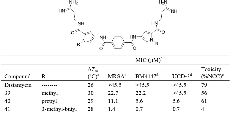
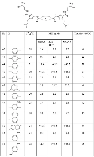
(7) Minor groove binders: Recent research in drug development. 59. Table 3. DNA binding affinity and in vitro antimicrobial activity of 23-32. O. No.. RHN. X. O. N. H N O. R N N. X. NHR. N. O. O. CH. KaATa 105M-1 1.0. VRSA (mg/L) 16. MRSA (mg/L) 8. CH. 0.108. >32. >32. CH. 0.391. >32. >32. X. 23 24. H N. N. 25. N. 26. N. CH. 0.679. 2. 1. 27. N. CH. 0.931. 1. 5. 28. N. CH. ND. 8. 4. N. ND. >32. >32. O. 29. N. 30. N. N. ND. 32. 32. 31. N. N. ND. 8. 4. 32. N. N. ND. >32. >32. O a. Association constant for binding to AT hairpin (5’-CGAAAAACAAAAAGTTTTTCG-3’) as determined by the ethidium bromide displacement assay.. observed. The authors found that strong DNA binding properties are necessary for antibacterial activity and that lipophilicity was a strong modulator of activity. They also optimised their library of compounds to reduce cytotoxicity toward human T-cells. Two groups of linked tetraamides shown in Tables 5 and 6 [24] were synthesized. Compounds 39-41: Table 5, which have linked pyrroles alkylated with methyl, propyl, and isoamyl groups, respectively, represent the first group. The second group (Table 6), compounds 42-53: Table 6, presents N-isoamylpyrrole units linked with different dicarboxylic acids.. This study showed that these short poly pyrroles with head-to-head geometry bind strongly to the minor groove of AT tracts of DNA. The comparison of the molecular modelling of these compounds to distamycin revealed improved hydrophobic interactions as well as improved hydrogen bonding to the floor of the groove. It appears that good DNA binding properties are a requirement for good antibacterial activity. The obvious conclusion from their studies is that lipophilic, alkyl substituents on the nitrogen atom of the pyrrole produced strong antibacterial activity as shown by submicromolar MIC values.
(8) 60. Abedawn I. Khalaf Table 4. DNA binding affinity and in vitro antimicrobial activity for 33-38. H N. RHN O. H N. X O. O. N. NHR O. N. KaATa 106M-1. VRSA (mg/L). MRSA (mg/L). N. 0.175. >32. >32. N. 0.0098. >32. >32. 35. N. 0.154. >32. >32. 36. N. 0.0238. >32. >32. No. X. 33. OH. 34. OH. R. 37. (CH2 )11. N. 0.021. >32. >32. 38. (CH2 )11. N. 0.0125. >32. >32. a. Association constant for binding to AT hairpin (5’-CGAAAAACAAAAAGTTTTTCG-3’) as determined by the ethidium bromide displacement assay.. Table 5. In vitro antimicrobial activity, cell toxicity, and DNA binding of terephthalamides. NH2. H2 N. HN. NH HN. NH. O. O R. HN. NH. N. N. R. O. O. MIC (µM)b Compound Distamycin 39 40 41 a. R -------methyl propyl 3-methyl-butyl. ∆Tm (oC)a 26 30 29 28. MRSAc >45.5 22.7 11.1 1.4. BM4147d >45.5 22.2 5.6 0.7. UCD-3d >45.5 >45.5 5.6 0.7. Toxicity (%NCC)e 79 56 61 4. ∆Tm, changes in the DNA melting temperature (∆Tm) of an AT-rich dodecamer, (CGATTATTAAGC).(GCTTAATAATCG), upon binding of an equimolar amount of compound. b MIC, minimal inhibitory concentration. cMRSA, methicillin resistant Staphylococcus aureus. d BM4147 and UCD3, two strains of VRE. eToxicity is measured as a fraction of growth as compared to no compound control (percent)..
(9) Minor groove binders: Recent research in drug development. 61. Table 6. In vitro antimicrobial activity, cell toxicity, and DNA binding for N-(3-methyl-butyl)-1H-pyrrole dimers with different linkers. NH 2. NH 2. HN. NH NH. HN. O. H N. NH N. X O. No. O. ∆Tm(oC). X. O N. MIC (µM) MRSA. BM 4147. Toxicity %NCC UCD-3. 42. 28. 1.4. 0.7. 0.7. 6. 43. 26. 0.7. 1.4. 1.4. 23. 44. 11. 11.4. >45.5. >45.5. 88. 45. 10. >45.5. >45.5. >45.5. 87. 46. 25. 1.4. 0.7. 1.4. 3. N. 47. N. 11. 2.8. 22.7. 22.7. 6. 48. N. 20. 2.8. 2.8. 2.8. 32. 25. 1.4. 1.4. 1.4. 42. 50. 20. 2.8. 2.8. 5.7. 15. 51. 24. >45.5. >45.5. >45.5. 0. N. 49. NO 2. 52. Me. 24. 0.7. 1.4. 1.4. 38. 53. Me. 12. 11.4. >45.5. >45.5. 75. Me.
(10) 62. Abedawn I. Khalaf. against both MRSA and VRE. From the above data, it appears that the lipophilicity of the compounds may be related to improved cellular uptake, which modulates antibacterial activity for those compounds that exhibit sufficient strong DNA binding. As uptake would also increase for cells other than the bacterial targets, significant toxicity would be expected. Strong toxicity against human T-cells was indeed observed for some compounds, but this toxicity did not correlate with antibacterial activity or DNA binding. Also, it was found that the degree of toxicity is very sensitive to the alkyl substituents on the pyrrole units.. 9. 10.. 11.. REFERENCES 1.. 2.. 3.. 4.. 5.. 6.. 7.. 8.. Khalaf, A. I., Pitt, A. R., Suckling, C. J., Scobie, M., Urwin, J., Waigh, R. D. Fishleigh, R. V., Young, S. C., and Fox, K. R. 2000, J. Chem. Research (S), 264-265. Khalaf, A. I., Pitt, A. R., Suckling, C. J., Scobie, M., Urwin, J., Waigh, R. D. Fishleigh, R. V., Young, S. C., and Fox, K. R. 2000, J. Chem. Research (M), 751-770. Fishleigh, R. V., Fox, K. R., Khalaf, A. I., Pitt, A. R., Scobie, M., Suckling, C. J., Urwin, J., Waigh, R. D., and Young, S. C. 2000, J. Med. Chem., 43 (17), 3257-3266. Khalaf, A. I., Pitt, A. R., Suckling, C. J., Scobie, M., Urwin, J., Waigh, R. D., Fishleigh, R. V., Young, S. C., and Fox, K. R. 2000, Tetrahedron, 56, 5225-5239. Suckling, C. J., Waigh, R. D., Khalaf, A. I., Parkinson, J. A., and Hunter, I. S. 2004, Drugs of the Future, 29 (Suppl. A), 109. Khalaf, A. I., Parkinson, J. A., Suckling, C. J., Anthony, N. G., J-Helsebeux, J., Mackay, S. P., Waigh, R. D., and Fox, K. 2004, Drugs of the Future, 29 (Suppl. A), 159. Antony, N. G., Fox, K. R., Johnston, B., Khalaf, A. I., Mackay, S. P., McGroary, I. S., Parkinson, J. A., Skellern, G. G., Suckling, C. J., and Waigh, R. D. 2004, Bioorg. Med. Chem. Lett., 14 (5), 1353-1356. Rehm, K. E., Connor, R. F., Jones, G. J. B., Yimbu, K., Mannie, M. D., and Roper, R. L. 2009, Immunology, 123 (3), 381-392.. 12.. 13.. 14. 15. 16.. 17. 18.. 19.. Pulido, D., Sánchez, A., Robles, J., Pedroso, E., and Grandas, A. 2009, Eur. J. Org. Chem., 1398-1406. Nahoum, N., Breen, D., Clarke, J., Donoghue, G., Drummond, A., Ellis, E. Gemmell, C., J-Helesbeux, J., Hunter, I., Abedawn I. Khalaf, A. I., Mackay, S., Parkinson, J., Suckling, C. J., and Waigh, R. D. 2007, J. Med. Chem., 50, 6116-6125. (a) Nagle, P. S., Rodriguez, F., Kahvedzic, A., Quinn, S. J., and Rozas, I. 2009, J. Med. Chem., 52, 7113-7121. (b) Jackson, A. P., Sanders, M., Berry, A., McQuillan, J., Aslett, M. A., Quail, M. A., Chukualim, B., Capewell, P., Annette MacLeod, A., Melville, S. E., Gibson, W., Barry, J. D., Berriman, M., and Hertz-Fowler, C. 2010, PloS neglegted tropical diseases, 4(4), e658, 1-13. (a) Ismail, M. A., Arafa, R. K., Brun, R., Wenzler, T., Miao, Y., Wilson, W. D., Generaux, C., Bridges, A., Hall, J. E., and Boykin, D. W. 2006, J. Med. Chem., 49, 5324-5332, (b) Bailly, C., and Chaires, J. B. 1998, Bioconjugate Chem., 9, 513-538. Nguyen, B., Lee, M. P. H., Hamelberg, D., Joubert, A., Bailly, C., Brun, R., Neidle, S., and Wilson, D. W. 2002, J. Am. Chem. Soc., 124, 13680-13681. Vooturi, S. K., Cheung, C. M., Rybak, M. J. and Firestine, S. M. 2009, J. Med. Chem., 52, 5020-5031. Bednarski, D., and Firestine, S. M. 2006, ChemBioChem., 7 (11), 1715-1721. Burli, R. W., Ge, Y., White, S., Baird, E. E., Touami, S. M., Taylor, M., Kaizerman, J. A., and Moser, H. E. 2002, Bioorg. Med. Chem. Lett., 12 (18), 2591-2594. Burli, R. W., Jones, P., McMinn, D., Le, Q., Duan, J. X., and Kaizerman, J. A. 2004, Bioorg. Med. Chem. Lett., 14 (5), 1259-1263. Burli, R. W., Kaizerman, J. A., Duan, J. X., Jones, P., Johnson K. W., Iwamoto, M., Truong, K., Hu, W., Stanton, T., Chen, A., Touami, S., Gross, M., Jiang, V., Ge, Y., and Moser, H. E. 2004, Bioorg. Med. Chem. Lett., 14 (9), 2067-2072. Burli, R. W., McMinn, D., Kaizerman, J. A., Hu, W., Ge, Y., Pack, Q., Jiang, V., Gross, M., Garcia, M., Tanaka, R., and Moser, H. E. 2004, Bioorg. Med. Chem. Lett., 14 (5), 1253-1257..
(11) Minor groove binders: Recent research in drug development 20.. 21.. 22.. 23. 24.. Kaizerman, J. A., Gross, M. I., Ge, Y., White, S., Hu, W., Duan, J. X., Baird, E. E., Johnson, K. W., Tanaka, R. D., Moser, H. E., and Burli, R. W. 2003, J. Med. Chem., 46 (18), 3914-3929. Khalaf, A. I., Ebrahimabadi, A. H., Drummond, A. J., Anthony, N. G., Mackay, S. P., Suckling, C. J., and Waigh, R. D. 2004, Org. Biomol. Chem., 2 (21), 3119-3127. Khalaf, A. I., Waigh, R. D., Drummond, A. J., Pringle, B., McGroarty, I., Skellern, G. G., and Suckling, C. J. 2004, J. Med. Chem., 47 (8), 2133-2156. Marini, N. J., Baliga, R., Taylor, M. J., White, S., Simpson, P., Tsai, L., and Baird, E. E. 2003, Chem. Biol., 10 (7), 635-644. Dyatkina, N. B., Roberts, C. D., Keicher, J. D., Dai, Y., Nadherny, J. P., Zhang, W.,. 25.. 26. 27.. 63 Schmitz, U., Kongpachith, A., Fung, K., Novikov, A. A., Lou, L., Velligan, M., Khorlin, A. A., and Chen, M. S. 2002, J. Med. Chem., 45 (4), 805-817. Hu, W., Burli, R. W., Kaizerman, J. A., Johnson, K. W., Gross, M. I., Iwamoto, M., Jones, P., Lofland, D., Difuntorum, S., Chen, H., Bozdogan, B., Appelbaum, P. C., and Moser, H. E. 2004, J. Med. Chem., 47 (18), 4352-4355. Dervan, P. B., Doss, R. M., and Marques, M. A. 2005, Curr. Med. Chem.: Anti-Cancer Agents, 5 (4), 373-387. Rodriguez, F., Rozas, I., Kaiser, M., Brun, R., Nguyen, B., Wilson, W. D., Garcia, R. N., and Dardonville, C. 2008, J. Med. Chem., 51, 909-923..
(12)
Figure
![Table 2. Results obtained from the thermal melting experiments showing the change in melting temperature in the presence of the monoamidine-like cations [14-21] to Salmon Sperm DNA (∆Tm(SS), oC); melting temperature in phosphate buffer (10 mM) is 69oC](https://thumb-us.123doks.com/thumbv2/123dok_us/1702318.123602/4.595.124.460.520.727/results-obtained-experiments-temperature-presence-monoamidine-temperature-phosphate.webp)
![Figure 1. Structure of furamidine (left) and general structure of the related compounds previously reported by Rozas’ group [27] that have shown good affinity toward DNA](https://thumb-us.123doks.com/thumbv2/123dok_us/1702318.123602/5.595.161.431.589.705/figure-structure-furamidine-structure-compounds-previously-reported-affinity.webp)
+3
Related documents
