City, University of London Institutional Repository
Citation
:
Dima, D., Stephan, K. E., Roiser, J. P., Friston, K. J. & Frangou, S. (2011).
Effective connectivity during processing of facial affect: evidence for multiple parallel
pathways. The Journal of Neuroscience, 31(40), pp. 14378-14385. doi:
10.1523/JNEUROSCI.2400-11.2011
This is the published version of the paper.
This version of the publication may differ from the final published
version.
Permanent repository link:
http://openaccess.city.ac.uk/15097/
Link to published version
:
http://dx.doi.org/10.1523/JNEUROSCI.2400-11.2011
Copyright and reuse:
City Research Online aims to make research
outputs of City, University of London available to a wider audience.
Copyright and Moral Rights remain with the author(s) and/or copyright
holders. URLs from City Research Online may be freely distributed and
linked to.
Behavioral/Systems/Cognitive
Effective Connectivity during Processing of Facial Affect:
Evidence for Multiple Parallel Pathways
Danai Dima,
1Klaas E. Stephan,
2,3Jonathan P. Roiser,
4Karl J. Friston,
2and Sophia Frangou
11Section of Neurobiology of Psychosis, Department of Psychosis Studies, Institute of Psychiatry, King’s College London, London SE5 8AF, United Kingdom, 2Wellcome Trust Centre for Neuroimaging, University College London, London WC1N 3BG, United Kingdom,3Computational Neuroeconomics Group,
Laboratory for Social and Neural Systems Research, Department of Economics, University of Zu¨rich, CH-8006 Zu¨rich, Switzerland, and4Institute of
Cognitive Neuroscience, University College London, WC1N 3AR, United Kingdom
The perception of facial affect engages a distributed cortical network. We used functional magnetic resonance imaging and dynamic
causal modeling to characterize effective connectivity during explicit (conscious) categorization of affective stimuli in the human brain.
Specifically, we examined the modulation of connectivity from posterior regions of the face-processing network to the lateral ventral
prefrontal cortex (VPFC) during affective categorization and we tested for a potential role of the amygdala (AMG) in mediating this
modulation. We found that explicit processing of facial affect led to prominent modulation (increase) in the effective connectivity from
the inferior occipital gyrus (IOG) to the VPFC, while there was less evidence for modulation of the afferent connections from fusiform
gyrus and AMG to VPFC. More specifically, the forward connection from IOG to the VPFC exhibited a selective increase under anger (as
opposed to fear or sadness). Furthermore, Bayesian model comparison suggested that the modulation of afferent connections to the
VPFC was mediated directly by facial affect, as opposed to an indirect modulation mediated by the AMG. Our results thus suggest that
affective information is conveyed to the VPFC along multiple parallel pathways and that AMG activity is not sufficient to account for the
gating of information transfer to the VPFC during explicit emotional processing.
Introduction
Emotions are core aspects of mental life. Several meta-analyses
(Phan et al., 2002; Murphy et al., 2003; Fusar-Poli et al., 2009;
Vytal and Hamann, 2010) have attempted to formulate models of
the neural circuitry underlying emotional processing. The
major-ity of studies included in these meta-analyses used facial affect as
a probe for emotional processing. Facial affect processing
in-volves a number of functionally and anatomically connected
cor-tical and subcorcor-tical brain structures, including the inferior
occipital gyrus (IOG) (Haxby et al., 2000), the fusiform gyrus
(FG) (Hoffman and Haxby, 2000; Haxby et al., 2002), the
amygdala (AMG), and the ventral prefrontal cortex (VPFC)
(Adolphs, 2002; Fairhall and Ishai, 2007). Within this network,
the AMG is thought to play a key role in the rapid detection of
facial affect and in biasing behavioral responses accordingly
(Le-Doux, 1998; Rolls, 1999). In contrast, the VPFC is thought to be
involved in a more detailed evaluation of emotional stimuli and
their contextual significance (Iidaka et al., 2001; Ochsner and
Gross, 2005; Quirk and Beer, 2006). Crucially, the processing of
facial features in this network is context sensitive. The AMG may
be more engaged when facial expressions are processed outside
the focus of attention (implicit processing) or when attention is
directed toward nonaffective cues (e.g., gender or facial features)
(Critchley et al., 2000; Hariri et al., 2003). In contrast, when
sub-jects are engaged in facial affect labeling (explicit processing),
AMG activation may be reduced (Critchley et al., 2000; Hariri et
al., 2000; Dyck et al., 2011). In addition, responses to emotional
faces are generally enhanced within visual cortices (Vuilleumier
and Driver, 2007). This phenomenon has been attributed to
modulation of early visual processing by prefrontal regions via
mechanisms of selective attention (Armony and Dolan, 2002),
although some models highlight the role of the AMG as the
source of signal amplification both for sensory processing and
prefrontal engagement (Anderson and Phelps, 2001; Pessoa et al.,
2002). Given such conflicting accounts, we wanted to
character-ize the functional interrelationships among the structures
in-volved in facial affect processing and infer the direction of effects
mediated by critical pathways in the network.
In this study, we combined conventional statistical parametric
mapping (SPM) with dynamic causal modeling (DCM) of
func-tional magnetic resonance imaging (fMRI) data (Friston et al.,
2003) to investigate effective connectivity within the face
percep-tion network and its modulapercep-tion by affect in 40 healthy adults
performing a face affect categorization task. The aims of the study
were twofold. First, we hypothesized that the explicit processing
or categorization of affective stimuli would increase the
connec-tivity from posterior regions of the face processing network to the
Received May 13, 2011; revised Aug. 9, 2011; accepted Aug. 14, 2011.
Author contributions: K.J.F. and S.F. designed research; D.D. and S.F. performed research; D.D., K.E.S., and J.P.R. analyzed data; D.D., K.E.S., J.P.R., K.J.F., and S.F. wrote the paper.
This work was supported by a National Alliance for Research on Schizophrenia and Depression Independent Investigator Award 2008 (S.F.) and the Neurochoice project of SystemsX.ch (K.E.S.).
The authors declare no competing financial interests.
Correspondence should be addressed to Dr. Danai Dima, Section of Neurobiology of Psychosis, Department of Psychosis Studies, Institute of Psychiatry PO66, King’s College London, De Crespigny Park, London SE5 8AF, UK. E-mail: [email protected].
DOI:10.1523/JNEUROSCI.2400-11.2011
VPFC. Second, we investigated the role of the AMG during
conscious processing of facial affect by testing the hypothesis
implied by previous work (Anderson and Phelps, 2001; Pessoa
et al., 2002) that AMG gates prefrontal connectivity during
affect processing.
Materials and Methods
Subjects
Forty healthy adults (Table 1) were recruited via advertisement in the local press and were included if they (1) were aged 18 – 65 years, (2) had no personal lifetime history of mental health problems, substance use, head injury, or medical disorders [as assessed following personal inter-view with trained psychiatrists using the Structured Interinter-view for DSM-IV-TR Axis I Disorders, Non-patient Edition (First et al., 2002)], (3) did not take any, prescription medication, and (4) were of self-reported Brit-ish white ancestry. An estimate of current intellectual function (IQ) was obtained using the Wechsler Adult Intelligence Scale–Revised (Wechsler, 1981). Educational level was scored on a five-point scale, where 1 indi-cated no formal qualifications and 5 indiindi-cated postgraduate-level qualifications.
The study was approved by the Ethics Committee of the Institute of Psychiatry and the South London and Maudsley National Health Service Trust. Written informed consent was obtained from all subjects.
Experimental design
We studied three negative emotions (anger, fear, and sadness) in three separate event-related facial affect recognition fMRI experiments con-ducted in a single acquisition session in a randomized order. Each exper-iment lasted for 5 min. In each experexper-iment, 10 different facial identities (six female, four male; www.paulekman.com) were presented; depend-ing on the experiment, they either depicted a negative emotion (anger,
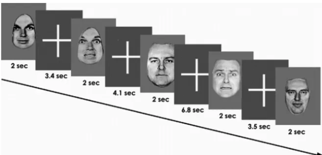
fear, or sadness) or a neutral facial expression. The facial identities were manipulated by computer software to depict 150% intensity of the emo-tion. The 150% level of intensity was chosen to minimize ambiguity and uncertainty about the nature of the stimuli (Calder et al., 1997; Phillips et al., 1997). Faces were presented in alternation with a fixation cross in a pseudorandom order. The neutral faces and affective faces were each displayed for 2 s and repeated 20 times, giving a total of 60 images (Fig. 1). The interstimulus interval followed a Poisson distribution and was varied between 3 and 9 s (mean interval, 5 s). Participants were instructed to press the right or left button with their dominant hand on an MRI-compatible response box to indicate whether the face was emotional or neutral. Subjects were familiarized with the task off-line 1 h before the scan. Response time and accuracy data were collected.
Image acquisition
Gradient echo planar MR images were ac-quired using a 1.5 T GE Sigma MR system (General Electric) fitted with 40 mT/m high-speed gradients. Foam padding and a forehead strap were used to limit head motion, and a quadrature birdcage head coil was used for ra-dio frequency transmission and reception. In each of the 16 noncontiguous planes parallel to the intercommissural (AC–PC) plane, T2*-weighted MR images reporting BOLD contrast were acquired (TR⫽2000 ms, TE⫽40 ms, flip angle ⫽70°, slice thickness ⫽ 7 mm, slice skip⫽0.7 mm, matrix size⫽64⫻64, voxel dimensions⫽3.75 ⫻3.75 ⫻7.7 mm). For each participant 3⫻150 fMRI images were acquired.
During the same session, a high-resolution T1-weighted structural image was acquired in the axial plane (inversion recovery prepared, spoiled gradient-echo sequence; TR⫽18 ms, TE⫽5.1 ms, TI⫽450 ms, flip angle⫽20°, slice thickness⫽1.5 mm, matrix size⫽256⫻ 192, FOV⫽240 ⫻180 mm, voxel dimen-sions⫽0.9375⫻0.9375⫻1.5 mm, number of excitations⫽1) for subsequent coregistration.
Image processing
Preprocessing.For image preprocessing and GLM analysis, we used the
SPM8 software package (Wellcome Trust Centre for Neuroimaging, London, UK; http://www.fil.ion.ucl.ac.uk); for effective connectivity analyses, DCM8 was used. Preprocessed images were realigned to correct for movement and normalized into MNI space using each subject’s struc-tural MRI image. The spatially normalized data were smoothed with an isotropic Gaussian filter (8 mm full-width half-maximum) to compen-sate for normal variation in structural and functional anatomy across subjects.
First level (within-subject) analysis.For each subject, the data from the
three experiments (emotions) were concatenated and modeled with a general linear (convolution) model, with additional regressors represent-ing potential confounds. Vectors of onset representrepresent-ing the correctly identified angry, fearful, and sad faces and correctly identified neutral faces were convolved with a canonical hemodynamic response function. Serial correlations were removed using a first-order autoregressive model and a high-pass filter (128 s) was applied to remove low-frequency noise. An explicit mask was used to ensure only voxels within the brain were included in the analysis. Six movement parameters were also en-tered as nuisance covariates. Additionally, the means of the three sessions were modeled as well as the transition at the end of each session. Contrast images of brain activations associated with correct categorization of emotional faces (angry, fearful, and sad) compared with neutral faces were produced for each participant.
Second level (between-subject) analysis. Group-level analyses were
based on random-effects analyses of the single-subject contrast images using the summary statistic approach. For facial affect, one-samplettests were used to investigate the main effect of task (correctly identified
[image:3.594.42.284.74.192.2]emo-Figure 1. The design of one facial affect recognition experiment is depicted (note that this example uses fear expressions). Subjects viewed pseudorandomized neutral expressions and affect-laden (angry or fearful or sad) facial expressions at 150% intensity during each of three separate experiments. Subjects judged the presence or absence of the facial emotion and pressed the corresponding button on an MRI-compatible response box.
Table 1. Demographic data and task information
Group (n⫽40)
Gender (male:female) 20:20
Age (in years) 31.5 (10.4)
Educational level 3.6 (0.8)
WAIS-R IQ 115.5 (15.9)
Response time to angry faces (in ms) 1085 (203) Accuracy for angry faces (% correct) 89.3 (6.3) Response time to fearful faces (in ms) 1048 (218) Accuracy for fearful faces (% correct) 97.6 (4.4) Response time to sad faces (in ms) 1185 (211) Accuracy for sad faces (% correct) 90.6 (9.3) Mean (SD). WAIS-R, Wechsler Adult Intelligence Scale–Revised.
[image:3.594.44.373.219.377.2]tional faces⬎correctly identified neutral faces). The statistical threshold was adjusted to provide a FWE ofp⫽0.05 (based on the spatial extent of clusters of voxels thresholded atp⬍0.001), corrected for multiple com-parisons across the whole brain. For all analyses, results are reported in the MNI coordinate system.
Volumes of interest
We selected a priori volumes of interest (VOIs) within a right-hemispheric network of regions implicated in face processing and its modulation by affect, following previous work (Fairhall and Ishai, 2007). These VOIs comprised the IOG (x⫽44,y⫽ ⫺78,z⫽ ⫺6), the FG (x⫽
24,y⫽ ⫺56,z⫽ ⫺12), the AMG (x⫽20,y⫽ ⫺2,z⫽ ⫺16), and the inferior frontal gyrus within the VPFC (x⫽52,y⫽20,z⫽ ⫺6). The coordinates for the visual regions, IOG and FG, were based on the group maxima from the contrast of all faces (minus crosshair), while the coor-dinates for the emotional regions, AMG and IFG, were specified from the contrast of emotional minus neutral faces. For each subject, we chose subject-specific maxima (in the appropriate SPM) in these regions that were within 4 mm of the group maxima and within the same anatomical area, as defined by the PickAtlas toolbox (Maldjian et al., 2003). Regional time series were summarized with the first eigenvariate of all activated (at
p⬍0.01) voxels within 5 mm of the subject-specific maxima.
Dynamic causal modeling
DCM (Friston et al., 2003) is a Bayesian model comparison procedure used to infer effective connectivity between brain regions. DCM
esti-mates directed interactions within neural systems. Crucially, it models these interactions at the neuronal level and distinguishes between endog-enous coupling and context-specific coupling, while accounting for the effects of experimentally controlled network perturbations (cf. stimulus-locked coupling) (Friston et al., 2003; Penny et al., 2004).
A four-area DCM was specified for all subjects with bidirectional en-dogenous connection between all regions (IOG, FG, AMG, VPFC) and the main effect of “all faces” as the driving input entering the IOG, the visual input region of our model. This base model (Fig. 2a) was then elaborated systematically to produce seven alternative variants. These variations were guided by our primary aim to define the role of the VPFC in facial affect recognition and the modulation of afferent connections to VPFC by affect (anger, fear, and sadness). Figure 2bshows the seven variants of four-area models, which include all possible combinations of how facial affect could modulate the forward connections to the VPFC. Note that while these figures show, for clarity, a single modulatory term labeled “Facial Affect,” the models contained distinct modulatory inputs for anger, fear, and sadness, allowing us to test the modulatory effects of these emotions (on connectivity) separately.
An additional (bilinear) model was constructed, where affective stim-uli directly entered AMG, to test whether affective information transfer to the VPFC is mediated by the AMG (Fig. 2b, Model 8). Furthermore, three additional nonlinear models were constructed that allowed for multiplicative interactions of postsynaptic inputs (Stephan et al., 2008). In these models, affective stimuli drove AMG activity directly, which
then modulated the forward connections (from IOG and/or FG) to the VPFC (Fig. 2c, Models 9, 10, 11). These nonlinear models replace the direct modulatory effect of experimentally defined facial affect with a vicarious physiological influence that is mediated by synaptic connec-tions from the AMG. All 11 models (Fig. 2) were constructed, fitted, and compared for each of the 40 subjects in this study.
Note that we limited our model comparisons to the right hemisphere, investigating the modulation of forward (afferent) connections to VPFC. This was motivated by the study by Fairhall and Ishai (2007), who high-lighted the predominance of the right hemisphere in the processing of emotional faces. Additionally, we assumed that visual input entered the IOG (as the lowest visual area). These constraints made it possible to define a relatively small model space (11 models per subject), in which we systematically investigated which of the forward connections was subject to modulation by facial affect and whether this modulation was best modeled by the direct effect of facial affect (bilinear models) or could be explained indirectly via by AMG modulation (nonlinear models) (Fig. 2).
Model comparison. Model comparison was implemented using
random-effects (RFX) Bayesian model selection (BMS) in DCM8 to compute exceedance and posterior probabilities at the group level (Stephan et al., 2009). The exceedance probability of a given model de-notes the probability that this model is more likely than any other model tested, given the data. Additionally, we made inferences about families of models (Penny et al., 2010; Stephan et al., 2010). Specifically, our models
were divided into a bilinear (models 1– 8) and a nonlinear (models 9 –11) family to test whether or not AMG activity could provide a sufficient account of the modulation of con-nections to VPFC. All models were included in the BMS procedure, both when comparing in-dividual models and when comparing model families. Finally, to summarize the strength of effective connectivity and its modulation quantitatively, we used random effects Bayes-ian model averaging (BMA) to obtain average connectivity estimates (weighted by their pos-terior model probability) across all models and all subjects (Penny et al., 2010).
Correlation with behavioral measures.To
ex-amine correlations between effective connec-tivity and behavior, we applied BMA on a subject-by-subject basis across all models and then extracted the resulting posterior means for each subject. These were entered into a sub-sequent correlation analysis with behavioral measures (accuracy and response time) with Bonferroni correction for multiple compari-sons (alpha⫽0.002).
Finally, we subjected the subject-specific BMA parameter estimates to one-sample tests to assess their significance in a classical sense (i.e., consistency across subjects). Behavioral and demographic data and parameter estimates were analyzed using SPSS 15 (SPSS) using one-samplettests, or appropriate nonparametric tests (one-sample Kolmogorov–Smirnov test) when data were not normally dis-tributed based on the Kolmogorov–Smirnov criterion, with␣⫽0.05. Since we tested 21 parameters of interest (i.e., all endogenous connec-tions and all bilinear modulaconnec-tions), we applied Bonferroni correction for multiple comparisons, resulting in an adjusted threshold of␣⫽ 0.002.
Results
Behavioral data
Subjects identified all facial affects with a high rate of accuracy, as
seen in Table 1.
SPM analysis
In accordance with previous meta-analytic studies (Phan et
al., 2002; Murphy et al., 2003; Fusar-Poli et al., 2009; Vytal and
Hamann, 2010), robust activation in response to emotional
faces (relative to neutral faces) was evident in the visual
asso-ciation cortices, the temporal gyrus as well as in frontal areas.
Details of the regional maxima are provided in Figures 3 and 4
and Table 2.
Figure 3. Image showing task-related brain activation in the group (N⫽40) during explicit (conscious) categorization of affective facial stimuli (FWE cluster-level corrected atp⬍0.05 across the whole brain, with voxel-level threshold ofp⬍0.001).
Figure 4. The graphs show the mean and the SEM of the GLM parameter estimates from the subject-specific maxima for all four conditions (neutral, anger, fear, sad) and for the four areas included in the models for dynamic causal modeling.
DCM analysis
Comparing the individuals models
Model 1 outperformed all other models with an exceedance
probability of 38% (Fig. 5). This optimal Model 1 contained
re-ciprocal endogenous connections between all four areas (IOG,
FG, AMG, VPFC), with affect only modulating the forward
con-nection from the IOG to the VPFC. Model 3 was the second-best
model with an exceedance probability of 20%. In Model 3, affect
modulated the forward connection from AMG to the VPFC.
Comparing the bilinear and nonlinear families
In a next step, we applied random-effects BMS at the family level
to clarify whether or not the AMG played a sufficient role in
modulating forward connections to VPFC during processing of
facial affect. Comparison between the bilinear (models 1– 8) and
nonlinear (models 9 –11) families showed that the bilinear family
was superior to the nonlinear family, with an exceedance
proba-bility of 100%. This suggests that the AMG alone does not
pro-vide a sufficient account for the gating of transfer of facial affect to
VPFC. Note that Bayesian model comparison eschews null
re-sults. In other words, we can be nearly 100% confident that the
(direct) modulatory effects of facial affect provide a better model
of empirical responses than a model in which the equivalent
modulation is mediated (indirectly) by the AMG.
Bayesian model averaging
The results from the BMA across all subjects and across all 11
models are shown in Table 3. The implementation of RFX BMA
in SPM8 employs an Occam’s window for computational
effi-ciency (Penny et al., 2010), excluding from the average those
models whose probability ratio (compared with the best model)
is
⬍
0.05. In our case, this applied to model 8 and the three
non-linear models (models 9 –11).
The BMA results highlight the importance of the efferent
con-nections from the IOG to FG, AMG, and VPFC in the network.
When BMA parameter estimates of the endogenous connections
originating from IOG were tested for consistency across subjects
with classical
t
tests, they were found to be highly significant, even
when correcting for multiple comparisons. Furthermore, the
IOG
3
VPFC connection was strongly and significantly
modu-lated by anger (
p
⬍
0.001; Table 3). It also showed a substantial
modulation by fear (
p
⬍
0.017), although this did not quite
sur-vive Bonferroni correction (
␣
⫽
0.002). By contrast, sadness did
not change this connection significantly (
p
⫽
0.076). Altogether,
these results emphasized the importance of this direct connection
to VPFC for the categorization of facial affect (Fig. 6).
Behavioral correlations
[image:6.594.302.543.83.352.2]We performed correlations between response time and accuracy
for angry, fearful, and sad faces, with the modulatory parameters
from BMA on a subject-by-subject basis over models. Mean
re-sponse time to angry faces showed a significant negative
correla-tion with anger modulacorrela-tion of the forward conneccorrela-tions from FG
Table 2. Voxel-based whole brain SPM analysis: Brain regions showing significantmain effects in terms of hemodynamic responses to the presentation of emotional faces compared to neutral faces
Brain region BA Laterality
Coordinates
Cluster size (voxels) Z-value
x y z
Middle occipital gyrus 18 R/L 38 ⫺84 ⫺4 147 5.94* Inferior occipital gyrus 19 44 ⫺78 ⫺6 5.38* Middle occipital gyrus 18 ⫺24 ⫺94 4 22 4.65* Lingual gyrus 18 R 10 ⫺82 2 240 5.6* Inferior temporal gyrus 37 L ⫺48 ⫺66 ⫺6 56 5.25* Middle frontal gyrus 8 R/L 54 12 42 64 5.11* ⫺50 8 44 4 3.29 Superior parietal gyrus 7 R 34 ⫺62 54 23 4.97* Fusiform gyrus 19 R 24 ⫺56 ⫺12 49 4.81*
Cuneus 17 L ⫺16 ⫺96 4 10 4.57*
Inferior frontal gyrus 47 R 51 20 ⫺6 3 4.55*
Precuneus 7 R 2 ⫺80 46 84 4.41
Amygdala N/A R/L 20 ⫺2 ⫺16 5 3.21** Superior temporal gyrus/ 38 ⫺32 6 ⫺22 332 4.29**
Amygdala N/A ⫺22 0 ⫺18
Inferior parietal lobule 40 L ⫺50 ⫺42 52 42 4.03 Postcentral gyrus 2 L ⫺62 ⫺22 28 13 3.65 Postcentral gyrus 1 L ⫺58 ⫺28 42 13 3.43 Inferior frontal gyrus 10 R 46 46 4 1 3.15 *p⬍0.05, FWE cluster-level corrected across the whole brain with a voxel-level cut-off ofp⬍0.001. **Survive small volume correction,p⬍0.01, FWE cluster-level.
MNI coordinates denote the distance (in mm) from the anterior commissure, with positivexvalues indicating a location right of midline, positiveyvalues indicating a location anterior to the anterior commissure, and positivez
[image:6.594.42.287.94.306.2]values indicating a location dorsal to a plane containing both the anterior and the posterior commissures. R, Right; L, left; BA, Brodmann area; N/A, Not Applicable.
[image:6.594.45.282.375.494.2]Figure 5. Expected probability and exceedance probability for the 11 models specified (N⫽40).
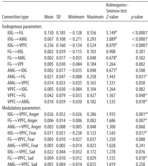
Table 3. Mean and SD DCM endogenous parameter and modulatory estimates for all connections across all subjects and across all models
Connection type Mean SD Minimum Maximum
Kolmogorov– Smirnov test
Z-value pvalue
Endogenous parameters
IOG3FG 0.130 0.185 ⫺0.128 0.556 5.149a ⬍0.0001**
IOG3AMG 0.067 0.108 ⫺0.271 0.293 3.889a ⬍0.0001**
IOG3VPFC 0.236 0.160 ⫺0.134 0.524 8.070a ⬍0.0001**
FG3IOG 0.002 0.039 ⫺0.115 0.103 0.908 0.381 FG3AMG 0.002 0.017 ⫺0.035 0.048 0.678a 0.502
FG3VPF 0.005 0.030 ⫺0.084 0.104 1.264 0.082 AMG3IOG 0.002 0.017 ⫺0.035 0.048 0.673a 0.508
AMG3FG 0.021 0.047 ⫺0.088 0.258 1.443 0.031* AMG3VPFC 0.014 0.033 ⫺0.035 0.165 1.331 0.058 VPFC3IOG 0.005 0.030 ⫺0.084 0.104 1.264 0.082 VPFC3FG 0.042 0.079 ⫺0.033 0.427 1.367 0.048* VPFC3AMG 0.018 0.039 ⫺0.030 0.182 1.535 0.018* Modulatory parameters
IOG3VPFC, Anger 0.026 0.052 ⫺0.026 0.286 1.935 0.001** FG3VPFC, Anger 0.006 0.014 ⫺0.006 0.082 1.686 0.007* AMG3VPFC, Anger 0.003 0.008 ⫺0.005 0.048 1.300 0.068 IOG3VPFC, Fear 0.011 0.051 ⫺0.238 0.123 1.543 0.017* FG3VPFC, Fear 0.003 0.010 ⫺0.027 0.037 1.250 0.088 AMG3VPFC, Fear 0.001 0.005 ⫺0.014 0.023 1.028 0.241 IOG3VPFC, Sad 0.022 0.044 ⫺0.032 0.172 1.278 0.076 FG3VPFC, Sad 0.004 0.010 ⫺0.012 0.029 1.533 0.018* AMG3VPFC, Sad 0.001 0.004 ⫺0.014 0.015 1.019 0.250 *Difference significant atp⬍0.05, uncorrected for multiple comparisons.
**Difference survives Bonferroni correction for multiple comparisons, corrected atp⫽0.002.
(
r
⫽ ⫺
0.336,
p
⫽
0.034) and from AMG to the VPFC (
r
⫽
⫺
0.337,
p
⫽
0.034). Mean response time to fearful faces showed
a significant positive correlation for fear modulation on the
for-ward connection from the IOG to the VPFC (
r
⫽
0.376,
p
⫽
0.017). However, these correlations did not survive Bonferroni
correction for multiple tests.
Discussion
In this study, we assessed effective connectivity during facial
af-fect processing using DCM as an established method for inferring
effective connectivity from fMRI data (Stephan et al., 2010). Our
aim was to examine the modulation of connectivity between
pos-terior regions of the face-processing network and the VPFC
dur-ing affect categorization and to evaluate the role of the AMG in
mediating this modulation. There are three key findings from our
study. First, we confirmed our hypothesis that facial affect
signif-icantly increased effective connectivity between posterior regions
of the face network and the VPFC. Our data were best explained
by a model in which facial affect modulated the connection from
the IOG to the VPFC. Second, our results suggest that affect may
modulate connections to the VPFC above and beyond any
puta-tive modulation by the AMG. This implies that the AMG is not
necessarily involved in gating prefrontal connectivity during
ex-plicit affective processing. Third, the effective connectivity
be-tween the IOG and VPFC showed evidence for differential
modulation according to valence, with the strongest modulation
observed for anger.
These findings are timely in view of the current revaluation of
existing models of affective stimuli processing in the brain
(Pes-soa and Adolphs, 2010). Our results contribute to this debate by
highlighting the role of the prefrontal cortex during affective
pro-cessing and by questioning the prevailing amygdalocentric model
of affective processing (LeDoux, 1996; Davis and Whalen, 2001).
Across all subjects, our modeling results suggest that the
VPFC receives information directly from regions within the face
network. Crucially, however, it was the coupling from the IOG to
the VPFC that emerged as the most significant effective
connec-tion within this network, which was further enhanced during the
processing of facial affect. A large body of literature has
estab-lished that attention enhances neural responses primarily within
extrastriate visual cortices (Moran and Desimone, 1985; Buffalo
et al., 2010). A similar enhancement has also been noted in
re-sponse to emotional valence in general (Lane et al., 1999) and
during presentation of emotional, particularly negative
(Vuil-leumier et al., 2004), facial expressions compared with neutral
ones (Critchley et al., 2000; Vuilleumier et al., 2001; Winston et
al., 2003). Electrical recordings have shown that during attentive
viewing, prefrontal and visual cortices show increased gamma
frequency synchrony suggestive of greater functional coupling
between these regions (Gregoriou et al., 2009). Our results
there-fore add support to the notion of increased coupling between
prefrontal and visual cortical regions during visual attention and
suggest that such coupling may be further increased when
atten-tion is directed to the emoatten-tional valence of the stimuli.
Research in affective neuroscience has traditionally
empha-sized the role of the AMG in emotional processing. The AMG is
considered a core component of a subcortical pathway for the
rapid detection of emotions during visual processing (LeDoux,
1998; Morris et al., 1999; Rolls, 1999) and is thought to influence
behavior through the modulation of PFC activity (Miller and
Cohen, 2001). There are innumerable examples from multiple
lines of research showing that the AMG modulates activity in the
ventral visual pathway in response to emotional signals (Sugase et
al., 1999), including facial expressions of affect (Pessoa et al.,
2002; Vytal and Hamann, 2010). Although the contribution of
the AMG to emotional processing is indisputable, our results,
together with those of others (Tsuchiya et al., 2009; Pessoa and
Adolphs, 2010; Piech et al., 2010), suggest that the
amygdalocen-tric model of affect processing overlooks the significant
contri-bution of other brain regions and the complexity of their
interactions. Our modeling results imply that the VPFC receives
information about facial affect directly from three distinct brain
regions—the IOG, the FG, and the AMG—and that AMG
mod-ulation of the connections from either the FG or the IOG to the
VPFC is not a complete or sufficient explanation for changes in
coupling. While the results of the present study do not disclose
the sources of this modulation, they imply that AMG activity does
not necessarily gate the transfer of facial affect information
to-ward the VPFC. Instead, they support a model in which affective
information from visual stimuli proceeds simultaneously along
parallel channels, creating multiple waves of activation across the
face-processing network (Pessoa and Adolphs, 2010). One may
speculate that the emotional modulation of these channels may
have arisen from multiple, and possibly diffuse, sources; these,
however, were not visible in the statistical parametric maps of our
present fMRI study. Finally, the implausibility of an
amygdalo-centric explanation of our data, as indicated by our model
com-parisons, may be due to the fact that the effective connectivity
(and its affect-sensitive changes) assessed by our paradigm
medi-ated the explicit or conscious processing of affective stimuli. This
contrasts with the implicit and automatic processes normally
as-sociated with AMG processing.
The inclusion of facial identities with three distinct negative
affects allowed us to explore the effect of valence on effective
connectivity within the face-processing network. Our results
sug-gest that anger showed the most potent modulation of the
cou-pling from the IOG to the VPFC. Anger is considered particularly
salient compared with other emotions, as it signals the need for
immediate action in response to perceived threat. The VPFC is
thought to exert a regulatory role in the expression of anger as
damage to the VPFC can increase violent and aggressive
behav-iors (Damasio et al., 1994; Grafman et al., 1996). Our findings
complement results from a recent meta-analytic review of the
Figure 6. Alterations in effective connectivity within the face-processing network across allsubjects (N⫽40), established by Bayesian model averaging across all models considered. Thick gray arrows indicate significant endogenous connections (p⬍0.0001) and the thick black arrow indicates a significant endogenous connection significantly modulated by anger (p⫽
0.001). Dashed arrows indicate backwards connections and thin black arrows indicate bidirec-tional connections.
discrete neural correlates of basic emotions, which suggested that
the rapid engagement of the VPFC during exposure to anger
stimuli may serve to avert potential overreaction, such as
unre-strained rage (Vytal and Hamann, 2010).
Finally, concerning the AMG, our results based on Bayesian
model averaging suggest that the differential responses to
affec-tive versus neutral faces in the AMG arise via two afferent
con-nections (Table 3), i.e., the IOG
3
AMG connection (
p
⬍
0.0001)
and the VPFC
3
AMG connection (
p
⬍
0.018, not quite
surviv-ing Bonferroni correction at
␣
⫽
0.002).
Our study focused on effective connectivity within the
face-processing network during attentive viewing and categorization
of facial affect. Since the ensuing estimates of effective
connectiv-ity are context- and paradigm-dependent, further studies are
required to explore the functional architecture of the
facial-affect-processing network during tasks that make additional
cog-nitive demands involving induction or suppression of emotional
responses. Our findings have significant implications for
pathophys-iological models of affective dysfunction in psychiatric disorders,
particularly mood disorders where current amygdalocentric
ap-proaches may need to be reevaluated.
In summary, our analyses have identified that during the
pro-cessing of facial affect, effective connectivity to the VPFC is
in-creased not only from the AMG but also from other regions in the
ventral visual stream, namely the FG and IOG. AMG modulation
of this coupling does not appear to be sufficient to account for
affect-dependent changes during explicit (conscious) processing.
Finally, the functional coupling of the IOG to the VPFC plays a
major role in the processing of facial affect and suggests a greater
contribution of visual cortical–prefrontal pathways to affect
pro-cessing than previously considered.
References
Adolphs R (2002) Neural systems for recognizing emotion. Curr Opin Neu-robiol 12:169 –177.
Anderson AK, Phelps EA (2001) Lesions of the human amygdala impair enhanced perception of emotionally salient events. Nature 411:305–309. Armony JL, Dolan RJ (2002) Modulation of spatial attention by fear-conditioned stimuli: an event-related fMRI study. Neuropsychologia 40:817– 826.
Buffalo EA, Fries P, Landman R, Liang H, Desimone R (2010) A backward progression of attentional effects in the ventral stream. Proc Natl Acad Sci U S A 107:361–365.
Calder AJ, Young AW, Rowland D, Perrett DI (1997) Computer-enhanced emotion in facial expressions. Proc Biol Sci 264:919 –925.
Critchley H, Daly E, Phillips M, Brammer M, Bullmore E, Williams S, Van Amelsvoort T, Robertson D, David A, Murphy D (2000) Explicit and implicit neural mechanisms for processing of social information from facial expressions: a functional magnetic resonance imaging study. Hum Brain Mapp 9:93–105.
Damasio H, Grabowski T, Frank R, Galaburda AM, Damasio AR (1994) The return of Phineas Gage: clues about the brain from the skull of a famous patient. Science 264:1102–1105.
Davis M, Whalen PJ (2001) The amygdala: vigilance and emotion. Mol Psy-chiatry 6:13–34.
Dyck M, Loughead J, Kellermann T, Boers F, Gur RC, Mathiak K (2011) Cognitive versus automatic mechanisms of mood induction differentially activate left and right amygdale. Neuroimage 54:2503–2513.
Fairhall SL, Ishai A (2007) Effective connectivity within the distributed cor-tical network for face perception. Cereb Cortex 17:2400 –2406. First MB, Spitzer RL, Gibbon M, Williams JBW (2002) Structured clinical
interview for DSM-IV Axis I disorders, research version, non-patient edi-tion (SCID-I/NP). New York: Biometrics Research, New York State Psy-chiatric Institute.
Friston KJ, Harrison L, Penny W (2003) Dynamic causal modelling. Neuro-image 19:1273–1302.
Fusar-Poli P, Placentino A, Carletti F, Allen P, Landi P, Abbamonte M, Barale F, Perez J, McGuire P, Politi PL (2009) Laterality effect on emotional
faces processing: ALE meta-analysis of evidence. Neurosci Lett 452:262–267.
Grafman J, Schwab K, Warden D, Pridgen A, Brown HR, Salazar AM (1996) Frontal lobe injuries, violence, and aggression: a report of the Vietnam Head Injury Study. Neurology 46:1231–1238.
Gregoriou GG, Gotts SJ, Zhou H, Desimone R (2009) High-frequency, long-range coupling between prefrontal and visual cortex. Science 324:1207–1210.
Hariri AR, Bookheimer SY, Mazziotta JC (2000) Modulating emotional re-sponses: effects of a neocortical network on the limbic system. Neurore-port 11:43– 48.
Hariri AR, Mattay VS, Tessitore A, Fera F, Weinberger DR (2003) Neocor-tical modulation of the amygdala response to fearful stimuli. Biol Psychi-atry 53:494 –501.
Haxby JV, Hoffman EA, Gobbini MI (2000) The distributed human neural system for face perception. Trends Cogn Sci 4:223–233.
Haxby JV, Hoffman EA, Gobbini MI (2002) Human neural systems for face recognition and social communication. Biol Psychiatry 51:59 – 67. Hoffman EA, Haxby JV (2000) Distinct representations of eye gaze and
identity in the distributed human neural system for face perception. Nat Neurosci 3:80 – 84.
Iidaka T, Sadato N, Yamada H, Murata T, Omori M, Yonekura Y (2001) An fMRI study of the functional neuroanatomy of picture encoding in younger and older adults. Brain Res Cogn Brain Res 11:1–11.
Lane RD, Chua PM, Dolan RJ (1999) Common effects of emotional valence, arousal and attention on neural activation during visual processing of pictures. Neuropsychologia 37:989 –997.
LeDoux JE (1996) The emotional brain. New York: Simon and Schuster. LeDoux JE (1998) The emotional brain: the mysterious underpinnings of
emotional life. New York: Touchstone.
Maldjian JA, Laurienti PJ, Kraft RA, Burdette JH (2003) An automated method for neuroanatomic and cytoarchitectonic atlas-based interroga-tion of fMRI data sets. Neuroimage 19:1233–1239.
Miller EK, Cohen JD (2001) An integrative theory of prefrontal cortex func-tion. Annu Rev Neurosci 24:167–202.
Moran J, Desimone R (1985) Selective attention gates visual processing in the extrastriate cortex. Science 229:782–784.
Morris JS, Ohman A, Dolan RJ (1999) A subcortical pathway to the right amygdala mediating “unseen” fear. Proc Natl Acad Sci U S A 96:1680 –1685.
Murphy FC, Nimmo-Smith I, Lawrence AD (2003) Functional neuroanatomy of emotions: a meta-analysis. Cogn Affect Behav Neurosci 3:207–233. Ochsner KN, Gross JJ (2005) The cognitive control of emotion. Trends
Cogn Sci 9:242–249.
Penny WD, Stephan KE, Mechelli A, Friston KJ (2004) Comparing dynamic causal models. Neuroimage 22:1157–1172.
Penny WD, Stephan KE, Daunizeau J, Rosa MJ, Friston KJ, Schofield TM, Leff AP (2010) Comparing families of dynamic causal models. PLoS Com-put Biol 6:1000709.
Pessoa L, Adolphs R (2010) Emotion processing and the amygdala: from a ‘low road’ to ‘many roads’ of evaluating biological significance. Nat Rev Neurosci 11:773–783.
Pessoa L, McKenna M, Gutierrez E, Ungerleider LG (2002) Neural process-ing of emotional faces requires attention. Proc Natl Acad Sci U S A 99:11458 –11463.
Phan KL, Wager T, Taylor SF, Liberzon I (2002) Functional neuroanatomy of emotion: a meta-analysis of emotion activation studies in PET and fMRI. Neuroimage 16:331–348.
Phillips ML, Young AW, Senior C, Brammer M, Andrew C, Calder AJ, Bull-more ET, Perrett DI, Rowland D, Williams SC, Gray JA, David AS (1997) A specific neural substrate for perception of facial expressions of disgust. Nature 389:495– 498.
Piech RM, McHugo M, Smith SD, Dukic MS, Van Der Meer J, Abou-Khalil B, Zald DH (2010) Fear-enhanced visual search persists after amygdala le-sions. Neuropsychologia 48:3430 –3435.
Quirk GJ, Beer JS (2006) Prefrontal involvement in the regulation of emo-tion: convergence of rat and human studies. Curr Opin Neurobiol 16:723–727.
Rolls ET (1999) The brain and emotion. Oxford: Oxford UP.
Stephan KE, Penny WD, Daunizeau J, Moran RJ, Friston KJ (2009) Bayesian model selection for group studies. Neuroimage 46:1004 –1017. Stephan KE, Penny WD, Moran RJ, den Ouden HE, Daunizeau J, Friston KJ
(2010) Ten simple rules for dynamic causal modelling. Neuroimage 49:3099 –3109.
Sugase Y, Yamane S, Ueno S, Kawano K (1999) Global and fine informa-tion coded by single neurons in the temporal visual cortex. Nature 400:869 – 873.
Tsuchiya N, Moradi F, Felsen C, Yamazaki M, Adolphs R (2009) Intact rapid detection of fearful faces in the absence of the amygdala. Nat Neu-rosci 12:1224 –1225.
Vuilleumier P, Driver J (2007) Modulation of visual processing by attention and emotion: windows on causal interactions between human brain re-gions. Philos Trans R Soc Lond B Biol Sci 362:837– 855.
Vuilleumier P, Armony JL, Driver J, Dolan RJ (2001) Effects of attention and emotion on face processing in the human brain: an event-related fMRI study. Neuron 30:829 – 841.
Vuilleumier P, Richardson MP, Armony JL, Driver J, Dolan RJ (2004) Dis-tant influences of amygdala lesion on visual cortical activation during emotional face processing. Nat Neurosci 7:1271–1278.
Vytal K, Hamann S (2010) Neuroimaging support for discrete neural cor-relates of basic emotions: a voxel-based meta-analysis. J Cogn Neurosci 22:2864 –2885.
Wechsler D (1981) Manual for the Wechsler Adult Intelligence Scale–Re-vised. New York: Psychological Corporation.
Winston JS, O’Doherty J, Dolan RJ (2003) Common and distinct neural responses during direct and incidental processing of multiple facial emo-tions. Neuroimage 20:84 –97.