The importance of the promoter in Drosophila dosage compensation : a thesis presented in partial fulfillment of the requirements for the degree of Doctor of Philosophy in Genetics at Massey University, Palmerston North, New Zealand
256
0
0
Full text
(2) The importance of the promoter in Drosophila dosage compensation. A thesis presented in partial fulfillment of the requirements for the degree of. Doctor of Philosophy in Genetics. at Massey University, Palmerston North, New Zealand.. Corey Laverty 2009.
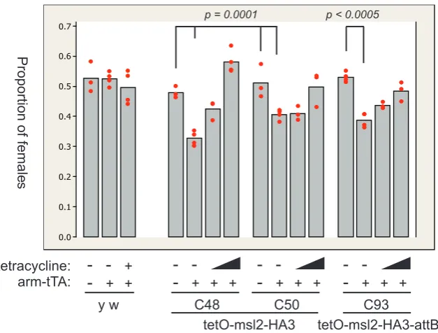
(3) Abstract Dosage compensation is the equalisation of gene expression from unequal doses of genes. Drosophila males up-regulate transcription from their single X chromosome to equal that from the two female X chromosomes. Five malespecific lethal (msl) genes are required in males, and encode the main agents of the up-regulation. At least these proteins, together with either or both of two noncoding RNAs, form the MSL chromatin-modifying complex. Female-specific translational repression of a key component, msl2, limits the complex to males. The MSL complex binds to the X chromosome at hundreds of distinct loci, acetylates nucleosomes, and de-condenses the chromatin. Together with possibly many co-factors, the transcriptional up-regulation caused by MSL complex appears to counteract repressive factors to achieve an average effect of transcriptional doubling. Here, I have studied the initiation of MSL regulation on the X chromosome with a variety of approaches. In order to study early events, dosage compensation was induced in females with ectopic expression of msl2 from the tetracycline system. However, low background expression without activation prohibited further studies. To identify novel factors that affect dosage compensation, a reporter gene system based on variable eye size was evaluated. The system provided a dose-dependent phenotype, but could not report additional up-regulation by the MSL complex, and was thus unsuitable for the proposed mutational screen. The quantifiable lacZ gene was measured in a strict comparison of expression from an eye-specfic (GMR) or a constitutive (armadillo) promoter. At defined locations on the X chromsome, armadillo-lacZ acquired local compensation, but GMR-lacZ did not. Further modifications upstream of GMR-lacZ increased the response, and confirmed the importance of the promoter in attraction of dosage compensation. To corroborate this with the established importance of genic sequences in MSL attraction, a combinatorial model of attraction is proposed. The relative importance of early or constitutive expression was also tested, by providing GMR-lacZ with extra expression through the tetracycline system. A burst of embryonic expression, and constitutive expression, were both insufficient to increase dosage compensation of the transgene. Finally, the compensation of GMRmediated transgenes was confounded by ‘transvection’ effects of chromosome pairing. This effect may have wider implications on the study of compensation at individual genes.. i.
(4) ii.
(5) Acknowledgements Thank you, Max Scott, my supervisor and mentor. Your ideas and advice throughout were invaluable. If I have done well, it is because of you. You’ve taught me how to work, how to research, how to think. Thank you, Kathryn Stowell for encouragement at all stages, and especially for your review of the figures. Al Rowland and David Penny, for tuning my mind to science philosophy, and evolution. Alasdair Noble, for arguing statistics. To all who helped, more thanks than I can muster: Fang Li for your many hours giving critical help with western blots, polytene preparations, and cloning. Your advice was always accurate and valued. Esther Belikoff for expert advice on micro-injection, and for tirelessly tending my flies when I couldn’t. Anja Schiemann for β-galactosidase assays. Carolina Concha for RNA advice. Vikki Weake for getting me started, and shared micro-injections. Helen Fitzsimons for discussions on the draft. All ‘Flyspot’ members, past and present, especially Abhimanyu Sarkar and Charles Ellen, for a co-operative and considerate work ethic, and a friendly, productive environment. Emma Brasell for making fly food, and for instant help at the spec. when the printer failed. Ava Handley, for countless helpful discussions and thoughts throughout the write-up. Ava, Sarah Lobb, and Jessika Charlton, for food and flywork. Sara Sutherland, for proofing my introduction. For supplying flies, plasmids, and antibodies: Andreas Bergmann, Johannes Bischof, Michele Calos, Mitzi Kuroda, Herman Steller, Ernst Wimmer. J. Bischof for adice on the ϕC31 system. Art Alekseyenko for advice on the MRE site in armadillo. For financial support: Massey University, the Institute of Molecular Biosciences, and the New Zealand Freemasons. To those that personally supported me: thank you, thank you, thank you. I could not do this without any of you. Most especially, my mother and father. Your love, generosity, and unquestioning, continual support is breathtaking. Jared and Gemma for your love and help, especially in Ireland. Ava Handley, for your love and encouragement, your strength when I had none, and for listening, advising, laughing. Sophie Borchert for your encouragement and friendship, and for support through the middle years. Lucy Pearse for the start. Angela Woodley for always being there. Emma Brasell, Charles Ellen, for distraction. Amanda Staddon-Smith, Sara Sutherland, Melle Steringa, for tolerance. Now it is done. I’ve learnt so much.. iii.
(6) iv.
(7) Contents 1. 2. Introduction 1.1 Dosage Compensation in Drosophila melanogaster . . . . . . . . . . . . . . . 1.1.1 Compensation of X chromosome genes . . . . . . . . . . . . . . . . . 1.1.2 Known factors linked to compensation . . . . . . . . . . . . . . . . . . 1.1.3 Sex-specific compensation . . . . . . . . . . . . . . . . . . . . . . . . 1.1.4 Targeting the male X chromosome . . . . . . . . . . . . . . . . . . . . 1.1.5 Potential mechanisms . . . . . . . . . . . . . . . . . . . . . . . . . . . 1.1.6 Summary . . . . . . . . . . . . . . . . . . . . . . . . . . . . . . . . . 1.2 Transvection . . . . . . . . . . . . . . . . . . . . . . . . . . . . . . . . . . . . 1.3 Basis of Experimental Design . . . . . . . . . . . . . . . . . . . . . . . . . . 1.3.1 Drosophila transformation and the ϕC31 system . . . . . . . . . . . . 1.3.2 Tetracycline-regulatable expression systems . . . . . . . . . . . . . . . 1.3.3 DNA replication-related element (DRE) . . . . . . . . . . . . . . . . . 1.3.4 The Glass Multimer Reporter . . . . . . . . . . . . . . . . . . . . . . 1.3.5 A reporter gene system to detect changes in MSL complex activity . . . 1.4 Aims . . . . . . . . . . . . . . . . . . . . . . . . . . . . . . . . . . . . . . . . 1.4.1 Determination of the relative order of key events in Drosophila dosage compensation . . . . . . . . . . . . . . . . . . . . . . . . . . . . . . . 1.4.2 Identification of novel factors involved in Drosophila dosage compensation . . . . . . . . . . . . . . . . . . . . . . . . . . . . . . . . . . . 1.4.3 Investigation of the deficiency in compensation of GMR-based expression. 1 2 2 4 8 9 14 16 17 18 18 18 19 20 20 22. Materials and methods 2.1 Bacterial methods . . . . . . . . . . 2.1.1 Growth and maintenance . . 2.1.2 Creation of competent cells 2.1.3 Transformation . . . . . . . 2.2 Drosophila methods . . . . . . . . . 2.2.1 Solutions . . . . . . . . . . 2.2.2 Stock maintenance . . . . . 2.2.3 Virgin collection . . . . . . 2.2.4 Genetic crosses . . . . . . . 2.2.5 Transformation . . . . . . . 2.2.6 Inverse PCR . . . . . . . . 2.3 Molecular biology methods . . . . . 2.3.1 Solutions . . . . . . . . . . 2.3.2 DNA Isolation . . . . . . . 2.3.3 DNA purification . . . . . .. 25 26 26 26 26 26 27 27 28 28 32 34 36 36 36 38. . . . . . . . . . . . . . . .. v. . . . . . . . . . . . . . . .. . . . . . . . . . . . . . . .. . . . . . . . . . . . . . . .. . . . . . . . . . . . . . . .. . . . . . . . . . . . . . . .. . . . . . . . . . . . . . . .. . . . . . . . . . . . . . . .. . . . . . . . . . . . . . . .. . . . . . . . . . . . . . . .. . . . . . . . . . . . . . . .. . . . . . . . . . . . . . . .. . . . . . . . . . . . . . . .. . . . . . . . . . . . . . . .. . . . . . . . . . . . . . . .. . . . . . . . . . . . . . . .. . . . . . . . . . . . . . . .. . . . . . . . . . . . . . . .. . . . . . . . . . . . . . . .. . . . . . . . . . . . . . . .. . . . . . . . . . . . . . . .. . . . . . . . . . . . . . . .. . . . . . . . . . . . . . . .. 22 23 24.
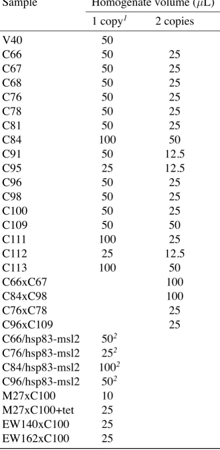
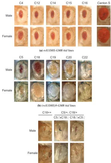
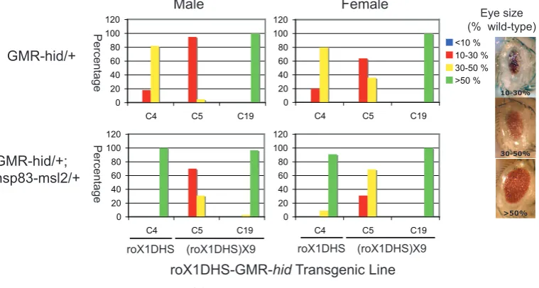
(8) 2.3.4 DNA precipitation . . . . . . . . . 2.3.5 DNA quantification . . . . . . . . . 2.3.6 Gel electrophoresis . . . . . . . . . 2.3.7 DNA Sequencing . . . . . . . . . . 2.3.8 Annealing of oligonucleotides . . . 2.3.9 Enzymatic manipulations of DNA . 2.3.10 PCR . . . . . . . . . . . . . . . . . 2.3.11 Molecular cloning . . . . . . . . . 2.4 Reverse Transcriptase (RT) -PCR . . . . . . 2.4.1 Precautions for RNA work . . . . . 2.4.2 RT-PCR reactions . . . . . . . . . . 2.4.3 Band quantification . . . . . . . . . 2.5 Western blot analysis . . . . . . . . . . . . 2.6 Female viability assay . . . . . . . . . . . . 2.7 Polytene chromosome immuno-fluorescence 2.8 Eye size assays . . . . . . . . . . . . . . . 2.9 β -galactosidase enzymatic assays . . . . . . 2.9.1 β -galactosidase assay . . . . . . . . 2.9.2 Total protein assay . . . . . . . . . 2.9.3 Enzymatic activity calculations . . 2.9.4 Statistical analyses . . . . . . . . . 2.9.5 Homogenate volume . . . . . . . . 2.10 β -galactosidase tissue staining . . . . . . . 2.11 Drosophila photography . . . . . . . . . . 2.11.1 Embryonic preparation . . . . . . . 2.11.2 Pupal dissection . . . . . . . . . . 2.11.3 Photography . . . . . . . . . . . . 3. 4. . . . . . . . . . . . . . . . . . . . . . . . . . . .. . . . . . . . . . . . . . . . . . . . . . . . . . . .. . . . . . . . . . . . . . . . . . . . . . . . . . . .. . . . . . . . . . . . . . . . . . . . . . . . . . . .. . . . . . . . . . . . . . . . . . . . . . . . . . . .. . . . . . . . . . . . . . . . . . . . . . . . . . . .. . . . . . . . . . . . . . . . . . . . . . . . . . . .. . . . . . . . . . . . . . . . . . . . . . . . . . . .. . . . . . . . . . . . . . . . . . . . . . . . . . . .. . . . . . . . . . . . . . . . . . . . . . . . . . . .. . . . . . . . . . . . . . . . . . . . . . . . . . . .. . . . . . . . . . . . . . . . . . . . . . . . . . . .. . . . . . . . . . . . . . . . . . . . . . . . . . . .. . . . . . . . . . . . . . . . . . . . . . . . . . . .. . . . . . . . . . . . . . . . . . . . . . . . . . . .. . . . . . . . . . . . . . . . . . . . . . . . . . . .. . . . . . . . . . . . . . . . . . . . . . . . . . . .. . . . . . . . . . . . . . . . . . . . . . . . . . . .. Development of an inducible dosage compensation system in female Drosophila 3.1 A two-component system for MSL2 induction was created . . . . . . . . . . 3.2 MSL2 was produced in females . . . . . . . . . . . . . . . . . . . . . . . . 3.2.1 MSL2 protein levels below detection limit . . . . . . . . . . . . . . . 3.2.2 MSL2 expressed sufficient to kill females . . . . . . . . . . . . . . . 3.3 Transgenic MSL2 localised to the female X chromosome . . . . . . . . . . . 3.4 Repressive chromatin effects reduced un-induced chromosome binding . . . 3.4.1 The 20C environment negatively affected gene expression . . . . . . 3.4.2 Less MSL complex bound the X chromosome when MSL2 was expressed from 20C . . . . . . . . . . . . . . . . . . . . . . . . . . . . 3.5 Evaluation of the inducible dosage compensation system . . . . . . . . . . . 3.6 Future directions . . . . . . . . . . . . . . . . . . . . . . . . . . . . . . . .. . . . . . . . . . . . . . . . . . . . . . . . . . . .. 38 38 39 39 40 40 41 44 45 45 45 46 46 48 48 50 51 51 51 52 52 52 53 55 55 55 55. . . . . . . .. 57 58 60 60 62 64 67 67. . 69 . 71 . 74. Development of the GMR-hid reporter gene system to search for novel factors involved in Drosophila dosage compensation 4.1 The roX1DHS-GMR-hid system displayed an unreliable sex-specific difference 4.1.1 Male-specific phenotype less significant than predicted . . . . . . . . . 4.1.2 New transformant locations did not improve the system . . . . . . . . . 4.1.3 The roX1DHS-GMR-hid system was dose responsive . . . . . . . . . . 4.2 The roX1DHS-GMR-hid system unreliably reported MSL complex activity . . 4.3 A replacement MSL binding site did not improve the reporting ability . . . . .. vi. 75 76 76 78 78 79 82.
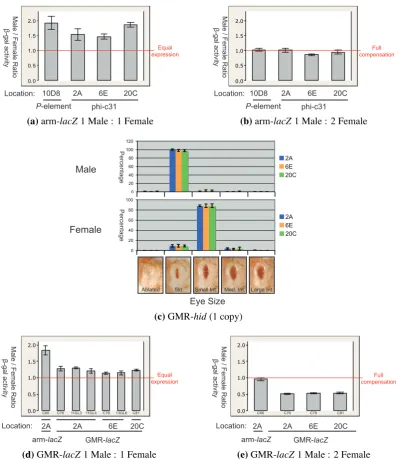
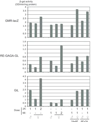
(9) 4.4. 4.5. 4.6 4.7 4.8. GMR-hid insufficient to report dosage compensation . . . . . . . . . . . . . . 4.4.1 X insertions of GMR-hid did not acquire full compensation . . . . . . . 4.4.2 GMR-hid on the X did not respond to altered MSL levels . . . . . . . . 4.4.3 A similar X-linked GMR-hid was also unaffected by MSL levels . . . . GMR-based expression only weakly affected by endogenous dosage compensation . . . . . . . . . . . . . . . . . . . . . . . . . . . . . . . . . . . . . . . 4.5.1 A targeted system was developed . . . . . . . . . . . . . . . . . . . . 4.5.2 Modified β-galactosidase assay to measure activity from the GMR-hsp70 promoter . . . . . . . . . . . . . . . . . . . . . . . . . . . . . . . . . 4.5.3 Acquired compensation detected at defined X chromosome sites . . . . 4.5.4 GMR-hid at compensation-permissive sites acquired very little compensation . . . . . . . . . . . . . . . . . . . . . . . . . . . . . . . . . 4.5.5 GMR-lacZ at compensation-permissive sites acquired very little compensation . . . . . . . . . . . . . . . . . . . . . . . . . . . . . . . . . Advantages of ϕC31 targeted integration for studying dosage compensation . . Critical issues for measuring β-galactosidase activity . . . . . . . . . . . . . . Summary and future directions . . . . . . . . . . . . . . . . . . . . . . . . . .. 82 83 83 84 86 86 92 93 95 95 98 101 102. 5. The deficiency in compensation of GMR-based expression 105 5.1 Identified differences between arm- and GMR-based vectors . . . . . . . . . . 106 5.2 Promoter modifications increased the male:female expression ratio of GMR-lacZ 108 5.2.1 SV40 3′ UTR altered expression level but not male:female ratio . . . . 108 5.2.2 Local MSL complex was not limiting . . . . . . . . . . . . . . . . . . 110 5.2.3 Promoter elements increased the male:female ratio . . . . . . . . . . . 110 5.2.4 Addition of an intron affected the male:female ratio . . . . . . . . . . 112 5.3 Females with two DRE-GAGA-GL doses were affected by transvection . . . . . 116 5.4 Increased GMR-lacZ male:female ratios observed specifically on the X chromosome . . . . . . . . . . . . . . . . . . . . . . . . . . . . . . . . . . . . . . 122 5.5 DRE-GAGA-GMR-lacZ responded to altered MSL levels . . . . . . . . . . . 124 5.6 Inefficient processing of ninaE intron . . . . . . . . . . . . . . . . . . . . . . 126 5.7 DRE and GAGA sites insufficient to alter GMR-hid expression pattern . . . . . 126 5.8 Effect of DRE and GAGA elements on GMR-GFP difficult to detect . . . . . . 129 5.9 An earlier burst of expression insufficient for compensation of GMR-lacZ . . . 130. 6. General discussion 6.1 Importance of the promoter in dosage compensation of a X-linked transgene . 6.2 Effect of promoter elements on dosage compensation . . . . . . . . . . . . . 6.3 Early expression insufficient for dosage compensation of a tissue-specific transgene . . . . . . . . . . . . . . . . . . . . . . . . . . . . . . . . . . . . . . . 6.4 On the combinatorial nature of Drosophila dosage compensation . . . . . . . 6.5 Dosage compensation and transvection . . . . . . . . . . . . . . . . . . . . .. 7. Conclusions. 137 . 138 . 139 . 142 . 143 . 145 147. Bibliography. 151. Appendices. 169. A Biological material. 171. vii.
(10) B Raw data for effect of msl2 expression on female viability. 181. C Raw data for effect of altered MSL levels on GMR-hid lines C60 and GMR-hidAla5 183 D Raw data for GMR-hid-attB at X-linked attP sites. 185. E Genomic sequences from inverse PCR of attP sites. 187. F Quantification of band intensitiies. 190. G Drosophila Transformation data. 192. H Transgenic Stocks. 195. I. 198. Plasmid maps. viii.
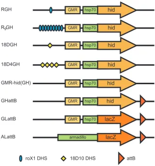
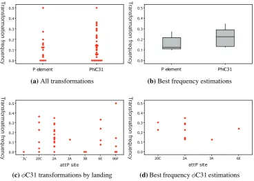
(11) List of Figures 3.1 3.2 3.3 3.4 3.5 3.6 3.7. Scheme to add epitope tags to msl-2 . . . . . . . . . . . . . . . . . . . Un-detectable induction of MSL2 by tTA . . . . . . . . . . . . . . . . Induction of MSL2 sufficient to lower female viability . . . . . . . . . Background level of MSL binding in transgenic females . . . . . . . . Modest increase in MSL binding to X chromosome with msl2 induction Position-dependent repression on the mini-white marker gene . . . . . . Reduced X chromosome binding of MSL complex with expression of from repressive 20C environment . . . . . . . . . . . . . . . . . . . .. 4.1 4.2 4.3 4.4. Schematic diagram of roX1 DHS-GMR-hid and related constructs . . . . . . Subtle sex-specific phenotype of roX1DHS-GMR-hid . . . . . . . . . . . . . Increased eye size of roX1DHS-GMR-hid with over-expression of msl genes . No increase in sex-specific phenotype of GMR-hid with use of 18D MSL binding site . . . . . . . . . . . . . . . . . . . . . . . . . . . . . . . . . . . . . . GMR-hid alone a poor reporter of dosage compensation . . . . . . . . . . . . Design of experiments to confirm transformation . . . . . . . . . . . . . . . Transformation frequencies . . . . . . . . . . . . . . . . . . . . . . . . . . . Acquired compensation of arm-lacZ, GMR-hid, and GMR-lacZ at defined X chromosomal sites . . . . . . . . . . . . . . . . . . . . . . . . . . . . . . .. 4.5 4.6 4.7 4.8. . . . . . . . . . . . . . . . . . . msl2 . . .. . . . . . .. 61 62 64 66 68 70. . 72 . 77 . 80 . 81 . . . .. 83 85 90 91. . 99. 5.1 5.2 5.3 5.4 5.5 5.6 5.7. Schematic diagrams of lacZ constructs . . . . . . . . . . . . . . . . . . . . . . 109 Modifications to GMR-lacZ altered the X chromosome expression ratio . . . . 115 Effect of two separate transgene doses on female expression levels . . . . . . . 120 Modifications to GMR-lacZ did not alter the autosomal expression ratio . . . . 122 DRE-GAGA-GL responded to altered MSL levels . . . . . . . . . . . . . . . . 125 Inefficient splicing of ninaE intron . . . . . . . . . . . . . . . . . . . . . . . . 127 No enhancement of GMR-hid sex-specific difference with addition of DRE and GAGA sites . . . . . . . . . . . . . . . . . . . . . . . . . . . . . . . . . . . . 128 5.8 DRE and GAGA sites were insufficient to alter GMR-GFP expression . . . . . 131 5.9 Unchanged compensation of tetO-GMR-lacZ with additional burst of early expression . . . . . . . . . . . . . . . . . . . . . . . . . . . . . . . . . . . . . . 134 5.10 Activity measurement of tetracycline transactivator drivers . . . . . . . . . . . 135 5.11 tetO-GMR-lacZ responded to tTA in brain and other tissues but not imaginal discs136 E.1 E.2 E.3 E.4. Genomic sequence from zh-attP-2A Genomic sequence from zh-attP-20C Genomic sequence from zh-attP-3Aa Genomic sequence from zh-attP-6E. . . . .. ix. . . . .. . . . .. . . . .. . . . .. . . . .. . . . .. . . . .. . . . .. . . . .. . . . .. . . . .. . . . .. . . . .. . . . .. . . . .. . . . .. . . . .. . . . .. . . . .. . . . .. . . . .. . . . .. 187 187 188 189.
(12) F.1. Intensity plots of the RT-PCR gel . . . . . . . . . . . . . . . . . . . . . . . . . 191. I.1 I.2 I.3 I.4 I.5 I.6 I.7 I.8 I.9 I.10 I.11 I.12 I.13 I.14 I.15 I.16 I.17 I.18 I.19 I.20 I.21 I.22 I.23 I.24 I.25 I.26 I.27 I.28 I.29 I.30 I.31 I.32 I.33 I.34 I.35 I.36 I.37 I.38 I.39 I.40 I.41 I.42 I.43. pHF10 . . . . . . . . . . . pW.T.P.-2 . . . . . . . . . pXL1 . . . . . . . . . . . pGMR-1 . . . . . . . . . . pCaSpeR-3-18D-monomer pTAattB . . . . . . . . . . pUASTattB . . . . . . . . pCaSpeR-arm-βgal . . . . pBS2N17mer HF12-1x12 . pBC-EGFP . . . . . . . . pCL04 . . . . . . . . . . . pSTPS′ . . . . . . . . . . pM2HA . . . . . . . . . . pM2HisB . . . . . . . . . pTM2HA . . . . . . . . . pTM2HA3 . . . . . . . . . pTM2HisB . . . . . . . . pTM2HA3attB . . . . . . pGMRhid . . . . . . . . . p18DGH . . . . . . . . . . p18D4GH . . . . . . . . . pBSattB . . . . . . . . . . pBSattB-X . . . . . . . . . pBClacZ . . . . . . . . . . pBSw+GMRattB . . . . . pALattB . . . . . . . . . . pGHattB . . . . . . . . . . pGLattB . . . . . . . . . . pGLattB-H . . . . . . . . pGLattB-RI . . . . . . . . pGLSV40 . . . . . . . . . pCL12 . . . . . . . . . . . pCL13 . . . . . . . . . . . pCL14 . . . . . . . . . . . pCL15 . . . . . . . . . . . pGiLattB . . . . . . . . . pGLSV4018DattB . . . . pGEG . . . . . . . . . . . pGiLattB-MfeI . . . . . . pCL19 . . . . . . . . . . . pCL21 . . . . . . . . . . . pCL24 . . . . . . . . . . . pCL25 . . . . . . . . . . .. . . . . . . . . . . . . . . . . . . . . . . . . . . . . . . . . . . . . . . . . . . .. . . . . . . . . . . . . . . . . . . . . . . . . . . . . . . . . . . . . . . . . . . .. . . . . . . . . . . . . . . . . . . . . . . . . . . . . . . . . . . . . . . . . . . .. . . . . . . . . . . . . . . . . . . . . . . . . . . . . . . . . . . . . . . . . . . .. . . . . . . . . . . . . . . . . . . . . . . . . . . . . . . . . . . . . . . . . . . .. . . . . . . . . . . . . . . . . . . . . . . . . . . . . . . . . . . . . . . . . . . .. x. . . . . . . . . . . . . . . . . . . . . . . . . . . . . . . . . . . . . . . . . . . .. . . . . . . . . . . . . . . . . . . . . . . . . . . . . . . . . . . . . . . . . . . .. . . . . . . . . . . . . . . . . . . . . . . . . . . . . . . . . . . . . . . . . . . .. . . . . . . . . . . . . . . . . . . . . . . . . . . . . . . . . . . . . . . . . . . .. . . . . . . . . . . . . . . . . . . . . . . . . . . . . . . . . . . . . . . . . . . .. . . . . . . . . . . . . . . . . . . . . . . . . . . . . . . . . . . . . . . . . . . .. . . . . . . . . . . . . . . . . . . . . . . . . . . . . . . . . . . . . . . . . . . .. . . . . . . . . . . . . . . . . . . . . . . . . . . . . . . . . . . . . . . . . . . .. . . . . . . . . . . . . . . . . . . . . . . . . . . . . . . . . . . . . . . . . . . .. . . . . . . . . . . . . . . . . . . . . . . . . . . . . . . . . . . . . . . . . . . .. . . . . . . . . . . . . . . . . . . . . . . . . . . . . . . . . . . . . . . . . . . .. . . . . . . . . . . . . . . . . . . . . . . . . . . . . . . . . . . . . . . . . . . .. . . . . . . . . . . . . . . . . . . . . . . . . . . . . . . . . . . . . . . . . . . .. . . . . . . . . . . . . . . . . . . . . . . . . . . . . . . . . . . . . . . . . . . .. . . . . . . . . . . . . . . . . . . . . . . . . . . . . . . . . . . . . . . . . . . .. . . . . . . . . . . . . . . . . . . . . . . . . . . . . . . . . . . . . . . . . . . .. . . . . . . . . . . . . . . . . . . . . . . . . . . . . . . . . . . . . . . . . . . .. . . . . . . . . . . . . . . . . . . . . . . . . . . . . . . . . . . . . . . . . . . .. . . . . . . . . . . . . . . . . . . . . . . . . . . . . . . . . . . . . . . . . . . .. . . . . . . . . . . . . . . . . . . . . . . . . . . . . . . . . . . . . . . . . . . .. . . . . . . . . . . . . . . . . . . . . . . . . . . . . . . . . . . . . . . . . . . .. . . . . . . . . . . . . . . . . . . . . . . . . . . . . . . . . . . . . . . . . . . .. 199 200 201 202 203 204 205 206 207 208 209 210 211 212 213 214 215 216 217 218 219 220 221 222 223 224 225 226 227 228 229 230 231 232 233 234 235 236 237 238 239 240 241.
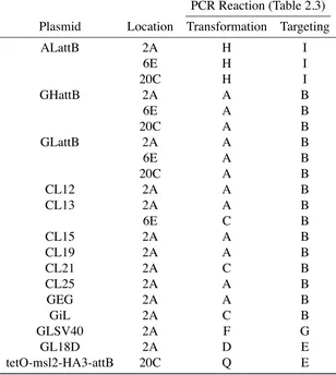
(13) List of Tables 2.1 2.2 2.3 2.4. Description of fly stocks C61–C65 for ϕC31 injections . . . . . . . . . . . Reaction guide for PCR confirmations of ϕC31 Drosophila transformations PCR Reaction Detail . . . . . . . . . . . . . . . . . . . . . . . . . . . . . Homogenate volume required for β -galactosidase assays . . . . . . . . . .. 4.1 4.2 4.3. Summary of ϕC31 transformations with arm-lacZ . . . . . . . . . . . . . . . . 89 β-galactosidase activity in hemisected flies carrying arm-lacZ at X-linked attPsites 94 β-galactosidase activity in heads of flies carrying arm-lacZ or GMR-lacZ at X-linked attP sites . . . . . . . . . . . . . . . . . . . . . . . . . . . . . . . . 97. 5.1. Altered β-galactosidase activity in flies carrying modified GMR-lacZ constructs at 2A . . . . . . . . . . . . . . . . . . . . . . . . . . . . . . . . . . . . . . . β-galactosidase activity in flies carrying constructs DRE-GAGA-GL or GiL at 6E attP site . . . . . . . . . . . . . . . . . . . . . . . . . . . . . . . . . . . β-galactosidase activity in flies with lacZ insertions at mixed sites . . . . . . Two copy β-galactosidase activities differed from expected two-fold increases Unaltered β-galactosidase activity in flies carrying modified GMR-lacZ constructs at 86F . . . . . . . . . . . . . . . . . . . . . . . . . . . . . . . . . . Effect on female β-galactosidase activity with over-expressed msl2 . . . . . . β-galactosidase activity from a single copy of tetO-GMR-lacZ, with or without an additional burst of early expression . . . . . . . . . . . . . . . . . . . . .. 5.2 5.3 5.4 5.5 5.6 5.7. . . . .. . . . .. 31 35 43 54. . 114 . 118 . 119 . 121 . 123 . 125 . 133. A.1 Fly strains used in this study . . . . . . . . . . . . . . . . . . . . . . . . . . . 172 A.2 Oligonucleotides used in this study . . . . . . . . . . . . . . . . . . . . . . . . 174 A.3 Plasmids used or created in this study . . . . . . . . . . . . . . . . . . . . . . 177 C.1 GMR-hid eye sizes with altered MSL levels . . . . . . . . . . . . . . . . . . . 184 D.1 Flies per category eye size . . . . . . . . . . . . . . . . . . . . . . . . . . . . 185 F.1. Calculations of band intensities for RT-PCR gel . . . . . . . . . . . . . . . . . 190. G.1 ϕC31 recombinase transformations . . . . . . . . . . . . . . . . . . . . . . . . 192 G.2 P element transformations . . . . . . . . . . . . . . . . . . . . . . . . . . . . 194 H.1 Transgenic Drosophila stocks created in this study. xi. . . . . . . . . . . . . . . . 195.
(14) xii.
(15) List of abbrv Aside from standard molecular biology abbreviations, and le Système international d’unités (SI) conventions, the following abbreviations are used: ˜ 6PGD abbrv a.k.a. arm BAD β-gal ChIP DRE FUM G G6PD GFP GMR H3K36 H3K36me3 H3S10 H4K16 HA HAT hid HIS hsp IRES LB mle mof. approximately 6-phosphogluconate dehydrogenase abbreviations also known as armadillo biotinylation activation domain β-galactosidase Chromatin Immuno-precipitation DNA replication-related element fumarase generation glucose-6-phosphate dehydrogenase green fluorescent protein Glass multimer reporter histone 3, lysine 36 tri-methylated H3K36 histone 3, serine 10 histone 4, lysine 16 hemaglutanin histone acetyl transferase head involution defective 6 histidine residues heat-shock protein internal ribosome entry site Luria-Bertani maleless males-absent on the first. xiii. MRE msl NURF p PCNA RFP roX RT rtTA SCF SV40 Sxl tet tetO TetR TRE TRF2 tsp tTA tTS UAS UTR X-gal y YFP w WT. MSL recognition element male-specific lethal nucleosome remodeling factor plasmid proliferating cell nuclear antigen red fluorescent protein RNA on the X reverse transcriptase reverse tTA DNA supercoiling factor Simian vacuolating virus 40 Sex-Lethal tetracycline tetracycline operator (TRE) tetracycline repressor tetracycline response element TBP-related factor transcription start point tetracycline-controlled transcriptional activator tetracycline-controlled transcriptional silencer upstream activation sequence un-translated region 5-bromo-4chloro-3indolyl-β D-galactopyranoside yellow yellow fluorescent protein white wild-type.
(16) Chapter 1 Introduction. 1.
(17) 1.1. Dosage Compensation in Drosophila melanogaster. A serious problem of gene deficiency exists in eukaryotic species with sex chromosomes. Where individuals of different sexes differ in their complement of chromosomes, compensation must be made for the accompanying gene dosage problems. The term “Dosage Compensation” is applied to a collection of mechanisms that have evolved to solve this problem, and has been most extensively studied in mammals, Caenorhabditis elegans, and Drosophila melanogaster. Essentially two problems must be solved: equilisation of the sex to autosome difference, and equilisation of the sex chromosomes between the sexes. Whereas all three systems appear to up-regulate the expression of X-linked genes to provide equality between the single male X chromosome and paired autosomes, they differ somewhat in their solutions to the second problem. The now over-expressed X chromosomes in mammalian females and C. elegans hermaphrodites (both XX) are down-regulated in a female-specific manner to achieve the correct expression level (Lyon, 1961; Meyer and Casson, 1986). The system in Drosophila, described below, has no need of the added complexity of female-specific down-regulation, as the up-regulation of the X is male-specific. Interesting biological questions thus arise: What mediates the up-regulation in Drosophila males? How is the process male-specific? How is the X chromosome targeted? What is the mechanism by which a precise two-fold regulation is attained?. 1.1.1. Compensation of X chromosome genes. X-linked expression is equal between sexes. Male Drosophila posses only one X chromosome, and yet mutations in X-linked genes display phenotypes equivalent to those observed in females, which have two X chromosomes. This was first observed with the apricot eye-colour produced by the mutant wa allele of the X-linked white gene (Muller et al., 1931). Males with only one wa allele, and females with two, both display the same level of eye pigmentation. Yet the eye colour is inherently capable of responding to differences in gene dose, as levels of pigmentation alter in response to gene duplications and deletions within a sex (Muller, 1932). Muller’s results suggest that males and females make equal amounts of X-linked gene products. This is also observed for X-linked genes that encode enzymes with easily measurable activities, such as fumarase (FUM), tryptophan 2,3-dioxygenase, glucose-6-phosphate dehydrogenase (G6PD) and 6-phosphogluconate dehydrogenase (6PGD) (Whitney and Lucchesi, 1972; Tobler et al., 1971; Seecof et al., 1969). More directly, amounts of protein are observed to be equal between the sexes, such as the peptide from the X-linked Sgs-4 gene (Korge, 1975). The equality in protein levels is a direct result of modifications to the process of transcrip-. 2.
(18) tion. Microscopically-visible polytene chromosomes of the salivary glands were incubated in (tritiated) [3 H]uridine and examined by autoradiography (Mukherjee and Beermann, 1965). The amount of radio-labelled uridine incorporated by the single male X was equal to the total incorporated by both female X chromosomes, implying an equivalent level of RNA synthesis. Similar results are observed in the quantification of specific RNA transcripts of X-linked genes, including the serine-4 tRNA, and the mRNAs from Sgs-4 and the G6PD gene Zw (Birchler et al., 1982; Breen and Lucchesi, 1986; Kaiser et al., 1986; Ganguly et al., 1985). The increased level of transcription observed from male X chromosomes could theoretically be a result of more X-linked DNA in males. However, measurements confirming twice the amount of X-linked chromosomal DNA in females as in males (Aronson et al., 1954; Rudkin, 1964) are convincing arguments against the possibilities of earlier replication or unequal replication of the male X (Roehrdanz and Lucchesi, 1977). Dosage compensation therefore acts at the level of transcription, to equalise the amount of X-linked RNA transcribed between the sexes. Transcription of male X chromosome is doubled. Although the simplest mechanism for the equalisation between sexes is an up-regulation of the male X chromosomal genes, the same result could be achieved through a down-regulation of the female X chromosomes. However, both female X chromosomes are active, as occasional observations of unpaired female X chromosomes in autoradiography experiments reveal that both chromosomes incorporate equal levels of tritiated uridine (Mukherjee and Beermann, 1965). Furthermore, the level of uridine incorporation to the X chromosomes by either sex is also equal to that incorporated by a pair of autosomes, suggesting that both female X chromosomes are transcribed at the expected level, and that two copies of all genes provide the required level of gene expression. The theory of up-regulation in males is also supported by the identification of male-specific factors that are implicated in dosage compensation (Section 1.1.2). Even the simple cytological observation that the width of the male X chromosome appears equal to that of the paired female X chromosomes (Offermann, 1936) now suggests a “hyperinflation” of the male X (Lakhotia and Mukherjee, 1969), given that no extra chromosomal material is present in the male X (Aronson et al., 1954; Rudkin, 1964). A modified male-specific model is also possible. Large, aneuploid deficiencies in chromosomal dose often lead to up-regulation of other genomic regions, presumably due to the loss of more negative than positive gene regulators (Birchler et al., 2001). As the single X chromosome in males resembles aneuploidy, we might expect all other autosomes to increase in expression, and the main mechanism of dosage compensation would therefore be to reduce the male autosomal level of expression, to restore the balance (Birchler et al., 2003). However,. 3.
(19) significant reductions in the amounts of known compensation machinery through the use of RNAi reduce the level of mRNAs produced from X-linked genes, but do not alter levels from autosomes (Hamada et al., 2005; Straub et al., 2005a). The change in the level of expression from specific X-linked genes is clear even when normalised to specific genomic DNA, thus removing the doubts present with normalisation to autosomal transcripts. Perhaps the gradual evolution of the sex chromosomes in Drosophila did not present equivalent biological dosage problems to those observed in spontaneous aneuploidy (Straub et al., 2005a). The simplest explanation remains the most likely: dosage compensation in Drosophila is achieved through a male-specific up-regulation of transcription from the single X chromosome.. 1.1.2. Known factors linked to compensation. Male-specific lethal proteins. Five proteins that are essential for male survival have been linked to dosage compensation, and are collectively termed the male-specific lethal or MSL proteins. Extensive screens of ethyl methane sulphonate-treated chromosomes identified male-specific lethal alleles of the genes msl-1, msl-2, and maleless (mle) on the second chromosome (Belote and Lucchesi, 1980b); msl-3 on the third (Lucchesi et al., 1982); and males-absent on the first (mof ) on the X chromosome (Hilfiker et al., 1997). The male-specific lethality caused by loss of these genes does not necessarily mean that they have no general, or even female-specific (but less critical) roles. Basic characteristics of the five proteins can be outlined, following the molecular cloning of the respective genes (Kuroda et al., 1991; Palmer et al., 1993; Bashaw and Baker, 1995; Gorman et al., 1995; Kelley et al., 1995; Zhou et al., 1995; Hilfiker et al., 1997). The roles of MSL1 and MSL2 appear to be largely structural, although MSL2 contains a RING finger domain that may have E3-ubiquitin ligase activity (Bashaw and Baker, 1995; Kelley et al., 1995; Zhou et al., 1995; Fang et al., 2003). MSL3 contains a chromodomain capable of binding methylated nucleosomes (Buscaino et al., 2006; Larschan et al., 2007; Sural et al., 2008). MOF is a histone acetyl transferase (HAT), but also has a zinc-finger motif important for nucleosome binding, and a chromo-barrel domain known to bind RNA (Hilfiker et al., 1997; Smith et al., 2000; Akhtar and Becker, 2000; Akhtar et al., 2000; Akhtar and Becker, 2001). Proteins with male-specific roles are good candidates for the effectors of dosage compensation. Indeed, MSL1, MSL2, and MLE are necessary for the unusually wide male X chromosome, and MLE for the increased tritiated uridine uptake by the male X, and the doubling of Sgs-4 mRNA (Gorman et al., 1993; Belote and Lucchesi, 1980a; Breen and Lucchesi, 1986). Likewise, mutations in MSL1, MSL2, and MLE eliminate the compensation of FUM, G6PD, and 6GPD (Belote and Lucchesi, 1980a). Perhaps most convincingly, fluorescent antibodies against all five MSL proteins reveal hundreds of binding sites specifically on the male X chro-. 4.
(20) mosome, although both MLE and MOF appear to bind autosomes and female X chromosomes to a lesser degree (Kuroda et al., 1991; Palmer et al., 1993; Bashaw and Baker, 1995; Kelley et al., 1995; Zhou et al., 1995; Gorman et al., 1995; Gu et al., 1998). Taken together, these observations suggest the involvement of the MSL proteins in dosage compensation. A male-specific chromatin-modifying complex. The sub-nuclear localisation of the MSL proteins to the male X chromosome suggests synergy of action, or even physical interactions between the proteins. Indeed, dual labelling of the male X chromosome by fluorescent antibodies against pairs of MSLs shows that they bind to the same locations (Bone et al., 1994; Bashaw and Baker, 1995; Gorman et al., 1995). Furthermore, the wild-type binding pattern of any one protein depends on the presence of all other MSLs (Gorman et al., 1993; Palmer et al., 1994; Gorman et al., 1995; Bashaw and Baker, 1995; Hilfiker et al., 1997; Gu et al., 1998; Buscaino et al., 2003). As amounts of protein also seem interdependent (Gorman et al., 1995; Chang and Kuroda, 1998; Buscaino et al., 2003), the stabilising interactions of a protein complex seem likely. A basic description of the interactions within the complex is possible. MSL1, 2, 3, and MOF co-immunoprecipitate, but MLE is more loosely associated (Kelley et al., 1995; Copps et al., 1998; Smith et al., 2000). A subset of X chromosomal sites is bound by MSL1 or MSL2 in the absence of MSL3, MLE, or MOF, suggesting the possibility of partial complexes consisting of just MSL1 and MSL2 (Palmer et al., 1994; Bashaw and Baker, 1995; Hilfiker et al., 1997; Lyman et al., 1997). Nevertheless, the structural core of the complex is MSL1, as direct, independent, in vitro associations are observed with MSL2, MSL3, and MOF, yet not amongst most other MSL proteins (Copps et al., 1998; Scott et al., 2000; Morales et al., 2004; Li et al., 2005). Recombinant MOF and MSL3 also bind weakly in vitro, but MSL1 and MSL3 together as a pair are necessary for MOF association, and specificity of MOF acetyl-transferase activity (Buscaino et al., 2003; Morales et al., 2004). An RNA component to the complex is suggested by the observation that MSL3, MLE and MOF (but not MSL1 or MSL2) do not localise to the X chromosome upon treatment with RNase (Richter et al., 1996; Akhtar et al., 2000; Buscaino et al., 2003). Accordingly, the two X-linked, non-coding RNAs roX1 and roX2 (RNA on the X) have been identified in a screen for sex-specific expression (Han et al., 1996; Amrein and Axel, 1997). The roX RNAs are implicated in dosage compensation, as they are only expressed in males, specifically coat the male X chromosome, co-localise to the same sites as MSL1, and can be detected by RT-PCR in immuno-precipitates of MSL1, MSL3, MLE, and MOF (Amrein and Axel, 1997; Meller et al., 1997; Franke and Baker, 1999; Meller et al., 2000; Akhtar et al., 2000; Smith et al., 2000; Buscaino et al., 2003). The known RNA helicase and dsRNA-binding abilities of MLE (Gibson and Thompson, 1994; Lee et al., 1997) make it the obvious candidate to bind roX RNA,. 5.
(21) and indeed neither RNA is present if MLE carries a mutation in the helicase domain (Gu et al., 2000). Nevertheless, the chromo-barrel domains of recombinant MSL3 or MOF both bind RNA non-specifically in vitro, and a specific in vivo interaction between roX2 and MOF seems likely, as the RNA is not co-immunoprecipitated when MOF is mutant in the chromo-barrel domain (Akhtar et al., 2000). The importance of the RNAs in dosage compensation is clear, as a double roX1 and roX2 mutation results in a characteristic drop in male viability, and rescues the lethality caused by inappropriate compensation when dosage compensation is induced in females (as discussed below) (Franke and Baker, 1999; Meller and Rattner, 2002). Despite large sequence and length differences, the two RNAs appear to be functionally redundant, as flies with either gene mutated display no phenotype whatsoever (Meller et al., 1997; Franke and Baker, 1999; Meller and Rattner, 2002). Given that MSL1, MSL2, and MLE do not localise to the X chromosome in double roX mutants (Franke and Baker, 1999), the main role of the RNAs appears to be in the targeting of the MSL complex. This is elaborated on in Section 1.1.4. The most well-defined role of the MSL complex is to acetylate the histones of the male X chromosome. Antibodies specific for the isoform of histone 4 acetylated at lysine 16 (H4K16Ac) bind at least three times higher to the male X than to the autosomes or female X chromosome (Turner et al., 1992). The acetylation is linked to dosage compensation, as the isoform colocalises with MSL1 and MLE, and the male X enrichment disappears with mutations in any of the MSLs (Bone et al., 1994; Hilfiker et al., 1997). Radio-labelled acetyl CoA is incorporated into free histones or nucleosomes when incubated with recombinant MOF, but not when the acetyl CoA binding site in MOF is mutated (Akhtar and Becker, 2000). Furthermore, a partially-purified MSL complex mediates the same specific histone acetylation, but not when MOF is mutated as before, implying that MOF is the only histone acetyl-transferase associated with the complex (Smith et al., 2000). Thus the ribonucleoprotein MSL complex specifically binds to and modifies the chromatin of the Drosophila male X chromosome. Associated proteins are difficult to detect. Additional factors involved in any aspect of dosage compensation have a more general role in both sexes, thus cannot be identified by male-specific lethality. Association with dosage compensation can sometimes be inferred if hypomorphic alleles affect male more than female viability, but this method relies on the fortuitous generation of these alleles. Other factors can instead be identified through their effects on the male X chromosome morphology or gene expression. Physical interactions with, or proximity to, known MSL components also implies guilt by association. The best characterised factor aside from the MSL proteins is the JIL-1 tandem kinase. Null alleles of JIL-1 are recessive lethal, but hypomorphs have a lower male than female viability,. 6.
(22) and a condensed chromatin structure most striking on the male X chromosome (Wang et al., 2001; Deng et al., 2005a). Indeed JIL-1 binds chromatin, and is enriched on the male X chromosome, where it co-localises and physically interacts with MSL proteins (Jin et al., 1999, 2000). The MSL complex appears to recruit JIL-1, as X-bound JIL-1 requires the MSL proteins, but not vice versa. JIL-1 phosphorylates both itself and histone 3 at serine 10 (H3S10), an epigenetic mark of active chromatin (Jin et al., 1999; Wang et al., 2001). Reduced levels of JIL1 increase the amount of histone 3 di-methylated at lysine 9, and associated HP1, on the male X chromosome, signaling an increased heterochromatic state (Zhang et al., 2006). Although the effect on X-linked transcription levels remains to be determined, JIL-1 is necessary for dosage compensation of white-mediated eye colour (Lerach et al., 2005). Thus recruitment of JIL-1 by the MSL complex causes enrichment of a second chromatin modification, beyond H4K16 acetylation, on the male X chromosome, and is likely a vital part of the dosage compensation mechanism. Similar lines of evidence also link the DNA supercoiling factor (SCF) with dosage compensation (Furuhashi et al., 2006). Like JIL-1, SCF binds chromatin and is enriched on the male X chromosome. SCF on the male X chromosome co-localises with, and is dependent upon, MSL proteins. Reduction of SCF by RNAi leaves MSL complex on the X chromosome, but abolishes the associated male-specific transcriptional increases, and reduces male viability. These results suggest that MSL complex also recruits SCF to aid in the up-regulation, although the consequent effect is less obvious than for the JIL-1 kinase. Indirect associations with several modifiers of chromatin are also apparent. Hypomorphic alleles of the GAGA factor affect males more severely than females, but do not co-localise on the male X chromosome (Greenberg et al., 2004). Mutated components of the nucleosome remodeling factor (NURF) complex give the polytenised male X chromosome a decondensed appearance, and require MSL proteins to do so (Deuring et al., 2000; Badenhorst et al., 2002; Corona et al., 2002; Bai et al., 2007). Similarly, mutated or reduced amounts of heterochromatin-associated proteins SU(VAR)3-7 and HP1 de-condense all chromatin, most noticeably that of the male X chromosome, and are more lethal to males (Seum et al., 2002; Delattre et al., 2004; Liu et al., 2005). As discussed below (Section 1.1.5), a balance between repressive and stimulatory effectors of the male X chromosome may be necessary for correct levels of expression. As a chromatin modifier, the MSL complex will necessarily interact with many other proteins. Mass spectrometry of MSL3 or MOF immuno-precipitates identifies several of these immediate contacts (Mendjan et al., 2006). In high-stringency conditions (such that MLE, JIL1 and SCF are not retained), several components of nuclear pores and regulatory exosomes are robustly detected. Reduction of two nuclear pore components (Mtor and Nup153) removes MSL proteins from the male X chromosome, and abolishes dosage compensation. This provides. 7.
(23) evidence for a structural aspect to dosage compensation; separable from chromatin regulation.. 1.1.3. Sex-specific compensation. MSL2 is male-specific. Given that dosage compensation is a male-specific up-regulation of X-linked gene transcription, some molecular control must exist to activate the mechanism in males. Presence of a Y chromosome does not lead to dosage compensation, as males or females with an incorrect number of Y chromosomes are not abnormal (Belote and Lucchesi, 1980b; Seecof et al., 1969). Dosage compensation also acts independently from the doublesex and transformer genes, which are involved in sex determination (Belote and Lucchesi, 1980b; Fukunaga et al., 1975). However, the MSL2 protein is absent in females, exhibits differential splicing between the sexes, and contains binding sites for the female-specific Sex-Lethal (SXL) protein in the 5′ and 3′ untranslated regions (UTR) of the msl-2 transcript (Bashaw and Baker, 1995; Kelley et al., 1995; Zhou et al., 1995). SXL is an RNA-binding protein that sex-specifically splices its own transcript and that of transformer, ultimately controlling the sex determination pathway (Sosnowski et al., 1989; Bell et al., 1991). SXL also binds to and represses translation of the msl-2 transcript in females, preventing MSL complex formation (Bashaw and Baker, 1997; Kelley et al., 1997). Sucrose gradient analysis of the assembly of the ribosome complex on msl-2 RNA reveals that the presence of SXL inhibits association of the 40S ribosomal subunit, and therefore acts to block at least the initiation of translation (Gebauer et al., 2003). Ectopic expression of an msl-2 gene lacking the SXL binding sites leads to MSL complex assembly in females, and a corresponding 80 % decrease in female viability (Kelley et al., 1995). MSL2 induction leads to dosage compensation. MSL2 is not the only component expressed in a sex-specific manner. Amounts of both MSL1 and MSL3 are lower in females, roX1 is not expressed after embryogenesis, and roX2 transcripts are completely absent (Gorman et al., 1995; Amrein and Axel, 1997; Meller et al., 1997; Meller, 2003). Although the translation of MSL1 in females is also weakly repressed by SXL, the main reason for the lower protein levels is the lack of stabilisation by MSL2 (Chang and Kuroda, 1998). Induced MSL2 expression in females increases at least MSL1 levels, and depletion of MSL2 by RNAi in males leads to a decrease in both MSL1 and MSL3 levels (Kelley et al., 1995; Buscaino et al., 2003). Over-expression of both MSL1 and MSL2 in females results. 8.
(24) in full dosage compensation, and corresponding 100 % female lethality (Chang and Kuroda, 1998). Expression of the roX RNAs is also directly regulated by the MSL proteins. Levels of roX1 RNA drop heavily with RNAi directed against MSL2, and no roX transcripts are detected when any of the MSLs are absent (Straub et al., 2005b; Amrein and Axel, 1997; Meller et al., 1997). MSL1 can be detected binding to translocations of the roX genes to the autosomes, and roX2 DNA can be detected in immunoprecipitates of either MSL1 or MSL2 (Kelley et al., 1999; Smith et al., 2001). A male-specific DNaseI hypersensitive site (DHS) is present in each of the roX genes, and small regions spanning either DHS are sufficient to bind MSL1 at ectopic locations (Kageyama et al., 2001; Park et al., 2003). Short stretches of residues, including three that resemble binding sites for GAGA factor, are conserved between both DHS regions in several related Drosophila species, and mutations in each site reduce the MSL-binding ability (Park et al., 2003). Deletions of either DHS abolish transgenic roX RNA expression, and addition of the DHS to a roX1 reporter gene provides male-specific up-regulation that is dependent on the MSL proteins (Bai et al., 2004). Accordingly, induction of MSL2 in females gives normal roX RNA levels (Amrein and Axel, 1997). The ability of the MSLs to regulate roX expression may be distinct from the complex’s normal dosage compensation function. In contrast to the known requirements for dosage compensation, roX regulation does not require presence of the roX RNAs, nor a functional RING finger in MSL2 (Bai et al., 2004; Rattner and Meller, 2004). Furthermore, roX1 induction at the endogenous location does not appear to require MSL3, MLE, or MOF, nor even the DHS (Rattner and Meller, 2004). As the DHS can attract MSL complex, this surprising result may indicate an otherwise high local concentration of MSL proteins at the roX1 location. In any case, the importance of MSL2 is clear. Given that MSL2 also stabilises the levels of the other MSL proteins, the key sex-specific control of dosage compensation is thus the regulation of MSL2.. 1.1.4. Targeting the male X chromosome. Dosage compensation involves the co-ordinate regulation of hundreds of male genes on a specific chromosome, so some means of identifying the X chromosome must exist. Translocations of large fragments of the X to autosomes retain compensation of their corresponding genes, and the characteristic puffed chromatin morphology indicitive of hyper-transcription (Seecof et al., 1969; Tobler et al., 1971; Oh et al., 2004). Likewise, the activity levels of genes on autosome to X translocations remain not compensated (Lakhotia and Mukherjee, 1969; Roehrdanz and Lucchesi, 1977). This reinforces the natural assumption that gene-specific elements mark X-linked genes for compensation. However, transpositions of individual genes do not fully support this model. Transpositions of some genes from the X to an autosome retain compensation,. 9.
(25) but autosomal genes inserted on the X often acquire a level of compensation (Krumm et al., 1985; Scholnick et al., 1983; Spradling and Rubin, 1983). This complicates the situation by further implying that the X chromosome environment is in some way special. Indeed, limiting the compensation to X-linked genes appears to involve several levels of control. The problem can be best addressed by identifying how the MSL proteins, the main effectors of dosage compensation, are targeted to the X chromosome. The roX RNAs aid MSL complex targeting. Although deletion of both roX genes is generally not tolerated by males, a varying number of developmentally-delayed adults can survive, depending on the severity of the roX mutation (Meller and Rattner, 2002; Deng et al., 2005b). As the severity of roX deletion increases, the number of X chromosome sites bound by the MSLs decreases, and the number of autosomal sites increases, to the point where most MSL complex binds to the centromeric heterochromatin. MSL binding to roX DHS sites on autosomes is also abolished in the absence of endogenous roX genes (Park et al., 2003). Note that in both cases the targeting ability of roX RNA is unrelated to the chromosomal location of the roX gene, as correct MSL binding is restored upon expression of transgenic roX RNA from another chromosome (Park et al., 2003). Structurally, the same region of MSL2 that is necessary to incorporate roX RNAs is also needed to correctly locate the complex to the X chromosome (Li et al., 2008). Inclusion of the roX RNA in the complex therefore increases the binding specificity of the MSL proteins, to aid correct compensation of the X chromosome. The roX genes as chromosome markers. As the genes for the roX RNAs are themselves on the X chromosome, and the regulation of these genes necessarily involves MSL binding (Section 1.1.3), they may serve as a molecular marker of the X chromosome. Once located to the X chromosome, the complex would then “spread” to the rest of the genes to be compensated. Indeed, the MSL complex is sometimes observed to spread up to 1 Mb into the chromatin surrounding an autosomal roX transgene, and can increase the transcription of a coupled reporter gene (Kelley et al., 1999; Meller et al., 2000; Henry et al., 2001). Spreading can even be observed from a multimerised nine-copy tandem repeat of the roX1 DHS (Kageyama et al., 2001). While spreading may contribute to targeting, roX gene localisation on the X is not necessary, as an autosomal roX transgene can rescue the lethality of a double roX deletion, and correctly target the MSL complex to the X (Meller and Rattner, 2002; Park et al., 2002). Much like the gene dosage problem that is being solved, the targeting of dosage compensation to the X appears to depend upon a delicate balance between roX RNAs and the MSL. 10.
(26) proteins themselves. Over-expressed MLE or MOF bind to all chromosomes, indicating an inherent chromatin-binding ability, whereas over-expressed MSL1 and MSL2 together bind in excess to the X, giving a shortened, hyper-inflated chromosome (Richter et al., 1996; Gu et al., 2000; Oh et al., 2003). The affinity of MSL1 and MSL2 for the X chromosome is so great that the over-expressed pair can even partly rescue the lethality of a double roX mutant, restore the correct chromatin appearance, and return mis-localised MSL proteins to the X (Oh et al., 2003; Deng et al., 2005b). However, under normal circumstances roX RNAs appear to compete for a limited pool of MSL proteins, as the extent of spreading from a roX transgene is inversely proportional to the number of roX genes (Park et al., 2002). Spreading is only seen from a roX gene when transcribed, and is most extensive with high levels of MSLs or a reduced number of roX genes (Park et al., 2002; Oh et al., 2003; Demakova et al., 2003; Bai et al., 2004). Abnormally high spreading may thus reflect a locally high concentration of assembling complexes on the nascent RNA, and under a normal MSL / roX ratio the contribution of this mechanism to targeting is likely low. A hierarchy of sites with affinity for the MSL complex. Although deletion of MSL1 or MSL2 removes MSL complex from the X under any circumstances, partial complexes lacking MSL3, MLE, or MOF bind a subset of sites in females with a modified MSL2 that allows complex formation (Palmer et al., 1994; Bashaw and Baker, 1995; Gu et al., 1998). These “high affinity” sites number about 60 to 70, differ slightly depending on which MSL component is lacking, and appear to overlap only moderately with the subset of sites bound with limiting roX RNA (Lyman et al., 1997; Demakova et al., 2003; Deng et al., 2005b; Dahlsveen et al., 2006). Strong MSL binding is also seen at the roX genes in these females, but as double roX mutations are lethal, and repeated searches for roX-like transcripts identify no new candidates, the other high affinity sites are unlikely to resemble the roX genes (Meller and Rattner, 2002; Deng et al., 2005b; Fujii and Amrein, 2002; Oh et al., 2003). Indeed, the best characterised high affinity site (at cytological position 18D101 ) produces no transcript, and has no GAGA-like sites (Oh et al., 2004). Similarities can nevertheless be seen to the MSL binding site in the roX genes, as the 18D10 site also spans a DHS that is more pronounced in males, and this DHS can attract MSL complex binding to an autosomal location, especially if multimerised. The MSL complex can occasionally spread into neighbouring chromatin from both the larger 18D10 fragment and the four-copy multimer of the DHS, and the spreading increases with reductions in roX genes, indicating that the high affinity sites also compete for the limiting MSL proteins. Several of the highest affinity sites (including that at 18D10) are detected in immuno-precipitates of MSL1, but the ability of each to attract MSL appears to differ (Dahlsveen et al., 2006). Systematic deletion analysis of the strongest sites 1. recently correctly located at 18D11. 11.
(27) shows the significance of general DHS elements containing degenerate sequences, including a GA nucleotide pair, for MSL attraction (Gilfillan et al., 2007). It is tempting to conclude that both roX genes and the high affinity sites serve to mark the X chromosome, and that the MSL proteins once bound can then diffuse to the remaining Xlinked chromatin, but the situation is yet more subtle than that. The large translocations of X segments to autosomes always retain their normal MSL binding pattern, even if they encompass no obvious high affinity nor roX site (Fagegaltier and Baker, 2004; Oh et al., 2004). The MSL complex is also unable to spread into surrounding autosomal chromatin, nor onto an autosomal fragment translocated to the X, even when the foreign chromatin is immediately adjacent to a roX gene (Fagegaltier and Baker, 2004). Furthermore, the number of X sites bound by the complex can also be controlled by limiting the amount of MSL complex (Demakova et al., 2003). Severe msl-2 alleles allow binding to only four sites, increasingly functional alleles bind more sites until a wild-type pattern is reached, and over-expressed MSL1 and MSL2 bind additional autosomal and heterochromatic sites. Thus a hierarchy of sites exist, with differing affinities for the MSL complex. Each site appears to individually recruit the complex, with no obvious “spreading” linearly from high to low sites. Low amounts of complex, or partial complexes lacking MSL3, MLE, or MOF, are sufficient to bind the sites of highest affinity, but the full binding pattern is only achieved with correct levels of all MSL proteins. Chromosomal studies of MSL targeting are inherently low resolution, with each fluorescent band representing many kilobases of chromatin-bound DNA. To accurately identify many high affinity sites, immuno-precipitates of MSL proteins in limited binding patterns can be passed over microarrays representing the X chromosome (ChIP on chip) (Alekseyenko et al., 2008; Straub et al., 2008). Many such sites possess a defined MSL recognition element (MRE) of 20–30 bp, encompassing a nearly exact eleven-copy repeat of GA di-nucleotides. Highthroughput sequencing of MSL3 immuno-precipitates (ChIP-seq) bound in a wild-type male pattern confirms that between 150 and 300 of the sites with highest affinity for MSL complex encompass an MRE (Alekseyenko et al., 2008). The MREs in the high affinity sites lie predominantly in intronic or non-coding sequences near genes. However, although very strongly bound at MRE elements, MSL proteins also bind robustly over tens of kilobases of surrounding DNA in a high affinity site. An additional mechanism must be responsible for the final stage of MSL attraction to the X chromosome. Targeting to active chromatin. As with the identification of high affinity sites, the nature of all bound sites can be resolved by microarray analysis of MSL immuno-precipitates from wild-type male tissues (Alekseyenko et al., 2006; Gilfillan et al., 2006; Legube et al., 2006). When at full occupancy, the MSL proteins bind over 700 sites. In contrast to the location of high affinity sites, the complete binding. 12.
(28) pattern almost exclusively resides within genes rather than intergenic sequences, and within exons not introns, and is clearly enriched for coding sequences over un-translated regions. On binding profiles of all genes scaled by gene length, MSL proteins peak towards the 3′ end of target genes. About 90 % of the MSL target genes are expressed, and 88 % of the target sites clearly coincide with RNA polymerase II sites. It appears that the act of transcription can attract the MSL complex, as MSL binding is also seen at GAL4-induced transcription of random X chromosomal sequences (Sass et al., 2003). Furthermore, transcription of the X-linked mof gene at autosomal insertions attracts MSL complex even when different promoters are used, or an antisense transcript of the gene is produced instead (Kind and Akhtar, 2007). Thus the MSL complex is attracted to active chromatin. The ability to identify active chromatin may be provided by an affinity of MSL3 for an associated chromatin epigenetic mark. Methylation of lysine 36 of histone 3 (H3K36) is generally associated with active chromatin (Saunders et al., 2006). Whilst di-methylation of H3K36 is enriched at active promoters, tri-methylation (H3K36me3) peaks towards the 3′ end of the CDS (Pokholok et al., 2005; Rao et al., 2005; Barski et al., 2007; Mikkelsen et al., 2007; Bell et al., 2008). This is reminiscent of the 3′ localisation of the Drosophila MSL proteins, which partially co-localise with H3K36me3 on polytene X chromosomes (Bell et al., 2008). At the high resolution of ChIP on chip, MSL3 binding co-localises with H3K36me3 on the male X chromosome, and binds autosomal genes surrounding a roX2 translocation proportional to H3K36me3 enrichment (Larschan et al., 2007). Recombinant MSL3 binds nucleosomes containing H3K36me3, and binding is impaired by deletions in its chromodomain (Larschan et al., 2007; Buscaino et al., 2006; Sural et al., 2008). Depletion of the relevant histone methylase Set2 (alias KMT3) by RNAi reduces MSL binding to target genes, and impairs consequent H4K16 acetylation and dosage compensation (Larschan et al., 2007; Bell et al., 2008). Indeed, tri-methylation of H3K36 is a better predictor of MSL binding than gene transcription (Larschan et al., 2007). Targeting of the MSL complex to the X chromosome is thus best described as a two-step process. Initial attraction of the complex to high affinity sites is likely a complex combination of sequence-specific MREs, nucleosome occupancy, and chromatin marks. Subsequent dispersal of the MSLs to all sites requires the recognition of active chromatin marks including H3K36me3 by the chromodomain of MSL3. This distinction in targeting steps is clearly seen in immunoprecipitates of MSL3, and MSL3 mutated in the chromodomain (Sural et al., 2008). Wild-type MSL3 binds around 1400 X-linked genes in 700 clusters, but MSL3 lacking the chromodomain binds on average only one gene per cluster. The direct effect on dosage compensation is a higher dependancy on MSL2 for compensation of genes close to a MRE, but more reliance on MSL3 (and MOF) for genes further away (Straub et al., 2008). Taken together with the demonstrated importance of transcription, it appears that the presence of active genes themselves leads to the final pattern of MSL occupancy.. 13.
(29) Establishment of MSL binding. Although the nature of the MSL binding sites is mostly determined, the dynamics surrounding the initiation of MSL binding are relatively poorly understood. MSL proteins are expressed early in the developing embryo, and bound complex is detected in all somatic cells from the blastoderm stage onwards (about 2.5 hours after egg laying) (Rastelli et al., 1995; Franke et al., 1996). Maternal MSL1, MSL3, and MLE are provided, but complex cannot form until MSL2 expression after fertilisation. Bound complex requires prior roX expression, but differences between the two roX genes mean the very early complex may differ slightly (Meller, 2003). roX1 is expressed at about 2 hours after egg laying; roX2 at around 6 hours. Thus bound complex in the first few hours may differ slightly from that in later stages. The earliest available evidence of dosage compensation is microarray analysis of late (12–14 hour) embryo transcripts (Straub et al., 2005a). The ability of the complex to disassociate with the X chromosome is also unclear. Bound sites on polytene chromosomes are mostly identical across developmental time and tissue type, but some tissue- and stage-specificity is also evident (Sass et al., 2003; Kotlikova et al., 2006). However, fluorescent signals of bound MSL2 do not recover in 20 minutes after photo-bleaching, suggesting a very stable association with the X chromosome (Straub et al., 2005b). Chromosomewide ChIP on chip results confirm that the complex mostly binds similar genes in different tissues, with the differentially-bound genes reflecting tissue-specific transcripts (Alekseyenko et al., 2006; Legube et al., 2006). Furthermore, the genes that remain bound in larval salivary glands are mostly a subset of those bound in early (4–6 hour) embryos, implying that MSL binding is mostly established in embryogenesis, then maintained through development.. 1.1.5. Potential mechanisms. Binding of the MSL complex does not necessarily lead to transcriptional up-regulation. Although undoubtedly the MSL complex and related proteins are responsible for the transcriptional increase, there are several observations that indicate MSL binding and up-regulation are separable events. The amount of bound MSL by a gene correlates to the transcriptional change when MSL is depleted, confirming the MSL proteins as the main agents (Gilfillan et al., 2006). However, the transcription of many un-bound genes are also affected by MSL depletion, and can score as ‘compensated’ in male to female transcript comparisons (Alekseyenko et al., 2006; Legube et al., 2006). The complex can also bind without up-regulation, as a minority of the high affinity sites that attract the complex to an autosomal location cannot also increase a corresponding reporter gene’s activity (Alekseyenko et al., 2008). What, then, is the consequence of MSL complex binding? How is transcription increased? MSL binding appears to indirectly affect transcription by modifying chromatin. The com-. 14.
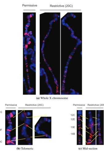
(30) plex does not increase polymerase initiation like a transcription factor would, but must rather facilitate transcription once begun. Although MSL binding correlates with transcription and active chromatin, the ChIP on chip results for MSL complex and RNA Polymerase II do not exactly overlap (Gilfillan et al., 2006). Neither do MSL proteins exactly co-localise with polymerase nor initiation factors on polytene chromosomes (Kotlikova et al., 2006; Legube et al., 2006). In fact the complex also binds several tRNA genes transcribed by RNA Polymerase III, and known to be compensated (Gilfillan et al., 2006; Birchler et al., 1982). There are no reports of MSL enrichment in promoter regions, no interactions with RNA Polymerase II or any general transcription factors, and no genetic or cytogenetic indication that any might be involved. Instead, MSL targets are predominantly 3′ coding sequences, and many of the proteins involved modify chromatin. As the acetylation of H4K16 by MOF also peaks towards the 3′ of coding sequences (Smith et al., 2001; Kind et al., 2008), it seems more likely that the complex facilitates increased transcription elongation through chromatin modification. Positive effects on transcription counteract repressive actions. As MSL complex de-condenses chromatin and increases transcription, it necessarily antagonises factors that negatively affect gene activity. In some cases this effect can be specifically observed. Insertions of a roX1 transgene marked by mini-white sometimes show position effect variegation of male eye colour that is affected by levels of heterochromatin-associated factors (Kelley and Kuroda, 2003). In these cases, female eyes are very pale or even white, implying specific repression of the mini-white gene. This repression is relieved in males or females expressing msl2 in the eye, due to roX1mediated attraction of MSL complex, and up-regulation of mini-white. More specifically, in vitro transcription can be repressed by assembly of nucleosomes on the template, but relieved by histone acetylation from recombinant MOF (Akhtar and Becker, 2000). Relief of repression is also evident at the roX1 locus itself, which MSL complex positively regulates (Section1.1.3). Basal expression of roX1 is constitutively suppressed in both sexes via the same DHS necessary for MSL up-regulation (Bai et al., 2004). The repression is dependent on the main component of the NURF complex, demonstrating direct antagonistic effects of the two chromatin-modifying complexes (Bai et al., 2007). If relief of repression is a common theme to MSL action, it may be that the apparent two-fold transcriptional increase is actually a product of a balance between repression and stimulation. The MSL complex may also have a global effect. By affecting the chromosome as a whole, the expression of individual genes could be altered without direct binding by the complex. The male X chromosome tends to locate towards the periphery of the nucleus in interphase cells (Rastelli et al., 1995; Rastelli and Kuroda, 1998), and directly interacts with nuclear pore components (Mendjan et al., 2006). As nuclear pore mutants de-localise MSL proteins from the X chromosome, their effect on MSL targeting necessarily also impacts dosage compensation. However, the association with the pores may also help euchromatic regions of the male. 15.
(31) X chromosome form sub-nuclear domains. Concentration of the genes to be compensated in a particular area would be an efficient use of MSL complex, JIL-1, SCF, and the corresponding chromatin modifications. The possibility of global regulation of the chromosome would explain the above discrepancy between MSL binding and gene regulation. Dosage compensation may thus be ‘action at a distance’; not just chromosomal in effect, but in approach.. 1.1.6. Summary. Dosage compensation is the equilisation of gene expression from unequal doses of genes. Drosophila males up-regulate transcription from their single X chromosome to match the level produced from XX females. Five male-specific lethal (MSL) genes are absolutely required in males, and encode the main agents of the up-regulation. At least these five proteins, together with either or both of two non-coding roX RNAs, form the MSL complex, which binds to and modifies the chromatin of the male X chromosome. Female-specific translational repression of a key MSL protein (MSL2), and subsequent loss of auto-regulatory and stabilising effects, limits the MSL complex to males. Attraction of the MSL complex to the X chromosome is a complicated and finely-balanced process. A hierarchy of sites with differing affinities for the MSL complex compete for a limiting pool of MSL proteins. The roX loci are two of the most potent attractors, likely due to MSL regulation of roX expression, and complex assembly on nascent roX transcripts. A further 70–300 high affinity sites share a common 21 bp MSL recognition element (MRE). The differing affinities of each site are probably due to differing strengths of MRE consensus, combined with nucleosome occupancy and surrounding chromatin state. Attraction to the X chromosome may be a two-step process: first to the roX genes and (predominantly non-coding) high affinity sites, then to surrounding active genes. Indeed most MSL sites are within active genes. The establishment of chromosome binding and the dynamics of the association remain relatively unknown. The mechanism by which gene dose is compensated is by modifying chromatin. The MSL complex and co-factors collectively alter X-linked euchromatin to create a de-condensed state that facilitates increased transcription. Rather than enhancing polymerase initiation at promoters, MSL proteins accumulate at 3′ coding sequences, where they likely affect transcription elongation or RNA processing. The characterised activities of histone acetylation and phosphorylation act at least in some cases to relieve repression, demonstrating that final two-fold transcriptional changes may be a product of antagonistic effects. Such chromatin alteration could also have a long-range effect on transcription. By creating sub-nuclear domains of decondensed chromatin, the modifying factors can be efficiently shared to globally modify the entire chromosome. Thus the effect on transcription is indirect: an average two-fold increase provided by a combination of local and global changes to chromatin structure.. 16.
(32) 1.2. Transvection. Homologous chromosomes of Dipteran species pair not just in meiosis but also through all stages of a mitotic cell cycle (McKee, 2004). Homologous pairing in somatic cells initiates during the maternal to zygotic transition of embryogenesis (Fung et al., 1998; Gemkow et al., 1998). The mechanism responsible for pairing remains unknown, but is likely a complex interaction of chromatin factors. A key contributing factor may be Drosophila topoisomerase II, as reduction of this enzyme by RNAi significantly reduces pairing in several cell lines (Williams et al., 2007). The tight association of homologous chromosomes can affect gene expression levels, in a variety of ways known collectively as ‘transvection’. Drosophila transvection effects were first observed in a study of Ultrabithorax (Lewis, 1954). Chromosomal re-arrangements with break points near Ultrabithorax affect the ability of certain alleles to complement others. In effect, such complementation only occurs if the alleles are homologously paired (Duncan, 2002; Ashburner et al., 2005). Several transvection pairing effects appear to share a common mechanism. Gans (1953) first noticed that the recessive allele of zeste z1 would produce yellow eyes only if flies carried two wild-type copies of white. This observation reflects a transvection effect on white, as both white alleles need to be homologously paired (Jack and Judd, 1979). The Zeste protein tends to form aggregates that suppress the eye enhancer of white, and the z1 allele enhances this suppression (Bickel and Pirrotta, 1990; Qian et al., 1992). Suppression is probably mediated through local chromatin modifications, as Zeste binds the three Polycomb group proteins: Psc, Scm, and E(z) (Saurin et al., 2001). Pairing-dependent repression is not unique to zeste repression of white, and affects at least cubitus-interruptus, Sex combs reduced, and brown (Locke and Tartof, 1994; Pattatucci and Kaufman, 1991; Henikoff and Dreesen, 1989). Pairing-dependent transcription enhancement, or relief of repression, similarly occurs at decapentaplegic, and eyes absent (Ashburner et al., 2005), but is most well-studied at yellow. As for Ultrabithorax, different alleles of yellow can complement each other, but only if homologously paired (Geyer et al., 1990). The yellow allele y82f29 lacks wing and body enhancers, but can be expressed in these tissues if the homologous allele (e.g. y1#8 ) supplies wild-type enhancer elements (Morris et al., 1998). Interestingly, the enhancers only work in trans when the cis promoter is inactive (Morris et al., 2004). The effect is also independent of the specific basal promoter, as wing and body enhancers can activate in trans a yellow transgene expressed from white or eve promoters (Lee and Wu, 2006). Transvection is thus the synergistic action of enhancers or repressors between homologous copies of a gene (Duncan, 2002; Ashburner et al., 2005). The exact nature of gene regulation in each instance may vary considerably, and can include heterochromatic effects. Subtle interactions with zeste occur with at least Ultrabithorax, decapentaplegic, eyes absent, and yellow. 17.
Figure
+7
Outline
Basis of Experimental Design
β galactosidase enzymatic assays
Drosophila photography
Repressive chromatin effects reduced un-induced chromosome binding
Evaluation of the inducible dosage compensation system
Future directions
GMR-hid insufficient to report dosage compensation
Promoter elements increased the male:female ratio
An earlier burst of expression insufficient for compensation of GMR-lacZ
Effect of promoter elements on dosage compensation
Related documents
It was decided that with the presence of such significant red flag signs that she should undergo advanced imaging, in this case an MRI, that revealed an underlying malignancy, which
The paper assessed the challenges facing the successful operations of Public Procurement Act 2007 and the result showed that the size and complexity of public procurement,
The scattergram represents the distribution with age of 69 determinations of concentration of potassium in serum of 39 premature infants with respiratory distress syndrome (Table
19% serve a county. Fourteen per cent of the centers provide service for adjoining states in addition to the states in which they are located; usually these adjoining states have
Assessing the Impact of Biodiversity Conservation in the Management of Maize Stalk Borer (Busseola f
Field experiments were conducted at Ebonyi State University Research Farm during 2009 and 2010 farming seasons to evaluate the effect of intercropping maize with
The paper is discussed for various techniques for sensor localization and various interpolation methods for variety of prediction methods used by various applications
In this PhD thesis new organic NIR materials (both π-conjugated polymers and small molecules) based on α,β-unsubstituted meso-positioning thienyl BODIPY have been
