DISSERTATION
“A STUDY ON THE ACCURACY OF ALVARADO SCORING SYSTEM IN THE DIAGNOSOSIS OF ACUTE APPENDICITIS”
Dissertation submitted to
THE TAMILNADU DR.M.G.R MEDICAL UNIVERSITY
In partial
for the award of the degree of
M.S.
THANJAVUR MEDICAL COLLEGE
THE TAMILNADU DR.M.G.R.MEDICAL UNIVERSITY
DISSERTATION ON
“A STUDY ON THE ACCURACY OF ALVARADO SCORING SYSTEM IN THE DIAGNOSOSIS OF ACUTE APPENDICITIS”
Dissertation submitted to
THE TAMILNADU DR.M.G.R MEDICAL UNIVERSITY
In partial fulfillment of the regulations
for the award of the degree of
M.S.-GENERAL SURGERY BRANCH – I
THANJAVUR MEDICAL COLLEGE THANJAVUR-613 004
THE TAMILNADU DR.M.G.R.MEDICAL UNIVERSITY CHENNAI – 600 032
APRIL – 2015
“A STUDY ON THE ACCURACY OF ALVARADO SCORING SYSTEM IN THE DIAGNOSOSIS OF ACUTE APPENDICITIS”
THE TAMILNADU DR.M.G.R MEDICAL UNIVERSITY
CERTIFICATE
This is to certify that the dissertation entitled “A STUDY ON THE ACCURACY OF ALVARADO SCORING SYSTEM IN THE DIAGNOSIS OF ACUTE APPENDICITIS”, is a bonafide record of work done by Dr.M.JEDIDIAH SAMRAJ, in the Department of Surgery, Thanjavur Medical College Hospital, Thanjavur, during his post-graduate
course 2012-2015. This is submitted in partial fulfillment for the award of
M.S., degree examination, Branch I ( General Surgery ) to be held in April
2015 under The Tamil Nadu Dr. M. G. R. Medical University, Chennai.
Prof. Dr. M. Elangovan. M.S., Unit Chief,
Department of General Surgery, Thanjavur Medical College,
Thanjavur. Prof. Dr. V. Balakrishnan. M.S., Head of the Department,
Department of General Surgery, Thanjavur Medical College, Thanjavur.
DEAN,
DECLARATION
I declare that this dissertation entitled “A STUDY ON THE ACCURACY OF ALVARADO SCORING SYSTEM IN THE DIAGNOSOS OF ACUTE APPENDICITIS” is a record work done by me in the Department of General Surgery,Thanjavur Medical College
Hospital, Thanjavur, during my Post-Graduate course from 2012-15 under
the guidance and supervision of Prof. Dr. M. Elangovan. M.S., my Unit Chief, Prof. Dr. V. Balakrishnan. M.S., Professor and Head of the Department, Department of General Surgery, Thanjavur Medical College.
It is submitted in partial fulfillment for the award of M.S., degree
examination, Branch I ( General Surgery ) to be held in April 2015 under
The Tamil Nadu Dr. M. G. R. Medical University, Chennai.
This work has not been submitted previously by me for the award of
any degree or diploma from any other university.
ACKNOWLEDGEMENT
I express my sincere gratitude to Prof. Dr. M. Elangovan. M.S., my unit chief and Professor of Surgery , Thanjavur Medical College Hospital for his constant guidance and encouragement throughout the
period of this study.
I am deeply indebted to Prof. Dr. V. Balakrishnan, M.S., Professor and Head of Department of Surgery for being a source of inspiration and
guidance. I express my thanks to Prof. Dr. Yeganathan, M.S.,Prof. Dr. Karunakaran, M.S., Prof. Dr. Rajendiran,M.S., Prof. Dr. K. Sathyabama, M. S., and also the former professors, Prof. Dr. Shanthini, M.S., M.S., Prof. Dr. Maragatha mani, M.S., Prof. Dr. Rajagopal, M.S.,
for their valuable guidance.
I thank, Dr. W. Premalatha Sharon Rose, M.S., Dr. V. Vimal, M.S., Dr. Ashok kumar, M.S., Dr. R. Aravindh, M.S., and other assistant professors for their guidance throughout the period of study.
I thank Prof. Dr. K. Mahadevan, M.S., Dean, Thanjavur Medical College and Prof. Dr. P.G. Sankara narayanan, M.D., Dean I/C, Thanjavur Medical College for permitting me utilize the hospital facilities
during this study.
I would like to thank my parents Dr.V.Manickaraj and
course of this study.
I am very much thankful to all the patients who, despite all their
ABSTRACT
Background is Objective
Acute appendicitis is the most common surgical emergency .Failure to
make an earlier diagnosis leads to complications like perforation and intra
abdominal abscess. The objectives of the study are
1) To study the accuracy of the Alvarado score system in the diagnosis
of acute apprndicitis by comparing with the histopathological
examination report of removed appendix.
2) To compare the negative laparotomy in this study against other study
Methods
The study population consists of patient admitted with pain in the right iliac
fossa and the diagnoses of acute appendicitis was confirmed by
investigations like ultra sonogram abdomen and CT abdomen. The severity
of the acute appendicitis is scored by Alvarado score and the patients were
managed according to the severity.
Results
Most cases acute appendicitis presented with right iliac fossa pain
and it was commoner in younger age group. Patients with higher Alvarado
score were considered to have acute appendicitis. Such patients were
surgical intervention reduced the morbitidy and mortality associated with
acute appendicitis. Those patients with lesser scores were managed
conservatively and discharged home.
Interpretation and Conclusion
Alvarado scoring system has a higher sensitivity and positive predictive
value. Thus scoring system is a dynamic one, allowing observation and re
evaluation of clinical picture. Its value in decision making is high both in
males and females. In females the reason is multifactorial and diagnostic
laparocopy is essential. Its application improves diagnostic accuracy and
considerably reduces the negative laparotomy rate. The score system is
quite and cost effective.
KEY WORDS
INDEX
s.no CONTENTS PAGE NO
1 INTRODUCTION 1
2 OBJECTIVES 7
3 REVIEW OF LITERATURE 8
4 METHODOLOGY 71
5 RESULTS 74
6 DISCUSSION 93
7 SUMMARY 99
LIST OF TABLES
S.NO TITLE PAGE
1 AGE AND SEX DISTRIBUTION 75
2 SEX DISTRIBUTION 77
3 FREQUENCY DISTRIBUTION 78 4 RESULTS OF APPLICATION OF ALVARADO 80 5 MEAN SCORES OF DIFFERENT GROUP 81 6 INDIVIDUAL FEATURES OF DIFFERENT
SCORES
82
7 RESULTS OF GROUP A 84
8 RESULTS OF GROUP B 86
9 RESULTS OF GROUP C 88
10 STATISTICAL ANALYSIS OF THE STUDY 90
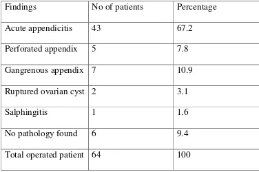
11 FINAL DIAGNOSIS 90
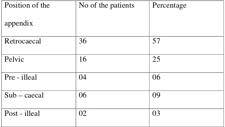
12 POSITION OF APPENDIX 91
LIST OF GRAPHS
S.NO TITILE PAGE NO
1 AGE AND SEX DISTRIBUTION
76
2 SEX DISTRIBUTION 77 3 FREQUENCY
DISTRIBUTION
79
4 RESULTS OF APPLICATION OF ALVARADO SCORE
80
5 MEANSCORE OF DIFFERENT GROUPS
81
6 INDIVIDUAL FEATURES OF ALVARADO SCORE
83
7 RESULTS OF GROUP A 84 8 RESULTS OF GROUP B 86 9 RESULTS OF GROUP C 88 10 FINAL DIAGNOSIS 91 11 POSITION OF APPENDIX 92 12 COMPARATIVE ANALYSIS
OF ACCURACY
INTRODUCTION
“Diagnosis of appendicitis is usually easy” – as told by Sir Zachary
Cope, but still there is difficulty in diagnosing acute appendicitis. It is
nothing but the challenge we face while diagnosing acute appendicitis on
clinical grounds.
Acute appendicitis being a common cause of surgical emergency needs to
be diagnosed with accuracy at the earliest to reduce the morbidity and
mortality associated with it.2
The question Does this patient have appendicitis? , an important
question for the following reasons:
For the common causes of abdominal pain appendicitis is a one
such condition.
Western literatures report that 6% of population have risk of
suffering from appendicitis during their lifetime.3
Although the mortality due to complications of acute
appendicitis has dropped less than 1% with the advent of
antibiotics and early surgical intervention in elderly it is
approximately 5 to 15%.
The morbidity due to appendiceal perforation (rupture) and
rate is higher in elderly and children.
Failure to make an early diagnosis leads on to complications
like perforation, which in turn leads on intra abdominal
abscesses.
Th e negativ e laparotomy rat e range s fro m 15 % t o 35 %
and i s associate d with significant morbidity.4,5 The negative
laparotomy rate is significantl y higher in young women
(up to 45%) because of prevalence of pelvinflammatory
disease (PID) and other common obstetrical and
gynaecological disorders.4,5
Thus, diagnosing acute appendicitis accurately is very
important to decrease complications following appendicitis and
the morbidity and mortality associated with it.
Routine history & physical examination remains the most
effective and practical diagnostic modalities.7The typical history is
onset of generalized abdominal pain followed by anorexia and
nausea. Typically, the patient presents with central abdominal pain
shifting to the right lower quadrant. Vomiting may happen at this
time, especially in children. Depending on the severity of
any acute intra-abdominal process-local rebound tenderness, muscle
guarding, rigidity, cutaneous hyperesthesia, and tenderness on rectal
examination. Since, about a third of all patients with acute
appendicitis present with atypical symptoms, 4,8 the differential
diagnosis is varied such as gastroenteritis, regional enteritis, ovarian
&tubal disorders (in young women), Ureteric colic, peptic ulcer,
diverticulitis, mesenteric adenitits , cholecystitis.
The routine laboratory examination of blood and urine is
mandatory. In old patients elevated leukocyte count with shift to left
may be absent and it is usual finding in others.4C - reactive protein is
a non specific indicator of acute inflammatory conditions.
Estimation of CRP may help to support surgeon’s clinical diagnosis
and to reduce negative appendicectomies.10, 11
The roentgenogram findings like
1) Faecolith
2) Dilated loop of ileum – due to local ileus
3) Air fluid level in caecum
4) Haziness in right lower quadrant
5) Blurring of Psoas shadow
Among these the important findings are air fluid level in the terminal ileum
and dilatation of a loop of ileum. Both have a speficity of around 95%and
78% and sensitivity around 51% and 62%.
Contrast studies like barium enema, the major risk being the caecal
perforation, findings are often negative in such condition. Such findings are
also negative if the appendix got perforated, moreover it is
Time consuming for the radiologist.
Uncomfortable for the patient.
Entails ionizing radiation
Ultrasonogram with high frequency probe is useful in diagnosis of
acute appendicitis but it has its own limitation. There are many
prospective studies published which showed that findings in
ultrasonogram were important and it is used to help the surgeons to
arrive at the decision to operate. These studies showed an overall
accuracy of 87 to 96% with a sensitivity of around 94% and specificity
of around 86% to 100%. Blind ending tubular structure will give clue
clue for diagnosis and probe tenderness is an additional feature in USG.
Computerized tomogram also ha s its limitation like radiation exposure
and presence of fluid in right iliac fossa to diagnose acute appendicitis.
Laparoscopy has been shown by some authors to be particularly
useful in young women in reproductive age because
gynaecological conditions may mimic acute appendicitis. The rate
of diagnostic error is twice as high in women of reproductive age as
that in men.
Inspite of the advanced imaging modalities, the rate of negative
laparotomies is around 15-25%. The complication rate of
appendicectomy for a non inflamed appendix is also same as that of
inflamed appendix. It is around 13%.
The mortality rate of appendicectomy is around 0.65 for every
100 surgeries. Considering the mortality and the complications
associated with appendicectomy, if the patient is managed
conservatively, the delay in the intervention leads to perforation of
appendix in around 28%.
Alvarado A described the scoring system in 1986. M. Kalan, D. Tabot,WJCulliffe and AJ Rier in 1994 later modified it by
taking one laboratory finding of the scoring system. The Alvarado
appendicitis has been useful in the early diagnosis of acute
appendicitis as demonstrated by various studies and was helpful in
reducing the incidence of negative appendicectomies without
OBJECTIVES
To study the accuracy of Alvarado scoring system in the
diagnosis of acute appendicitis by comparing with the
histopathological examination report of removed appendix
specimen
To compare the negative laparotomies in this study against other
REVIEW OF LITREATURE
The word “appendicitis” refers to inflammation of appendix
veriformis. The literal meaning of appendix is an appendage – anything
that is attached to a larger or major part as a tail or limb. The Latin word,
Appendices vermiformis is a worm shaped tubular structure araisng from
the posteriomedial aspect of the caecum and about 2cm below the terminal
ileum. It is confined almost entirely to humans and the higher primates,
and occasionally be absent in humans.
HISTORICAL NOTE:
Though the presence of the appendix has been known for
centuries, the credit for its first description goes to the
physician-anatomist, BerengarioDaCapri, in the year 1521. In 1492 Leonardo
davinci clearly depicted the appendix in his anatomic drawings.
Though it was depicted in 1492 it came to light in 18tn century,
and was well illustrated in the AndreasVesalius work, “De
HumaniCorporisFabrica,” published in 1543.
EVOLUTION OF APPENDICITIS:
The disease appendicitis has been known for centuries. Aretaeus in
the second century A.D. described a case in which he drained an abscess
of the right part of the abdomen near the liver. This might have been a
Jean Fernel, the great French Physician, described a case of
perforated appendicitis in his UniversaMedicina, which was published
in 1554. He gave an account of a seven- year old girl who had diarrhea
for several days and her grandmother gave her a large quince. It stopped
her diarrhoea, but the girl began to have severe abdominal pain and
eventually she died. At autopsy the “caecum intestinum was narrow and
constrticted; also quince was found adherent to the inside and stopping of
the lumen”.
In 1711 Lorenz Heister, professor of surgery at Helmstadt
discovered a case of appendicitis when he was called to dissect the
body of a criminal who had been executed. In account he wrote later
that as he was “about to demonstrate the situation of the great
guts, found the vermiform process of the caecum preternaturally black,
adhering closer to the peritoneum than usual.”29
William Ballonius, in his Consiliorum Medicinalium published in
Geneva in 1734, gave the description of gangrenous appendicitis in the
living patient, although he did not use this term.
Sir Zachary Cope in his book “A history of Acute
& Oliver Prescott of New England reported perforation of appendix in
1812. However, J.B.Louyer-Villermay in 1824 emphasized the
importance of the condition in his paper, “Observations of Use in the
inflammatory Conditions of the Caecal Appendix” which was presented in
the Royal academy of medicine in Paris. Walcott Richard’s diagnosis of
perforation of appendix, which he described as “ulceration of the appendix
veriformis” in 1838, was confirmed on autopsy.29
During the nineteenth century, the caecum was considered as the
chief cause of trouble .All the diseases in the right lower quadrant was
attributed to caecum. The diseases of caecum and appendix were
considered to be same.All the troubles of the right lower quadrant were
termed under the term typhlitis, or inflammation of the caecum. Husson
and Dance in 1827, Goldbeck in 1830 and Dupuytren in 1835
developed the concept of inflammation arising in the cellular tissue
surrounding the caecum. It was Goldbeck who confined the term
“perityphlitis”26. Later J.F.H.Albers of Bonn described four varieties of
Frederick Merling in the study of the pathologic anatomy of the
appendix published in 1838 reported that a foreign body has been found
in the appendix and was thought to have caused gangrene. Since then
much has been written about foreign bodies in the appendix and are
blamed for perforations.29In 1965 R.E.Shaw reported that the stones
found in the appendix are true calculi, not just faecoliths. He said that
calculous appendicitis was more apt to gangrene and perforation.29
Reginald Fitz of Boston gave his classical paper on appendix before
the Association of American Physicians in 1863. His paper was based on
an analysis of 257 cases of perforating ulcer of appendix and of 209
cases clinically diagnosed as typhlitis and perityphliticabcess. The disease
was found to be most common in youngadults, especially males. A
faecal concretion or foreign body was present in three-fifths of cases.
He went on to discuss the origin of the term typhlitis, perityphlitis and
paratyphlitis abscess and concluded that in vast majority of cases the
primary cause was inflammation of the appendix. He preferred the term
“appendicitis” to all others. He wrote “in most cases of typhlitis, the
caecum is intact whilst the appendix is ulcerated and perforated.”
Surgeons in the United States discarded the old term of typhlitis in the
cause of inflammations in the right iliac fossa, and the previous concept of
caecum was discarded.
In1899 Charles Mcburney of New York illustrated that “exact
locality of the maximum tenderness, when one examines with the
fingertips in adults, is one-half to two inches inside the right anterior
spinous process of the ileum on the line drawn to the umbilicus. The
accuracy of this sign (Mcburney’s point), I have demonstrated in every
case operated upon by me since I first made the observation”29. This
point corresponds to the base of the appendix and therefore does not move
with the tip.
EVOLUTION OF APPENDICECTOMY:
According to R.G.Richardson in “The Surgeons Tale”, the first
appendicectomy was performed at St.Georges Hospital, London, in 1726
by Claudius Amyand. The patient, a boy, had hernia and a faecal
fistula. Richardson reported: “When he opened the scrotum he found
the appendix in the unusual position and moreover, that the appendix
was perforated by a pin. He removed the appendix and then dealt with
Hancock in London successfully drained an appendix abscess in
a female patient aged 30 years that was in her eighth month of
pregnancy in 1848. After incising the peritoneum, fluid was drained
and he made no search for the appendix.29Willard Parker, an
American surgeon, started draining appendiceal abscesses since 1867.
He did not remove the appendix and his technique is still used but the
appendix is removed later on.29
Lawson Tait, the great English surgeon, was the first to remove an
acutely inflamed appendix.26.He thought that his patient had a general
peritonitis resulting from rupture of caecum or appendix. However,
when he opened the abdomen he found “a large abscess which
extended deeply down towards the brim of the pelvis lying bare was the
vermiform appendix which was black and discoloured and gangrenous”.
The patient made a perfect recovery following appendicectomy and
drainage of abscess.29
Abraham Groves performed the first elective appendicectomy in
Canada in 1883. His patient was a twelve- year old boy. The appendix
was removed and the stump was cauterized with a heat probe heated
over the flame of a lamp. The patient recovered. Early operation for
(1855-1931), Charles Mcburney (1845-1913) and Murphy of Chicago.25
In 1894, Mcburney described his incision for appendicectomy. Though he
wasthe first to describe this incision, L.L.McArthur, who had used the
incision in more than 60 cases29, had used it for a longer time. Later
McBurney gave McArthur credit for using the incision first, but despite
this, it is still known as the Mcburney’s incision.
Later others modified the incision like Rutherford Morison in
1896, A.E.Rockey in 1905, and G.G.Davis in 1906.28 Noteworthy as
these various dates are, it is doubtful whether any of them areas
important in the history of the appendicectomy as 24th June 1902. The
coronation of King Edward VII had been arranged to take place on
26thJune 1902, but the king fell ill with abdominal pain and fever only a
few days before, At a consultation of some of the most distinguished
surgeons in the land, including Lord Lister, it was decided that the only
chance to save his life lay in urgent operation. Frederick Treves, who
had performed his first successful appendicectomy in 1887, opened
the abdomen and drained an appendix abscess on 24th June 1902. The
king made a good recovery and the operation was entirely successful.
After the postponed coronation on 9thaugust 1902, Treves received a
original members of the Order of Merit. When welcoming Lister to his
Council, the king is supposed to have said, ‘I know that is it had not been
ANATOMY:
Embryologically, the vermiform appendix is the part of the
caecum, which forms the blind end. It develops from the caudal part of
the midgut loop. A line is dawn from the anterior superior iliac spine and
the umbilicus. The junction of the medial two third and the lateral one
third is considered to be the proposed site of the base of the appendix as
described by McBurneys and it is called as Mc Burneys point. Intra
operatively the confluence of the taenia is used to identify the base of the
appendix.
Its length varies from 2cm to 20cm, with average length of
9cm. It may occupy one of the several positions, thus it may be
retrocaecal, retrocolic, pelvic or descending over the pelvic brim, in close
relation to the right uterine tube and ovary. Other positions are
occasionally seen especially when there is a long appendix mesentery
allowing greater mobility which include subcaecal, preileal and postilieal.
It has a mesoappendix with which it is attached to the ileal mesentery.
The lumen of the appendix is small (admits a matchstick). The opening
of the appendix into the caecum usually lies below and posterior to the
illeocaecal opening. The illeocaecal valve is nothing but a mucosal fold
which gaurds it and is not patent in all.
the free border of the meso appendix. It is usually the only supply for the
appendix. The base of the appendix lies in close association with the
appendicular artery and hence any inflammation will cause gangrene of
the appendix.
The recurrent appendicular artery araises from the posterior
caecal artery and it usually lies near the base of the appendix. If
recurrent appendicular artery is present it may anastamose with the
appendicular artery.
The venous drainage of the appendix is by appendicular vein
which drains into the illeocolic vein and in turn drains into the superior
mesenteric vein.
There is an about four to six lymphatic channel that drains into
the illeocolic node.
The symphathetic and parasymphathetic nerve supply to the
appendix is from superior mesenteric plexus.
Histologically appendix contains the following layers
1) Mucosa
2) Submucosa
4) Serosa
Mucosa:
The epithelium of the mucosa contains the following cells
1) Columnar cells
2) Mucous cells
3) Stem cells
4) Microfold cells
5) Neuroendocrine cells
Crypts of the appendix is larger and numerous. Each crypt is lined
columnar epithelium with mucous cells, neuroendocrine cells, microfold
cells and stem cells at the base.
Lamina propria:
It is made up of connective tissue that supports the epithelium. The
speciality is lamina propria of the appendix is rich in solitary lymphoid
follicles.
Muscularis mucosa: Muscularis mucosa contains circular and longitudinal muscle fibres.
Muscularis externa: it contains inner circular and outer longitudinal muscle
Serosa:
The appendix contains serosa except the place where the
mesoappendix is attached.
Though the physiologic role of the appendix is unproved and
immunologic function is suggested by its content of lymphoid tissue.
Nevertheless, it is a useful organ for surgeons as it can be used for on
table lavage of large bowel. It can also be used as a conduit for
permanent continent urinary diversion.
The position of the appendix can be anywhere along the arc with the
centre at the base of the caecum.23It is the only organ in the body that
has no constant anatomic position; in fact, its only constant feature is its
mode of origin from the ceacum. The various positions of the appendix
are: paracolic, retrocolic, preileal, postileal, promontoric, pelvis and
subcaecal. In situs inversus the appendix may lie in the left iliac fossa.
The position of the appendix as given by Sir C wakeley
1) Retrocaecal 74%
2) Pelvic 21%
3) Paracaecal 2%
4) Sub caecal 1.5%
ACUTE APPENDICITIS:
Incidence
Acute Appendicitis is one of the most common causes of the
acute surgical abdomen.8,26 .But since the disease is not notifiable, its
exact incidence is not known. There is an increase in the incidence of
acute appendicitis in Europe, America, and Australia. The rate of
appendicectomies in this population is around 16%. In the recent past there
is a decline in the incidence of acute appendicitis in these countries with the
appendicectomy rate of around 8.6%and 6.7% for males and females
respectively.
In England the total number of appendicectomies falls from
1,13,000 to 48,000 in the 20th century. There has been an annual
decrease of 17% in the numbers of appendicectomies performed
between 1987&1996 in Sweden. Appendicitis has shown an
association with western diet habits. It is also believed that there is a
familial tendency in this disease that could be explained to be due to
an inherited malformation of the organ. Anderson & colleagues
compared 29 children between the ages of 5 and 15 years suffering
from appendicitis with 29 controls. Twenty in the study group
parents and siblings.28However, family history of appendicitis has no
diagnostic value.
Pathology
Acute appendicitis is thought to arise from infection superimposed
on luminal obstruction. The lumen of the appendix becomes obstructed
by hyperplasia of submucus lymphoid follicles, fecolith, stricture, tumor,
or any pathological condition. Once obstruction occurs, continous mucus
secretion and inflammatory exudation increases intraluminal pressure,
obstructing lymphatic drainage. Oedema and mucosal ulceration develops
with bacterial translocation to the submucosa. Resolution may occur at
this point either spontaneously or in response to antibiotic therapy. If
this condition progresses, further distention of the appendix may cause
venous obstruction and ischemia of the appendix wall. With ischemia,
bacterial invasion occurs through the muscularispropria and sub mucosa,
producing acute appendicitis. Finally ischemic necrosis of the appendix
wall produces gangrenous appendicitis, with free bacterial
contamination of the peritoneal cavity. Alternatively, the greater
omentum and loops of small bowel become adherent to the inflamed
appendix, walling of the spread of peritoneal contamination, resulting in
normal appendix is similar to that of the normal colon. The appendiceal
flora remains constant throughout life with the exception of
Porphyromons gingivalis, which is seen in adults. The principal
organisms seen in the normal appendix, in acute appendicitis, and in
perforated appendicitis are Escherichia Coli and Bacteroides fragilis.
However, a wide variety of both facultative and anaerobic bacteria and
mycobacteria may be present. Appendicitis is a polymicrobial infection
with some series reporting up to 14 different organisms cultured in
patients with perforation. According to a study by Pieper and colleagues of
the bacteriology of 50 inflammed appendices, both aerobic and
anaerobic bacteria were isolated in all patients. Anaerobic isolates were
more than aerobic, 141 versus 96 isolates. E.Coli were the most common
aerobic bacterium (45 out of 50). Other gram negative aerobes like
klebsiella, and proteus and pseudomonas were isolated in ten patients.28
Enterococci were found in 15 patients and streptococci in 21
paitents. Among the anaerobes, the most common was Bacteroides
fragilis. Next in frequency were gram positive cocci. Clostridium
perfingeus was isolated from 9 patients.30There are two types of acute
appendicitis, Catarrhal & Obstructive appendicitis. Catarrhal appendicitis
is initially a mucosal and submucosal inflammation. Externally; the
appendix may be quite normal, or hyperemic in early stages. However
studded with dark brown hemorrhagic infarcts, patches of green
gangrene, or small ulcers. Eventually the appendix becomes swollen
and turgid and the serosa becomes roughened coated with fibrinous
exudates, in these cases the lumen of appendix is patent and these
cases rarely progress to gangrene. However the lymphoid hyperplasia
may lead to obstruction of the lumen and proceed to gangrene.
Furthermore, if the episode of catarrhal appendicitis resolves, adhesion
formation and kinking of the appendix may lead to a final episode of
acute obstructive appendicitis.28
Obstructive appendicitis is the dangerous type, since the appendix
becomes a closed loop of bowel containing feacal matter. When the
appendix gets obstructed, the appendix becomes distended with mucus in
which the bacteria proliferate. Because of increase in intraluminal
pressure, there is pressure atrophy of the mucosa and the bacteria
invade the deeper tissue plane. The inflammation of the wall of the
appendix leads to thrombosis of the vessels, as the appendix has an end
arterial blood supply, gangrene occurs inevitably followed by perforation
of the necrotic appendix wall.
Wilkie demonstrated the relationship between obstruction of the
appendicitis followed ligation of the appendix in the
rabbit.31Wangensteen and colleagues documented in 1937 and 1940
that combined obstruction and bacterial infection resulted in acute
appendicitis.
In two third of all gangrenous appendicitis, feacolith is in the
appendiceal lumen. A true fecolith is ovoid, about 1 to 2 cms in
length, and fecal coloured. The great majority of these fecoliths are
radioopaque and, in 10% of cases, contain sufficient calcium to be
demonstrated on plain x-ray film of the abdomen. Other foreign bodies
like food, debris, worms, or even gallstones have been found to obstruct
the appendix lumen.26one of the rare causes of obstructive appendicitis is
the appendix becoming strangulated in hernial sac. Thomas et al (1982)
reported seven such cases.34
The most frequent site of perforation is along the antimesenteric
border, usually near the tip, as the Appendicular artery is subserosal
at this point and more prone to be involved in the inflammatory process
and become thrombosed. After perforation a localized abscess may form
in the right iliac fossa or the pelvis, or diffuse peritonitis may ensue.
depends on many factors, including age of the patient, the virulence of the
invading bacteria, the rate at which he inflammatory condition has
progressed within the appendix and the position of the appendix.28 It is
usually stated that the poorer localization of the infection occurs in
infants because the omentum of the child is filmy and less able to form
a protective sheath around the inflamed appendix. A more likely
explanation is that delays in diagnosis are more prone to occur in
infants. Similar delays occur in the management of elderly persons.
Gangrenous appendix is more dangerous than the catarrhal type of
appendicitis. An appendix situated in the retrocaecal position is more
likely to form a local abscess than one in the pre ilieal or subcaecal
position.35
The consequences of a perforated appendix are potentially severe in
women of child bearing age. The relative risk of infertility is increased
three to five times in a female patient with a history of a ruptured
The entity of chronic or grumbling appendicitis is
controversial.28It has been well said that “the appendix does not
grumble – it either screams or remains silent.” Both the clinical and
experimental data support the belief that some patients have repeated
attacks of appendicitis. In fact, it is not unusual for one or more such
episodes to precede a full blown acute appendicits. In such cases, surgical
specimens have shown chronic inflammatory infiltrates depending on
whether the appendicectomy was performed during the attack or in
between the bouts.37Thus the term chronic appendicitis has been used.
But, it definitely does not mean prolonged abdominal pain lasting weeks
or months.
CLINICAL MANIFESTATIONS
The diagnosis and management of acute abdominal pain remains
one of the last bastions of clinical medicine. There is no other common
situation where clinical features, accurate diagnosis, and immediate
decision are of such importance. The diagnosis of acute appendicitis is
made primarily on the basis of the history and the physical findings,
with additional assistance from laboratory and radiographic
examinations. In appendicitis, there is highly characteristic sequence of
signs and symptoms.
localized colicky abdominal pain. This is due to the midgut visceral
discomfort in response to appendiceal inflammation and obstruction. The
pain is frequently initially noticed in the epigastric or periumbilical
region, presumably due to the distention of the appendix. This central
abdominal pain is followed by anorexia, nausea and vomiting. With
progressive inflammation of the appendix, the parietal peritoneum in the
right iliac fossa becomes irritated, producing more intense, constant and
localized somatic pain that begins to predominate. During the first 6
hours, there is rarely any alteration in temperature or pulse rate, after
some time, slight pyrexia with corresponding increase in pulse rate is
usual. Though the patient frequently complains of constipation
especially during early phase of visceral pain, many patients particularly
children may present with diarrhea. If the temperature is considerably
raised (i.e.>103°F) at the very beginning attack then appendicitis is less
likely unless there is perforation. And perforation is extremely
uncommon before 24-36 hours of onset of symptoms.38
Physical findings are determined by the anatomic position of the
inflamed appendix, as well as by whether the organ has already
ruptured when the patient is first examined. The order of occurrence of
recognized the importance of the sequence of symptoms. The march of
event is
Pain, usually epigastric or umbilical
Anorexia
Nausea or vomiting
Tenderness
Fever
Leucocytosis
The sequence of symptoms of pain abdomen followed by
vomiting and then by fever is termed as “Murphy’s syndrome”. If
vomiting occurs before pain abdomen then the diagnosis of acute
appendicitis is questionable and a peaceful night is assured to the
surgeon.24Murphy stated: “The symptoms occur almost without
exception in the above order, and when the order varies I always
question the diagnosis.” This dictum is usually true with occasional
exceptions.
Tenderness in the right iliac fossa (RIF) is a very important sign. The early deep tenderness is almost always detected just below the joining of
anterior superior iliac spine and the umbilicus. Tenderness over the
appendix, as the tenderness appears to be located actually in the
appendix itself. In fact, the site of the tenderness varies somewhat
according to the position of the appendix. Tenderness may be less in case
of retrocaecal or post ileal appendix. With a retrocecal or a post ileal
appendix, the anterior abdominal findings are less striking and
tenderness maybe most marked in the flank. When the inflamed
un-perforated appendix hangs over the brim of the pelvis or is lying
wholly within the pelvis; In the so called ‘silent appendix’, abdominal
findings may be entirely absent, and the diagnosis may be missed unless
the rectum is examined, pain is felt in the suprapubic area ,as well as
locally within the rectum.24,26
Peritoneal signs:
A)Mc Burney’s sign: Finger tip pressure is made over the Mc Burney’s point (i.e, at the junction of lateral third with medial two thirds of the
right spino-umbilical line), which if the sign is positive, registers the maximum abdominal tenderness.
B)Pointing test: When the patient is asked to point the site of pain this usually corresponds with the site of localized tenderness in
C) Rovsings sign: Palpation of the left iliac fossa may produce pain in the right iliac fossa (crossed tenderness). This sign appears to be due to
the shift of coils of ileum to the right impinging on an inflamed focus in
the right iliac fossa
D)Cough Test: When the patient coughs vigorously and holds his or her right lower quadrant of the abdomen or refuses to cough because of
pain, right lower quadrant peritonitis is confirmed.
E)Blumberg’s sign or Rebound tenderness or Release sign: Pain on abrupt release of the palpating hand in the right iliac fossa suggests
localized peritoneal irritation. However, since this exam causes severe
pain to the patient, it should not be elicited frequently.
F) Cope’s Psoas test: A retrocaecal appendix lies on the psoas major muscle. Inflammation of this causes irritation of psoas major muscle
which is concerned with flexion of hip joint. The patient is turned to the
G)Cope’s obturatortest :Internal rotation of hip in a patient with pelvic appendicitis, initiates pain as it lies over the obturator internus muscle.
H)Baldwing’s sign : A hand is placed over the right flank and the patient is asked to raise the right lower limb with knee extended, in retrocaecal
appendicitis this initiates pain and indicates the retrocecal position of the
appendix.
Local hyperesthesia in the Sherren’s triangle ( this is formed by lines joining the umbilicus, right anterior superior iliac spine and symphysis
pubis) is regarded as a good guide in diagnosis of gangrenous
appendicitis. This nearly always lies in the area of distribution of the
nerves from tenth, eleventh and twelfth dorsal and first lumbar spinal
segments. Hyperaesthesia signifies that the inflamed appendix is, as yet,
unperforated; when perforation occurs it passes off.
Guarding- a state of voluntary contraction and rigidity- a state of involuntary contraction are uncommon findings in the early stage.
Rigidity is usually present in case of diffuse peritonitis due to perforation.
However, the accuracy of these signs in diagnosing appendicitis is
regarding evaluation of the accuracy of the clinical presentation of
appendicitis. Three findings show a high positive likelihood ratio
(LR+) and, when present are most useful for identifying patients at
increased likelihood for appendicitis: right lower quadrant pain (LR+=8.0),
rigidity (LR+=4.0) and the migration of pain to right lower quadrant
(LR+=3.1). Unfortunately, no single component consistently provided a
low negative likelihood ratio (LR-) that would rule out appendicitis. The
absence of right lower quadrant pain and the presence of similar pain in
the past demonstrate powerful negative LRs (0.2and 0.3, respectively)
In another prospective study39, the diagnostic value of 21
elements of the history, clinical findings, body temperature and
laboratory examinations were assessed and compared in 496 patients
with suspected appendicitis. No single variable had sufficiently high
discriminating or predicting power to be used as a true diagnostic test.
But, the independent predictors of appendicitis were total leukocyte and
differential counts, CRP concentrations, rebound tenderness, abdominal
guarding and patient gender.
This study showed that the element of disease history had low
power in discriminating for appendicitis and advanced appendicitis.
power than history except the site of tenderness. A family history of
appendicitis, previous experience of similar symptoms, anorexia, nausea,
constipation, diarrhea or the progression of pain had no diagnostic value
for appendicitis. Right sided rectal tenderness was found to be a
DIFFICULTY IN DIAGNOSIS
SPECIAL FEATURES RETROCAECAL:
Localised rigidity is often absent and tenderness may not be elicited
by deep pressure. In retro-caecal appendix, it lies above the caecum, which
is filled with gas, prevents the pressure exerted by the hand from reaching
the inflamed structure. Rigidity of Quadratus lumborum and Psoas muscle
can occur. Flexion of the hip can occur due to the contact of the inflamed
appendix with the psoas muscle.
PELVIC:
If the appendix being pelvic in position, abdominal rigidity, Mc
Burneys point tenderness will be absent. Diarrhoea can occur due to
irritability of the rectum by the inflamed appendix which lies close to it. If
the inflamed appendix lies close to the bladder, it can cause increased
frequency of micturition.
POST ILLEAL:
Inflamed appendix lies behind the ileum. Migration of pain to right
iliac fossa will not occur in post illeal appendix. It may present like
diarrhoea with marked retching. There will be illdefined tenderness at the
SPECIAL FEATURES ACCORDING TO AGE Infants:
Appendicitis is rare in infants below 36 months of age. If acute
appendicitis occurs in infants it is severe because of delay in the diagnosis,
which leads to the occurrence of perforation and postoperative
complications.
If the appendicitis in children causes localized peritonitis, it will eventually
lead on to generalized peritonitis due to underdeveloped greater omentum.
Children:
In children with acute appendicitis, vomiting will be an important
symptom. Other important symptom is complete aversion to food.
Elderly:
In elderly patient due to lax abdominal wall, guarding may not be that
much manifested. Acute appendicitis with gangrene and perforation are
common in elderly. Sometimes in elderly the clinical picture may be like
that of subacute intestinal obstruction.. All the above said reasons lead to
delay in the diagnosis of acute appendicitis in elderly population leading to
Pregnancy:
In pregnancy acute appendicitis is one of the most common
emergency conditions. The classical Obstetric concept is caecum and
appendix are pushed upwards due to the enlarged uterus. The pain in acute
appendicitis is usually in the right iliac fossa only. Acute appendicitis in
pregnancy can occur in 1in 500 – 2000 pregnancies. The estimated fetal
loss is 3-5% and it can be even upto 20%, if perforation occurs.
Differential diagnosis:
Children:
In children the differential diagnoses are
1) Acute gastroenteritis
2) Mesenteric lymphadenitis
3) Meckels diverticulum
4) Henoch schonlein purpura
5) Lobar pneumonia
Acute gastroenteritis:
In acute gastroenteritis there will be pain and diarrhoea. It may
mimic acute appendicitis. There will be fever and dehydration.
Mesenteric lymphadenitis:
fossa. Pain is usually colicky in nature. Cervical lymphnodes may be
enlarged in it. In meckels due to the presence of ectopic gastric mucosa,
there may be frequent abdominal pain as intraluminal gastrointestinal
bleeding.
Intussusception:
Intussusception is much more common than acute appendicitis in
children. The age of presentation will be usually around 18 months. The
presentation of intussusception will be red currant jelly stools. The
management of it will be enema or open reduction.
Henoch schnolein purpura:
It is usually preceded by sore throat or respiratory tract infection.
There will be echymotic lesion in the extensor surface of the buttocks.
Microscopic hematuria with normal bleeding count is the common
presentation.
Lobar pneumonia:
Right sided abdominal pain due to right lower lobe pneumonia
and pleurisy may mimic acute appendicitis. In pneumonia, abdominal
symptoms and signs will be minimal. Respiratory system examination will
IN ADULTS:
1) Terminal ileitis
2) Ureteric colic
3) Rt sided pyelonephritis
4) Perforative peritonitis
5) Terminal ileitis
6) Rectus sheath haematoma
Terminal ileitis:
Terminal ileitis may be due to
1) Non specific
2) Specific
In specific type it may be due to the Chrons and Yersinia. A chronic
history of abdominal pain, weight loss, diarrhoea suggests regional ileitis
rather than acute appendicitis.
Yersinia enteroclitica can cause ileitis and it can cause inflammation
of caecum and appendix and mesenteric lymphadenopathy.
If mesenteric lymphadenopathy was there, node was divided into
two, one was sent for histopathological examination and other was sent for
Ureteric colic:
Pain due to ureteric calculus will be radiating from right loin to
groin. Pain will be colicky type of pain. Routine urine examination should
be done. Urine should be examined for red cells, pus cells, deposits, and
albumibn. X ray KUB may reveal renal stone or ureteric stone. Renal
USG or Intravenous urogram is usually diagnostic.
Right pyelonephritis:
Right sided pyelonephritis, pain occurs in right loin and also in right
iliac fossa Patient will have high grade fever and associated co
morbidities like diabetes. USG abdomen is the investigation of choice. CT
abdomen can also be used to differentiate the pyelonephritis and acute
appendicitis.
Perforated peptic ulcer:
If there is a duodenal perforation, the contents of perforation
passes on to the paracolic gutter and then to the right iliac fossa. If the
perforation got sealed , there will be collection in the right iliac fossa
which will lead to pain, tenderness in the right iliac fossa. Since it is
initially a duodenal perforation there will be previous history of abdominal
pain in the epigastric region. There will be usually findings of free air
Testicular torsion:
Testicular torsion is an important differential diagnosis in the acute
appendicitis. Since the patients are usually of young age, they may have
shyness to reveal the testicular pain. In such a situation it is necessary to
examine the external genitalia, which is tender on palpation.
Acute pancreatitis:
Acute pancreatitis is also an important differential diagnosis in
adults. In acute pancreatitis pain will be more on the epigastric region, may
radiate to back, but it can be confirmed by serum amylase or lipase. CT is
the investigation of choice for acute pancreatitis
Rectus sheath hematoma:
It is a rare differential diagnosis. The presentation may resemble
that of acute appendicitis but it usually follows an episode of strenuous
physical exercise. The gastrointestinal discomfort is usually absent in
rectus sheath hematoma.
On those people who are on anticoagulants, rectus sheath
Adult female:
In reproductive age group, the females can have gynaecological
diseases like pelvic inflammatory disease, torsion or haemorrhage or
rupture of ovarian cyst and ectopic pregnancy. The common differential
diagnoses are
1) Pelvic inflammatory disease
2) Mittelschmerz
3) Torsion / haemorrhage of ovarian cyst
4) Ectopic pregnancy- ruptured or unruptured.
Pelvic inflammatory disease:
It includes a group of diseases like
1) Salphingtis
2) Endometriosis and
3) Tubo ovarian sepsis
These disorders are commoner in reproductive age group. Patient
may give a history of discharge per vagina, dysmenorrhea, and burning
micturition.
On examination the patient, may have adnexal and cervical
tenderness. If pelvic inflammatory disease is suspected a high vaginal
swab should be taken for Chlamydia trachomatis and Neisseria
gonorrhoeae. Trans vaginal ultrasound can be done. If still there is a
antibiotics like metronidazole and ofloxacin for 14 days is the drug of
choice.
Mittelschmerz:
Rupture of ovarian follicle during mid cycle in the menstrual period
produces abdominal pain which may mimic appendicitis. Systemic
symptoms like fever may be absent.
Urine pregnancy test will be negative. If still there is a doubt in the
diagnosis of acute appendicitis, diagnostic laparoscopy may be needed.
There is an entity called retrograde menstruation which may mimic like
that of acute appendicitis.
Ectopic pregnancy:
Unruptured tubal pregnancy can mimic that of acute appendicitis.
Ruptured ectopic pregnancy with haemoperitoneum is unlikely to be like
that of acute appendicitis with perforation
• Tubal pregnancy,
• Tubal abortion,
Can mimic exactly that of acute appendicitis.
In such situation the urine pregnancy test will be positive and a
history of period of ammenorhea. Severe pain will be felt in the cervix on
vaginal examination
Patient should be asked for any pain in the right iliac fossa which is
Elderly:
In elderly people, the following are the differential diagnosis
1) Diverticulitis
2) Intestinal obstruction
3) Carcinoma caecum
In patients with long sigmoid loop, the colon may come and lie in
the right iliac fossa and diverticulitis of sigmoid colon may misdiagnosed
as acute appendicitis. The investigation of choice to differentiate the
diverticulitis and appendicitis is CT abdomen. If such a condition is
suspected conservative management with iv antibiotics and iv fluids
should be considered. Right colon diverticulitis is a rare entity and it is
difficult to distinguish between the diverticulitis and acute appendicitis. If
diverticulitis is the diagnosis, it should be treated conservatively and if it
fails laparoscopy or laparotomy can be considered.
Intestinal obstruction:
Only in elderly acute appendicitis and intestinal obstruction are
considered as differential diagnosis. If the diagnosis of intestinal
obstruction is made, it has to be managed conservatively followed by
Carcinoma caecum:
A perforation of caecum due to malignancy of caecum mimics
exactly that of perforated appendicitis.
History of altered bowel habits,
unexplained anaemia,
may raise the suspicion of carcinoma caecum.
On examination a mass may be palpable. The investigation of choice for
carcinoma caecum is CT abdomen.
Rare differential diagnosis:
1) Preherpetic pain
2) Tabetic crisis
3) Spinal condition
4) Porphyria
5) Diabetic ketoacidosis
6) Typhlitis
7) Leukemic illeocaecal syndrome
Preherpetic pain:
Herpes involving the d10 and d11 spinal nerves can cause severe
pain in the right iliac fossa. The pain in herpes will be severe and static not
as in appendicitis where it is migratory in nature. Heretic eruptions can
occur 3-8 hours after pain.
Tabetic crisis:
In tabetic crisis severe abdominal pain and vomiting can occur.
Additionally other symptoms and signs of tabes can occur.
Spinal conditions:
Spinal conditions causing abdominal pain that mimics that of acute
appendicitis can
Occur in childrens and elderly namely
• Tubercolosis of spine,
• Multiple myeloma,
• Metastatic deposits,
• Osteoporotic lesions,
All the above conditions can cause compression of nerve roots
leading on to pain. Usually in the above said conditions gastrointestinal
Porphyria:
Acute intermittent porphyria is an acute abdominal emergency.
Abdominal pain can mimic that of acute appendicitis. It is a rare
differential diagnosis in the children. There will be usually similar history
of abdominal pain in porphyria.
Diabetic ketoacidosis:
In diabetic ketoacidosis there will be severe pain in the abdomen. In
diabetic ketoacidosis the patient will be diabetic and plasma acetone will
be positive.
Usually diabetic ketoacidosis is common in insulin dependent
diabetes mellitus thereby it is commoner in childrens.
Typhilitis:
Initially the cause for acute appendicitis was thought to be due to
thyphoid. Thyllitis is still the differential diagnosis for acute appendicitis.
Leukemic syndrome:
It is a rare and a potentially life threatening condition.
Clostridial septecemia:
Clostridial septecemia is a rare progressively fatal condition.
Treatment is with appropriate antibiotics. Surgical intervention is rarely
Appendicitis in pregnancy, the risk is similar to that of non
pregnant woman of the same age. Appendicitis occurs more frequently
during the first two trimesters, and during this time period the symptoms
of appendicitis are similar to those seen in non pregnant women. During
the third trimester, the cecum and appendix are displaced upwards. This
results in localization of pain either more cephalad or laterally in the
flank, leading to delay in diagnosis and an increased incidence of
perforation and diffuse peritonitis as displacement of the omentum by
the uterus impairs localization of the inflamed appendix. It is the
peritonitis, and not the appendectomy, that poses the risk to the mother
and fetus alike, and therefore, early operation is the rule.
Nothing can be so easy or as difficult as the diagnosis of acute
appendicitis.The clinical examination and the investigations are
non-specific. Thus, the list of differential diagnosis is long.24-28. Some of
the entities in the differential diagnosis of appendicitis also require
operative therapy and are not made worse by an exploratory laparotomy,
but it is necessary to eliminate pancreatitis, myocardial infarcation, and
basal pneumonia for which surgery would be a blunder. The disease in
young children that are most frequently mistaken for acute appendicitis
pyelitis, small intestinal intussusception,enteric duplication, and basilar
pneumonia. In teenagers and adults, the differential diagnosis is different
in men and women. In young women, the differential diagnosis include
ruptured ectopic pregnancy, mittelschmertz, endometriosis, ureteric colic
and salpingitis. Chronic constipation also needs a considerationIn older
patients, the differential diagnosis include diverticulitis, a perforated
peptic ulcer, acute cholecystitis, acute pancreatitis, intestinal obstruction,
perforated caecal carcinoma, mesenteric vascular occlusion, rupturing
aortic aneurysm, and the disease entities already mentioned for young
DIAGNOSTIC STUDIES
Routine history and physical examination remain the most practical
diagnosis modalities. No laboratory or radiological test yet devised is
diagnostic of this condition.
White cell count:
The polymorpholeucocytosis is an important feature of acute
appendicitis. In three quarters of patients the white cell count is raised
above 12,000/cmm.4However, in others, the count may be slightly
raised or normal, especially in children.38Neutrophilia is also one of
the features of appendicitis. In 1982,Pieper et al40noted that 66.7% had
white cell count of 11,000/cmm or more and in only 5.5% it was raised
above 20,000/cmm. Anderson et al39 reported that the WBC and
neutrophils count had higher power in discriminating for advanced
appendicitis than for all appendicitis. Appendicitis was unlikely at lowest
level of the WBC and neutrophils count and rate (LR0.16-0.28 at WBC
count <8000/cmm, neutrophils count <7000/cmm, or rate<70%) and
likely at the highest WBC Count. Neutrophils count >13,000/cumm and
rate >85%. However, Coleman C et al reported that WBC is a poor
predictor of the severity of the disease in the diagnosis of acute
Urine examination:
The presence of hematuria or pus cells in the urine does not rule
out appendicitis. Irritation of ureter or urinary bladder by the inflamed
appendix may cause microscopic hematuria or
pyuria.24-26Graham(1965) quantitatively analysed midstream urine specimens in
71 patients operated upon with the diagnosis of acute appendicitis. Of
these, 62 had an acutely inflamed appendix removed and nine patients
had normal appendix. In this whole group, nine female patients had
microscopic pyuria and one also had hematuria. One male patient had
microscopic hematuria.26
C-reactive protein
CRP is a non specific acute phase reactant, which appears in
the sera of individuals in response to a variety of inflammatory
conditions and tissue necrosis. It is a non-specific indicator for acute
appendicitis. There have been various studies regarding the importance
of CRP in differentiating appendicitis from other non inflammatory
conditions of the abdomen.11One of the such studies showed that CRP
value is increased markedly only after appendiceal perforation or abscess
early marker of appendiceal inflammation. This study reported
that the CRP concentration and temperature had high power in
discrimating advanced appendicitis than all appendicitis. Also the CRP
concentration >10mg/L was found to be one of the independent predictors
of appendicitis.39
Radiography:
Plain films of abdomen in supine and erect position are of value in
differential diagnosis of acute abdominal pain. However, they are non
specific. Brookes and Killen42have described a number of radiological
signs in patients with acute appendicitis:
Fluid level localized to the caecum and to the terminal ileum
Localized ileus, with gas in the caecum, ascending colon or
terminal ileum
Increased soft tissue density in the right lower quadrant.
Blurring of right flank stripe, the radiolucent line
produced by fat between the peritoneum and transverse
abdominals.
A faecolith in the right iliac fossa
A gas filled appendix
Free peritoneal gas
Deformity of caecal gas shadow due to an adjacent inflammatory
mass They reviewed the x-rays of 200 patients underwent
laparotomy for acute appendicitis without knowing the diagnosis.
80% of patients with acute appendicitis had one or more of these
signs positive. However 37% of patients who had normal
appendix had similar x-ray findings. Thus, plain films of abdomen
are neither sensitive or specific to alter the maxim “If the
diagnosis of appendicitis remains in doubt ,still appendicectomy is
the accepted treatment ”.43
Ultrasonography :
In 1989, Julien B.C.M. Puylaert described the value of graded
compression sonography in the evaluation of acute appendicitis.
The accuracy afforded by sonography should keep negative laparotomy
rates at approximately 10%, clearly an improvement over the rate
achieved by instinct alone. Ultrasound proved most useful for those
patients who have an indeterminate probability to the disease upon
initial clinical examination. The sonographic hallmark of appendicitis is
direct visualization of the inflamed appendix. The typical appearance is
that of a concentrically layered, almost incompressible, sausage like