DESIGN, SYNTHESIS, CHARACTERIZATION AND
BIOLOGICAL EVALUATION OF SOME NEW HETEROCYCLIC DERIVATIVES AS ANTI-TUBERCULAR AGENTS
A dissertation submitted to
THE TAMIL NADU Dr. M. G. R MEDICAL UNIVERSITY CHENNAI-600032.
In partial fulfilment of the requirements for the award of degree of
MASTER OF PHARMACY IN
PHARMACEUTICAL CHEMISTRY
Submitted by261415710
Under the guidance of
Dr. A. JERAD SURESH M.Pharm., Ph.D., M.B.A., Principal, Professor and Head
Department of Pharmaceutical Chemistry
COLLEGE OF PHARMACY
MADRAS MEDICAL COLLEGE
CHENNAI – 600 003
CERTIFICATE
This is to certify that the dissertation entitled “DESIGN, SYNTHESIS,
CHARACTERIZATION AND BIOLOGICAL EVALUATION OF SOME NOVEL
HETEROCYCLIC DERIVATIVES AS ANTI- TUBERCULAR AGENTS” submitted by
the candidate bearing the register No:261415710 in partial fulfillment of the requirements for
the award of degree of MASTER OF PHARMACY in PHARMACEUTICAL
CHEMISTRY by the Tamilnadu Dr. M.G.R Medical University is a bonafide work done
by him during the academic year 2015-2016 at the Department of Pharmaceutical
Chemistry, College of Pharmacy, Madras Medical College, Chennai- 600 003.
Dr. A .JERAD SURESH Principal,
Professor and Head,
Department of Pharmaceutical Chemistry, College of Pharmacy,
CERTIFICATE
This is to certify that the dissertation entitled “DESIGN, SYNTHESIS,
CHARACTERIZATION AND BIOLOGICAL EVALUATION OF SOME NOVEL
HETEROCYCLIC DERIVATIVES AS ANTI- TUBERCULAR AGENTS” submitted by
the candidate bearing the register No: 261415710 in partial fulfillment of the requirements for
the award of degree of MASTER OF PHARMACY in PHARMACEUTICAL
CHEMISTRY by the Tamilnadu Dr. M.G.R Medical University is a bonafide work done
by him during the academic year 2015-2016 under my guidence at the Department of
Pharmaceutical Chemistry, College of Pharmacy, Madras Medical College,
Chennai- 600 003.
Dr. A .JERAD SURESH, Principal,
Professor and Head,
Department of Pharmaceutical Chemistry, College of Pharmacy,
CONTENTS
S.NO
TITLE
PAGE NO
1
INTRODUCTION
a.
TUBERCULOSIS
b.
ENYME PROFILE
c.
BASIC NUCLEUS PROFILE
1
12
16
2
REVIEW OF LITERATURE
18
3
AIM AND OBJECTIVE
28
4
MATERIALS AND METHOS
a.
DOCKING STUDIES
b.
SYNTHETIC METHODOLOGY
c.
CHARACTERIZATION STUDIES
d.
BIOLOGICAL EVALUATION
30
41
46
48
5
RESULTS AND DISCUSSION
50
6
SUMMARY AND CONCLUSION
71
LIST OF ABBREVIATIONS
WHO World Health Organisation Mtb Mycobacterium Tuberculosis MDR-TB Multi Drug Ressistant Tuberculosis XDR-TB Extensively Drug Ressistant Tuberculosis HIV Human Immuno Deficiency Syndrome AIDS Acquired Immuno Deficiency Syndrome NSAID Non Steriodal Anti Inflammatory Drugs GLIDE Grid Based Ligand Docking Energetics QSAR Quantitative Structure Activity Relationship NMR Nuclear Magnetic Resonance Spectroscopy
3D 3 Dimentional
Pdb Protein Data Bank
ADME Absorption Distribution Metabolism and Excretion BBB Blood Brain Barrier
PSA Polar Surface Area
OSIRIS Optical, Spectroscopic and Infra red Remote Imaging System IR Infra-red Spectroscopy
KBr Potassium bromide
NMR Nuclear Magnetic Resonance Spectroscopy DMSO Di Methyl Sulfoxide
GC-MS Gass Chromatography and Mass Spectroscopy MABA Microplate Alamar Blue Assay
MIC Minimum Inhibitory Concentration
ACKNOWLEDGEMENT
“Gratitude makes sense of our past, brings peace for today and creates a vision for tomorrow”.
I consider this as an opportunity to express my gratitude to all the dignitaries who have been
involved directly or indirectly with the successful completion of this dissertation.The satisfaction that
accompanies the successful completion of any task would be incomplete without mention of the
people who made it possible with constant guidance, support and encouragement that crows all effort
with success.
I express my immense gratitude to Govt of Tamilnadu for providing me the Monthly
scholarship.
I express my thanks to the Dean Dr. R. Vimala M.D., Madras Medical College, for
permitting me to undertake the project during the period of my academic study.
It is with great pleasure that I place on record a deep sense of gratitude and heartfelt thanks to
my guide Prof. Dr. A. Jerad Suresh M,Pharm. ,Ph.D.,MBA, Principal, Head, Professor,Department of Pharmaceutical chemistry,College of Pharmacy,Madras Medical College
,Chennai-03 for his help , support and constant encouragement throughout the progress of this work
.It was really a great experience working under him. His guidance was of immense help in my project
work without which it would have been an unachievable task.
I am extremely happy to place on record my sincere gratitude and thanks to my esteemed
teacher Dr. V. Niraimathi, M.Pharm., Ph.D., Assistant Professor, Department of pharmaceutical Chemistry, College of pharmacy ,Madurai Medical College , Madurai for her valuable suggestions,
immense help and constant encouragement throughout the project work.
It is a great pleasure for me to thank all the teaching staff members Dr. R. Priyadharshini,
M.Pharm.,Ph.D., Mrs. T. Saraswathy, M.Pharm., Dr.P. G. Sunitha, M.Pharm., Dr. M. Sathish, M.Pharm., Ph.D., Tutors in pharmacy,Department of Pharmaceutical chemistry for their gracious support and encouragement in making this work successful.
I express my thanks to The Tamilnadu Pharmacy Welfare Trust, for awarding price
I extend my thanks to all non-teaching staff members Mr. Baskar, Mrs. Maheshwari,
Mrs,Murugeshwari, Mrs. Mala , Mr. R. Sivakumar Department of pharmaceutical Chemistry, College of pharmacy,Madras Medical College ,Chennai-03, for assistance during my come work.
My sincere thanks to all the research scholars Mr. K. M. Noorulla, Mrs. R. Devi, Ms. P. R.
Surya Department of pharmaceutical Chemistry, College of pharmacy, Madras Medical College, Chennai-03.
I am grateful thanks to Dr. Kishore G.Bhat, Mr. Madhesh, and Mr. Sathyamoorthy for
his support in carrying out the in-vitro evaluation of anti-tubercular activity and GC-MS and NMR
studies.
My heartful thanks to my loveable friend Mr. D. Aravindan for their motivation and also
thank my sister Mrs. R.Kalaiyarasi for her kind support and encouragement without which I could
not have completed this work successfully.
I express my special thanks to my classmates Mrs. B. Karunya , Mrs . N.Ramya, Mr. K.
Madhesh , Mrs. G. Sathyavani, Mr. M. Neelakandan, Mr. R. Ravikumar, Ms. S. Mala and all others for them co-operation.
I would like to thank my friends Ms. N. Shantha sheela, Ms. S. Nandhini , Ms. P.Elamathi
who stood beside me in each and every step during my project and given me constant support.
I would like to thank my seniors and my juniors and UG friends for their kind support and
co-operation.
Most of all I would like to thank my beloved parents and my dearest friends for their
Introduction
Department of Pharmaceutical chemistry Page 1 1. INTRODUCTION
Tuberculosis is an infectious disease usually caused by the bacteria Mycobacterium tuberculosis. Tuberculosis may infect any part of the body, but most commonly occur in the lungs (known as pulmonary tuberculosis). Extrapulmonary TB occurs when tuberculosis develops outside of the lungs. Extrapulmonary TB may co exist with pulmonary TB. The classic symptoms of active TB are chronic cough with blood- tinged sputum, fever, night sweats and weight loss.Tuberculosis is spread through the air when people who have active TB in their lungs cough, spit, speak or sneeze.(1-2) Active infection occurs more often in people with HIV/AIDS and in those who smoke.
One third of the world population is thought to be infected with TB .(1) New infection occur in about 1% of the population each year.(3) In 2014 , there where 9.6 million case of active TB which resulted in 1.5 million deaths .More than 95% of deaths occurred in developing countries (1). About 80% of the people in many Asian and African countries test positive while 5-10 % of people in the United Status population tests positive by the tuberculin test .Tuberculosis has been present in humans since ancient times .(4)
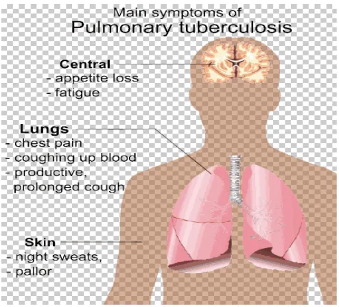
GENERAL SIGNS AND SYMPTOMS
Introduction
[image:9.612.202.449.152.376.2]Department of Pharmaceutical chemistry Page 2 Figure. 1 (6)
BACKROUND
Tuberculosis or TB is the most common infectious disease. In the past Tuberculosis also called as phthisis or phthisis pulmonalis. TB is second only to HIV/AIDS as the greatest killer worldwide due to the single infectious agent.(7)
Introduction
Department of Pharmaceutical chemistry Page 3 HISTORY
M. Tuberculosis, then known as the "Tubercle Bacillus", was first described on 24 March 1882 by Robert Koch, WHO subsequently received the Nobel Prize in physiology or medicine for this discovery in 1905; The Bacterium is also known" Koch's Bacillus".(10) Tuberculosis has existed throughout history, but the name has changed frequently over time. In 1720, though, the History of Tuberculosis started to take shape into what is known of it today, as the physician Benjamin Marten described in his A Theory of Consumption, Tuberculosis may be caused by small living creatures transmitted through the air to other patients. (11)
MYCOBACTERIA
The main cause of TB is Mycobacterium tuberculosis which is a rod shaped, small, aerobic, non motile bacillus. The high lipid content of this pathogen accounts for many of its unique clinical characteristics. It divides every 16 to 20 hours .Which is an extremely slow rate compared with other bacteria. Mycobacteria have an outer membrane lipid bilayer. If a gram stain is performed, MTB either stains very weekly gram – positive or does not retain dye as a result of the high lipid and mycolic acid content of its cell wall (12)
. In nature the bacterium can grow only within the cells of a host organism, but
Introduction
Department of Pharmaceutical chemistry Page 4 `Figure. 2 (14)
SCIENTIFIC CLASSIFICATION Kingdom : Bacteria Phylum : Actinobacteria Class : Actinobacteria Class : Actinomycetalus Sub Order : Corynebacterineae Family : Mycobacteriaceae Genus : Mycobacterium
Species : Mycobacterium tuberculosis (15) CELL WALL
Introduction
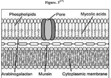
Department of Pharmaceutical chemistry Page 5 The peptidoglycan polymer confers cell wall rigidity and just external to the bacterial cell membrane another contributer to the permeability barrier of mycobacteria. The peptidoglycan polymer confers cell wall rigidity.
[image:12.612.150.505.327.577.2]Another important component of the cell wall is lipoarabinomannan, a carbohydrade structural antigen on the organism that is immunogenic and facilitates the survival of mycobacteria within macrophages. The cell wall is key to the survival of mycobacteria and a more complete understanding of the biosynthetic pathways and gene functions and the development of antibiotics to prevent formation of the cell wall are areas of great interest.(16)
Figure. 3(17)
DRUG RESISTANT TB
Introduction
Department of Pharmaceutical chemistry Page 6 regimen or did not take the prescribed regimen appropriately, or because of other conditions such as drug malabsorption or drug-drug interactions that led to low serum levels.
MDR TB is caused by organisms resistant to both isoniazid and rifampicin, which are the two most effective anti-TB drugs .These drugs are considered first-line drugs and are used to treat most persons with TB disease.
XDR TB is relatively rare type of drug- resistant TB .XDR TB is resistant to isoniazid and rifampicin ,plus any fluoroquinolone and at least one of three injectable second-line drugs (i.e., amikacin, kanamycin, orcapreomycin).Because XDR TB disease is resistant to first-line and second-line drugs ,patients are left with treatment options that are more toxic ,more expensive, and much less effective.
THE NEED FOR NOVEL TUBERCULOSIS DRUGS
1. To improve current treatment by shortening the total duration of treatment
2. To improve the treatment of MDR-TB[18]
3. To provide for more effective treatment of latent tuberculosis infection (16)
4. New drugs to improve current drugs that facilitate compliance by providing for less intensive supervision are also of great interest
Introduction
Department of Pharmaceutical chemistry Page 7 6. MDR TB must be treated with a combination of “second line” drugs which are not only more expensive but also much more toxic and less effective than the drugs used
in standard therapy.(19)
GENOME
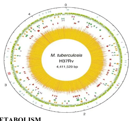
The Mycobacterium Tuberculosis genome encodes about 190 transcriptional regulators, including is sigma factors, two-component system and more than 140 transcription regulators. Several regulators have been found to respond to environmental distress, such as extreme cold or heat, iron starvation, and oxidative stress. To survive in these harsh conditions for a prolonged period in the host, Mycobacterium tuberculosis had learned to adapt to the environment by allowing an inhibiting transcription according to it’s surrounding. (20)
GENOME STRUCTURE
Introduction
[image:15.612.208.431.158.367.2]Department of Pharmaceutical chemistry Page 8 Figure. 4 (22) The chromosome of M.Tuberculosis H37Rv and the gene synthesise
mycolic acids.
CHOLESTROL METABOLISM
Cholesterol metabolism has been studied extensively because of its possible therapeutic application in TB infections. It has been shown numerous times that TB requires cholesterol for virulence in vivo, because Mycobacterium Tuberculosis (Mtb); the causative agent, utilizes cholesterol as a source of carbon, energy, and steroid-derived metabolites throughout the course of infection. (23)
ROLE OF CHOLESTROL IN TB INFECTION
Introduction
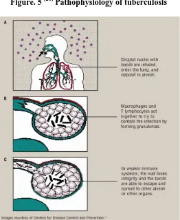
Department of Pharmaceutical chemistry Page 9 PATHOPHYSIOLOGY OF TUBERCULOSIS
Mycobacterium tuberculosis requires high levels of oxygen to grow. Primarily a pathogen of the mammalian respiratory system, it infect the lungs .The most frequently used diagnostic methods for TB are the tuberculin test, acid- fast stain, and chest radiographs.(25)
M.tuberculosis is divides every 15-20 hours, which is extremely slow, compared to other bacteria. It is a small bacillus that can withstand weak disinfectants and can survive in a dry state for weeks. Its unusual cell wall, rich in lipids (e.g. mycolic acids).(26) Humans are the only known reservoirs of Mycobacterium tuberculosis. When in the lungs, mycobacterium tuberculosis is taken up by alveolar macrophages, but they are
unable to digest and eradicate the bacterium. It cell wall prevents the fusion of the
Introduction
[image:17.612.195.457.84.404.2]Department of Pharmaceutical chemistry Page 10 Figure. 5 (28) Pathophysiology of tuberculosis
(A) inhalation of bacilli, (B) containment in the granuloma ,( C) Breakdown of the granuloma in less immunocompetent individuals .
Need for new anti-TB drugs
The recent rise in TB cases and especially the increase of drug resistant
mycobacteria indicate an urgent need to develop new anti-TB drugs.
The long duration of TB therapy is a consequence of persistent mycobacterium
tuberculosis, not effectively killed by current anti-TB agents. (29)
Recent advances in the knowledge of the biology of the organism and the
Introduction
Department of Pharmaceutical chemistry Page 11 It is expected that the application of functional genomic tools, such as structure
based drug design and combinatorial chemistry will lead to the development of new drugs that are active against drug resistant TB. (30)
There is a need to design new drugs that are more active against slowly growing
Introduction
Department of Pharmaceutical chemistry Page 12 ENZYME PROFILE
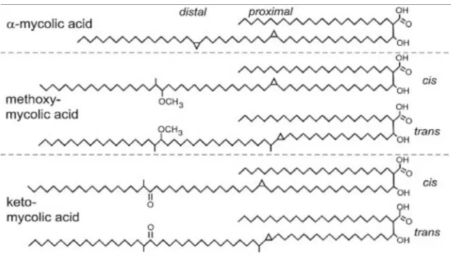
[image:19.612.162.486.408.592.2]Mycolic acids, a homologous series of C60-C90 long-chain alpha-alkyl- and beta hydroxyl fatty acids, represent essential components of the mycobacterial cell wall. They are important for mycobacterial growth, survival, and pathogenicity. They are found as esters of anarabinogalactan as well as free lipids in the form trialosedimycolate (TDM).Arabinogalactanmycolate is covalently linked to the cell wall peptidoglycan via a phosphodiester bond located on the inner leaflet of the outer membrane. Both arabinogalactan and TDM provide a protective thick cell wall and protect the tubercle bacillus from antibiotics and host‘s immune system.TDM also inhibits phagolysosome fusion and is often considered to be an indicator of virulent strains.
Figure. 6(31)
Introduction
Department of Pharmaceutical chemistry Page 13 15% of the mycolic acids in the organism. The alpha-mycolic acid is a cis, cis-dicyclopropyl fatty acid. Both methoxy-and keto- mycolic acids have either cis- or trans- cyclopropane rings. Cyclopropane rings in mycolic acids protect the bacillus from oxidative stress.
Several front-line drugs used for treating tuberculosis inhibit mycolic acid synthesis. Understanding the pathway of mycolate biosynthesis and the underlying molecular mechanisms of the disease tuberculosis a well as the identification of new antituberculosis drug targets is important. InhA(ECn1.3.1.9,enoyl-[acyl-carrier-protein]reductase), involved in mycolic acid synthesis ,is a target of front-line anti-tubercular drugs, such as isoniazid and ethionamide. Enzymes needed for biosynthesis of mycolic acids, such as methoxy mycolic acid synthase2, cyclopropane mycolic acid synthase 2, methyl transferase (PcaA),beta-ketoacyl-acyl carrier protein synthase (KasAB and FabH),acyl-AMP ligase (Fad32) and polyketide synthase(Psk13) are promising drug targets for new anti-TB agents.(31)
The presence of mycolic acids gives M.tuberculosis many characteristics that defy medical treatment.Theylend the organism increased resistance to chemical damage and dehydration, and prevent the effective activity of hydrophobic antibiotics .In addition, the mycolic acids allow the bacterium to grow readily inside macrophages, effectively hiding it from the host's immune system. Mycolate bio- synthesis is crucial for survival and pathogenesis of M. Tuberculosis.(32-33)
Five distinct stages are involved in biosynthesis of mycolic acid these were summarized as follows
Introduction
Department of Pharmaceutical chemistry Page 14 acid synthase -I (FAS-I) to provide the α-alkyl branch of the mycolic acids.
Synthesis of the C56 fatty acids by FAS-II providing the meromycolate
backbone.
Introduction of functional groups to the meromycolate chain by numerous
Cyclopropane synthases.
Condensation reaction catalysed by the polyketide synthase Pks13 between the α-branch and the meromycolate chain before a final reduction by the enzyme
Corynebacterineae mycolate reductase A (CmrA) to generate the mycolic acid; and Transfer of mycolic acids to arabinogalactan and other acceptors such as trihalose via the antigen 85 complex.
The fatty acid synthase-I and fatty acid synthase-II pathways producing
mycolic acids are linked by beta-ketoacyl-(acyl-carrier-protein) synthase III enzyme, often designated as mtFabH. Novel inhibitors of this enzyme could potentially be used as therapeutic agents.(34)
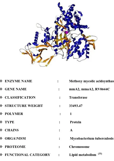
MmA2 is required for introduction of the distal cyclopropane ring in the
formation of meroacids. Analysis of a mmA2 deletion mutant of Tuberculosis revealed that mycolic acid lacks a distal cyclopropane group and instead contains a cis unsaturation. Thus, mmA2 is required for the distal cyclopropane
Introduction
[image:22.612.128.511.128.670.2]Department of Pharmaceutical chemistry Page 15 Figure .7 (33) Methoxy Mycolic Acid Synthase 2
ENZYME NAME : Methoxy mycolic acidsynthase2 GENE NAME : mmA2, mmaA2, RV0644C CLASSIFICATION : Transferase
STRUCTURE WEIGHT : 33493.47 POLYMER : 1
TYPE : Protein CHAINS : A
ORGA\NISM : Mycobacterium tuberculosis PROTEOME : Chromosome
Introduction
Department of Pharmaceutical chemistry Page 16 BASIC NUCLEUS PROFILE
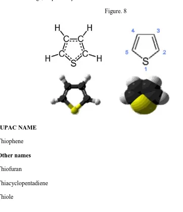
THIOPHENE
[image:23.612.113.475.285.704.2]Tiophene, a heterocyclic nucleus has attracted a wide attention of the chemist in search for the new therapeutic molecules. Thiophene, also called as thiofuran, is a heterocyclic compound with the formula C4H4S. It consists of a flat five membered ring, it is aromatic as indicated by its extensive substitution reactions. Related to thiophene are benzothiophene, and dibenzothiophene, containing the thiophene ring fused with one and two benzene rings, respectively (36).
Figure. 8
IUPAC NAME Thiophene Other names Thiofuran
Introduction
Department of Pharmaceutical chemistry Page 17 PROPERTIES
Chemical formula : C4H4S Molar mass : 84.14 g/mol Appearance : colorless liquid Density : 1.051g/ml Melting point : - 38c Boiling point : 84c Refractive index : 1.5287(36) USES
Review of literature
Department of Pharmaceutical chemistry Page 18
2. REVIEW OF LITERATURE
Literature review on Tuberculosis research
1. Robert Koch et. a.l., (2008) (10) History of Tuberculosis
2. Williams B. G et.al., (2010) (57) studied about the “The Population Dynamics and Control of Tuberculosis.
3. Vander Geize R. et.al., (2007) “(24) A Gene Cluster Encoding Cholesterol
Catabolism in a Soil Actinomycete Provides Insight into Mycobacterium Tuberculosis Survival in Macrophages.”
4. De Souza MVN, et.al.,(2006) (58) Current status and future prospects for new therapies for Pulmonary Tuberculosis.
5. Duncan k et.al., (2004) (59) Prospects for New Anti-Tubercular drugs.
Literature review on target enzyme Methoxy Mycolic Acid Synthase2
6. Asselineau. J et.al., (1950)(31) structure of the Mycolic acids of Mycobacteria.”
7. Takayama K et.al., (2005) (32) "Pathway to Synthesis and Processing of Mycolic
acids inMycobacterium tuberculosis".
8. Bhatt A.M.et.al., (2007) (34) studied about the biosynthesis of mycolic acid.
9. Raman K. R et.al., (2005)(33) studied that Flux Balance Analysis of Mycolic Acid
Review of literature
Department of Pharmaceutical chemistry Page 19
10. Michael S. G et.al.,(2002)(35) studied about acid cyclopropane synthase of the alpha mycolic tuberculosis encodes the distal the mma A2 gene of mycobacterium.
Literature review on drug design
11. Madsen et al., (2002) (60) Textbook of Drug Design and Discovery.
12. Tollenaere JP et.al (1996) (61) "The role of Structure based ligand design and molecular modeling in drug discovery".
13. Sajujoy Parvathy S Nair et.al., (2006) (46) “Detailed comparison of protein-ligand docking efficiency of GOLD, a commercial package and Argus lab, a licensable freeware”(Insilico biology ).
14. Mickey Sahu et.al., (2013) (43) Computer Aided Drug Design: The Most Fundamental Goal is to Predict Whether a Given Molecule will bind to a Target.
15. Lipinski C A et.al., (2001) (62) “Experimental and computational approaches to estimate solubility and permeability in drug discovery and development settings.”
16. Tarbit M H et.al., (2002) “The emerging importance of predictive ADME simulation in drug discovery”.
17. Lipinski C A et.al (2004) (62) “Lead and Drug-like compounds: the rule-of-five revolution.”
Review of literature
Department of Pharmaceutical chemistry Page 20
Literature review for spectroscopy
19. Gurdeep R Chatwal et.al (2005) (64) wrote a book on, Instrumental methods of chemical analysis.
20. P S Kalsi (65) Text book on Spectroscopy of organic compounds.
21. D Kealey et al., (66) Text book on Instant notes Analytical Chemistry.
22. Y. R. Sharma (55)Tezole chalconext book on Elemental Organic Spectroscopy.
Literature review on chalcones
Review of literature
Department of Pharmaceutical chemistry Page 21
N N H
R N H
NH O
O O
24. Rajarkur R. B et.al.,(2012) reported that study various chalcones were synthesized by the base catalyzed reaction between substituted aromatic ketones and substituted aromatic aldehydes. These chalcones were then subjected to the reaction with hydroxyl amine hydrochloride, guanidine hydrochloride and isoniazid to give 3,5- disubstituted isoxazoles, 4,6-disubstituted pyrimidine-2-amines and 3,5-disubstituted pyrazole derivatives respectively. These compounds were evaluated for their good antimicrobial, antifungal and antitubercular activity.
N N
N
Review of literature
Department of Pharmaceutical chemistry Page 22
25. Naik A V et.al .,(2005) reported that, The chalcones are associated with different biological activities like insecticidal, anticancer, anti-inflammatory, bactericidal, fungicidal, antiviral, antitumor, antimalarial and antiulcer. Literature shows that lieochalcone and oxygenated chalcone has strong antileishmanial activity. It is reported that chalcones exhibited potent activity against human malarial parasite. Many workers have reported the different pharmaceutical activities of chalcones and its derivatives.
Br
HO
C2H5
O
R
Review of literature
Department of Pharmaceutical chemistry Page 23
O
O C H3 CH3
N N
CH3
H
H O
CH3
27. Babasaheb et.al.,(2010)11 have reported synthesis and biological evaluation of β-chloro vinyl chalcones. All synthesized compounds were evaluated for their anti-inflammatory activity and antimicrobial activity. Most of compounds showed very good antibacterial and antifungal activity.
OH
O O
CH3 CH3
O Cl R1
R2
Review of literature
Department of Pharmaceutical chemistry Page 24
28. Jen-Hao et.al.,(2008)14 have reported synthesis of 2,5-dialkoxylchalcones. The new chalcones were prepared by Claisen–Schmidt condensation of appropriate acetophenones with suitable aromatic aldehyde. The novel 2,5-dialkoxylchalcones were evaluated for their cytotoxic, anti-inflammatory, anti-oxidant ,anti tubercular activity.
S OR
OR
O
CH3
Literature review on thiophene nucleus
Review of literature
Department of Pharmaceutical chemistry Page 25
Cl
S
Cl
NH N
O
S
O R H
H
SH H2
30. Parvesh Singh P S et.al .,(1999) Sulphur compounds are of great chemical and pharmaceutical significance and display diverse properties such as antifungal, anti-HIV,antipsoratic,and antimicrobial activities. Some imidazo[2,1-b]-[1,3]thiazines and pyrimido[2,1-b]-[1,3]thiazines are well known anti-inflammatory agents. Likewise, thiophene compounds are well known to exhibit various biological and medicinal activities such as BACE1 inhibitors, antitubercular, anti-depressant, anti-inflammatory, anti-HIV PR inhibitors,and anti-breast cancer activities.
O
S
O
OH
Review of literature
Department of Pharmaceutical chemistry Page 26
31. Ashish Das et.al., (2014) studied that A variety of biological and pharmacological importance of thiophene makes it an essential pharmacophore in the field of medicinal chemistry. Already marketed drugs like Clavix, Plavix which acts by irreversible inhibition of P2Y12 receptor, Tioconazole, a proven fungicidal agent which acts by inhibition of cell wall synthesis having thiophene core has been proven to be efficacious drugs in present respective disease scenario. Thus the synthesis and characterization of novel thiophene moieties with wider therapeutic consequences is a topic of interest for the medicinal chemist. This mini review enumerates the reported synthetic strategy to synthesize thiophene and its major therapeutic field as exploited in the literature.
S OH
O
Review of literature
Department of Pharmaceutical chemistry Page 27
class of heterocyclic compounds and their applications in ever challenging chemotherapy of various ailments/ infections etc.
S
R
OR
R1
33. Sahar M I et.al .,(2010) It has been reported in the literature that heterocyclic compounds such as thiophenes exhibited potent anti-inflammatory activity. Likewise, thiazole, 1, 3, 4-thiadiazole, and their derivatives were found to possess anti tubercular and anti inflammatory activities.
S
NO2
N S
O
NH
N+
N H
R
Aim and objective
Department of Pharmaceutical chemistry Page 28
3. AIM AND OBJECTIVE
AIM
The aim of this project is to discover molecules with potential anti -tubercular activity.
OBJECTIVE
Design compounds and docked against a specific crucial target, Methoxy Mycolic acid Synthase 2, which is involved in the cell wall synthesis. The synthesized compounds are expected to act on this enzyme.
THE PLAN OF WORK
Design of methoxy mycolic acid synthase 2 inhibitors by docking studies. Insilico Drug likeness prediction.
Insilico Toxicity Assessment.
Laboratory synthesis of the compounds with top Docking Scores. Characterization of the synthesized compounds by
Infrared Spectroscopy.
H1 Nuclear Magnetic Resonance Spectroscopy. C13 Nuclear Magnetic Resonance Spectroscopy. Mass Spectroscopy.
Aim and objective
Department of Pharmaceutical chemistry Page 29
The whole study was carried out according to this flow chart
Identification of basic nucleus
↓
Structural Modification of the basic Nucleus ↓
Docking the molecule in the target protein ↓
Optimization of the highly Interacting molecule ↓
Top G score compounds selected ↓
Synthesis ↓
Characterization-Spectroscopy(IR,NMR,MASS) ↓
Acute toxicity studies ↓
Materials And Methods
Department of Pharmaceutical chemistry Page 30
4. MATERIALS AND METHODS DOCKING STUDIES
Drug design
Drug design is carried out using an automated docking program like GLIDE (grid based ligand docking with energetics) maestro 9.0 Schrodinger suites, Auto Dock or Argus Lab. It helps search molecules (ligands) having maximum favorable interactions with a receptor (target) usually a protein. Ligand is a single molecule whereas receptor may include proteins, metals and cofactors. It runs on rigid and flexible docking modes. The later which one generates conformations automatically for the input of each ligand and gives out the best fit pose of the molecule been docked on the receptor. (38)
Types
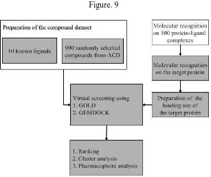
There are two major types of drug design. The first is referred to as ligand based drug design and the second, structure based drug design.
1. Ligand based
Materials And Methods
[image:38.612.173.480.99.357.2]Department of Pharmaceutical chemistry Page 31
Figure. 9
2. Structure based
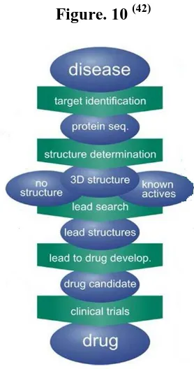
Structure based drug design (or direct drug design) relies on knowledge of the three dimensional structure of the biological target obtained through methods such as x-ray Crystallography or NMR spectroscopy. (40) If an experimental structure of a target is not available, it may be possible to create a homology model of the target based on the experimental structure of a related protein. Using the structure of the biological target, Candidate drugs that are predicted to bind with high affinity and selectivity to the target may be designed using interactive graphics and the intuition of a medicinal chemist. Alternatively various automated computational procedures may be used to suggest new drug candidates.
Materials And Methods
Department of Pharmaceutical chemistry Page 32
[image:39.612.232.371.152.416.2]ligands are obtained from calculations. This has encouraged the rapid development of the structure based drug design. (s
Figure. 10 (42)
COMPUTER-AIDED DRUG DESIGN
Computer-aided drug design uses computational chemistry to discover, enhance, or study drugs and related biologically active molecules. Molecular mechanics or molecular dynamics are most often used to predict the conformation of the small molecule and to model conformational changes in the biological target that may occur when the small molecule binds to it. Molecular mechanics methods may also be used to provide semi-quantitative prediction of the binding affinity. Also, knowledge-based scoring function may be used to provide binding affinity estimates.
Materials And Methods
Department of Pharmaceutical chemistry Page 33
1. Hit identification using virtual screening (structure- or ligand-based design)
2. Hit-to-lead optimization of affinity and selectivity (structure-based design, QSAR, etc.) 3. Lead optimization: optimization of other pharmacokinetic properties while maintaining affinity.
In order to overcome the insufficient prediction of binding affinity calculated by recent scoring functions, the protein-ligand interaction and a compound’s 3D structure information are used for analysis. (43)
Active site identification
Active site identification is the first step in this program. It analyzes the protein to find the binding pocket, derives key interaction sites within the binding pocket, and then prepares the necessary data for Ligand fragment link. The basic inputs for this step are the 3D structure of the protein and a pre docked ligand in PDB format, as well as their atomic properties. Both ligand and protein atoms need to be classified and their atomic properties should be defined, basically, into four atomic types:
1. Hydrophobic atom: All carbons in hydrocarbon chains or in aromatic groups. 2. H bond donor: Oxygen and nitrogen atoms bonded to hydrogen atom(s).
3. H bond acceptor: Oxygen and sp2 or sp hybridized nitrogen atoms with lone electron pair(s). 4. Polar atom: Oxygen and nitrogen atoms that are neither H bond donor nor H bond acceptor, sulfur, phosphorus, halogen, metal, and carbon atoms bonded to heteroatom(s).(44)
DOCKING
Materials And Methods
Department of Pharmaceutical chemistry Page 34
usually interacts with protein’s binding sites.(45) Binding sites are areas of protein known to be active in forming of compounds. There are several possible mutual conformations in which binding may occur. These are commonly called binding modes. It also predicts the strength of the binding, the energy of the complex; the types of signal produced and calculate the binding affinity between two molecules using scoring functions.(46)
TYPES OF DOCKING
Lock and key or rigid docking- In lock and key docking, both the internal geometry of the receptor and ligand is kept fixed and docking was performed.
Induced fit or flexible docking- An enumeration on the rotations of one of the molecules (usually smaller one) is performed. For every rotation the surface cell occupancy and energy is calculated; later the most optimum pose was selected. (45)
ARGUS LAB
Argus lab 4.0 distributed freely and is made available for windows platforms by Planaria Software. It is an introductory molecular modelling package for academics. The Argus docking engine approximates an exhaustive search method with similarities to AUTO DOCK and GLIDE.
Flexible ligand docking possible with Argus lab where the ligand is described as torsion tree or free and grids are constructed that overlay the binding site. The key features such as “the nature of binding site and the number of rotatable bonds to the ligand, can be determined. (46) STEPS INVOLVED IN DOCKING
Docking is done by using ARGUS LAB Software. 1. Protein preparation.
Materials And Methods
Department of Pharmaceutical chemistry Page 35
3. Ligand Preparation. 4. Docking Procedure.
5. Visualization / Interpretation of Docking. 1. PROTEIN PREPARATION
Step: 1
Enter protein (pdb) ID in the protein data bank. (1TPY) Go to download files and select pdb as text file.
Save the download pdb (text file) to the desktop.
Step: 2
Open Argus lab file Open Import pdb file from the desktop.
3D Structure of the protein will appear in the workspace of Argus lab. Left side of the screen shows molecular tree view.
Open pdb Open ‘residues’ ‘Open misc’
From ‘Misc’ delete the inhibitor and hetero residues [Note: Do not delete Co
factor]
Open water press shift, select all water molecules and delete. Add hydrogen atoms.
Go to Calculation on the toolbar energy by UFF method start. Save the prepared protein as *.agi file format in the desktop.
2. Q-SITE FINDER (47) Step: 1
Open Q-Site finder through online.
Materials And Methods
Department of Pharmaceutical chemistry Page 36
Find all the active site and make a list out of the common amino acid residues.
Step: 2
Open residues open Amino acids.
Press control and select the amino acids which were listed from the Q-Site
finder.
Make sure that all amino acid residues listed are selected.
Right click on the mouse make a group from the selected residues give name
Binding site Ok.
3. LIGAND PREPARATION
Draw the structure from Chem sketch and save as MDL Mol format. Import the ligand into workspace of Argus lab.
Clean Geometry Clean Hybridization.
Select the ligand, Right click on the mouse Make a group from the residues give name
ligand Ok.
4. DOCKING PROCEDURE
Select the set up a Dock Ligand calculation from the toolbar. Argus Dock as the Docking Engine.
Dock was selected as calculation type. Flexible for the scoring function. Calculation size.
Start docking.
Materials And Methods
Department of Pharmaceutical chemistry Page 37
5. VISUALIZATION / INTERPRETATION OF DOCKING
Molegro Molecular viewer will help in analyzing the energies and interaction of the
binding.
View Docking view & Secondary Structure view. View Hydrogen bond interaction.
Ligand map Interaction overlay.
IN-SILICO SCREENING OF DRUG LIKENESS
A drug to be pharmacologically active and exert the action it should possess
Pharmacokinetic properties like absorption, distribution, metabolism and excretion. In the field of drug research and development many drug failures occur due to unfavorable ADME properties. This has to be ruled out earlier in the process of drug discovery. Some computational methods have been evolved to investigate the most suitable drug molecules before synthesis. (48)
“Lipinski’s rule of five” it is also known as Pfizer’s rule of five is rule to evaluate drug likeness. It is used to predict whether a molecule is likely to be orally bio-available or to evaluate drug likeness.
Lipinski’s rule
Lipinski’s rule is used to predict if a molecule is likely to be orally bio-available or to evaluate drug likeness. The rule was formulated by Christopher A. Lipinski in 1997. The rule states that for drug likeness the molecule should have the following properties.
Molecular weight less than 500 Daltons. Calculated log P value less than 5.
Materials And Methods
Department of Pharmaceutical chemistry Page 38
Less than 10 rotatable bonds.
The designed and docked molecules are screened insilico using Molinspiration
Cheminformatics software to evaluate drug likeness. This tool is quick and easy to use. It is a software available online for calculation of important molecular properties (log P, polar surface area, number of hydrogen bond donors and acceptors and others),as well as prediction of bioactivity score for the most important drug targets(GPCR ligands, kinase inhibitors, ion channel modulators, nuclear acceptors. (49)
ADME ANALYSIS
A deeper understanding of the relationships between important ADME parameters and molecular structure and properties has been used to develop in silico models that allow the early estimation of several ADME properties. Among other important issues, prediction of properties that provide information about dose size and dose frequency such as oral absortion, bioavailability, brain penetration clearance and volume of distribution (for frequency) also needed. (50)
Absorption
Materials And Methods
Department of Pharmaceutical chemistry Page 39
Bioavailability
Important properties for determining permeability seem to be the size of the molecule, as well as its capacity to make hydrogen bonds, its overall lipophilicity and possibly its shape and flexibility.
Blood –Brain Barrier penetration (BBB)
Drugs that act in the CNS need to cross the blood-brain barrier (BBB) to reach their molecular target by contrast, for drugs with a peripheral target, little or no BBB penetration might be required in order to avoid CNS side affections. Rule –of –five like recommendations regarding the molecular parameters that contribute to the ability of molecules to cross the BBB have been made to aid BBB- penetration predictions; for example molecules with a molecular mass of <450Da or with polar surface area (PSA) <100A0 are more likely to penetrate the BBB. Dermal and ocular penetration
The existing transdermal models are typically a function of the octanol/water partition coefficient and dermas that have been associated with aqueous solubility, including hydrogen – bonding parameters, molecular weight and molecular flexibility. Commercial models for the prediction of solute –permeation rates through the skin are available, for example qikrop and Derm Win programs.
METABOLISM
Insilico approaches to predicting metabolism can be divided into QSAR and three
Materials And Methods
Department of Pharmaceutical chemistry Page 40
on the crystal structure of the metabolizing enzymes. Ultimately, such programs might be linked to computer-aided toxicity prediction on the basis of quantitative structure-toxicity relationships and expert systems for toxicity evaluation. (51)
TOXICITY PREDICTION
All the data set molecules were subjected to the toxicity risk assessment by using Osiris program, which is available online. The OSIRIS property Explorer shown in this page is an integral part of Actelion's in house substance registration system. It allows drawing chemical structures and also calculates various drug relevant properties whenever a structure is valid. Prediction results are color coded in which the red color shows high risks with undesired effects like mutagenicity or a poor intestinal absorption and green color indicates drug-conform behavior. (52)
Molecular property prediction includes Toxicity risk assessment
clog P prediction Solubility prediction Molecular weight
Materials And Methods
Department of Pharmaceutical chemistry Page 41
SYNTHETIC METHODOLOGY SYNTHESIS
Synthetic scheme was framed for the hit compounds from docking and the procedure for synthesis was collected from literatures. The necessary chemicals of laboratory grade for the synthesis where procured from Sigma Aldrich and Synthesis was carried out.
Scheme 1( 53)
2-Acetyl thiophene (0.01mol) and appropriately substituted N, N Dimethyl amino benzaldehyde (0.012 mol) were mixed in ethanol (20ml) containing 10% aq. Potassium hydroxide (8ml) and magnetically stirred the solution constantly at room temperature for 10 hours. The whole mixture was transferred in to 100ml ice cold water and acidified with dil.Hydrochloric acid.The solid form was washed, filtered and dried, recrestallised from absolute ethanol. Compound 1 S O CH3 N CH3 CH3 O S O N CH3 CH3
+
Ethanolic KOH
Compound 2:
Materials And Methods
Department of Pharmaceutical chemistry Page 42
S O CH3 N+ O -O O S
O N+ O
-O
+
Ethanolic KOH
Compound 3:
The compound was synthesized by two a step reaction. Step1: (53)
2-Acetyl thiophene (0.01mol) and appropriately substituted 2,4 Di Chloro benzaldehyde (0.012 mol) were mixed in ethanol (20ml)containing 10% aq. Potassium hydroxide (8ml) and magnetically stirred the solution constantly at room temperature for 10 hours . The whole mixture was transferred in to 100ml ice cold water and acified with dil.Hydrochloric acid.The solid form was washed, filtered and dried , recrestallised from absolute ethanol .
Materials And Methods
Department of Pharmaceutical chemistry Page 43
Step 2(54)
A mixture of chalcone (0.02mol),thiosemicarbazide (0.02mol)were dissolved in ethanolic
sodium hydroxide solution (10ml) was stirred for 3hrs.then it was poured into 400ml of cold water
with continuous stirring for 1hour then left overnight. The precipitate formed was filtered, washed
and recrystallised from ethanol.
S N N NH2 Cl Cl S Cl O
+
N H2 S NHNH2
NaOH\Ethanol
REACTANT PROFILE 2 Acetyl thiophene
S
CH3
O
Molecular formula : C6H6OS Molecular weight : 126.18
Materials And Methods
Department of Pharmaceutical chemistry Page 44
N, N Dimethyl amino benzaldehyde
N CH3
CH3 OHC
Molecular formula : C9H11N0 Molecular weight : 149.19
Appearance : Yellow white powder Melting point : 74°c
O-Nitro benzaldehyde
OHC
O2N
Molecular formula : C7H5NO3 Molecular weight : 151.11
Materials And Methods
Department of Pharmaceutical chemistry Page 45
2, 4 Di chloro benzaldehyde
Cl
Cl OHC
Molecular formula : C7H4Cl2O Molecular weight : 175.01
Appearance : White crystalline solid Melting point : 64-69°C
Thiosemicabazide
N H2
S
NH NH2
Molecular formula : CH5N3S Molecular weight : 91.14
Appearance : White crystalline solid Melting point : 177-179°C
RECRYSTALLISATION:
Materials And Methods
Department of Pharmaceutical chemistry Page 46
CHARACTERISATION STUDIES MELTING POINT
The melting point of the synthesized compound was determined by tone end open capillary
tube method. The temperature at which the compound starts losing its crystallinity and changes from
solid to liquid form was found recorded.
IR SPECTROSCOPY
IR spectroscopy helps to ascertain the presence and absence of the functional group. The
synthesized compound was made into a pellet with potassium bromide by pressed pellet technique
using pellet press (Model No: M15). The pellet was mounted on the pellet disc and percentage
transmittance was recorded in ABB IR Spectrophotometer (Model No: 3000). IR Spectroscopy is an
important tool for structure elucidation and compound identification.
EXAMPLE:
OH groups : 3600-3200 cm-1
C=Ogroups : 1710 cm-1
Ar C-H str : 3050 -3000cm-1
C=C str : 1600 cm-1
N-S str : 3540-3300 cm-1
C-H aliphatic str : 2795-2840 cm-1
NMR SPECTROSCOPY
Proton NMR Spectroscopy helps us to study the number of equivalent protons and their
environment thereby we can ascertain the structure of the molecule. The NMR spectra was recorded
on 300 MHz BRUKER Advance III NMR Spectrometer DMSO was used as a solvent.
MASS SPECTROSCOPY
Materials And Methods
Department of Pharmaceutical chemistry Page 47
Technique and was quantified using Lab Solutions Software 7.0, Samples were prepared by dissolving a minute quantity of pure compounds in methanol. The fragmentation patterns were reported in m/z values. (55)
HYPHENATED TECHNIQUE
Materials And Methods
Department of Pharmaceutical chemistry Page 48
BIOLOGICAL EVALUATION ANTI-TB ACTIVITY
There are various high through put assays available for screening of new chemical entities against Tuberculosis. They are
Microplate Alamar blue Assay
BACTEC Assay
Luciferous reporter phage assay
REMA Assay
Broth Dilution Assay
Middle brook (7H9,7H10,7H11) Agar Dilution Assay
PRINCIPLE
The micro plate Alamar blue assay (MABA) is an indirect colorimetric DST method for determining the MICs of TB drugs for strains of mycobacterium tuberculosis . in this assay , the redox indicator alamar blue monitors the reducing environment of the living cell . It turns from blue to pink in the presence of mycobacterial growth.
PROCEDURE
The anti-mycobacterial activity of compounds (M13,M14) were assessed against
M.tuberculosis using micro plate Alamar blue assay(MABA)
This methodology is non – toxic, uses a thermally stable reagent and shows good
correlation with proportional and BACTEC radiometric method.
Briefly, 200ml of sterile de-ionised water was added to all outer perimeter wells of sterile
Materials And Methods
Department of Pharmaceutical chemistry Page 49
The 96 wells plate received 100 ml of the middle brooke 7H9 broth and serial dilution of
compounds was made directly on a plate.
The final drug concentrations tested were 100 to 0.8mg/ml
Plates are covered and sealed with paraflim and incubated at 37c was dive days
After this time 25 ml of freshly prepared 1:1 mixture of alamar blue reagent and 10%
tween 80was added to the plate and incubated for 24 hours.
A blue colour in the well was interpreted as no bacterial growth, and pink colour was
scored as growth.
The MIC was defined as lowest drug concentration which prevented the colour change
from blue to pink .(56) ADVANTAGES
It has accurate time-course measurement It has highly sensitivity and linearity
It is ideal for use with post-measurement functional assays It is flexible and it can be used with different cell modes
It is scable and it can be used with flourescence and /or absorbance-based
instrumentation platforms
It is non toxic , on –radioactive and is safe for the user,
APPLICATIONS
Results and discussion
Department of Pharmaceutical chemistry Page 50
5. RESULTS AND DISCUSSION
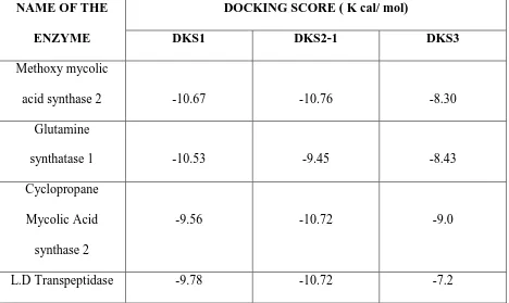
Nearly 200 molecules were sketched using chemsketch. Were docked against the enzyme using A. L 4.0.1 Software. The molecules were also docked against the following targets. The molecule with best docking score and interaction were selected and synthesis.
The molecules were also docked against the following targets:
1. Methoxy mycolic acid synthase 2
2. Glutamine synthatese 1
3. Cyclopropane mycolic acid synthase 2
[image:57.612.68.536.426.705.2]4. Decaprenylphosphoryl-b-d-ribose2’-Epimerase1 (DprE1)
Table no: 1
NAME OF THE ENZYME
DOCKING SCORE ( K cal/ mol)
DKS1 DKS2-1 DKS3
Methoxy mycolic
acid synthase 2 -10.67 -10.76 -8.30
Glutamine
synthatase 1 -10.53 -9.45 -8.43
Cyclopropane Mycolic Acid synthase 2
-9.56 -10.72 -9.0
Results and discussion
Department of Pharmaceutical chemistry Page 51
INTERACTIONS OF THE DOCKED MOLECULE USING THE ENYME METHOXY
[image:58.612.99.523.119.723.2]MYCOLIC ACID SYNTHASE 2
Table no: 2 SAMPLE
CODE DOCKING VIEW
INTERACTION WITH AMINO ACIDS
DKS1
DKS2-1
DKS3
Results and discussion
Department of Pharmaceutical chemistry Page 52
RESULTS OF SCHEME COMPOUND NAME: DKS1
IUPAC NAME:( 2E)-3-[4-(dimethylamino)phenyl]-1-thien-2-ylprop-2-en-1-one
Molecular formula : C15H15NOS
Molecular weight : 257.35g/mol
Appearance : Reddish orange
Melting point : 69°c
Composition : C(70.01%)H(5.87%)N(5.44%)O(6.22%)S(12.46%)
Molar refractivity : 79.80±0.3cm3
Molar volume : 217.2±3.0cm3
Surface tension : 49.4±3.0dyne/cm
Density : 1.184±0.06g/cm3
Parachor : 31.63±0.5,10-24cm3
Index of refraction : 1.655±0.02
Results and discussion
Department of Pharmaceutical chemistry Page 53
COMPOUND NAME: DKS2-1
IUPAC NAME : 5-(2,4-dichlorophenyl)-3-thien-2-yl-4,5-dihydro-1H-
pyrazole-1-carbothioamide
Molecular formula : C14H11CL2N3S2
Molecular weight : 356.29
Appearance : yellowish brown
Melting point : 72°c
Composition : C(47.19%)H(3.11%)C(19.90%)N(11.79%)S(18.00%)
Molar refractivity : 92.68±0.5cm3
Molar volume : 226.5±7.0cm3
Surface tension : 58.8±7.0dyne/cm
Density : 1.57±0.1g/cm3
Parachor : 627.5±8.0cm3
Index of refraction : 1.754±0.05
Results and discussion
Department of Pharmaceutical chemistry Page 54
COMPOUND NAME: DKS3
IUPAC NAME : (2E)-3-(2-nitrophenyl)-1-thien-2-ylprop-2-en-1-one
Molecular formula : C13H9NO3S
Molecular weight : 259.38
Appearance : Light brown
Melting point : 67°c
Composition : C(60.22%)H(3.50%)N(5.40%)O(18.51%)S(12.37%)
Molar refractivity : 72.03±0.3cm3
Molar volume : 191.1±3.0cm3
Surface tension : 58.9±3.0dyne/cm3
Density : 1.346±0.06g/cm3
Parachor : 529.6±4.0cm3
Index of refraction : 1.677±0.02
Results and discussion
Department of Pharmaceutical chemistry Page 55
IR SPECTROSCOPY
The sample were prepared by the KBr pellet techniques of spectrum. The spectra were examined for the absence of the functional groups of the parent compounds and examined for the presence of the vibrational absorption band for the new functional group.
[image:62.612.68.532.329.669.2]The synthetic reaction involves, two compounds invoved in the reaction between ketone and aldehydes to yield chalcones . Another one compound invoved in the reaction between chalcones and thiosemicarbazide to yield benzimidazole derivative.
Table no :3
ABSORBANT BAND
DKS1 DKS2-1 DKS3
C=0 Stretching
Ar C-H Stretching × ×
C=C Stretching
Results and discussion
Department of Pharmaceutical chemistry Page 56
SAMPLE CODE : DKS1
Results and discussion
Department of Pharmaceutical chemistry Page 57
Results and discussion
Department of Pharmaceutical chemistry Page 58
[image:65.612.74.543.108.704.2]NMR SPECTROSCOPY SAMPLE CODE :DKS1
Table no. 4
S.NO § VALUE NATURE OF PEAK NUMBER OF
PROTONS
1 6.62 Doublet 2 protons
2 6.70 Doublet 2 protons
3 7.24-7.25 Multiplet 3 protons
4 7.42 Singlet 1proton
5 7.45 Singlet 2protons
6 7.60 Doublet 2 protons
7 7.75 Singlet 2protons
Results and discussion
Department of Pharmaceutical chemistry Page 59
[image:66.612.73.522.146.350.2]SAMPLE CODE : DKS2-1
Table no: 5
S.NO § VALUE NATURE OF PEAK
NUMBER OF PROTONS
1 7.84 ppm Triplet 1proton
2 7.85ppm Singlet 1proton
3 7.90-7.97ppm Multiplet 1proton
4 7.33-7.23 Multiplet 3protons
5 7.17-7.22 Multiplet 3protons
6 1.166-1.22 Multiplet 1proton
Results and discussion
Department of Pharmaceutical chemistry Page 60
[image:67.612.78.549.127.713.2]SAMPLE CODE : DKS3
Table no: 6
S.NO § VALUE (PPM) NATURE OF PEAK
NUMBER OF PROTONS
1 8.05ppm Doublet 1 proton
2 7.55-7.78ppm Multiplet 1proton
3 7.80-7.84ppm Multiplet 2proton
4 7.87 ppm Doublet 1proton
5 7.95ppm Doublet 1proton
6 7.36ppm Doublet 2proton
Results and discussion
Department of Pharmaceutical chemistry Page 61
GC-MS SPECTROSCOPY
[image:68.612.64.547.204.389.2]The molecular weight of the synthesised compounds were comfirmed by GC-MASS analysis
Table no:7
SAMPLE CODE CALCULATED MASS ACTUAL MASS
DKS1 257.35g/mol 257.0 g/mol
DKS2-1 352.29g/mol 352.20g/mol
DKS3 259.38g/mol
Results and discussion
Department of Pharmaceutical chemistry Page 62
Results and discussion
Department of Pharmaceutical chemistry Page 63
Results and discussion
Department of Pharmaceutical chemistry Page 64
Results and discussion
Department of Pharmaceutical chemistry Page 65
IN-SILICO TOXICITY PREDICTION
All the data set molecules were subjected to the toxicity risk assessment by using Osiris
program, which is available online. The OSIRIS property Explorer shown in this page is an integral
part of Actelion's in house substance registration system. It allows drawing chemical structures and
also calculates various drug relevant properties whenever a structure is valid. Prediction results are
color coded in which the red color shows high risks with undesired effects like mutagenicity or a
[image:72.612.66.549.300.440.2]poor intestinal absorption and green color indicates drug-conform behavior.
Table no: 8
SAMPLE DKS1 DKS2-1 DKS3
MUTAGENIC - + +
TUMORIGENIC - + +
IRRITANT + + +
Results and discussion
Department of Pharmaceutical chemistry Page 66
SAMPLE CODE : DKS1
Results and discussion
Department of Pharmaceutical chemistry Page 67
Results and discussion
Department of Pharmaceutical chemistry Page 68
BIOLOGICAL EVALUATION
[image:75.612.69.547.207.402.2]The final pure compounds were screened for Anti-mycrobial activity by in vitro method called Microplate Alamar Blue Assay (MABA)
Table no: 9
S.N O SAMP LE CODE 100µg/ ml 50µg/ ml 25µg/ ml 12.5µg/ ml 6.25µg/ ml 3.12µg/ ml 1.6µg/ ml 0.8µg/ ml 50µg/ ml
1 DK
S1 S S R R R R R R R
2 DKS
2-1 S S R R R R R R R
3 DK
S3 S S R R R R R R
R
NOTE: S- Sensitive
R-Resistant
Strain used:M.Tuberculosis(H37RV strain)
Here are the standard values for the anti –TB test which was performed.
Results and discussion
Department of Pharmaceutical chemistry Page 69
Strain used:M.Tuberculosis(H37RV strain)
Here are the standard values for the anti –TB test which was performed. Pyrazinamide-3.125µg/ml
Streptomycin-6.25µg/ml Ciprofloxazin-3.125µg/ml
Table no: 10
SAMPLE DRUG PHOTOGRAPH
S.NO SAMPLE CODE 100µg/ ml 50µg/ ml 25µg/ ml 12.5µ g/ml 6.25µg/ ml 3.12µg/ ml 1.6µg/m l 0.8µg /ml 50µg/ ml
1 DKS1
2 DKS2-1
[image:76.612.71.590.283.444.2]3 DKS3
Table no: 11
Results and discussion
Department of Pharmaceutical chemistry Page 70
DISCUSSION
Finally all the reports were discussed, that the purity of the compounds are confirmed by sharp melting point and TLC. Then the molecular weights of the compounds are confirmed by NMR and the functional groups of the formed structure were confirmed by absorption bands obtained in the spectra. Among 3 compounds, one compound was 100% pure in nature it was concluded by GC-MS.
Toxicity of the compounds are reported, that 2 compounds are non-toxic and one compound was slightly toxic in nature.
Summary and conclusion
Department of pharmaceutical chemistry Page 71 6.SUMMARY AND CONCLUSION
Methoxy mycolic acid synthase 2 (1TPY) a critical enzyme for the growth of
Mycobacterium tuberculosis was chosen for our study after review of literature.
A database of 200 scaffolds with high prospect of inhibiting the target 1TPY were
carefully chosen by making changes to the known hit molecules , here the chalcones
and thiophene nucleus.
Candidate molecules were designed and docked against 1TPY protein using Argus
lab 4.1 software.
Three molecules with good Docking score (lower binding energy) and interactions
were shortlisted for synthesis. The reaction conditions were optimized.
The selected molecules were subjected to Toxicity Prediction assessment by OSIRIS
software. The results are color Coded as green color which confirms the drug likeness The molecules were labeled as DKS1, DKS2-1,DKS3 and synthesized with
satisfactory yield.
Purity of the synthesized compounds was ensured by repeated recrystallization .The
compounds were evaluated by TLC. and Melting point determination.
The characterization of the synthesized compounds was done using Infra-red, Nuclear
Magnetic Resonance and Mass spectroscopic methods.
The final pure compounds were screened for Anti- mycobacterial activity by in vitro
Summary and conclusion
Department of pharmaceutical chemistry Page 72 CONCLUSION
Our work concludes that our synthesized molecules are effective in inhibiting enzyme
Methoxy mycolic acid synthase 2 (1TPY) which is important for the growth of
Mycobacterium tuberculosis.
All the three compounds gave Docking score between -7.00 to -10 kcal/mol.
Pyrazinamide gave Docking score of -5.6 for 1TPY, Streptomycin gave Docking
score of -7.4 for 1TPY and Ciprofloxacin gave Docking score of -5.9 for 1TPY.
There is correlation between the core and activities of all the three compounds which
were tested and compared with the standard drugs. This goes to prove that Methoxy
Mycolic Acid Synthase2 (1TPY) is a critical enzyme for anti-mycobacterial activity.
The minimum inhibitory concentration of 3 of the synthesized compounds ranged
from 50μg/ml which is compared to that of the known anti-TB agents.
Pyrazinamide - 3.125μg/ml,
Ciprofloxacin - 3.125μg/ml and
Streptomycin - 6.25μg/ml.
A further refinement to the structure of the synthesized compounds is expected to
yield new outlook to the development of promising molecules against the pathogen
References
Department of pharmaceutical chemistry Page 73 7.REFERENCES
1. “Tuberculosis Fact sheet N ° 104”. WHO. October 2015.Retreived11 Febraury 2016.
2. “Basic TB Facts “.CDC. March 13, 2012;Retreived 11 Febraury 2016. 3. “Tuberculosis “. World Health Organisation. 2002.
4. Lawn, SD; Zumla, Al (2July 2011)”.Tuberculosis “. Lancet378 (9785):57-72. 5. Dolin,[Edited by]Gerald L.Mandal,Douglas, and Bennatt’s principles and practice
of infectious diseases ( 7th ed.). Chapter 250. 6. Wiki/Signs and Symptoms . image..Jpg.,
7. AlimuddinZumla, M.D., Ph.D., Mario Raviglione M.D., Richard Hafner M.D., Tuberculosis, the new Ingland journal of medicine 2013; 368:745-55.
8. Sepra N.Rampersad Multiple Application of Alamar Blue As an indicator of Metabolic Function and Cellular Health in Viability bioassays.Sensors 2012, 12, 12347-123.
9. Development of new vaccines and drugs for TB: limitations and potential strategic errors, Furthur Microbiology, 2011 February;6(2):161-177.
10."Robert Koch and Tuberculosis: Koch's Famous Lecture" . Nobel Foundation. 2008. .
11."Tuberculosis History Timeline"
References
Department of pharmaceutical chemistry Page 74 13.Kumar V, Abbas AK,Fausto N, Mitchell RN (2007).Robbins Basic Pathology(8th
ed.) .Saunders Elsevier.pp.516-522. 14.Wiki/ Mycobacteria.image..jpg..
15.IsmaelKasim,RayCG(Editors)(2004).SherrisMedicalMicrobiology(4th edition.MC Graw Hill. ISBN 0-83 85-85 29-9.
16.Nancy A Knechel, Tuberculosis: Pathophysiology, Clinical features and diagnosis, Crit Care Nurse 2009; 29:34-43.
17.http://en.Wikipedia.Wiki/Mycobacterium Tuberculosis Cell Wall., Image...jpg 18.Ahmed Kamal, Shaik Azeeza, M.Shaheer Malik, Ahmad Ali Shaik And
Maddamsetty V. Rao Efforts Towards The Development Of New Antitubercular Agents: Potential For Thioactomycin Based Compounds, J Pharm Pharmaceutical Science 11(2):56s-80s,200
19.Melvin K.Spigelman, New Tuberculosis Therapeutics: A Growing Pipeline , Journal of infectious disease, (2007) 196