Spectrum of gastrointestinal and hepato-pancreato-biliary tract
malignancies at a tertiary centre in south India- A prospective
observational study
A dissertation submitted in partial fulfillment of the requirements for
DM (Gastroenterology) examination of the
Tamil Nadu Dr. M.G.R. Medical University, Chennai,
to be held in August 2015.
Acknowledgement
I take this opportunity to express my sincere thanks to my guide, Dr Uday George Zachariah, Department of Hepatology and Dr. CE. Eapen, Professor and Head, Department of Clinical Gastroenterology and Hepatology, for their guidance, encouragement and constant support in undertaking and completing this project.
I express my sincere thanks to my co-guides Dr. AJ Joseph, Professor and Head, Department of Gastroenterology and Dr. Ebby George Simon, Professor, Department of Gastroenterology for guidance and support for the project. I take pleasure in thanking Dr. Ashish Goel, Department of Hepatology for helping me in the statistical analysis.
CONTENTS
SL NO.
TOPIC
PAGE NUMBER
1
INTRODUCTION
1
2
AIM AND OBJECTIVE
3
3
REVIEW OF LITERATURE
4
4
MATERIAL AND METHODS
27
5
RESULTS
34
6
DISCUSSION
67
7
SUMMARY AND
CONCLUSIONS
89
8
BIBLIOGRPGHY
93
9
APPENDIX
-Mastersheet
-Proforma
Introduction
Cancer is a major health problem worldwide. Prevalence of gastrointestinal malignancies vary in geographical area and depends upon the genetic, socioeconomic and dietary factors(1). Monitoring time trends and incidence is essential for cancer research and health care planning. Cancer registries provide the best means of evaluating the cancer burden on the general population(2).
Therefore studying the pattern of gastrointestinal malignancies helps identify people at high risk and recommend appropriate preventive and surveillance strategies(1).
The incidence of cancers is low in India and this is one third to one half of the USA. The rates are expected to rise as the life expectancy increases along with urbanization and, within the next few decades, may reach those recorded in Indians living abroad. The incidence of malignancy of the esophagus and stomach is declining spontaneously in India. It may be possible to accelerate this decline by reducing the use of tobacco and improving the diet. But at the same time the incidence of cancers of the colon, pancreas, liver and gall bladder is rising, largely due to urbanization that leads to major changes in the diet and personal habits. A preventive approach is needed by public health education. Individuals should be encouraged to retain their traditional protective diets, eat more fruits and vegetables, do more physical activity, and abstain from tobacco. Gastroenterologists can help in secondary prevention by screening high-risk individuals, e.g., patients with chronic liver disease for liver cancer and relatives of patients with familial bowel cancer(3).
Aims and objective
Aim: To study the spectrum of gastrointestinal and hepato-pancreato-biliary tract malignancies at a tertiary centre in south India.
Objective: To analyse the clinico-pathological profile of gastrointestinal and hepato-pancreato-
Review of literature
Cancer is a major health problem worldwide. Prevalence of gastrointestinal malignancies vary in geographical area and depends upon the genetic, socioeconomic and dietary factors(1). Monitoring time trends and incidence is essential for cancer research and health care planning. The analysis of cancer registry databases reveals incidence trends and provides information on changes in the characteristic features of the cancers. Cancer registries provide the best means of evaluating the cancer burden on the general population(2). Therefore studying the pattern of gastrointestinal malignancies helps identify people at high risk and recommend appropriate preventive and surveillance strategies(1).
The incidence of cancers is low in India and this one third to one half of the USA. The cancers occur more frequently in the middle aged. The rate is expected to rise in India as life expectancy increases. Inspite of advances in diagnostic techniques, surgery, radiation and chemotherapy, gastrointestinal malignancy remains undefeated. In absence of screening most gastrointestinal malignancies are diagnosed in late stage with regional and distant metastasis. The real hope for control and early diagnosis is by screening or primary prevention. Gastrointestinal malignancies being deeply seated require investigational modalities like endoscopy, ultrasound or CT/MRI abdomen for diagnosis. In western countries tobacco is implicated in the causation of nearly 30% of the cancer death. The incidence of malignancy of the esophagus and stomach appears to be declining spontaneously in India(3,4). This decline may be aided by reducing tobacco use further and dietary improvement. Simultaneously, malignancies of the colon, pancreas, liver and gall bladder is rising, probably due to urbanization which leads to changes in lifestyle(3,4).
In a retrospective study by Rawal KK et al published in 1995 on 1751 patients over period of ten years (1984-93), showed that relative distributions of gastrointestinal malignancies according to site was esophagus 36.0%, stomach 19.9%, liver secondaries 13.9%, colon 9.8%, periampullary 5.6%, gall bladder 4.5%, duodenum 3.0%, malignant ascites 2.6%, HCC 2.3% and pancreas 1.1%. Out of 83380 patients registered in the gastroenterology department of GB Pant Hospital, New Delhi over 10 years, a total of 1751 (2.1%) patients with gastrointestinal malignancies were identified. The mean age for esophageal malignancy was 53.5 +/- 11.4 year, stomach malignancy 51.8 +/- 12.9, colon malignancy 49.1 +/- 16.7, duodenum malignancy 45.3 +/- 11.4, malignant ascites 51.8 +/- 13.1, pancreatic malignancy 46.9 +/- 15.3 and HCC 52.5 +/- 12 years. Male were more affected as compare to female except for cancer of the gall bladder. The overall mean age of all GI malignancies was 49.7 +/- 13.4 years(5).
In another retrospective pathology centric study by Kalyani R et al from Kolar, Karnataka, India, revealed that gastro-intestinal neoplasms constituted 22.96% of all the cancers diagnosed. Adenocarcinoma was the most common histology type. There was increased incidence of GI cancers over time. The male to female ratio was 1:0.6. The maximum incidence occurred in the 7th decade of life. Stomach was the commonest site (48.4%) followed by esophagus (27.7%), rectum (6.5%), colon (5.0%) and hepatocellular carcinoma (HCC) (4.76%)(1).
per 100,000 for the cancers of colon, rectum and liver. In Indian registries, in the ranking of cancer incidence in men, cancer of esophagus ranks third in Mumbai, Chennai and Bangalore Registries, fourth in Bhopal registry and eighth in Delhi registry. Cancer of the stomach ranks first for Bangalore Registry, second for Chennai Registry and ninth for Mumbai Registry. It has been observed that there was a statistically significant decrease in the incidence of esophagus cancer for Mumbai and Delhi registries. There was an increase in the incidence for Chennai registry. There has been statistically significant decrease in the incidence of stomach cancer over a period of time in most of the registries, highest decrease was observed for Delhi registry. The trend in the incidence of colon, rectum and liver has been shown to be increasing in all the registries. Statistical significant increase was noticed in Chennai, Bangalore and Delhi registries for cancer of colon. For rectum, there has been increase in the incidence for Chennai and Bangalore registries. For liver cancer, increase in Mumbai, Chennai and Bangalore registries has been noted over the time period examined. While esophagus and stomach malignancies are decreasing, malignancies of colon, rectum and liver are increasing in the Indian population(6).
group of 51-60 year (58 cases, 23.77%). Nine cases (3.68%) were present below 20 year of age. The rate of malignancies increased after the age of 30 year. The male: female ratio was 1.39:1. Most of these patients came from a lower socioeconomic status. The most common GI malignancy was esophageal carcinoma (38.93%) followed by colorectal carcinoma (32.79%) and carcinoma of stomach (16.80%). Of forty one cases of stomach malignancy, twenty two cases were adenocarcinoma, nine cases of signet ring adenocarcinoma and four cases of lymphoma. Most of the cases of squamous cell carcinoma presented from the esophagus whereas adenocarcinoma was from the colon. Microscopically, adenocarcinoma was the commonest malignancy (45.90%), followed by Squamous cell carcinoma (33.60%)(8).
Esophagus malignancy:
Esophageal Cancer is the second most common cancer in India responsible for about 0.3 million deaths a year. This is the second most common cancer among males and the fourth most common cancer among females(9). Esophageal malignancies increase with age and peak in the seventh and eighth decade of life. Squamous-cell carcinoma and adenocarcinoma are the two main histological subtypes. The endoscopic appearance is similar in both adenocarcinoma and squamous cell carcinoma. Seventy-five percent of cases of adenocarcinoma are found in the lower esophagus. Squamous-cell carcinoma is more common in the proximal and middle esophagus. Adenocarcinoma is three to four times common in men while being more equally distributed for squamous-cell carcinoma(10).
The etiology of squamous cell esophageal cancer has been linked to excess alcohol consumption and/or cigarette smoking, smoked opiates, and fungal toxins in pickled vegetables, the ingestion of lye, radiation-induced strictures, chronic achalasia, Plummer-Vinson syndrome and, tylosis palmaris et plantaris. Male are more affected than female. The etiology of adenocarcinoma is related to diets high in cholesterol, smoking, obesity, Barrett’s esophagus(13,14).
According to a study by Lal N et al and Gado A et al, the biopsy yield increased from 95.8% when two biopsy specimens were taken to 100% when five to six were taken(15,16).
A retrospective study by Cherian JV et al from south India on 994 patients with esophageal carcinoma revealed that squamous cell carcinoma was the most common malignancy, 912 (92%). Eighty two patients (8%) had adenocarcinoma. Sixty five of these eighty two patients (79%) had gastroesophageal junction malignancy and seventeen (21%) had tumor in the lower third of the esophagus. Sixty-five of the 82 patients with Adenocarcinoma had an esogastric junction (EGJ) malignancy and in seventeen the lesion was in the lower third of the esophagus. None involved the middle or upper third of the esophagus. In squamous cell carcinoma, the male to female ratio was 2.16:1. Most common location of squamous cell carcinoma (SCC) was lower esophagus (N-413, 41.6%). In middle and lower esophagus, there were 379 (38.1%) and 120 (12.1%) cases of squamous cell carcinoma. In adenocarcinoma, male to female ratio was 3.6:1. The mean age was 54.7 ± 11.4 and 49.3 ± 13.5 in squamous cell carcinoma and adenocarcinoma respectively. The author concluded that squamous cell carcinoma is the most common type of esophageal malignancy cancer in the India. Adenocarcinoma is not common and affects more frequently men younger than forty years old(17).
esophagus, five in upper thoracic (3%), sixty four in middle thoracic (38.6%), sixty eighty in lower thoracic (41%), and cardia in twenty two cases (13.2%). SCC (81.1%) was the most common type of esophageal cancer. In male, carcinoma of esophagus usually involves the lower parts, while in female it involves the upper parts of esophagus. SCC was the most common type of esophageal cancer in all age groups. The symptoms of esophageal cancer were dysphagia in 155 subjects (93.4%), loss of appetite in 5 (3%), and weight loss in 6 (3.6%). Three patients (1.8%) had a past medical history of achalasia(18).
A retrospective study by Tony J et al was done in the Department of Gastroenterology at Calicut Medical College from January 1985 to December 2004. Among 29,926 gastroscopies done over 20 years, 909 cases of esophageal cancers were identified. Mean age was 63.12 ±11.2 years (range 28–87). The male to female ratio was 2.77:1. Malignancy were seen in 41 (4.7%) in the upper, in middle in 341 (39.7%) and in lower third in 476 (55.4%) cases. All malignancies involving the upper and the middle thirds of esophagus were SCC. 476 cases of biopsy proven malignancy of lower oesophagus were included for analysis. Two hundred forty nine (52.3%) were adenocarcinomas and 227 (47.7%) were squamous cell. There was an increase in the frequency at the gastro-oesophageal junction but it was not of statistical significance (p value < 0.15). Fifty five percent of malignancies affected the lower third of the oesophagus and gastroesophageal junction. Forty percent of malignancies were located in the middle third and five percent were located in the upper third. Adenocarcinoma were located in lower end than squamous cell carcinomas(19).
Gastric malignancy:
Gastric carcinoma is the third most common type of carcinoma in South India. In northern India, some states have high incidence of stomach malignancy. In India, carcinoma of stomach was estimated to be the fifth commonest site in males and the seventh in females overall. Rates in India are lower than in other countries around the world(21–23). Possible etiological factors for gastric cancer are consumption of smoked, poorly preserved and salted foods, low consumption of vegetables and fruits, infection (Helicobacter pylori), previous gastric resection and pernicious anemia(24).
Ninety per cent of stomach cancers are adenocarcinomas and other rare gastric malignancies include lymphomas, gastrointestinal stromal tumours(GIST), and neuroendocrine tumours(NET)(25). Endoscopy with biopsies is the standard of care for the diagnosis of gastro-oesophageal cancer(16,26).
followed by left supraclavicular lymph node (7.6%). About seventy patients (44.3%) had poorly differentiated, fifty five (35.8%) moderately differentiated and thirty one patient (19.6% ) well differentiated adenocarcinoma(27).
A retrospective study by Pulimood A et al from CMC, Vellore (India) on 361 cases of gastrointestinal lymphomas over a period of 10 years (2001–2010) showed that primary GI lymphoma was present in 267 males (79.64%) and 68 females (20.24%). The mean age was 45 years. Diffuse large B-cell lymphoma was the commonest type in 222 patients (66.71%). Thirty four patients (10.12%) had low-grade marginal zone lymphoma and 35 patients (10.48%) had Burkitt’s lymphoma. The commonest site was stomach in 180 patients
(53.57%), followed by small intestine in 79 patients (23.51%) and large intestine in 68 patients (20.23%). Of large intestine lymphomas, 34 cases were located in ileocecal region, 23 case in the colon, and 12 cases in the rectum(28).
A prospective study by Saha AK et al from West Bengal on 23851 patients underwent upper GI endoscopy over a period of five years (2006–2010) showed that four hundred and sixty two patients were detected to have gastric malignancy. Male to female ratio was 2.7: 1. In this study, the median age was 55 years. Abdominal pain was the most common symptom in 306 patients (66.23%) followed by indigestion in 212 patients (45.88%) and weight loss in 200 patients (43.29%). Melena was present in 44 patients (9.52%). H. pylori was positive in 80.09% of gastric carcinoma. The antrum was commonest site of involvement (51.9%). It was followed by body (18.6%) and fundus (16.5%). Incisura(12.9%) was least common site(30).
A retrospective study by Cherian JV et al from Chennai, India on 1719 patients with adenocarcinoma from 1989 to 2004 showed that the mean age was 55.50 ± 12.68 years. The study period was divided into four cohorts of 4 years each (1989-1992, 1993-1996, 1997-2000 and 2001-2004). The most common sites of gastric cancer was antrum in 1157 patients (67.3%) followed by the body in 400 patients (23.3%), proximal stomach in 97 patients (5.6%) and OGJ in 65 patients (3.8%)(31).
Small intestine malignancy:
Small intestinal malignancies are rare group of GI tumours (3-6%) that extend from the duodenum to the ileum(32–34).
Risk factors for small intestine tumors are Crohn’s disease, celiac disease, and inherited disease (Hereditary nonpolyposis colorectal cancer , Familial adenomatous polyposis, Peutz-Jeghers syndrome)(32,35).
Another retrospective study by Egberts J-H et al from Europe on 43 patients with small bowel cancer showed that 37.2% had adenocarcinoma (n=16), neuroendocrine tumors (n=12; 27.9%), gastrointestinal stroma tumor (GIST) (n=10; 23.3%), lymphoma (n=3; 7%) and desmoid tumor (n=2; 4.6%). Tumor localizations were within duodenum (46.5%), jejunum (16.3%) and ileum (37.2%)(34).
A retrospective analysis by Dabaja BS et al from Texas, USA on 217 patients with small intestine adenocarcinoma showed that the mean age was 55 years. The male to female ratio was 1.58:1. Eighty three patients (38%) were more than 60 years. Most common clinical presentation was abdominal pain (66%), obstruction in (40%), and bleeding (24%). Ninety patients (60%) had well or moderately differentiated malignancies, 56 patients (37%) had signet ring and poorly differentiated tumors, two patients had papillary carcinoma, one patient had villous carcinoma, and one patient had small cell carcinoma. Nine (4%) patients had Stage I disease, forty three (20%) patients had Stage II disease, eighty six (39%) patients had Stage III disease, and seventy five (35%) patients had Stage IV disease. The liver was the most common site of metastasis in forty four (59%) patients. Tumors were located in duodenum in 52% patients, in jejunum in 25% patients, in ileum in 13% patients, and in unspecified sites in 10% patients(36).
tumors of the small bowel most commonly arise in the ileum (60%) followed by the jejunum (35%)(37).
A retrospective study by Cunningham JD et al from New York, USA on 73 patients of small intestinal malignancies showed that the male to female ratio was 1.5:1. The median age was 57 years (range 26-90 years). The location of tumors in the study was 25% in the duodenum, 37% in the jejunum, and 38% in the ileum. Adenocarcinomas were located most commonly in the duodenum and jejunum. Thirteen of eighteen duodenal tumors were adenocarcinoma. Tumors located in the jejunum were most likely adenocarcinomas or lymphomas. Half of the ileal tumors were carcinoids. Lymphomas and sarcomas were located most commonly in the jejunum or ileum. The most common symptom was abdomen pain (53%). Other symptoms included weight loss (12.3%), anemia (8.21%), gastrointestinal bleeding (13.6%), or the carcinoid syndrome (9.58%)(38).
A retrospective study by Koo DH et al on 91 patients with advanced small bowel adenocarcinoma showed that the median age was 58 years (range, 25- 82 years). Male to female ratio was 2.7:1. In seventy one patients, tumor was located in duodenum and twenty patients, jejuno-ileal. Forty four patients had well-differentiated/moderately differentiated adenocarcinoma. Thirty eight patients had undifferentiated adenocarcinoma. Thirty nine cases had liver metastasis and 42 cases had peritoneal metastasis at time of presentation(39). Colorectal malignancy:
Risk factors for CRC include are environmental risk factors such as obesity, cigarette smoking, hereditary factors, ureterosigmoidostomy, previous colorectal cancer and inflammatory bowel disease(43).
A retrospective study by Hussain N et al from Raipur, India on 233 patients with colonic malignancies revealed that 39.05% of the patients were less than 40 years of age and 4.29% were less than 20 years of age. One hundred and thirty four patients (57.51%) were males and ninety nine (42.48%) were females (ratio-1.35:1). In patients who were less than 40 years of age, majority were males (63.73%), mostly located in the rectum (84.61%). One hundred patients (42.91%) had well differentiated adenocarcinoma; fifty four patients (23.77%) had moderately differentiated. Poorly differentiated adenocarcinoma was seen in 79 (33.90%) patients and most of had advanced stage (71.42%)(44).
A retrospective study by Peedikayil MC et al from Kerala, India on 220 cases of CRC showed the median age at diagnosis was 58 years (range 23–85 years). Majority of the patients were in seventh decade (31.8%), followed by sixth decade (26.4%). Twenty eight (12.7%) patients of colorectal carcinoma (CRC) were below the age of 40 years. The male to female ratio was 2.09:1. One hundred and fifty five (70.5%) patients presented with bleeding per rectum, 86 (39.1%) had significant weight loss, 72 (33%) had constipation, and another 55 (25%) had intermittent diarrhea. Hundred (45.4%) and 51(23.1%) patients had tumor in rectum and sigmoid colon. One hundred sixty three of the tumors (74%) were located in left side of colon(45).
and poorly differentiated adenocarcinoma respectively. CEA levels were more 4 ng/ml in 162 (65.3%) patients and more than >10 ng/ml in 108 (43.5%) patients(46).
A prospective study by Zargar SA et al from Kashmir, India on 212 cases of colorectal cancers showed that 113 (53.3%) cases were located in the colon and 99 (46.7%) cases in rectum. The male to female ratio was 1.2:1. Thirty four patients were detected in the age group 55–59 years and 30 patients in age group 50–54. The incidence rate of colorectal cancer was 3.65/100,000; it was 3.78 in males, and 3.50/100,000 in females(47).
A prospective study by Halder SK et al from West Bengal on 192 patients with colorectal malignancy showed that 78 patients were of younger age group (<35 years) and 114 patients were older (>35 years). The mean age of was 44.1 years. The male to female ratio in younger and older group was 1.68:1 and 1.85:1 respectively. Rectal bleeding was the commonest symptom. Pain in abdomen (39.7%) and intestinal obstruction (21.8%) were the predominant presenting features in the patients of younger group whereas weight loss was commonest presenting feature in the patients of older age group. Most common histological type was adenocarcinoma (93.8%). Moderately differentiated adenocarcinoma was the commonest subtype affecting 45% of patients. Poorly differentiated adenocarcinoma was seen in 26.7%. Right sided colonic growth was more common in women while rectum was the commonest site in man(48).
rectosigmoid, 20% in transverse colon, ascending colon (12.1%), and descending colon (11.2%). Six percent of the cases had synchronic lesion(40).
A retrospective study by Mukherji A et al on 32(9.9%) young patients with colorectal cancer between ages of 10-25 years treated between January 2000 and December 2006 were studied. In this same period, the total number of colorectal carcinomas registered was 324. The median age of presentation was 21.5 years. The male to female distribution was almost equal, with a slightly higher female predominance. The mean duration of symptoms was 11.7 months. Four-fifths of the cases presented in late stage. Nodal involvement was seen in 38% cases and metastatic disease in 12% cases. Rectum was the most commonly involved site (81%), followed by the sigmoid colon (19% cases) and then the descending colon. Anal canal was also involved in 12% cases. A majority of the patients presented with pain (81%), altered bowel habits (72%) and bleeding per rectum (78%). Most common type of malignancy was the mucinous or mucin secreting adenocarcinoma (31%). cases. Well differentiated adenocarcinoma was seen in 5 patients (16%). Poorly differentiated carcinomas were seen in 3 cases (9%) and moderately differentiated adenocarcinoma was seen in only six (19%) cases(49).
Gall bladder malignancy:
Gall bladder carcinoma (GBC) is the most common tumour of the biliary system. GBC is ten times more common in North India as compared to Southern part of India. Risk factors for GBC are Anomolous union of the pancreaticobiliary ductal system, Adenomyomatosis, Cholelithiasis ( >1 cm), first-degree relatives, inflammatory bowel disease (IBD), primary sclerosing cholangitis and porcelain gallbladder(50).
present in 42% followed by jaundice in 27% of cases. Only twelve patients had associated gallstones (19.6%). Only six patients (9.8%) had operable disease while forty patients (50%) had distant metastasis. Fundus of the gallbladder (40%) was the most common site for GBC followed by the neck (3%)(51).
A prospective study by Khan I et al from Kolkata, India on 63 cases of gall bladder cancer showed incidence of four cases per one lakh population per year. Forty nine percent were in fifth decade of life. The male to female ratio was 1:3.8. Gall stones were present in 45 out of 63 cases (71.42%). Abdominal pain was the commonest complaint (87.30%), followed by pallor, lump in right upper quadrant, nausea & vomiting and jaundice in 71.42%, 69.84%, 66.66%, 31.74% patients respectively(52).
A retrospective study by Batra Y et al from AIIMS, New Delhi on 634 patients with GBC over ten years revealed a mean age of 51 ± 11 years and male: female ratio of 0.36:1.00. 81% of patients had abdominal pain and 73% had jaundice. Cholangitis was present in 18.0%. Gallstones were noted in 54% of patients and the majority of patients had unresectable disease. Two percent had stage I GBC, 3.4% stage II, 17.5% stage III, 47.0% stage IVa and 29.8% stage IVb(53).
Hepatocellular carcinoma (HCC):
Worldwide, HCC is the fifth most common cancer in men and the eighth most common in women ranking fourth in annual cancer mortality rates(55,56).
Chronic hepatitis B virus (HBV) and hepatitis C virus (HCV) infections, alcoholic cirrhosis, among other causes of chronic liver disease are strongly associated with HCC(57). In India there are 19,000 new cases of HCC and 17,000 deaths due to HCC annually. Given the prevalence of two major risk factors for HCC in India, chronic hepatitis C (HCV) (1–1.9% population) and active hepatitis B (HBV)(2.4%), the incidence of HCC in India can be expected to increase(58).
On autopsy studies from India, the prevalence of HCC is low, and varies from 0.2 to 1.9%. On autopsy, presence of cirrhosis in patients with HCC ranges from 70-80%. In India the hepatitis-B surface antigen (HBsAg) positivity in patients with HCC varies from 36 to 74% (mean of 47%)(55). The male: female ratio for HCC in India is 4:1. The age of presentation varies from 40 to 70 years. In India, almost half patient with HCC has underlying HBV infection and one quarter have HCV infection(59,60).
HCC leading to decompensation of cirrhosis was the first presentation of the liver disease in nearly one third of the cases. The mean size of HCC was 5.3±2.9 cm. HCC was multicentric in 57% of patients, and portal vein thrombosis was present in 34.4% patients. About 66% of the patients belonged to BCLC stage C or D. The median AFP value in patients with an HCC size <2 cm was 69 ng/mL, with an HCC size of 2–5 cm was 380 ng/mL, and with an HCC size >5 cm was 1,000 ng/mL. Serum alpha-fetoprotein was >200 ng/mL in 67.2% of the patients, while it was normal in 18.7% of the patients(61).
A retrospective study by Paul SB et al from AIIMS, India on 324 case of HCC showed mean age of 52.4 years. The male to female ratio was 7.1:1. The right upper quadrant pain was the predominant symptom in 53% of the patients. Ascites was the main complaint in 13% and weight loss was present in 9%. 165 (51%), 110 (34%) and 49 (15%) patients were in CTP A, B and C respectively. In 265 (85%) patients, the dominant lesion was more than 3 cm in size. The etiology of HCC was: hepatitis B virus 165 (51%), hepatitis C virus 38 (12%), alcohol 20 (6%), combined 31 (10%) and unknown 70 (21%). 316(97.5%) had underlying cirrhosis. There was a past history suggestive of acute hepatitis in 34 (11%) and chronic hepatitis in 26 (8%) patients. for HCC. In this study, AFP was elevated in 71% of patients. Serum alpha– fetoprotein (AFP) was > 400 ng in 36%(62).
one lobe or both lobes were seen in 36% of patients. Very large tumors (>5 cm) were seen in three-fourth of cases. Small HCC (<2cm lesion) was seen in only 0.8% of patients. Approximately 8.4% cases were detected incidentally. Fine needle aspiration was available in 118 patients and liver biopsy in 12 patients. Sixteen patients (17%) had normal AFP value. The median serum AFP value was 320 ng/ml (range 2.5–900 000). Diagnostic level of serum AFP was seen in only 46%(63).
A case–control study was performed by M Kumar et al from GB Pant hospital, New Delhi on 213 patients with HCC where 254 control subjects not affected with hepatic diseases or neoplasm were recruited. They looked at chronic viral markers and alcohol as risk factors for HCC. Among patients with HCC, one hundred and fifty (70.42%) patients were positive for one or more of the HBV markers, 26 (12.21%) patients for HCV markers and 10 (4.69%) patients had markers for both. History of significant ethanol intake was found in 25 (15.24%)(64). They concluded that any of the HBV markers were a risk factor for HCC even in the absence of cirrhosis, unlike HCV and alcohol were the presence of cirrhosis was essential for development of HCC(64).
Pancreatic malignancy:
cancer most commonly present with abdominal pain and weight loss. Jaundice is a most common symptom of tumors in the head of the pancreas(65).
Pancreatic cancer is associated with environmental factors like smoking, obesity and several genetic syndromes including hereditary pancreatitis syndrome, chronic pancreatitis and Peutz–Jeghers syndrome(65,66).
The sensitivity of helical CT in revealing pancreatic carcinoma is high, ranging between 89% and 97%. EUS-guided fine-needle aspiration biopsy (FNAC) is a safe and highly accurate method for tissue diagnosis of patients with suspected pancreatic carcinoma(67).
An analysis of data from National Cancer Database in USA by Sener SF et al showed that peak age was 70-79 years. A total of 100,313 patients were analysed. 6% of all patients were less than 50 years of age, and 18% were over 80 years at the time of presentation. The anatomic site distribution was: head, 78%; body, 11%; and tail, 11%. 83% did not have a surgical procedure and 58% did not have cancer-directed treatment, reflecting the advanced nature of disease at presentation(68).
A prospective study by Yeo CJ et al on 650 patients who underwent pancreaticoduodenectomies showed pancreatic cancer (n = 282; 43%), ampullary cancer (n =70; 11%), distal common bile duct cancer (n = 65; 10%), duodenal cancer (n = 26; 4%), chronic pancreatitis (n = 71; 11%), neuroendocrine tumor (n = 31; 5%), periampullary adenoma(n=21;3%), cystadenocarcinoma (n = 14; 2%), cystadenoma (n = 25; 4%), and other (n = 45; 7%). The mean age was 63 ± 12.8 years, with 54% male. Six ,sixty three and thirty one patients had well, moderately and poorly differentiated adenocarcinoma(69).
age of 63.1 years with 31 (86%) of patients having pancreatic malignancy in the head and five (14%) in body and tail. None of the patient had underlying chronic pancreatitis(70).
A nationwide survey by Ito T et al from Japan on 3379 patients with gastrointestinal neuro-endocrine tumors (GI-NET) showed an overall prevalence of 2.69 per 100,000 people. The locations of GI-NETs varied: foregut, 26.1%; midgut, 3.6%; and hindgut, 70.3%. 65.5% were non-functioning tumor (NF)-PNETs followed by insulinoma (20.9 %) and gastrinoma (8.2 %)(71).
Periampullary carcinoma:
The area within 2 cm of the ampulla is called periampullary region. Ampullary carcinomas are rare, accounting for less than 1% of all gastrointestinal cancers and 4% to 8% of periampullary carcinomas. The etiology of periampullary carcinomas is unknown in the majority of cases. Most periampullary tumors are adenocarcinomas(72).
A retrospective study by Chauhan A et al from Manipal, India on 24 patients with periampullary carcinomas revealed that the majority were in the 50-60 years age group (28.3%), followed closely by the 60-70–year age group (27.2%). Males and females were 58.7% and 41.3% respectively. All of the patients (100%) had jaundice as a presenting complaint. Other complaints were pruritus (66.7%), abdominal pain (54.2%), weight loss (54.2%), and vomiting (29%)(73).
epigastric pain. Twenty-nine (20%) patients described a weight loss of more than 10 kg in the three months preceding the presentation(74).
A retrospective study by Yeo CJ et al form USA on 242 patients with periampullary adenocarcinoma showed that 149 (62%) were pancreatic primaries, 46 (19%) arose in the ampulla, 30 (12%) were distal bile duct cancers, and 17 (7%) were duodenal cancers. Fifty five percent were male. Twelve percent were well-differentiated adenocarcinoma. 88% of the tumors were moderately or poorly differentiated adenocarcinoma was most common histology(75).
A retrospective study by Riall TS et al from USA on 890 patients who underwent pancreaticoduodenectomy for periampullary adenocarcinoma revealed that mean age was 65 years. Fifty five percent were male. Sixty eight percent were moderate to well differentiated and thirty two percent were poorly differentiated .The carcinoma origin was pancreatic in 46%, ampullary in 25%, distal bile duct in 17%, and duodenal in 12%(76).
Cholangiocarcinoma:
Cholangiocarcinoma is a rare malignancy that can occur anywhere along the intrahepatic or extrahepatic biliary tree. The most common site (60% to 80%) of cholangiocarcinomas are in the perihilar region(77).
Predisposing causes of cholangiocarcinoma are Infestation with Opisthorchis viverrin in northeast Thailand, Infestation with Chlonorchis sinensis in China, Intrahepatic stones, Primary sclerosing cholangitis, Choledochal cysts, and Ulcerative colitis(78,79).
A retrospective study by Nakeeb A et al from USA on 294 patients with cholangiocarcinoma showed that 18 (6%) had intrahepatic, 196 (67%) had perihilar, and 80 (27%) had distal tumors. Mean age was 62.2 ± 0.7 years. Fifty five percent patients were male. Age, gender, race, and associated diseases were similar among the three groups. The most common presenting features were jaundice (84%) followed by abdominal pain (35%), weight loss (33%), and fever (10%). Patients with intrahepatic tumors, by definition, were less likely (p < 0.01) to be jaundiced and more likely (p < 0.05) to present with abdominal pain(77).
A retrospective study by Yusoff AR et al analysis from Malaysia on 69 patients with cholan-giocarcinoma showed that 38 (55%) were male. Mean patient age was 61 ± 14.2 years. Twelve patients (17%) had intrahepatic, 38 (55%) had perihilar and 19 (28%) had distal tu-mors. The most common symptoms were jaundice (78%), anorexia (57%), weight loss (52%), abdominal pain (44%), abdominal mass (44%), itching (25%), vomiting (9%) and fever ( 7%)(82).
A retrospective analysis by DeOliveira M et al on 564 patients with Cholangiocarcinoma showed that 44 (8%) had intrahepatic, 281 (50%) had perihilar, and 239 (42%) had distal tumors. The mean age was 65 years and 58% were males. The most common symptoms were jaundice(84% ),abdomen pain (30% ) and wt loss(35%)(83).
Material and methods
Study design
This study was a prospective observational study performed in Christian Medical College (CMC), Vellore (which is a tertiary care centre in southern India).
Study setting and population
The study was conducted in the department of Gastrointestinal Sciences in CMC Hospital, Vellore from August 2013 to December 2014.
All patients seen in the outpatient / inpatient and endoscopy areas of the departments of Gastroenterology and Hepatology with a diagnosis of GI or HPB malignancy during the study period were enrolled after an informed consent. The diagnosis was based on the histopathological report of endoscopic biopsies in case of gastrointestinal cancers (esophageal, stomach, periampullary, small intestinal and colo-rectal). A combination of radiological findings, laboratory markers and if available, histopathology were used to diagnose HPB malignancies. Data regarding clinical presentation, age distribution, site of involvement, life-style factors, socio-economic status and histological types where available were collected and analysed. These results were compared with other studies.
Variables: Broad groups of variables were included: Diagnostic criteria
- Esophagus Cancer: The diagnosis required endoscopy with biopsy(85). Esophagus tumor can be located in the cervical (15-20 cm), upper (20-25 cm), middle (25-30 cm), lower (30-40cm), or GE junction (I- esophagus, II- cardiac, III-subcardiac)(86).
-Small intestine Cancer: The diagnosis requires endoscopy with biopsy(89). Small intestine can be located in duodenum (excluding periampullary region, as defined below), jejunum or ileum(88).
- Colorectal cancer: The diagnosis required colonoscopy with biopsy. In all patients with colonic malignancy, colonoscopy was done till cecum unless because of stricture or growth, the scope could not negotiate the lesion(90). Colonic carcinoma can be located in different part of the colon as defined by anatomical location(88).
-Hepatocellular carcinoma: The diagnosis of HCC based on a combination of radiological imaging techniques, S. alfa-feto protein (AFP) and/or biopsy. S. AFP > 200 U/ml
was considered significant(91). Contrast-enhanced study of the liver (Dynamic CT-scan or MRI) showing intense arterial uptake followed by ‘‘washout’’ of contrast in the venous-delayed phases was considered diagnostic of HCC. HCC staging was done according to Barcelona Clinic Liver Cancer staging system (BCLC)(88).
For surgical resection, tumor should be confined to one lobe and nontumorous liver tissue should not be cirrhotic. Surgical option can also be considered if the tumor is limited to the left lobe or a portion of the right lobe, CTP class A cirrhosis, normal serum bilirubin level, and portal hypertension is not present. Radiofrequency ablation (RFA) is considered most effective for a lesion less than 3 cm in largest diameter(88).
The Milan criterion was used to define patients with HCC as potential candidates for liver transplantation. The Milan criteria include: a single lesion upto 5 cm or 2 or 3 lesions, each upto 3 cm, with no large vessel vascular invasion or metastasis.
-Non-Alcoholic Fatty Liver Disease (NAFLD)- NAFLD is designated when clinical, anthropometric and pathological evidence exists and significant alcohol intake has been excluded(92).
-Carcinoma of the gallbladder: The diagnosis was based on radiological imaging (USG /CECT abdomen) and / or histopathology where available(93). Surgery is a curative
option for gallbladder carcinoma. Stage Tis (carcinoma in situ) and T1a gallbladder (tumor invades lamina propria) cancer can be treated with simple cholecystectomy. A simple cholecystectomy can be done for stage 1b gallbladder carcinoma but higher recurrence rates have been observed after simple cholecystectomy. Therefore, radical cholecystectomy is preferred for stage 1b gallbladder carcinoma (tumor invades muscularis propria). Invasion of the muscularis propria, as in stage T2 (tumor invades perimuscular connective tissue without extension beyond serosa or into liver), requires radical cholecystectomy. Contraindications to surgery includes hepatic/peritoneal/distant metastases, malignant ascites, encasement of major vessels, or poor performance status(88). A gall bladder carcinoma was considered inoperable if there was liver/peritoneal/distant metastasis or major vessel involved. A gall bladder carcinoma was considered locally advanced if vessel encased or locally spread to lymph node seen on imaging.
-Cholangiocarcinoma: The diagnosis was based on radiological imaging (MRCP/ERCP) and / or histopathology where available(94). Surgical resection is the
A CCA was considered inoperable if there was liver metastasis, secondary biliary radical or major vessel involvement.
-Periampullary carcinoma: Periampullary carcinomas arise from within 2 cm of the ampulla of Vater. The diagnosis required side-view scopy with biopsy(95). On imaging (CECT abdomen and MRCP and if deemed necessary, an EUS) absence of mass lesion in the head of pancreas or distal common bile duct was necessary to classify these patients under the periampullary malignancy group. Hepatic metastasis, serosal implants, ascites, lymph node involvement outside the resectional field, and major vessel invasion all are contraindications to surgical resection(88). A periampullary carcinoma was considered inoperable if there was liver metastasis, periaortic lymph nodes or major vessels involved. A periampullary carcinoma was considered locally advanced and inoperable in this study if vessel encased or locally spread to lymph node seen on imaging.
-Pancreatic cancer: The diagnosis was based on imaging (CECT Abdomen/Endoscopic ultrasound-EUS) and / or EUS guided Fine-needle aspiration cytology or post-operative histopathology(96). Pancreatic malignancies can be located in head, body, neck, tail or uncinate process(65). Surgery is absolute contraindicated in extrapancreatic disease (malignant ascites or metastasis to the liver, peritoneum, or periaortic lymph nodes). Relative contraindications includes encasement or occlusion of the major vessels(88). A pancreatic malignancy was considered inoperable if there was liver metastasis, major vessel encased or periaortic lymph node involved. A pancreatic carcinoma was considered locally advanced and inoperable in this study if there was vessel encasement or local spread to lymph node as seen on imaging.
All radiological images were reported by one of three specialised abdominal radiologists and reviewed if necessary by one of the others.
For BMI, cut-off" for an Indian population were used(98).
Alcohol intake of >40 gm per day in males and >20 gm per day in females for 10 years was considered significant intake(88).
Standard definition of urban and rural area was used as defined by government of India(99).
Weight loss was considered significant if weight loss more than >5% in one month or >10 % in 6 months(100).
Site of involvement
Relevant Tumor markers
History of relevant risk factors for each malignancy
Family history of malignancy
Biopsy report and radiological findings
Inclusion criteria:
All patients diagnosed with GI or HPB malignancies (on the basis of histopathology or a combination of radiological findings, laboratory markers and if available histopathology) who presented to the Gastroenterology and Hepatology departments (Outpatient / Inpatient / Endoscopy areas) and who give informed consent were included in this study.
Exclusion criteria:
Those who were unwilling to participate in study
Ethics Committee review
Sample size:
Survey of OP / IP and endoscopy reports for the month of June 2013 showed that out of 3680 patients, 111 had GI or HPB Malignancies [Cancer(Ca) stomach-36 (32.43%), Ca Colon-21 (18.91%), Periampullary -17 (15.31%), Ca pancreas-11 ( 9.90%), Ca esophagus-9(8.10%), HCC-6(5.40%), Ca gallbladder-5(4.50%), Ca small intestine-3(2.70%), Ca cholangiocarcinoma-3(2.70%)]. We estimated a sample size of 1200 cases.
Statistical analysis:
Results
The present study included various gastrointestinal (GI) and hepato-pancreato-biliary (HPB) tract malignancies which came to the departments of Medical Gastroenterology and Hepatology. Out of 41543 patients newly registered in the departments of Medical Gastroenterology and Hepatology from August 2013 to December 2015, a total of 1200 cases of newly diagnosed GI and HPB tract malignancies were included during the study period (2.8%).
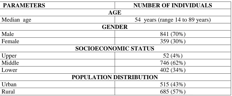
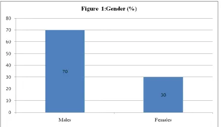
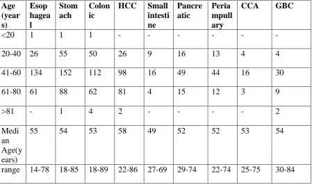
Table 1 shows median age of 54 years (ranging from age 14 to 89 years). In the present study, 3 cases were below 20 years of age. Most of the malignancies were seen in the age group of 41-60 years (58%). The rate of malignancies increased after the age of 20 years (table 6). Out of 1200 cases, 841 (70%) were males and 359 (30%) were females. The male: female ratio was 2.34:1. Six hundred eight five cases (57%) were from a rural area and five hundred fifteen (43%) were from an urban area. Sixty two percent patients belong to middle class of socioeconomic status.
Demographic profile
Table 1: Demographic profile of the individuals with GI and HPB malignancy
PARAMETERS NUMBER OF INDIVIDUALS
AGE
Median age 54 years (range 14 to 89 years)
GENDER
Male 841 (70%)
Female 359 (30%)
SOCIOECONOMIC STATUS
Upper 52 (4%)
Middle 746 (62%)
Lower 402 (34%)
POPULATION DISTRIBUTION
Urban 515 (43%)
Rural 685 (57%)
Table 2: Anthropometric data for individuals
BMI (Kg/M2) Number of individuals (N-1025, %)
Underweight (< 18) 226 (22%)
Normal (18-22.9) 543 (53%)
Overweight (23-25) 116 (11%)
Table 3: Gender distribution of GI and HPB malignancy
Gend er
Esop hagus
Stom ach
Colon ic
HCC Small intesti ne
Pancre atic
Peria mpull ary
Cholan giocarc inoma
Gall bladder cancer
Male 164 185 155 184 21 47 50 18 18
Femal e
58 112 74 23 8 33 19 5 27
M:F ratio
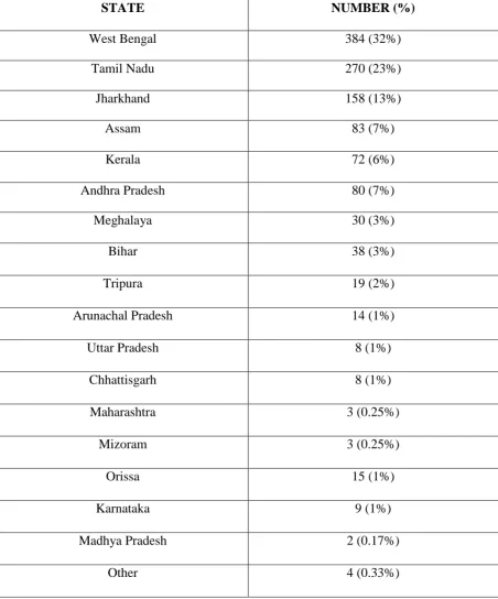
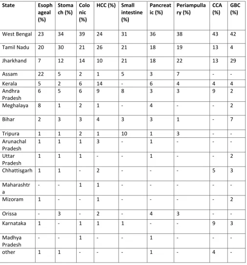
The distribution of type of malignancy according to state is shown in table 4. Most cases were from West Bengal 384(32%) and Tamil Nadu 270 (23%). These were followed by Jharkhand 158 (13%) and Andhra Pradesh 80 (7%).
Table 4: Distribution of GI and HPB malignancy according to state
STATE NUMBER (%)
West Bengal 384 (32%)
Tamil Nadu 270 (23%)
Jharkhand 158 (13%)
Assam 83 (7%)
Kerala 72 (6%)
Andhra Pradesh 80 (7%)
Meghalaya 30 (3%)
Bihar 38 (3%)
Tripura 19 (2%)
Arunachal Pradesh 14 (1%)
Uttar Pradesh 8 (1%)
Chhattisgarh 8 (1%)
Maharashtra 3 (0.25%)
Mizoram 3 (0.25%)
Orissa 15 (1%)
Karnataka 9 (1%)
Madhya Pradesh 2 (0.17%)
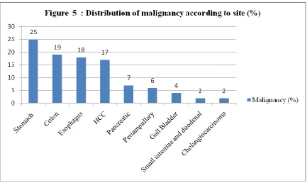
The distribution of type of malignancy according to sites is shown in figure 5. Most common malignancy was stomach (25%), and second most common malignancy was colorectal (19%). These were followed by malignancy of esophagus (18%), hepatocellular carcinoma (17%), pancreas (7%), periampullary (6%), gall bladder (4%), small intestine (including duodenal malignancy) (2%) and cholangiocarcinoma (2%).
[image:51.595.73.507.217.475.2]Table 5: Distribution of type of GI and HPB malignancy according to state (N-1200)
State Esoph
ageal (%) Stoma ch (%) Colo nic (%)
HCC (%) Small intestine (%) Pancreat ic (%) Periampulla ry (%) CCA (%) GBC (%)
West Bengal 23 34 39 24 31 36 38 43 42
Tamil Nadu 20 30 21 26 21 18 19 13 4
Jharkhand 7 12 14 10 21 18 22 13 29
Assam 22 5 2 1 5 3 7 - -
Kerala 5 2 6 14 - 6 4 4 4
Andhra Pradesh
6 5 6 9 8 3 3 9 2
Meghalaya 8 1 2 1 - 4 - - 2
Bihar 2 3 3 4 3 3 1 - 7
Tripura 1 1 2 1 10 1 3 - -
Arunachal Pradesh
1 1 1 3 - 1 - - -
Uttar Pradesh
1 1 1 - - 1 - - 2
Chhattisgarh 1 1 - 2 - - - 5 3
Maharashtr a
- - 1 1 - - - - -
Mizoram 1 - - 1 - - - - 2
Orissa - 3 - 2 - 4 3 - -
Karnataka 1 - 1 1 1 - - 9 3
Madhya Pradesh
- - 1 - - 1 - - -
other 1 1 - - - 1 - 4 -
Table 6: Incidence of GI and HPB malignancy according to age
Age (year s)
Esop hagea l
Stom ach
Colon ic
HCC Small intesti ne
Pancre atic
Peria mpull ary
CCA GBC
<20 1 1 1 - - - -
20-40 26 55 50 26 9 16 13 4 4
41-60 134 152 112 98 16 49 44 16 30
61-80 61 88 62 81 4 15 12 3 9
>81 - 1 4 2 - - - - 2
Medi an Age(y ears)
55 54 53 58 49 52 52 53 54
range 14-78 18-85 18-89 22-86 27-69 29-74 22-74 25-75 30-84
Esophagus malignancy:
As shown in the table 3, there were 222 (18%) cases of esophageal malignancy. Male: female ratio was 2.82:1.
Table 7: Percentage of occurrence of symptoms in esophagus cancer (N-222)
Symptoms Total incidence (%)
Dysphagia 207 (93%)
Weight loss 182 (81%)
Hematemesis 10 (5%)
Vomiting 140 (64%)
Odynophagia 10 (5%)
Chest pain 11 (5%)
As shown in the table 7, most common presenting symptoms were dysphagia (93%) and weight loss (81%).
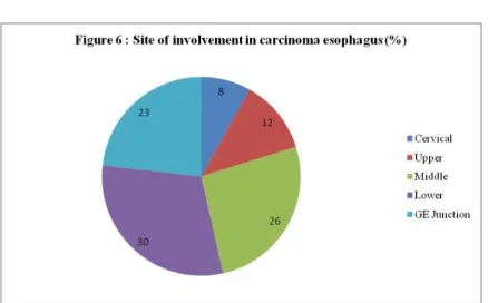
As showed in figure 6, esophageal malignancy was centred around the lower and mid esophagus (30% and 26% respectively), followed by GE junction (23%). The cervical site was least common site for esophageal malignancy.
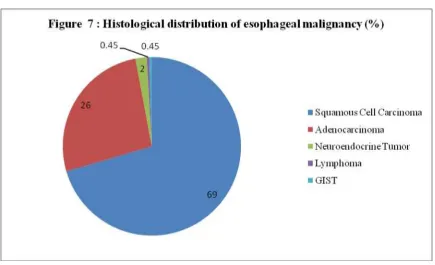
Distribution of type of esophageal cancer according to histology type:
Figure 7 showed that squamous cell carcinoma was most common histology type (69%). Sixty three (26%) patient had Adenocarcinoma. Four (2%), one (0.45%) and one (0.45%) patient had Neuroendocrine tumour (NET), gastrointestinal stromal tumor (GIST) and B-cell lymphoma respectively.
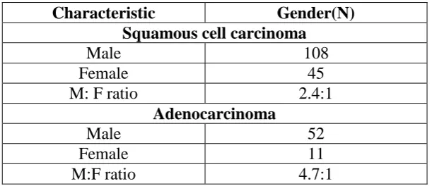
[image:56.595.72.508.194.459.2]Figure 8: Distribution of squamous cell carcinoma and adenocarcinoma (N):
Table 8: Male to female distribution:
Characteristic Gender(N)
Squamous cell carcinoma
Male 108
Female 45
M: F ratio 2.4:1
Adenocarcinoma
Male 52
Female 11
M:F ratio 4.7:1
Table 8 showed male:female ratio was 2.4:1 in squamous cell carcinoma. In adenocarcinoma, the male to female ratio was 4.7:1.
Stomach malignancy:
As shown in the table 6, the median age was 54 years (range from 18-85). Most of the cases were in 41-60 years age group (51%).
Table 9: Percentage of occurrence of symptoms in stomach cancer (N-297)
Symptoms Total incidence (%)
dysphagia 16 (5%)
Weight loss 254 (86%)
Abdomen pain 249 (84%)
vomiting 257 (87%)
anorexia 214 (72%)
Gi bleed 41 (14%)
Abdomen distension 23 (8%)
As shown in the table 9, most common presenting symptoms were vomiting (87%) and weight loss (86%). The least common symptoms were abdomen distension (8%) and dysphagia (5%).
Seven patients had associated H. pylori infection in the biopsy. Three patients had family history of gastric malignancy. One patient had pernicious anaemia and another history of gastric resection.
The adenocarcinoma (N-260, 88%) was most common histology type followed by B-cell lymphoma (N-23, 8%). Seven (2%) patient had NET and GIST.
Colonic malignancy:
As shown in the table 3, there were 229(19%) case of colonic malignancy. Male:female ratio was 2.09:1.
Table 10: Percentage of occurrence of symptoms in colonic cancer (N-229)
Symptoms Total incidence (%)
GI bleed 148 (65%)
Abdomen pain 111 (48%)
Weight loss 154 (67%)
Loss of appetite 152 (66%)
Diarrhea 67 (29%)
Constipation 38 (17%)
As shown in the table 10, most common presenting symptom was weight loss (67%) and anorexia (66%). The least common symptom was constipation (17%).
The figure 11 showed rectum (51%) was most common location of colonic malignancy, followed by the ascending colon (21%). The descending colon (4%) was least common site of colonic malignancy.
Nine Patients (0.39%) had family history of colonic malignancy. Six patients (0.26%) had
polyposis coli, possibly familial adenomatous polyposis (FAP). None of the patient had
Histological distribution of colonic malignancy (%)
As shown in figure 12, adenocarcinoma was the most common histology (N-214), the majority of which were moderately differentiated (N-137). Twelve patients had Lymphoma (B-cell type) and two patients had neuroendocrine tumor (NET).
Carcinoembryonic Antigen (CEA) was tested in 193 of 214 (90%) patients with colonic malignancy. Median CEA value was 5 ng /ml (range 1 to 5270). In 98 patients (46%), it was within normal limits (<5 ng /ml).
Small intestine malignancy including duodenal malignancy:
Twenty nine patients (2%) were diagnosed with a small intestinal malignancy including duodenal malignancy. As shown in the table 3, male:female ratio was 2.62:1.
Table 11: Percentage of occurrence of symptoms in small intestine cancer (N-29)
Symptoms Total incidence (%)
Weight loss 25 (86%)
Abdomen pain 24 (83%)
Vomiting 26 (90%)
Anorexia 23 (79%)
GI bleed 5 (17%)
Early satiety 16 (55%)
As shown in the table 11, most common presenting complaints were vomiting (90 %) followed by weight loss (86 %). The least common symptom was GI bleed (17 %).
Figure 13 showed duodenum (N-26) was most common location for small intestine tumor,
most being located in D1 followed by D2. None of the patients had tumor in the jejunum,
[image:62.595.71.510.313.571.2]As shown in figure 14, the adenocarcinoma (59%) was most common histology. 17% had
B-cell lymphoma and NET.
Hepatocellular carcinoma (HCC):
As shown in the table 3, there were 207 cases (17%) of hepatocellular carcinoma. Male: female ratio was 8:1.
[image:63.595.77.507.61.332.2]As shown in the table 6, the median age was 58 years (range from 22-86). Most of the cases were in 41-60 years age group (47%).
Table 12: Percentage of occurrence of symptoms in HCC cancer (N-207)
Symptoms Total incidence (%)
Abdomen pain or discomfort 98 (47%)
Weight loss 113 (43%)
Abdomen distension (ascites) 88 (55%)
Gi bleed 35 (17%)
Jaundice 75 (36%)
As shown in the table 12, most common presenting complaints were abdominal distension (55%) followed by abdominal pain (47%). The least common symptom was altered sensorium (6%).
[image:64.595.71.509.160.422.2]As shown in figure 15, most of the patients were in CTP-A class. Median MELD score was 12 (range from 6 to 36).
Table 13: AFP value
AFP value( IU/ml) Number (204)
<6 38 (19 %)
7-200 52 (25 %)
>200 114 (56 %)
As shown in figure 16, most common etiology for HCC was hepatitis B (n-74, 37%). Fifty two (26%) patients had cryptogenic CLD related HCC. Twenty (10%) patients had HCV related HCC.
Four patients had family history of HCC. Twenty nine (14%) patients had biopsy proven HCC. In 72 patients with HBsAg related HCC, twenty two were HBeAg positive. HBV-DNA was done in 42 patients and median value was 145000 (range from 10 to 100000000).
In cryptogenic HCC, ten patients were anti-HBc-Ag positive.
Pancreatic malignancy:
As shown in the table 3, there were 80 cases (7%) of pancreatic malignancy. Male:female ratio was 1.42:1.
Table 14: Percentage of occurrence of symptoms in pancreatic cancer (N-80)
Symptoms Total incidence (%)
Weight loss 63 (79%)
Abdomen pain 71 (89%)
Vomiting 64 (80%)
Anorexia 56 (77%)
Jaundice 30 (38%)
Cholangitis 6 (8%)
Early satiety 45 (56%)
Abdomen distension 4 (5%)
As shown in the table 14, most common presenting symptoms were abdominal pain (89%) and vomiting (80%). Thirty patients (38%) had jaundice and six patients (8%) had cholangitis. The least common symptom was abdominal distension (5%).
Figure 20: Type of pancreatic malignancy (%)
As shown in the figure 20, 77% had adenocarcinoma. Nine (12%) patient had NET.
Four (5%) had IPMN and three (4%) had mucinous cystadenocarcinoma.
One patient had family history of pancreatic malignancy. Thirteen patients (16%) had associated chronic pancreatitis. Sixty one patients (77%) had biopsy proved pancreatic malignancy.
CA 19-9 was done in 64 (83%) cases. The median CA 19-9 values were 336 U/ml (range 1- 33900).
Periampullary carcinoma:
As shown in the table 3, there were 69 cases (6%) of periampullary malignancy. Male:female ratio was 2.63:1.
[image:69.595.65.508.74.323.2]As shown in the table 6, the median age was 52 years (range from 22-74). Most of the cases were in 41-60 years age group (64%).
Table 15: Percentage of occurrence of symptoms in periampullary carcinoma (N-69)
Symptoms Total incidence (%)
Weight loss 64 (93%)
Abdomen pain 58 (84%)
Vomiting 51 (74%)
Jaundice 62 (90%)
Cholangitis 15 (22%)
Early satiety 41 (59%)
As shown in the table 15, the most common presenting symptoms were weight loss (93%) and jaundice (90%). Fifteen patients (22%) had cholangitis. The least common symptom was abdominal distension (1%).
One patient had family history of periampullary malignancy.
As shown in the figure 22, forty (58%) patients had moderately differentiated adenocarcinoma. Twenty seven (39%) patients had well differentiated adenocarcinoma and two (3%) patients poorly differentiated adenocarcinoma.
CA 19-9 was done in 47(68%) cases (normal value upto 32 U/ml) and median CA 19-9 value was 99 U/ml (range 2-71400).
Cholangiocarcinoma (CCA):
As shown in the table 3, there were 23 cases (2%) of cholangiocarcinoma. Male to female ratio was 3.60:1.
[image:71.595.65.508.74.322.2]As shown in the table 6, the median age was 53 years (range from 25-75). Most of the cases were in 41-60 years age group (70%).
Table 16: Percentage of occurrence of symptoms in cholangiocarcinoma (N-23)
Symptoms Total incidence (%)
Weight loss 22 (96%)
Abdomen pain 21 (91%)
Vomiting 18 (78%)
Anorexia 15 (65%)
Jaundice 19 (83%)
Cholangitis 6 (26%)
Early satiety 18 (18%)
As shown in the table 16, most common presenting symptoms were weight loss (96%) and abdominal pain (91%). The least common symptom was abdominal distension (13%). Six patients (26%) were presented in cholangitis.
Ten patients (39%) had biopsy proven cholangiocarcinoma. Twenty one (91%) had extrahepatic and two patient (9%) had intra hepatic CCA.
CA 19-9 was done in 19 (82%) cases. The median CA 19-9 values were 345 U/ml (range 2- 20100).
Gall bladder cancer:
As shown in the table 3, there were 45 cases (4%) of gall bladder carcinoma. Male to female ratio was 0.66:1.
[image:73.595.65.466.277.409.2]As shown in the table 6, the median age was 54 years (range from 30-84). Most of the cases were in 41-60 years age group (67%).
Table 17: Percentage of occurrence of symptoms in gall bladder cancer (N-45)
Symptoms Total incidence (%)
Weight loss 44 (98%)
Abdomen pain 39 (87%)
vomiting 41 (91%)
anorexia 33 (73%)
Jaundice 34 (76%)
Cholangitis 10 (22%)
Early satiety 27 (60%)
Abdomen distension 2 (4%)
As shown in the table 17, most common presenting symptoms were weight loss (98%) and vomiting (91%). The least common symptom was abdominal distension (4%). Ten patients (22%) presented with cholangitis.
Sixteen patients (38%) had gall bladder stones and one patient had porcelain gallbladder. Twenty three patients (51%) were biopsy proven gall bladder malignancy.
Distribution of lymphoma according to site:
As shown in the figure 26, of 41 cases of lymphoma, all were B-cell origin, most common sites stomach (N-23, 57%), colon (N-12, 29%). The least common site was esophagus (N-1, 2%).
2
57 12
[image:74.595.71.505.73.321.2]29
Figure 26: Location of lymphoma(%)
esophagus
stomach
small intestine
Distribution of gastrointestinal stromal tumor (GIST) according to site:
Distribution of neuroendocrine tumor (NET) according to site:
Discussion
The present study was an observational study which was intended to analyse the clinico-pathological profile of gastrointestinal (GI) and hepato-pancreato-biliary (HPB) tract malignancies. Out of 41543 patients newly registered in the departments of Medical Gastroenterology and Hepatology from August 2013 to December 2015, a total of 1200 cases of newly diagnosed GI and HPB tract malignancies were included during the study period (2.8%).
In the present study, most malignancies were seen in age group of 41-60 years (58%), peaking in the sixth decade. The overall male : female ratio was 2.34:1. These findings were compared with multiple studies (Table 18). Theses finding were comparable with other studies except the study by Kalyani R et al (1). The male preponderance is similar to that seen in many studies (1, 5, 7 and 8).
In present study, the most common malignancy was gastric (25%), and second most common malignancy was colorectal (19%). These were followed by malignancy of the esophagus (18%), liver, ie HCC (17%), pancreas (7%), periampullary (6%), gall bladder (4%), small intestine (2%) and cholangiocarcinoma (2%). These findings were compared with earlier Indian studies (Table 18). Most studies from India analysing GI malignancies in particular were hospital based pathology centric and retrospective in design.
North-East, a high incidence of cancers in general from different sites, especially tobacco related cancers, have been reported.
Interestingly, in the present study, the second and third position was shared by colo-rectal and esophageal malignancies. Colo-rectal malignancies were also prominent in the study from Karnataka by Kalyani et al (1). Rectal neoplasms far outweighed colonic, as seen in other studies as well. It must be highlighted that both this and our study were one in the 21st Century, where lifestyle changes and better diagnostic facilities exist.
The fourth position was occupied by HCC (17%) in our study. In studies done earlier in the 1960-90s’, HCC occupied a much lower position (2-4%). This again could be related to both diseases related to life style changes and better awareness and detection strategies.