1
DISSERTATION ON
CLINICO-PATHOLOGICAL CORRELATION AND ASSESSMENT OF BURN
WOUNDS
Dissertation submitted to
THE TAMILNADU DR. M.G.R. MEDICAL UNIVERSITY
In partial fulfillment of the regulations for the award of the degree ofMASTER OF SURGERY
IN
GENERAL SURGERY
THANJAVUR MEDICAL COLLEGE,
THANJAVUR
-
613 004
THE TAMILNADU DR. M.G.R. MEDICAL UNIVERSITY
2
CERTIFICATE
This is to certify that this dissertation entitled “ CLINICO-PATHOLOGICAL CORRELATION AND ASSESSMENT OF BURN
WOUNDS” is the bonafide work of Dr.J.Rajarajan in partial fulfilment of the
requirements for M.S Branch -I (General Surgery) Examination of the Tamilnadu Dr. M.G.R. Medical University to be held in APRIL - 2015 under my guidance and supervision during the academic year January- 2014 to July - 2014.
Prof.Dr.V.BALAKRISHNAN,M.S.,
Head of the Department,
Department of General surgery, Thanjavur Medical College, Thanjavur - 613 004.
Prof. Dr. K.MAHADEVAN M.S.,
DEAN,
Thanjavur Medical College,Thanjavur - 613 004. Prof. Dr.G.RAJENDRAN, M.S., FICS.,
Unit Chief S-VI,
3
DECLARATION
I, Dr.J.RAJARAJAN, solemnly declare that the dissertation titled “CLINICO-PATHOLOGICAL CORRELATION AND ASSESSMENT OF
BURN WOUNDS” is a bonafide work done by me at Thanjavur Medical College,
Thanjavur during January - 2014 to July - 2014 under the guidance and supervision of Prof. Dr. G.RAJENDRAN M.S., F.I.C.S., Thanjavur Medical College, Thanjavur.
This dissertation is submitted to Tamilnadu Dr. M.G.R Medical University towards partial fulfilment of requirement for the award of
M.S. degree (Branch -I)in General Surgery.
Place: Thanjavur.
5
ACKNOWLEDGEMENT
I gratefully acknowledge my sincere thanks to Prof. Dr. K.Mahadevan M.S., Dean, Thanjavur Medical College, Thanjavur, for allowing me to do this dissertation and utilize the
institutional facilities.
I am extremely grateful to Prof. Dr. V. Balakrishnan M.S., Head of the Department, Department of General surgery, Thanjavur Medical College, for his full-fledged support
throughout my study and for his valuable suggestions and guidance during my study and my post
graduation period.
I am greatly indebted to my Prof. Dr.G.Rajendran M.S. F.I.C.S., my guide in this study, for his timely suggestions, constant encouragement and scholarly guidance in my study
and in my post graduation period.
I am also extremely grateful to Retired Prof.Dr.Maragathamani Elangovan MS for initiating this dissertation and guiding me in right path.
I profoundly thank my respected professors, Prof. Dr. M.Elangovan MS.,Prof.
Dr.Yeganathan MS.,DA., and Prof. Dr. Karunaharan M.S, for their advice and valuable
criticism which enabled me to do this work effectively.
My sincere thanks to assistant professors, Dr.K.Anbarasan M.S, Dr.V.Bharathiraja
M.S.,and Dr.S. Jeevaraman M.S.,DLO., for their motivation, encouragement and support.
6 CONTENTS
S.NO CONTENTS PAGE NO
1 INTRODUCTION 1
2 AIMS AND OBJECTIVES 3
3 MATERIALS AND METHODS 4
4 REVIEW OF LITERATURE
Anatomy and Function of skin Initial burn management Burn Wound assessment Burn Wound categorization
Laboratory and complementary tests Fluid Resuscitation
Monitoring and Patient control Nutritional support
General Patient support
Treatment of Inhalational Injury Pain management
Infection control
Burn wound Excision and Grafting Topical antimicrobials
Synthetic and Biological Dressing
6-101
5 OBSERVATION AND RESULTS 102
6 DISCUSSION 105
7 CONCLUSION 113
8 BIBLIOGRAPHY 114
9 PROFORMA 116
10 CONSENT FORM
ABSTRACT
CLINICO-PATHOLOGICAL CORRELATION AND ASSESSMENT OF BURN WOUNDS
INTRODUCTION:
Trauma can be defined as bodily injury severe enough to pose a threat to life, limbs, and tissues and organs, which requires the immediate intervention of
specialized teams to provide adequate outcomes. Burn wound biopsies classified as to the depth of infections,confirm the frequent occurrence of bacterial, fungal and viral infection in burn wounds and also provide document for the importance of increasing severity of infection on successive biopsies. Burn wound biopsy can distinguish microbial colonization from invasive infection which can guide patient’s treatment.
AIMS AND OBJECTIVES: 1.The study is designed to analyse the clinico-pathological profile of burn patients with wound sepsis.
2. To carry out histo pathological assessment of burn wounds and burn wound infections.3. To correlate histo pathological findings with the clinical
findings.
MATERIALS AND METHODS:
The present study was conducted in Thanjavur Medical College,
Tamil Nadu . 132 patients with varying extent of burns, starting from 30% of Total Body Surface Area (TBSA), were studied prospectively over a period of
two years from September 2012 to September 2014. All the relevant demographic, clinical and laboratory data required were obtained from clinical records. Serial wound evaluation along with general condition of the patient, serial wound swabs for culture and sensitivity and biopsies were taken.
OBSERVATIONS AND RESULTS:
The maximum number of patients comprised of age group 21-30 years
(37.88%). The commonest cause of burn was flame burn (93.93%) followed by scald (2.27%), chemical burn(2.27%) and electrical burn (1.51%) respectively. Maximum cases had burns in the range of 30-40% TBSA (24.24%). This was followed by burns in the range of 41-50%(15.15%), 51-60%(12.88%),
71-80%(12.88%), 61-70%(12.12%),81-90%(11.36%),91-100%(11.36%) TBSA respectively. Patients having burn more than 60% TBSA had 100% mortality when followed up.
involvement. Presence of bacterial growth on the surface as well as in the eschar and sub-eschar tissue was seen in most biopsies. This represented‘colonization’ and not true ‘invasion’ which was seen as presence of bacteria in the adjoining viable tissue.
The microbiological studies revealed that Pseudomonas is predominant organism isolated from burn wound followed by Klebsiella, Proteus, Staphylococcus and Escherichia coli in decreasing order. There was a good correlation between surface swab culture reports and tissue biopsy culture reports as far as the
type of organism isolated from the burn wound is concerned. However the surface swab culture reports showed growth in almost all cases, which was
probably due to surface contamination.Whereas tissue biopsy culture reports showed positive findings (82.5%) mostly in cases with suspicion of
infection. This finding suggested that tissue biopsy culture is a more reliable indicator of wound infection than surface swab culture where chances of
contamination are more.The calculation of bacterial load per gram of tissue, done in 45 cases, showed positive results (count>105) in 35 cases and all the 35 cases went into septicemia.
CONCLUSION
7
CLINICO-PATHOLOGICAL CORRELATION AND ASSESSMENT OF BURN WOUNDS
INTRODUCTION
Trauma can be defined as bodily injury severe enough to pose a threat to life, limbs, and tissues and organs, which requires the immediate intervention of
specialized teams to provide adequate outcomes. Burn injury, unlike other traumas, can be quantified as to the exact percentage of body injured, and can be viewed as a paradigm of injury from which many lessons can be learned about critical illness involving multiple organ systems. Proper initial management is critical for the survival and good outcome of the victim of minor and major thermal
trauma. Tissue burns involve direct coagulation and microvascular reactions in the surrounding dermis that may result in extension of the injury. Large
injuries are associated with a systemic response caused by a loss of the skin barrier,
the release of vasoactive mediators from the wound and subsequent infection.
Infection is an inevitable complication of extensive burns (more than 30% body
surface) and a major cause of morbidity and mortality. In burns,skin barrier is
replaced by eschar. This moist, protein rich avascular environment encourages
8
there is a release of intermediaries that impede the immune response. Eschar also
restricts distribution of systemically administered antibiotics because of
its avascularity. Burn wound infection has to be differentiated between
colonization of the burn wound and burn wound sepsis which is characterized by
microbial invasion of viable tissue beneath the eschar. Gauging
burn wound sepsis by clinical signs and symptoms is difficult. Burn wound
biopsies classified as to the depth of infections,confirm the frequent occurrence of
bacterial, fungal and viral infection in burn wounds and also provide
document for the importance of increasing severity of infection on successive
biopsies.Wound swab culture /sensitivity cannot differentiate
between wound colonization and wound sepsis. Burn wound biopsy can
9
AIMS AND OBJECTIVES:
1.The study is designed to analyse the clinico-pathological
profile of burn patients with wound sepsis.
2. To carry out histo pathological assessment of
burn wounds and burn wound infections.
3. To correlate histo pathological findings with the clinical
10
MATERIALS AND METHODS:
The present study was conducted in Thanjavur Medical College,
Tamil Nadu . 132 patients with varying extent of burns, starting from 30% of Total
Body Surface Area (TBSA), were studied prospectively over a period of
two years from September 2012 to September 2014. All the relevant demographic,
clinical and laboratory data required were obtained from clinical records. Serial
wound evaluation along with general condition of the patient, serial wound swabs
11 REVIEW OF LITERATURE
ANATOMY AND FUNCTION OF SKIN
The skin is the largest organ in the body, making up 15% of body weight,and covering approximately 1.7 m2 in the average adult. The function of the skin is complex: it warms, it senses, and it protects. A burn injury implies damage or destruction of skin and/or its contents by thermal, chemical, electrical, or radiation energies or combinations thereof. Thermal injuries are by far the most common and frequently present with concomitant inhalation injuries. Of its two layers,only the epidermis is capable of true regeneration. When the skin is seriously damaged, this external barrier is violated and the internal milieu is altered.Following a major burn injury, myriad physiological changes occur that together comprise the clinical scenario of the burn patient. These derangements include the following:
Fluid and electrolyte imbalance: The burn wound becomes rapidly edematous.In burns over 25% BSA, this edema develops in normal noninjured tissues. This results in systemic intravascular losses of water, sodium,
albumin, and red blood cells and henceby shock if untreated.
Metabolic disturbances: This is evidenced by hypermetabolism and muscle catabolism. Unless early enteral nutrition and pharmacological intervention restore it, malnutrition and organ dysfunction develop.
12 Complications from vital organs.
The successful treatment of burn patients includes the intervention of a
[image:14.612.91.479.120.502.2]multidisciplinary burn team. The philosophy of care is based on the concept that each patient is an individual with special needs. Each patient’s care, from the day of admission, is designed to return him or her to society as a functional, adaptable, and integrated citizen.
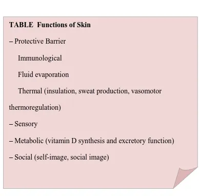
TABLE Functions of Skin
– Protective Barrier
Immunological Fluid evaporation
Thermal (insulation, sweat production, vasomotor thermoregulation)
– Sensory
13 INITIAL BURN MANAGEMENT
The general trauma guidelines apply to the initial burn assessment. A primary survey should be undertaken in the burn admission’s room or in the Accidents
and Emergency Department, followed by a secondary survey when resuscitation is underway.
The primary survey should focus on the following areas:
Airway (with C-spine control): Voice, air exchange, and patency should be noted.
Breathing: Check breath sounds, chest wall excursion, and neck veins. Circulation: Mentation should be noted. Check skin color, pulse, blood pressure, neck veins, and any external bleeding.
Disability-Neurological assessment: Check Glasgow coma score. Expose the patient and environment control-keep warm
14
INITIAL ASSESSMENT OF THE BURNED PATIENT
Treatment of the burn injury begins at the scene of the accident. The first priority is to stop the burning. The patient must be separated from the burning source. For thermal burns, immediate application of cold compresses can reduce the amount of damaged tissue. Prolonged cooling, however, can precipitate a
dangerous hypothermia. For electrical burns, the source should be removed with a nonconducting object. In cases of chemical burns, the agent should be diluted with copious irrigation, not immersion. The initial physical examination of the burn victim should focus on assessing the airway, evaluating hemodynamic status, accurately determining burn size, and assessing burn wound depth. Immediate assessment of the airway is always the first priority. Massive airway edema can occur, leading to acute airway obstruction and death. If there is any question as to the adequacy of the airway, prompt endotracheal intubation is mandated. All burn victims should initially receive 100% oxygen by mask or tube to reduce the likelihood of problems from pulmonary dysfunction or carbon monoxide poisoning. The next step is to place two large-bore peripheral intravenous
15
BSA burn requires IV access. Begin infusion of Ringer’s lactate solution of about
1000 ml/h in adults, 400–500 ml/m2 BSA/h in children, until more accurate assessments of burn size and fluid requirements can be made. An indwelling Foley catheter should be placed to monitor urinary output. A nasogastric tube is inserted for gastric decompression.It is also imperative during the initial
assessment to make a brief survey of associated injuries. A thorough secondary survey can be postponed, but lifethreatening injuries such as cardiac tamponade, pneumothorax, hemothorax, external hemorrhage, and flail chest must be identified and treated promptly.
Patient evaluation should include what is termed an AMPLE history: allergies, medications, pre-existing diseases, last meal, and events of the injury, including time, location, and insults. In children the developmental status should be
investigated and any suspicious injuries should raise the possibility of child abuse. A history of loss of consciousness should be sought. A complete physical
16
for pulses, especially in patients with circumferential burns. Evaluation of pulses can be assisted by use of a Doppler ultrasound flowmeter. If pulses are absent, and fluid resuscitation is adequate, the involved limb should undergo urgent
escharotomy to release the constrictive eschar. It must be noted, however, that the most common cause of pulseless limbs is inadequate resuscitation. Therefore, the intravascular status of the patient must be assessed before proceeding with
escharotomies.
17 Burn Wound Assessment
After the patient’s stabilization and initial resuscitation, physicians should focus
on the burn wound. Burns are gently cleansed with warm saline and antiseptics, and the extent of the burn is assessed. Burn injury must be categorized as the exact percentage of BSA involved. The rule of nines is a very good approximation as an initial assessment .Another good rule of thumb is measuring
the extent of the injury with the palm of the burn victim, which is estimated as 1% BSA. The area burned is transformed as the number of hand palms affected and then multiplied by 1%.
18 Secondary Assessment
1. Initial trauma assessment and primary assessment completed. 2. Thorough head-to-toe evaluation.
3. Careful determination of trauma other than obvious burn wounds. 4. Use cervical collars, backboards, and splints before moving the patient. 5. Examine past medical history, medications, allergies, and mechanism of injury.
6. Establish intravenous access through large peripheral catheters and administer intravenous fluids through a warming system.
19
Lund and Browder Chart
The best way to measure the area burned accurately is the Lund and Browder Chart In this method, the areas burned are plotted in the burn diagram,and every area burned is assigned an exact percentage. The Lund and Browder method takes into consideration the differences in anatomical location that exist in the pediatric
20
different ages. After the burn size is determined, the individual characteristics of the patient should be plotted in a standard nomogram to determine the body surface area and burned surface area of the patient .Measuring and weighing the patient in centimeters and kilograms provides the surface area of the patient in square
meters. This measurement will help to calculate metabolic needs, blood loss, hemodynamic parameters, and skin substitutes.
At this point, the specific anatomical location of the burn should be noted
21
In ventilated patients or patients with suspected smoke inhalation injury,
direct bronchoscopy should be performed to determine the extent of the injury, and also aids in diagnosis and therapeutic lavage of soot and damaged epithelium. After direct bronchoscopic examination is completed, a definitive diagnosis is made based on clinical, laboratory, and bronchoscopic findings.After definitive assessment in the burn center, a final diagnosis regarding the burn wounds (extent and depth), accompanying injuries, and smoke inhalation injury is reached. At this point burn wounds should be covered with a clean burn wound dressing.
Compressive dressings should be avoided, because they can induce further hypoperfusion and conversion of partial-thickness wounds to full-thickness. When the treatment of choice of full-thickness burns is early burn wound excision in 72 h, after resuscitation is completed, burns can be treated either with 1% silver sulfadiazine during the period between the accident and the definitive surgery. Partial-thickness burns are treated in a similar initial manner.
Determining burn depth requires experience. It is an important part of the
burn assessment because the depth of the burn will determine the treatment option and the patient’s outcome. It must be noted, however, that even in the hands of
22
burn wound. Laser Doppler scanning has emerged as a good tool in the proper assessment of depth but its expensive and available in only few centres. The
evaluation of degree (depth) of burn was found to be more accurate by histological
assessment than by clinical assessment in many studies and is economical .
A B
The laser Doppler scanner (A) is helpful for the diagnosis of burn wound depth. Its sensitivity and specificity are best between 48 and 72 h after the injury.
23 Partial-Thickness Burns
Cover burns with clean dressing until definitive diagnosis by experienced burn surgeon .
Minor burn: semiocclusive dressing (special locations: silver sulfadiazine)
Large burn: Synthetic artificial skin or pig skin Full-Thickness Burns
Immediate burn wound excision protocol (24 h): – Telfa clear or plastic film Early burn wound excision protocol (72 h): silver sulfadiazine or cerium nitrate–silver sulfadiazine
Staged excision protocol (first week): cerium nitrate silver sulfadiazine
24 BURN WOUND CATEGORIZATION
Burn wound have been classically categorized as first-, second-, and third degree. First-degree burns are superficial and involve just the epidermis. Typified
by sunburn, first-degree burns are inconsequential in subsequent burn management.They heal in 5–7 days. Oral intolerance and severe discomfort requiring hospitalization may accompany large first-degree burns. These burns have a red,hyperemic appearance of the surface, which, along with the
hypersensibility and discomfort, is typical of these injuries
Second-degree burns, also called partial-thickness burns, involve variable
amounts of dermis .Second-degree burns are subdivided into superficial and deep second-degree wounds.
25
In deep second-degree burns, however, the epidermis, papillary dermis, and various depths of the reticular (deep) dermis have been damaged. Regeneration occurs much more slowly than in superficial burns. Complete healing take more than 3 weeks and scarring and infection are common. These injuries are best treated surgically, since excision of the dead tissue and skin grafting shorten hospital stay and improve outcomes. Deep second-degree burns tend to be
26
First-degree burns. Only the epidermis has been damaged. Typical appearance is that of a hyperemic area with severe discomfort and hyperesthesia.
27
Second-degree burn injuries (or partial-thickness burns) present with
different degrees of damage to the dermis. Pain is very intense . They usually blanch with pressure and do not usually leave any permanent scarring.
28
Deep second-degree burns present with lesser degrees of pain
29
In contrast to the former injuries, third degree burns or full-thickness burns never heal spontaneously, and treatment involves excision of all injured tissue In these injuries, epidermis, dermis, and different depths of subcutaneous and deep tissues have been damaged. Pain involved is very low (usually with marginal partial-thickness burns) or absent. The potential for infection if left non excised is very high. A dry, white, or charred appearance is common. In infants and patients with immersion scalds, the burns may appear cherry red, and they may be misleading .
30
Burns that affect deep structures, such as bones and internal organs, are
31
After a definitive diagnosis has been made regarding size and depth, burns can be classified as minor, moderate, or major injuries A major burn injury is defined as greater than 25% BSA involvement (15% in children) or more than 10% BSA full-thickness involvement. Major burns require aggressive resuscitation, hospitalization, and appropriate burn care. Additional criteria for major burns include deep burns of the hands, feet, eyes, ears, face, or perineum;
inhalation injuries; associated medical conditions; extreme age; and electrical burns.
Moderate thermal burns of 15–25% BSA or 3–10% BSA full-thickness often require hospitalization to ensure optimal patient care. Other criteria for admission include concomitant trauma, significant pre-existing disease, and suspicion of child abuse.
32
LABORATORY AND COMPLEMENTARY TESTS
Routine admission laboratory evaluations should include the following: Complete blood count
Coagulation tests, including D-dimmers and fibrinogen Blood group type and screen
Glucose
Blood urea nitrogen (BUN) Creatinine
Total proteins, albumin, and globulins Serum electrolytes
Calcium, phosphorus, and magnesium Osmolality
Liver function test C-reactive protein Total CO2
Arterial blood gas, including lactate and Carboxyhemoglobin (HbCO) Urine analysis, including urine electrolytes
33
Other complementary tests include chest x-ray and other x-ray examinations performed on an individual basis. A 12 lead electrocardiogram should be obtained in all patients on admission and should repeated periodically in all electrical
injuries.Routine cultures are obtained on admission as part of the infection control protocol. They are then repeated twice per week unless dictated otherwise by the patient’s clinical picture. Cultures should include blood, urine, sputum, throat,
34 FLUID RESUSCITATION
The most crucial aspect of early care of the burn patient is prompt initiation of volume replacement of large quantities of salt-containing fluids to maintain adequate perfusion of vital organs. Many formulas for burn resuscitation have proven clinically efficacious, and each differs in volume, sodium, and colloid content. The aim of any fluid resuscitation is to have a lucid, alert, and cooperative patient with good urine output.
Guidelines for correct resuscitation include the following: Do not delay resuscitation.
Estimate burn size and calculate fluid requirements.
Fluid formulas are only a guideline; monitor urine output and tailor intravenous fluids to the response of the patient.
Monitor peripheral pulses, blood pressure, respiration rate, heart rate, urine output, oxygen saturation, and temperature (core/peripheral).
Monitor central venous pressure and/or cardiac output and hemodynamic parameters in severe burns or patients at risk for complications.
Achieve a urine output of 0.5 ml/kg/h in adults and 1 ml/kg/h in children, no more, no less
35 Start enteral feeding on admission.
The aim is to obtain an awake, alert, conscious, and cooperative patient. Do not obtain a replica of the Michelin Man; prevent edema.
The recommended resuscitation formulas for adults and children are the modified Parkland formula for adults and the Galveston formula for children. In each, half of the volume is administered in the first 8 h and the rest in the second 16 h. Adult burn patients are resuscitated with the modified Parkland formula.
It calls for the infusion of 3 ml/kg/% burn in the first 24 h postburn of Ringer’s lactate solution. In the subsequent 24 h, transcutaneous evaporative losses from burn wounds are replaced at 1 ml/kg/% burn daily. The rate is adjusted hourly to ensure a urinary output of 0.5 ml/h.
RESUSCITATION IN PEDIATRIC BURNS
Resuscitation of burned children differs in two aspects. First, the Parkland
formula commonly underestimates fluid requirements in a burned child and may not provide even the usual daily maintenance requirements. There is great
variability between body surface area and weight in a growing child. More
accurate estimation of resuscitation requirements in burned children can be based on BSA determined from nomograms of height and weight .For children,
36
ml/m2 BSA total/day of Ringer’s lactate. Again, one-half is given over the first 8 h and the rest in the next 16 h during the first 24 h postburn. Due to small glycogen stores, infants require glucose since they are prone to hypoglycemia in the initial resuscitation period; therefore, the basal maintenance fluid administration is given as 5% glucose-containing solutions. In the subsequent 24 h fluid requirements are 3750 ml/m2 BSA burned/day plus 1500 ml/m2 BSA total/day. Care should be taken to avoid rapid shifts in serum sodium concentration, which may cause cerebral edema and neuroconvulsive activity. The rate is adjusted to ensure a urinary output of 1 ml/kg/h . Patients in air-fluidized beds should receive 1000 ml/m2 BSA/24 h extra fluids to replace the evaporative fluid loss produced by the bed.
Enteral feeding is usually started on admission and gradually increased
until the maximum full rate is achieved. As the enteral feeding volume is increased and absorbed by the patient, intravenous fluid are diminished at the same rate, so that the total amount of resuscitation needs are met as a mixture of IV fluids and enteral feeding. By 48 h, most of the fluid replacement should be provided via the enteral route.
37
Pediatric Patients
First 24 h:
5000 ml/m2 BSA burned/day + 2000 ml/m2 BSA total/day of Ringer’s lactate (give half in first 8 h and the second half in the following 16 h)
Subsequent 24 h:
3750 ml/m2 BSA burned/day + 1500 ml/m2 BSA total/day (to maintain urine output of 1ml/kg/h)
Adult Patients
First 24 h: 3 ml/kg/% BSA burned of Ringer’s lactate (give half in first 8 h and the second half in the following 16 h)
Subsequent 24 h: 1 ml/kg/% burn daily (to maintain urine output of 0.5 ml/kg/h)
Additional fluids are commonly needed in patients with inhalation injuries, electrical burns, associated trauma, and delayed resuscitation. Fluid resuscitation should be started according to the fluid resuscitation formula. Fluid administration needs then to be tailored to the response of the patient based on urine output
38
insult to pulmonary, renal, and mesenteric vascular beds. Fluid overload can produce undersized pulmonary or cerebral edema. It will also increase wound edema and thereby dermal ischemia, producing increased depth and extent of cutaneous damage.
Fluid requirements in patients with electrical injuries are often greater than those in patients with thermal injury. The main threat in the initial period is the development of acute tubular necrosis and acute renal insufficiency related to the precipitation of myoglobulin and other cellular products. A common finding in patients with electrical injuries is myoglobinuria, manifested as highly
concentrated and pigmented urine. The goal under these circumstances is to maintain a urine output of 1–2 ml/kg/h until the urine clears. In nonresponding patients,alkalization of the urine and the use of osmotic agents may prevent death. The use of colloid solutions for acute burn resuscitation remains debated.
Development of hypoproteinemia in the early resuscitation period increases edema in nonburned tissues. In the absence of inhalation injury, however, lung water content does not increase. Early infusion of colloid solutions may decrease overall fluid requirements in the initial resuscitation period and reduce nonburn edema. However, injudicious use of colloid infusion may cause iatrogenic
39
g/100 ml after the first 8 h. In selected cases, it can be supplemented in the first 8 h postburn. Albumin solution 5% should be used instead of 25% solution in unstable patients with hypovolemia.
Fluid resuscitation should be monitored using clinical parameters. The best single indicator is urine output. Hypotension is a late finding in burn shock; therefore, pulse rate is much more sensitive than blood pressure. Normal
sensorium,core temperature, and adequate peripheral capillary refill are additional clinical indicators of adequate organ perfusion. Fluid shifts are rapid during the acute resuscitation period (24–72 h), and serial determinations of hematocrit, serum electrolytes, osmolality, calcium, glucose, and albumin can help to direct appropriate fluid replacement.
40 MONITORING AND PATIENT CONTROL
Patients with major burns should receive full monitoring, including: Continuous electrocardiograph monitoring
Continuous respiratory rate monitoring Pulse oximetry
Central venous pressure Arterial line
Foley catheter and urine output Temperature probes
Capnometry (ventilated patients)
Pulmonary artery catheter (unstable severe burn patients)
Esophageal Doppler monitoring (alternative to Swan-Ganz catheters) Doppler monitor for compartment syndromes
Central lines and arterial lines do carry some morbidity in burned patients. Judicious use of these otherwise helpful monitoring devices is advised.
41
40%BSA without significant pre-existing diseases can be managed without central line catheters. Control of blood pressure, pulse rate, pulse oximetry, respiratory rate,temperature, weight, and urine output should suffice in most of the patients. Blood pressure is often monitored using arterial lines. In most cases, however, indirect measure of blood pressure along with the physiological parameters mentioned earlier and the valuable addition of pulse oximetry are more than
enough to monitor the patient. Arterial lines should be reserved for use in unstable patients,those with inhalation injury, unstable patients receiving ventilatory
support, and patients who will need repeated blood gas analysis. Catheter-related sepsis has plagued burn patients for decades. With the advent of modern
indwelling catheters,and strong policies for periodical line change, the incidence of catheter related sepsis has declined dramatically. The usual recommendation is to change all lines every 7 days. Nevertheless, increasing evidence suggests that lines do not need to be changed unless they become infected. Care of the line should include daily inspection of entry point and daily dressing with dry compresses. Occlusive dressings and antibiotic creams are not effective to control infection, and there are reports that they may even increase the risk of infection.
42
temperature (24–28 deg C) and at least 50%humidity. Patients are covered and placed under thermal panels. These panels provide a central area just over the patient with a high temperature (ideally 36 deg C) whereas in the rest of the room the environmental conditions, although still warm, are cool enough to allow
reasonable comfort for health personnel. Head, limbs, and genitalia are to be elevated, and the patient should be positioned comfortably over the patient, allowing a lower temperature in the rest of the environment for staff and visitor comfort.
Patients should be encouraged to walk and mobilize as soon as possible, and early physiotherapy should be started. Patients should never be at bed rest unless absolutely necessary. Patients must be comfortable and pain free and patients and families should be trained in wound care and rehabilitation.
All patients need to be closely monitored by burn physicians. A formal
morning round should be established, with review of all systems and wounds when deemed necessary. All altered parameters need to be corrected. Performing an informal evening round to check the daily progress of the patient, and
43
Emergency treatment focuses on stabilization of patients, treatment of associated injuries, fluid resuscitation, initial respiratory support, and emergency treatment of the burn wound. Soon after stabilization and resuscitation, a formal
discharge plan (treatment plan, rehabilitation plan, and social support) is
established.Focus of burn treatment is then shifted to the definitive burn wound treatment and to the general support of the patient, which include:
Nutritional support General patient support
Support of the hypermetabolic response Treatment of inhalation injury
Pain management and psychosocial support
44 NUTRITIONAL SUPPORT
The hypermetabolic response to burns is the greatest of any other trauma or infection.A major burn injury provokes a complex disruption of hormonal homeostasis that induces an increased resting metabolic rate and oxygen
consumption, increased nitrogen loss, increased lipolysis, increased glucose flow, and loss of body mass. To meet postburn energy demands, all main metabolic pathways are utilized. Carbohydrate stores are small; therefore, carbohydrate intermediate metabolites, which are also essential for fat catabolism, are obtained from skeletal muscle breakdown, thus increasing muscle catabolism. Prolonged inflammation,pain or anxiety, environmental cooling, and sepsis can further exaggerate this postburn hypermetabolic response.
One of the main principles underlying successful management of the postburn hypermetabolic response is providing adequate nutritional support. In general, patients affected with more than 25% body surface area (BSA) burned and
those patients with malnutrition or who cannot cope with their metabolic demands as a result of concomitant injuries or diseases should receive nutritional support. The nutritional formula of choice is enteral nutrition. Total parenteral nutrition should be abandoned and reserved for patients who cannot tolerate the enteral route. It carries a high mortality in burn patients.
45
regimen, the gastric residuals should be checked regularly. Once the residual has been checked, it is then infused back to the stomach to avoid
electrolytic imbalances and alkalosis. If these residuals are more than a 2 h tube feeding infusion rate, the feeding should be stopped and the cause investigated. The most common cause of enteral feeding intolerance is tube malposition, although important causes of intolerance that all physicians should bear in mind are sepsis and multiple organ failure. The enteral feeding should be started on admission and continued until the wounds are 90% healed and the patient can maintain an oral intake of his or her caloric demand.
Enteral feeding is started on admission and, if absorbed, it is increased until full strength is obtained, ideally in the first 24 h. The hourly absorbed nutrition is subtracted from the total resuscitation hourly fluids the patient is receiving, in order to avoid overloading. When patients are scheduled for surgery, nutrition is stopped 2–4 h before surgery, and the stomach is aspirated prior to the induction of anesthesia. In ventilated patients, enteral nutrition is not stopped but is
continued during surgery.Caloric requirements in burn patients should be ideally calculated by means of indirect calorimetric measurement. This is an easy
46
reassessed on a daily basis.Patients should be weighed regularly.Inadequate intake necessitates an alteration of the regimen .
Initial Nutritional Assessment
Determine the caloric and protein needs of patients immediately upon admission
Assessment by physician and dietician – Personal background
– Chronic conditions
– Hypermetabolic conditions
– Physical conditions that may interfere with food intake – Predisposing factors
– Recent weight loss or gain – Food preference and allergies
– Weight and height for age and gender – Total lymphocyte count
– White blood cells
– Hemoglobin and hematocrit/ Mean corpuscular volume
Perform indirect calorimetry if available Calculate daily calorie and protein needs
47
Measurement of these variables, together with indirect calorimetry and the weight gain/loss of the patient will give a good estimate of his or her nutritional status. Burn patients who can eat normally receive a high-protein, high-calorie
diet. Liquids should be supplemented in the form of high-calorie fluids, such as milk or commercial milkshakes. Patients with burns over 25%BSA burned cannot cope with the caloric demands that trauma imposes on them, so that in all of them enteral supplementation is indicated.
Different enteral formulas are available. The most popular ones are Curreri and Harris Benedict for adults, and Galveston formula for children. Some of them (Curreri) may overestimate calorie requirements, whereasothers (Harris-Benedict) may underestimate these needs. Therefore, they should be used as initial estimates, with patients needs titrated to their hypermetabolic response as measured by
48
component is also important. Caloric replacement should be based on nonprotein calories only.
Nutritional Formulas
Nutritional formulas for pediatric burn patients
0–12 months: 2100 kcal/m2 surface area + 1000 kcal/m2 surface area burned 1–11 years: 1800 kcal/m2 surface area + 1300 kcal/m2 surface area burned > 12 years: 1500 kcal/m2 surface area +1500 kcal/m2 surface area burned Nutritional formulas for adult burn patients
Curreri: 16–59 years: 25 kcal/kg + 40 kcal for each percentage of burn area 60 years and more: 20 kcal/kg + 65 kcal for each percentage of burn area Harris-Benedict: Basal energy expenditure (BEE) in kcal/day (at rest):
Males: BEE = 66 +13.7 (kg) +5 (cm)-6.8 (years) Females: BEE = 655 + 9.6 (kg) +1.8 (cm)- 4.7 (years) Activity/injury factor:
Bed rest 1.2, ambulatory 1.3, minor surgery 1.2, trauma 1.35, sepsis 1.6, severe burn 2.1
Calculated caloric requirements
= BEE * activity/injury factor
49
needs are 4.4 g/kg in infants (less than 6 months), 4.0 g/kg in small children (6–24 months), and a calorie to nitrogen ratio of 120:1 (kcal/N) in the rest of patients. For a balanced daily diet, administration of vitamins C, A, and E, B complex, zinc, iron, folate,and trace minerals is essential.
When the patient’s wounds are virtually covered, the diet should transition
from one in which the majority of the nutrition is supplied via tube feedings to a total oral diet. The transition should be slow and may take several days. The following steps should be followed:
1. Reduce tube feedings to a rate that with the oral intake equals 100% of reassessed goal.
2. As oral intake increases, provide only nocturnal tube feedings to equal 100% of goal.
3. When oral intake is at least 50% of goal, begin 3 day trial of oral diet with tube feedings held.
4. If goal is not being met at the end of the trial, re-evaluate feeding methods and, if necessary, resume tube feedings.
50 PATIENT SUPPORT
Hypoproteinemia and Anemia
Hypoproteinemia due to malnutrition, continuing serum protein losses, and postburn sepsis and hepatic dysfunction will persist until wound closure is achieved.
Intravascular proteins can be replaced by processed albumin (generally preferred), or fresh frozen plasma if a significant coagulopathy is also present. The efficacy of colloids is the subject of debate; therefore every effort should be made to correct hypoproteinemia with early enteral nutrition. Colloids should be reserved for patients with severely low protein levels and those with significant clinical effects of such hypoproteinemia.
Anemia is very common among burn patients. The initial thermal injury is accompanied by direct destruction of red cells. Other cells are not initially destroyed, but they are structurally damaged and the half-life of the red cell pool is significantly decreased. Repeated trips to the operating room makes this anemia even more profound, as a joint effect of hemorrhage and blood replacement,
51 Environmental Control
Control of the surrounding environment is a well-recognized part of appropriate burn care. Burn patients lose some of their thermoregulatory abilities and are prone to hypothermia. An ambient room temperature of 28–33ºC keeps the patient more comfortable and reduces his or her heat losses from evaporation. Natural light and large windows help patients to maintain their well being. Strong noises should be avoided; and the area needs to be kept pleasant, clean, and relaxing. Stress Ulcer Prophylaxis
The acid pH of the stomach plays an important role in infection control in the human body. This acid serves as a topical treatment for all foods that enter the digestive tube. This acid pH can be problematic when different problems collide in the same clinical situation. Tissue hypoperfusion (frequently measured by gastric tonometry) and the depletion of reduction agents and free radical scavengers promote a progressive damage of gastric mucosa. This erodes and progresses to small ulcers by the action of gastric acid and digestive enzymes. Maintaining good patient support and preventing sepsis and multiple organ dysfunction is extremely important to prevent stress ulcers.
The best prophylaxis for stress ulcers is enteral nutrition. Burn patients
52
and serves as a scavenger for gastric acid. Patients who are not receiving any enteral nutrition at any given time should receive sucralfate, which coats the gastric mucosa and prevents ulceration. H2 blockers and antacids should be reserved for patients whose condition cannot be managed properly with enteral nutrition and sucralfate. Changing the gastric pH encourages bacterial overgrowth and leads to a higher incidence of pneumonia. Acid kills bacteria, which is the most important action of acid in our stomachs.
Deep Venous Thrombosis Prophylaxis
Burn patients should be encouraged to be mobile except in the immediate
postoperative period or if they are attached to a ventilator. Even ventilated patients, should be exercising. The rest of the patients should be up with their limbs
53 TREATMENT OF INHALATION INJURY
Inhalation injury is evident in over 30% of hospitalized burn patients and in 20–84% of burn-related deaths. Heat can result in damage and edema to the upper airway, but uncommonly produces injury below the vocal cords except with steam burns. Acute asphyxia can occur due to environmental oxygen consumption or by reduction of oxygen transportation by carbon monoxide or cyanides.
The majority of tissue damage attributed to inhalation injury is mediated by a chemical injury from incomplete combustion products carried by smoke, including aldehydes, oxides, sulfur, nitrogen compounds, and hydrochloric gases. This chemical damage to the lower airways and parenchyma is propagated by neutrophils. Lung vascular permeability is increased, promoting pulmonary
edema. Desquamation of small airways along with inflammation produces airway casts, leading to areas of atelectatic lung alternating with emphysematous regions, acute pulmonary insufficiency, and bronchopneumonia.
Diagnosis of inhalation injury should be suspected in patients with facial
burns, singed nasal hair, cough, carbonaceous sputum, or evidence of upper airway edema, including hoarseness, stridor, or wheezing. It should be considered
54
be determined, but it may be misleading if initially normal. Diagnosis of inhalation injury is best confirmed by results of fiberoptic bronchoscopy. Chest x-ray is an insensitive initial test.
Treatment of inhalation injury should begin at the scene of the accident
with immediate administration of 100% oxygen. Carbon monoxide poisoning produces asphyxia by binding competitively to hemoglobin and reducing oxygen carrying capacity. Hemoglobin has a 210-times greater affinity for carbon monoxide than oxygen. On room air, carboxyhemoglobin (CO-Hgb) has a half-life of about 4 h in the bloodstream. The half-life of CO-Hgb is reduced to 20 min when the subject is breathing 100% oxygen. If oxygen supplementation is started promptly, anoxic cerebral injuries are reduced. Levels of CO-Hgb greater than 15% are clinically significant, and levels above 40% can produce coma.
Great debate still exists regarding intubation in patients with suspected
55
56
The nebulization of various substances and different respiratory therapy maneuvers have proved beneficial in the prevention of progression to tracheobronchitis,
pulmonary edema,ARDS, and bronchopneumonia. The protocol is universal, and
Inhalation Injury Protocol
1. Titrate high-flow humidified o2 to maintain arterial o2 saturation >90%
2. Cough, deep breathing exercises every 2 h
3. Turn patient from one side to the other every 2 h 4. Chest physiology and physiotherapy every 4 h
5. Nebulize 3 ml N-acetylcysteine 20% solution every 4 h for 7 days 6. Nebulize 500 units of heparin with 3 ml normal saline every 4 h for 7 days
7. Nebulize bronchodilators every 4 h for 7 days 8. Nasotracheal suctioning as needed
9. Early ambulation
10. Sputum cultures Monday, Wednesday, and Friday
11. Pulmonary function studies before discharge and at scheduled outpatients visits
57 can be applied to patients with any sort of burn .
In general, aggressive bronchial toilet with direct bronchoscopy and lavage to remove bronchial casts is one of the pillars of such strategy. In addition, tailoring ventilatory support to the individual needs helps to prevent barotrauma and other complications. When patients can no longer maintain their normal gas exchange, ventilatory support is necessary.. In general, the physician should choose a
58 PAIN MANAGEMENT
Burns hurt, and so does burn treatment. Pain is the most immediate concern of the burn patients. Suffering a combination of physical discomfort and mental torment increases the postburn hypermetabolic stress response. Treatment for a patient’s suffering, however, involves more than control of pain. Emotional
support is essential, and uninterrupted sleep is beneficial. Other problems burn patientsoften experience are anxiety, itching, and posttraumatic stress disorder. There are two types of burn pain. The first type is background pain. Background pain is always present and its range of fluctuation is very small. The second type of pain is the excruciating, intolerable pain that occurs when something is done to the patient, such as procedural pain during dressing changes, line change, or physiotherapy. It is the worst pain a patient can encounter, and patients cannot make any comparison to other experiences in life. Pain control is one of the great challenges in the burn unit, and it is an unsolved problem. Anxiety, sleep disorders, and posttraumatic stress are problems often encountered along with pain.
59
responses to pain stimulus vary significantly. The patient’s pre-existing
psychological make-up, ethnocultural background, the experience of the injury, and its meaning modulate the individual response to pain.
General recommendations for Pain control
1. If patient says he or she has pain, then he or she has pain.
2. Analgesics are most effective when given on a regular basis (not as needed or required).
3. Intramuscular injections are not usually appropriate because the patient fears the injection and intramuscular flow may be altered.
4. Bowel management begins with the narcotic pain management.
5. Pain management protocol should be initiated with the starting doses, which can be modified as the situation dictates.
Pain Assessment
The patient’s pain can be assessed using the 10-point scale or the Faces scale
In the 10-point scale, the sliding scale is moved until the patient feels that the number expressed matches the pain he or she is experiencing. For children less than 3 years old, the Faces pain-rating scale is best used.
60
Both background and procedural pain occur in the emergency, acute, and rehabilitation phase. Therefore different methodologies should be applied depending on the patient’s phase of the disease in order to obtain good pain control. In general, liberal use of opioids and benzodiazepines is advised. Unless patients have a pre-existing drug abuse problem, they will not become opioid dependent from its use during the acute phase of the burn.
Burn patients should always receive baseline analgesia. Drugs need to be scheduled in order to maintain a basal level of painkillers that will make
controlling the pain much easier. Therefore, do not order analgesics at the patient’s request (PRN). The best practice in patients who are candidates for opioid
analgesia is patient-controlled analgesia (PCA) pumps. They decrease patients’ anxiety by putting them in control of the pain regimen. Children of 8 years and older are candidates for PCA. Patients receiving opioids need to be started on a bowel regimen to prevent the development of constipation.In general, prune juice or lactulose will suffice in most. When patients get constipated,mineral oil should be started, followed by enemas if a good response is
61 Pain vs. Anxiety
Anxiety can be very debilitating in the acute situation, and it often accompanies pain. It can be very diffcult to differentiate between them. It can not be
overemphasized,however, that pain should not be treated with anxiolytics, and, likewise,anxiety is not treated with opiates. Pain must be always addressed first as well as acute stress disorder problems. When pain is under control, anxiety may disappear in some patients. Anxiety can be measured similarly to pain. Anxiety can be defined in a simplistic manner as fear. Often it is experienced as an anticipation of extreme pain. Patients learn quickly their routine in the burn unit and anticipate the pain with enormous amounts of stress and anxiety. The fear thermometer is a good way of measuring anxiety. A fear scale of 1 means no fear at all, a fear scale of 5 means very much fear. Patients point at the scale that best represents their fear at a specific time, and treatment is given accordingly. It is important to note that anxiolytics are also not to be given PRN. In order to achieve a good level of anxiolysis, patients need to receive a regular dose of drugs. Anxiolytics can be added to the procedural pain management regimen to decrease fear and add a certain degree of amnestic properties to the analgesia.
Pain Management Protocol
Background Pain Management
62
additional drugs on top of the previously administered drug. Consider posttraumatic stress disorder and anxiety if pain is not well controlled. For ICU patients:
Morphine or fentanyl drips or morphine via PCA, and Midazolam or lorazepam for anxiety or agitation
When patients are stable and in an subacute phase, change to slow-release oral morphine
For patients not in the ICU:
Slow-release oral morphine or morphine via PCA, or Acetaminophen +codeine, or Tramadol
Consider ibuprofen when anti-inflammatory action is also indicated (not in young children)
Sympathetic discharge symptoms such as sweating, palpitations, and abdominal pain can be blocked by clonidine. Hypotension may occur; thus monitoring is a must.
Procedural Pain Management
63
For ICU and non-ICU patients, the general regimens available include: Morphine or fentanyl +lorazepam or midazolam; and Morphine + nitrous
oxide. When the patient is undergoing major dressing change with debridement, regimens include ketamine (+ bezodiazepine in patients >16 years or >50 kg); and propofol.
It is important to mention that when patients receive procedural pain management regimens, they require full monitoring (pulse oxygen, electrocardiogram,
blood pressure) monitoring of vital signs every 5 min. A physician or registered anesthesia nurse should be present. Procedural pain medication should be
scheduled 30 min to 1 h pre-procedure rather than PRN. Management of Anxiety
Burns hurt. This is well known, and, as such, people fear pain. Besides the fear that patients experience when they are confronted with multiple, repetitive, painful procedures, they may feel that they have lost control of life events. Both pain and loss of control are intensely anxiety-provoking situations. The anticipation of pain provokes a rise in the anxiety level, which is normally highest when health
64
management and acute stress disorder needs to be addressed first. The following agents are used:
1. Lorazepam: first choice in the acute phase
2. Diazepam: useful for rehabilitation therapy because it relaxes skeletal muscle
3. Midazolam: only when a short-acting agent is needed.
Patients who receive lorazepam for more than 15 days will need to have their dosage tapered. Diazepam has a longer half-life than lorazepam, and no taper in dosage is necessary.
Management of Acute Stress Disorder
A significant number of burn survivors will experience symptoms of posttraumatic stress disorder, including intrusive memories of the injury, during their
acute recovery. If anxiety is associated with other symptoms of posttraumatic stress, such as hypervigilance or poor sleep, treatment should be considered. Symptoms commonly described include nightmares, flashbacks (re-experiencing the trauma while awake), difficulty falling sleep, difficulty staying asleep,
hypervigilance,startle response, and dissociative feelings. Pharmacological
65 INFECTION CONTROL
Despite improvements in antimicrobial therapies and programs of early excision and grafting, sepsis continue to account for 50–60% of deaths in burn patients today. The burn wound is an ideal substrate for bacterial growth and provides a wide portal for microbial invasion. Microbial colonization of the open burn wounds, primarily from an endogenous source, is usually established by the end of the first week. Organisms isolated after the burn injury are predominantly gram positive. Seven days after the injury the burn wounds are colonized by the patient’s endogenous flora, predominantly hospital-acquired gram-negative flora.
Infection is promoted by loss of the epithelial barrier, by malnutrition induced by the hypermetabolic response to burn injury, and by a generalized postburn suppression of nearly all aspects of immune response. Postburn serum levels of immunoglobulins, fibronectin, and complement levels are reduced, as is the ability for opsonization. Chemotaxis, phagocytosis, and killing function of neutrophils, monocytes, and macrophages are impaired, and cellular immune response is impaired.
66
ensue. When bacterial wound counts are > 105 micro-organisms per gram of tissue, risk of wound infection is great, skin graft survival is poor, and wound closure is delayed.
The goals of wound management are the prevention of desiccation of viable tissue and the control of bacteria. It is unrealistic to expect to keep a burn wound sterile. Bacterial counts less than 103 organisms per gram of tissue are not usually invasive and allow skin graft survival rates of more than 90%. The isolation of
Streptococcus in the wound should be considered an exception to the former, since bacterial counts of less than 103 bacteria per gram of tissue can provoke invasive burn wound infection and should be treated.
Great debate still exists regarding the appropriate isolation regimen for burn patients. For decades, burned patients were treated in dedicated burn
centers with strict isolation techniques. It is now common knowledge, however, that burned patients do become infected from endogenous gram-negative flora. Cross-contamination among patients is minimal; therefore, the standard practice of strict isolation is no longer needed. In general, barrier nursing and hand
67
does not decrease the incidence of infection. It increases strains of multiple
resistant organisms and challenges the postoperative management of burn patients. It is advisable to administer anti streptococcal antibiotics in infants and small children for 24–48 h when surgery or application of synthetic dressing is
considered. Children are often colonized by these organisms and are very sensitive to their growth. Perioperative systemic broad-spectrum antibiotics are advised when major surgery is performed.It is advised to add this perioperative
prophylaxis, which should be based on endogenous flora surveillance and include an anti staphylococcal agent in the acute period. Several studies have shown that burn patients experience sepsis 72 h after surgery if no antibiotics are used during major burn surgery. These agents should only be continued after surgery if
evidence of sepsis is confirmed.
Bacterial surveillance through routine surface wound cultures is strongly advised. When patients become septic, cultures are helpful to direct antimicrobial therapy. Knowledge of local bacterial flora and local sensitivities patterns helps to
rationalize antibiotic use, but they do not provide definitive data for the diagnosis of sepsis. Quantitative wound biopsies are a better determinant of significant pathogens than qualitative surface swabs. If bacterial counts are >105 (103 in
Streptococcus isolates), wound infection should be suspected. Burn
68
examination.Diagnosis of sepsis in burn patients can be difficult to differentiate from the usual hyperdynamic, hyperthermic, hypermetabolic postburn state. Any significant burn will start an SIRS in patients. Fever spikes are not always related to underlying infection, and blood cultures are commonly negative. Close
monitoring and daily physical examination of burn patients are crucial for the prompt diagnosis of septic complications. In general, the most clinical subjective sign of infection is a sudden unexpected change in the patient’s progress. An
increase in metabolic rate, feeding intolerance, change in mental orientation or gas exchange, increasing pain scores, or change in biochemistry will signal impending infections.Once this change has been detected, the cause should be investigated. Infection is the leading cause. Local evidence of invasive wound infection includes the following: Black or brown patches of wound discoloration
Rapid eschar separation
Conversion of wounds to full thickness Spreading periwound erythema
Punctuate hemorrhagic subeschar lesions
Violaceous or black lesions in nonburned tissue (ecthyma gangrenosum)
69
The diagnosis of sepsis is made when at least five of the clinical criteria below are met, in addition to the documentation of a septic source such as:
burn wound biopsy with > 105 organisms/g tissue and/or histological evidence of viable tissue invasion,
positive blood culture,
urinary tract infection with > 105 organisms/ml urine,
pulmonary infection with positive bacteria and white cells on a class III or better sputum specimen
Clinical criteria for diagnosis of sepsis include the presence of at least five of the following:
1. Tachypnea (> 40 breaths/min in adults) 2. Prolonged paralytic ileus
Definition of SIRS
Two or more of the following conditions must be present: Body temperature >38 °C or < 36 °C
Heart rate > 90 beats/min
Respiratory rate >20/min or PaCO2 <32 mmHg
70
3. Hyper- or hypothermia (< 36.5ºC or > 38.5ºC) 4. Altered mental status
5. Thrombocytopenia (<50,000 platelets/mm3)
6. Leukocytosis or leukopenia (>15,000 or <3500 cells/ mm3) 7. Unexplained acidosis
8. Hyperglycemia
Other parameters often seen associated with sepsis are enteral feeding intolerance, hypernatremia, and coagulopathy.
In the absence of a confirmed organism or site, antibiotic selection should
be based on routine surveillance cultures. Empirical antibiotic choice should also be based on sensitivities of the burn facility’s endogenous organisms. Routine
perioperative antibiotics should also take ward-endogenous organisms into
account.Systemic empirical antibiotics should be continued until micro-organisms are identified; use of agents is changed based on microbiology results. Treatment is continued for at least 72 h after evidence of sepsis has resolved.
71
Pneumonia or bronchopneumonia is the most frequent site of infection in burn patients after burn wounds.
Gram-positive sepsis
1. Burn wound biopsy with >105 organisms/ g tissue and/ or histological evidence of viable tissue invasion
2. Symptoms develop gradually
3. Increased temperature to > 40°C or higher 4. Leukocytosis >20,000/µl
5. Decreased hematocrit
6. Wound macerated in appearance with exudates 7. Anorexic and irrational
8. Decreased bowel sounds
9. Decreased blood pressure and urinary output Gram-negative sepsis
1. Burn wound biopsy with > 105 organisms/ g tissue and/ or histological evidence of viable tissue invasion
2. Rapid onset (8–12 h)
3. Increased temperature 38–39°C (may be normal) 4. Normal or high white cell count
5. If not controlled, patient become hypothermic (34–35°C) plus leukopenia
6. Decreased bowel sounds
7. Decreased blood pressure and urinary output 8. Wounds develop focal gangrene
72 Pneumonia
The diagnosis of pneumonia in severely burned patients is exceedingly
problematic.Many of the usual signs and symptoms of pneumonia are unreliable in burn patients. Fever, leukocytosis, tachypnea, and tachycardia may all be present in the absence of infection. Sputum examination is often contaminated with
oropharyngeal flora. A class III sputum sample should be obtained in order to make a diagnosis. More invasive sampling techniques such as bronchoalveolar lavage have been advocated; however, these have been also shown to be less than ideal for establishing a diagnosis of pneumonia. Concomitant inhalation injury and changes in pulmonary vascular permeability result in diffuse nonspecific
radiographic changes. Radiographic findings can only be helpful if they reveal lobar consolidation. Pneumonia can result from descending infection of the
tracheobronchial tree or from hematogenous dissemination of microbial pathogens. Inhalation injury is associated with descending infection. Patients with inhalation injury who sustain nosocomial pneumonia have concomitant atelectasis,
ventilation–perfusion mismatch, arterial hypoxia, and respiratory failure. Critically ill burned patients present with a high risk for respiratory infections. Besides the already mentioned inhalation injury, patients have consecutive septic showers, during subsequent trips for surgery, dressing changes, or septic episodes.
73
aspiration pneumonia. Sudden changes in the patient’s hospital course and in his or her respiratory status should alert the physician to seek respiratory complications. Aggressive respiratory toilet and empirical systemic antibiotics should be started and ventilatory support reserved for cases of frank respiratory failure. Nosocomial pneumonia is generally a gram-negative infection and systemic antimicrobial therapy with multiple agents is generally required until the infection resolves clinically. Burn wound bacterial surveillance is of added value to direct empirical antibiotics, since organisms isolated in respiratory infections reflect burn wound flora in many instances. On the other hand, patients with ventilatory support
present with a microbial spectrum that resembles the typical ventilatory-dependent patient pneumonia. Tracheobronchitis presents with a heavy gram-positive
colonization, putting patients at risk for gram-positive pneumonia. Urinary Tract Infections
Urinary tract infections can be classified into upper and lower urinary tract
infection.True pyelonephritis is very rare in burn patients; however, lower urinary Diagnosis of Pneumonia
Systemic inflammatory response syndrome
Radiographic evidence of a new or progressive infiltrate
74
tract infection can occur as a result of a chronic indwelling Foley catheter. Urinary tract infections are diagnosed based on positive culture greater than 1 × 10⁵
organisms cultured from a urine specimen. Urinalysis may reveal white cells and cellular debris associated with active infection. Positive urinary cultures are common during the course of sepsis, and they are also treated in the general context of that particular septic episode. It must be noted, however, that the association of clinical signs of sepsis with burn wound cultures or blood cultures with positive urinary cultures make the final diagnosis of sepsis. If there is
suspicion of an ascending infection or sepsis, more aggressive treatment with prolonged systemic antimicrobials is warranted.
Catheter Related Infections
Catheter-related sepsis is associated with prolonged indwelling