EARLY
DETECTION OF COPD IN ASYMPTOMATIC
SMOKERS USING SPIROMETRY
Dissertation submitted to
The Tamil Nadu Dr. M.G.R. Medical University In partial fulfillment of the regulations for
The award of the degree of M.D. General Medicine [Branch- 1],
K.A.P.VISWANATHAM GOVERNMENT MEDICAL COLLEGE & M.G.M.GOVERNMENT HOSPITAL,
TIRUCHIRAPPALLI.
THE TAMILNADU DR.M.G.R MEDICAL UNIVERSITY
CHENNAI
CERTIFICATE
This is to certify that the dissertation entitled “EARLY
DETECTION OF COPD IN ASYMPTOMATIC SMOKERS USING
SPIROMETRY is a bonafide original work of
Dr.T.MOHANASUNDARAM in partial fulfillment of the requirements of M.D General Medicine [Branch- 1] examination of THE TAMILNADU Dr. M. G. R. MEDICAL UNIVERSITY to be held in April 2015.
Prof.Dr.P.KANAGARAJ.M.D, Prof.Dr.P.KARKUZHALI,M.D.,
HOD & UNIT-I CHIEF DEAN
Department of Medicine K.A.P.V Govt. Medical College
K.A.P.V. Govt. Medical College M.G.M.Govt. Hospital, M.G.M. Govt. Hospital, Tiruchirappalli
Tiruchirappalli.
DECLARATION
I Solemnly declare that the dissertation titled “EARLY DETECTION OF COPD IN ASYMPTOMATIC SMOKERS USING SPIROMETRY” is done by me at K.A.P.VISWANATHAM GOVT
MEDICAL COLLEGE, TIRUCHIRAPPALLI under the guidance and supervision of Prof. Dr. P.KANAGARAJ. M.D., This dissertation is submitted to The Tamil Nadu Dr. M.G.R. Medical University towards the
partial fulfillment of requirements for the award of M.D Degree [Branch-1] in General Medicine
Place: Tiruchirappalli Dr.T.MOHANASUNDARAM
Date: Post Graduate Student
M.D. General Medicine
K.A.P.V Government Medical college & M.G.M.Govt. Hospital,
ACKNOWLEDGEMENT
I express my sincere gratitude to The DEAN Prof.Dr.P.KARKUZHALI.,M.D, for allowing me to utilize the clinical material for this study
I am extremely grateful to Prof. Dr.P.KANAGARAJ.,M.D., Professor and Head of the Department, Department of Internal medicine, K.A.P.V.Govt Medical College and M.G.M.Govt. Hospital for permitting me to carry out this study and for his constant encouragement and guidance.
I whole heartedly thank Prof. Dr.A.MAHESH KUMAR., M.D (T.B.R.D)., Associate professor & H.O.D i/c , Department of Thoracic medicine for his constant motivation and valuable guidance throughout my dissertation work.
I sincerely thank our registrar Dr.N.K.Senthilnathan.,M.D., for his continuous support and guidance.
I express my sincere gratitute to Dr.V.P.ARIVUDAINAMBI,MD and Dr. P. MAHALAKSMI, D.T.C.D., Assistant Professors of Thoracic Medicine for guidance and help during this study.
I thank my unit Assistant professors Dr.K.NAMASIVAYAM, M.D., Dr.U.B.PADMANABAN,M.D., and Dr.M.SUBRAMANI, M.D., for their continuous motivation and valuable guidance throughout my work. I whole heartedly thank my parents, colleagues, friends and staff of our hospital for their support for this work.
CONTENTS
i. PROFORMA 108
ii. MASTER CHART 110
iii. ABBREVIATIONS 117
iii. ETHICAL COMMITTEE APPROVAL 119
iv. PLAGIARISM REPORT 120
SL.NO TITLE PAGE NO
1. INTRODUCTION 1
2. REVIEW OF LITERATURE 4
3. AIMS AND OBJECTIVES 64
4. MATERIALS AND METHODS 66
5. RESULTS 72
6. SUMMARY 85
7. DISSCUSSION 90
8. CONCLUSION 96
9 LIMITATION AND RECOMMENDATION 99
10 BIBLIOGRAPHY 101
ABSTRACT
KEY WORDS – EARLY DETECTION,COPD,ASYMPTOMATIC SMOKERS,PFT AIM OF THE STUDY:
Smokers with suspected COPD seek medical attention when they become dyspnoeic on mild to moderate exertion, but by than half of the ventilator reserves are lost irreversibly. Hence it seems logical to diagnose COPD early before development of significant
symptoms. Since smoking cessation in COPD is found to reduce rapid decline of ventilator function in smokers and to make an attempt to quit smoking in south Indian population
MATERIALS AND METHODS:
Patients attending outpatient department at MGM Govt. Hospital attached to KAPV Govt. Medical College , Tiruchirappalli
EASY ONE spirometer TYPE OF STUDY:
Unicentric Prospective description study. USEFULNESS OF THE STUDY:
INTRODUCTION
Among Non communicable diseases COPD is emerging as one of
the leading cause of mortality in INDIA. The incidence and prevalence of
COPD is increasing throughout the world with more population is
reaching the age group,above 60 years at which the disease normally
develops.
COPD is now ranked sixth among the leading cause of death
worldwide according to 1990 world burden of disease study. It is also
projected to become third leading cause of mortality by 2020.1
Prevalence of COPD in people aged more than 30 years in India is
2.7% in females and 5% in males according to meta analysis of
population based study by Jindal SK et al. 2
Smoking not only causes health hazard to individual also produces
environmental tobacco smoking (ETS) to non smokers as it contaminates
the atmosphere
Among various causes of COPD tobacco smoking is leading cause.
Smokers often ignore the early symptoms of COPD such as cough and
sputum production. Even treating physician often ignores it as normal in
Smokers seek physicians attention only when they develop mild to
moderate exertional dyspnoea by the time 50% of their ventilatory
reserves lost. The loss is also irreversible. 3
Therefore it is necessary to diagnose COPD incidence earlier in
smokers . Sothat measures such as smoking cessation can be initiated at
appropriate time and preserve the ventilatory capacity in smokers.4
Post bronchodilator Spirometry remains the gold standard for diagnosis
and follow up of COPD patients .5
Air flow limitation is the hallmark of COPD. It is most objectively
measured and reproduced by spirometry
Spirometry is best standardized and according to GOLD criteria it
can categorize the severity of disease.
This study is done to detect COPD earlier in south Indian smokers
and to analyze the association of age of onset, duration, pack years,
smoking index and severity of disease according to GOLD criteria.
REVIEW OF LITERATURE
Smoking cessation dates back to 1975 when Norwegean tobacco
act banned all forms of advertisements.6
The relationship between cigarette smoking and decline in lung
function was studied by Fletcher and Peto et al in 1977 and the same was
published in British medical journal in 1977 as the natural history of
chronic airflow obstruction.
They published the graphical representation of smoking and smoking
cessation and effects on lung function.7
The earlier 1977 Fletcher and Peto curve demonstrated slower
decline in lung function during early stages. The curve still holds land
mark in understanding the natural history of COPD.
Screening for Early detection of COPD among smokers started in
Finland in 1998 through program for chronic bronchitis and COPD. In
this program early detection of COPD using spirometry was done and
patients were followed by smoking cessation clinics.8
In Swedes study, 40 to 55-year-old inhabitants who smoked and
symptomatic or non symptomatic COPD were Invited to participate in
the study. The participants were given placards and subjected to have
spirometry. Of the approximately 5332 eligible smokers in that area, only
27% had COPD. In this study only 40-55 years age group included. They
also included already existing COPD patients. 43% males and 57%
females participated in this study but female smokers are low in our
country like India. 6 out of 147 participants had asthma during the study.
Bronchial asthma patients are not excluded from study. Stralelis G, et al
designed this study to assess a method to detect COPD at an early
stage.9
Kohansal, Martinez-Camblor, Agustı´, et al. done The
Framingham Offspring cohort which was started between 1971 and 1975
includes 5,124 males and females all of which had reliable spirometric
measurements and appropriate clinical information according to internal
National Heart, Lung, and Blood Institute–National Institutes of Health
standards smoking increases the rate of reduction of FEV1 in both males
and female. They also established that there is considerable variability in
the rate of decline of pulmonary function in constant smokers, both in
males (from 8 to 63 ml/yr) and females (from 14 to 49 ml/yr). However,
this is the first time that a huge cohort of both males and females, with a
extensive age range, is followed for up to 26 years to depict pulmonary
function changes from adolescence to Old age in healthy never-smokers
and to investigate the effects of smoking and smoking cessation. the
results were published in American journal of respiratory and critical care
values available for each participant and several confounders like
occupational exposure and environmental tobacco smoke were not
excluded.10
Gorecka, et al from Poland demonstrated that diagnosis of
Airflow limitation shared with smoking cessation advice increased
smoking cessation rate. High risk population screening for COPD have
been investigated and implemented in Poland in this study out of 11027
smokers more than 40 years screened for airflow obstruction was found
in 24.3% of smokers.4
In Lung health study (LHS), a large multi-centric study
conducted in USA and Canada, spirometry screening of more than 73,000
smokers aged 35 to 60 years was performed in 10 centers. Air flow
limitation was noted in 21.8% to 35.7% (mean 25%) cases and severe
obstruction (FEV1 <50% of predicted) was seen in 5% of total cases. In
LHS study symptomatic smokers were also included. Hence prevalence
was high in that group.11
Zielinski et al performed a study called “Know the age of your lung
study group” evaluated the usefulness of mass spirometry in detection of
airflow limitation in high risk smoking population above 39 years of age.
Outof 11027 subjects were screened with mean age of 51.8 ± 12.5 years.
The average smoking history of 26.1 ± 16.8 pack-years (equal to smoking
Mild obstruction was seen in 9.5%, moderate in 9.6% and severe in 5.2%
subjects. Analysis of sub-groups in the study threw light that obstruction
seen in 30.6% of smokers above 40
years of age. This correlate with smoking history of more than 10
pack years (equivalent to smoking index 200) .In contrary only 8.3% of
smokers below 40 years and having smoking history of less than 10 pack
years (equivalent to smoking index 200).12
P.L. Enright, M. Studnicka, J. Zielinski, from The University of
Arizona, Tucson, USA conducted study using handy or office spirometer
along with primary care physicians for early detection of COPD in
smokers. They have demonstrated that recent spirometers are weightless,
less space occupying, easy to handle and can be used by primary care
physicians. Spirometry to detect and manage COPD and asthma in the
primary care setting study also done according to GOLD criteria.
FEV1/FVC or FEV1/FEV6 ratio is taken as reference for airflow
obstruction detection. So our study we utilized microprocessor enabled
spirometer. Microprocessor enabled spirometer can be connected to
computer to analyze the data as done in the above study.13
In North India Barthwal MS,and Singh S studied the occurrence of
COPD in high risk population in Military institutions in Pune, Maharastra
using office spirometry. They have analyzed 460 patients and found air
been calculated based on the data available from foreign authors. They
also pointed out that prevalence of COPD increases as age advance. In
this study they used 40 years as age cut off. With the available data from
India we planned to conduct the study in South Indian population. In our
study sample size was calculated based on this study. So prevalence of
12.6% has been used to calculate sample size.14
In DIDASCO study which was designed to analyze diagnostic
criteria for COPD and asthma. They have divided the patients into two
groups . One group was screened with spirometry after inhaled steroids
and another with beta agonist.
They screened patient between 35-70 years. GOLD criteria was
used to categorize the patients for airflow obstruction. Patients were
analyzed for airflow reversibility of 12% from pre to post 400 mcg
inhaled salbutamol in FEV1.Patients with reversible airflow limitations
were diagnosed as asthma. This study clearly demonstrated that
spirometry can be used to detect obstructive lung disease in general
practitioners office using FEV1/FVC ratio as reference value. This study
also thrown light that 42% of patients with obstructive disease could not
have been diagnosed with out spirometry. Thus they recommended
routine use of spirometry for obstructive lung disease diagnosis and
In India Moorthy and Sastry published the report called burden of
diseases in India.In that report on the economic burden of COPD in our
country pointed out that prevalence of obstructive lung disease is high
compared to north India. Prevalence in Madras (Chennai) is 10.2%. So it
is rationale to have sample size of around 12% prevalence for our study.
That comes roughly 160-170 subjects. 16
SMOKING INDEX :
In countries like India smoking of tobacco varies between
individuals. It may be either cigarette or bidis. Quantity or number also
differs.
Cigarettes are 10 per pack and bidis 15-24 per pack. So quantum of
smoking by pack years is not applicable to our country. Thus evolved
smoking index. Which is expressed as number of cigarettes/bidis
smoked per day multiplied by years of smoking. It is not the amount of
tobacco present per bidi or cigarette but total particulate matter (TPM)
matters when degree of exposure is concerned. TPM per bidi is 23-30 mg
which is equal to nicotine content of 1.72-2.05 mg. in cigarette TPM is
21.16-21.94 mg and nicotine content is 1.04-1.21 mg. mean while as we
compare pack year and smoking index for example 1 bidi per day for 10
years is same as 10 bidis / day for 1year. It clearly indicates the amount of
SI is also categorized as mild 100, moderate 100-300 and severe
300 from Lung India, smoking index- a measure to quantify cumulative
smoking exposure,1988.17
With all these literature review the original curve of Fletcher and
Peto has been modified and published as follows in natural history of
obstructive lung disease. The earlier curve predicted that lung damage
occurs at old age but it was proved wrong. The newer concept is damage
to lung volumes in smokers occur at much early as predicted by
spirometry.
This modified curve of Fletcher and Peto clearly shows that
smoking cessation started at earlier age is more beneficial than at later
age in regard to obstructive lung pathology. The curve also gives clear
evidence that smoking cessation started before forty years is more
beneficial than later. So we included from age 25 years in this study. Any
intervention that is done after half of lung volume is lost is not useful for
improving the quality of life in COPD patients. So our aim is to identify
the smokers at risk in earlier stage so that they can be motivated for
smoking cessation.
TOBACCO SMOKING
Half of all regular cigarette smokers will eventually be killed by
their habit.18. Death due to tobacco is one million in developing
countries. This will increase to 10 million in 2020.19
Smoking causes increase in incidence of death due to carcinoma
lungs, respiratory tract carcinoma, COPD and corpulmonale.
It also advances the incidence of coronary artery disease and
cerebrovascular disease related death.
Smoking cessation decreases the morbidity and mortality related to
disease of tobacco.
Smoking habit starts for psychosocial reasons like parental
smoking, peer stress, feel of independence, rebelliousness usually during
adolescence.20
Once started pharmacological aspects of nicotine makes it as habit
which causes psychological advantages in mood of an individual and
Rusell described smoking as “ probably the most addictive and
dependence producing form of object specific self gratification known to
man”.21
In developing countries the pattern of smoking is different from
developed world. 50% of men and only 10% females smoke in
developing countries. Asia is accounting for about 50% 0f world’s
cigarette smoke.
SAFE CIGARETTES !!
NO CIGARETTE IS SAFE including substitutes like synthetic
HEALTH EFFECTS OF SMOKING
RESPIRATORY SYSTEM:
COPD, carcinoma of lungs, carcinoma of upper respiratory tract
particularly laryngeal carcinoma.18
CARDIOVASCULAR SYSTEM:
Incidence of death related to myocardial infarction is 2-3 times
higher in smokers. Cerebrovascular events like stroke, sub arachnoid
hemorrhage is also 2-3 times higher . Almost 90% of patients are
smokers when peripheral vascular disease is concerned.23, 24
GASTROINTESTINAL SYSTEM:
Peptic ulcer disease, carcinoma esophagus and Crohns disease have
strong association with smoking.25
Carcinoma stomach and pancreas also significant in smokers.
GENITOURINARY SYSTEM:
Increases the risk of infertility, renal and bladder cancer in men.
In women cervical carcinoma is 4 times more common than non
MISCELLANEOUS SYSTEMS:
Cadmium in tobacco causes cataract
Post menopausal fractures due to reduced bone density
Palmoplantar pustulosis and premature facial wrinkling .2728 29
MECHANISM OF HARM
1. SMOKERS
2. NON SMOKERS
IN SMOKERS:
LUNGS:-
Toxins like polycyclic aromatic compounds, nitrosoamines, and
radioactive polonium causes the following effects that leads to various ill
effects in lungs.30
BRONCHITIS EMPHYSEMA CARCINOMA
CARDIOVASCULAR: - increase in systemic vascular permeability to
lipids causes atherosclerosis.30
The above chain of events occur in smokers that leads to myocardial
infarction, cardiac arrythmias and sudden cardiac death.
NON SMOKERS:- it is variously known as Passive smoking, second
hand smoking or environmental tobacco smoking.
INCREASED CARBOXY
HEMOGLOBIN
DECREASED OXYGEN
CARRYING CAPACITY
INCREASED
CATECHOLAMINES
PLATELET STICKINESS
AND AGGREGATION
EFFECTS DUE TO PARENTAL SMOKING
1.Low birth weight
2.Sudden infant death
3.Pneumonia and bronchitis in children
4. Decreased lung function
5.Asthma in children
6. Increased risk of childhood cancers
7. Learning difficulties in children.31,32
EFFECTS DUE TO ENVIRONMENTAL SMOKING
1. Chest colds and loss of work days
2. Increased risk of lung cancer in spouse
3. Worsening of angina
4. Increased rate of death from ischemic heart disease
5. Decreased productivity and economic burden due to illness among
workers exposed to environmental tobacco smoke .33,34
SMOKING CESSATION
Health education regarding ill effects of smoking to general
Strict laws like Norwegian tobacco act 1975 should be implemented to
control smoking.
Advertising the financial savings of the individuals who quit
smoking is also important. This is done in Norway during 1975 – 1980
Which brought predictable drop in smoking . The Norwegian council on
smoking and health also made legislation that offered jobs in asbestos
industry only to non smokers. They also raised the price as well as tax on
cigarretes. So multi deciplinary approach alone will bring down the
incidence of smoking. Early detection of air flow obstruction in smokers
offers important tool to educate the community.
CHRONIC OBSTRUCTIVE PULMONARY DIASEASE
Definition : chronic obstructive pulmonary disease is defined
according to GOLD criteria as airflow limitation that is not fully
reversible.5
COPD includes the following
1. EMPHYSEMA: Anatomically defined condition in which there is
destruction and enlargement of alveoli
2. CHRONIC BRONCHITIS: Clinically defined disease which is
associated with chronic cough and phlegm
3. SMALL AIRWAYS DISEASE: Characterized by narrowing of Small
bronchioles.
Criteria to diagnose COPD is presence of chronic airflow obstruction.
RISK FACTORS
1. Cigarette smoking: Association between cigarette smoking and
COPD is proved absolutely. There is also correlation between FEV1
2. AIRWAY RESPONSIVENESS
Two hypothesis has been formulated based on airway responsiveness
of patients with COPD
i. DUTCH HYPOTHESIS: It states that bronchial asthma, chronic
bronchitis and emphysema are variations of same disease. These
are modulated by genetic and environmental factors to produce
ii. BRITISH HYPOTHESIS: It states that asthma and COPD are
two different disease entities .Asthma is due to allergic
phenomenon and COPD is due to smoking related inflammatory
disease.
Validation of these hypothesis awaits analysis of predisposing
factors like genetic and environmental factors.
3. RESPIRATORY INFECTIONS: This entity awaits to be proven
since there is no data available to correlate. These are now important
cause of acute exacerbations
4. OCCUPATIONAL EXPOSURE: The following occupational
exposures have been suggested as risk factors for COPD Coal mining,
cotton mill dust and gold Mining.
5. AMBIENT AIR POLLUTION: Urban living and biomass
combustion in rural areas are proposed but not proven.
6. PASSIVE OR SECOND HAND SMOKE EXPOSURE: Maternal
smoking is associated with adverse neonatal outcome like low birth
weight, reduced lung growth and reduction in post natal lung function.
7. GENETIC FACTORS:
i. Alpha one antitrypsin deficiency: Many protease inhibitors have
M allele is most common associated with normal a1AT
S allele with slightly reduced a1AT
Z allele with severely reduced a1AT
PiZ is known to be either individual with one Z and one null allele
or two Z allele and it is the common form of a1AT deficiency.
COPD occurs in earlier age in a1AT deficiency smokers than non
smokers.36
Other genes responsible for COPD are MMP12 and HHIP.38
NATURAL HISTORY OF COPD
The association of cigarette smoking and lung function depends on
duration of smoking, intensity of smoking, baseline pulmonary function
of individual and environmental factors.
Reduced levels of FEV1 is closely associated with mortality in
GRAPHICAL REPRESENTATION OF NATURAL HISTORY OF
COPD ON FEV1 TRACKING
Early interventions like smoking cessation in young age will
provide more morbidity and mortality benefits than measures done after
significant decline in pulmonary function.
Genetic and environmental factors play a crucial role along with
PATHOPHYSIOLOGY
Airflow obstruction in the form of persistent reduction in forced
expiratory flow rate is classical finding in patients with COPD.
AIRFLOW OBSTRUCTION:
Also known as airflow limitation which is measured by spirometry
uses forced expiratory maneuvers. Key parameters obtained are FEV1
and FVC. Patient with COPD have chronic reductions in FEV1/FVC
ratio.
Post bronchodilator FEV1 will show improvements up to 15%
which is helpful in differentiating from asthma. In asthma there will be
larger response to inhaled bronchodilator.
In initial stages of COPD the airflow abnormality is evident at or
below the functional residual capacity. It gives scooped out appearance
to lower part of flow volume loop. In advanced disease the curve assumes
reduced expiratory flow compared to normal
HYPERINFLATION:
It compensates for airflow limitation but has deleterious effects on
Diagrammatic representation of effects of hyperinflation on lung function
GAS EXCHANGE:
PaO2 falls below normal range only when FEV1 is less than 50%
of predicted. Rise in PaCo2 occurs only with FEV1values below 25% of
predicted.
Ventilation perfusion mismatch occurring in COPD is
characteristic as it is non uniform.
DECREASED ZONE OF OPPOSITION
• POSITIVE ABDOMINAL PRESSURE DURING
INSPIRATION IS NOT APPLIED TO CHEST WALL
• REDUCED RIB CAGE
MOVEMENT AND INSPIRATION
SHORTENING OF DIAPHRAGMATIC MUCLE FIBRE
• LESS CAPABLE OF GENERATING INSPIRATORY PRESSURES
LAPLACE LAW p=2t/r
• REDUCED RADIUS-r • REQUIRES GREATER
TENSION-t • TO DEVELOPE
TRANSPULMONARY PRESSURE-p
Supplemental oxygen is useful in treating hypoxemia due to
COPD because shunting is minimal.
PATHOLOGY
LARGE AIRWAYS: Mucous gland enlargement , goblet cell
hyperplasia and squamous cell metaplasia of bronchial mucosa
SMALL AIRWAYS: Airways <2 mm are major sites for resistance in
COPD. Reduction in surfactant secreting type 2 cells increases surface
tension.
LUNG PARENCHYMA: Destruction of respiratory bronchioles, alveolar
ducts and alveoli results in reduction in gas exchanging air spaces in
emphysema. Types of emphysema are cenriacinar and panacinar.
PATHOGENESIS
Fibrosis and collagen accumulation around small airways is
significant contributor. Schematic representation of chain of events that
Figure showing the pathogenesis of emphysema
Prominent steps are
1. Smoking causes recruitment of inflammatory cells to airways
2. Elastase and antielastase hypothesis
4. Cell death
5. Ineffective repair
CLINICAL PRESENTATION
Majority of smokers are asymptomatic until 50% of their lung volume
is lost.
Cough, sputum production and exertional dyspnea are common
symptoms in COPD.
Patients usually have symptoms years before and come for medical
attention only after worsening.
Physical examination show prolonged expiration and expiratory
wheeze.
Pink puffers or emphysematous patients are thin and non cyanotic
at rest.
Blue bloaters or chronic bronchitis patients are heavy and cyanotic
Hoovers sign- in advanced disease there is inward movement of rib
cage during inspiration.
LABORATORY FINDINGS
1. Pulmonary function testing.
Pulmonary function tests are defined as a series of tests both invasive
and non invasive done with standardized equipment .
They are used to identify and quantify many structural and functional
abnormalities of the respiratory system .
These include
1. Spirometry
2. Lung volumes by helium dilution or by body plethysmography
- Tests for ventilatory function
3. Peak Expiratory flow rate using breathometer
4. Pulse oximetry
- Bedside tests
6. Diffusing capacity for carbon monoxide
- Tests for diffusion
7. PI max and PE max
- Test for respiratory muscle function
8. Arterial blood gases
9 . Bronchial challenge tests
10. Exercise test
11. Polysomnography
1. Test for sleep related disorder
12. Other tests
1. Chest radiograph
2. Computed tomography of chest
3. a1AT levels in serum
4. Molecular genotyping of PI alleles (M,S ,and Z)
GOLD: Global Initiative for Obstructive Lung Disease requires
FEV1FVC ratio less than 0.7 post bronchodilator.
CRITERIA FOR COPD SEVERITY:
1. Mild COPD - FEV1/FVC <0.7 and FEV1p> 80%
2. Moderate COPD - FEV1/FVC <0.7 and FEV1p 50% - 80%
3. Severe COPD - FEV1/FVC <0.7 and FEV1p 30% - 50%
4. Verysevere COPD- FEV1/FVC <0.7 and FEV1p< 30%
or
FEV1p <50% and chronic respiratory
failure.
In our study after identifying the patients with COPD the
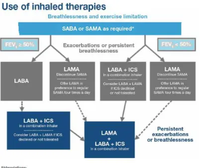
TREATMENT
1. General measures like smoking cessation, oxygen therapy.
2. Bronchodilators – beta agonist, anticholinergics, inhaled
corticosteroids
3. Oral glucocorticoids
4. Theophylline
5. N-acetyl cystine
6. a1AT augmentation therapy
7. Pulmonary rehabilitation
8. Lung volume reduction surgery
9. Lung transplantation
SPIROMETRY
HISTORY
1800 Hutchinson developed simple water sealed spirometer used
measure vital capacity
1930 barach developed kymograph or rotating chart drum that
displated changes in vital capacity as spirogram.
1941 Cournand and Richard described MVV
1947 Tiffeneau described FEV1/IVC ratio as index of airflow
limitation called Tiffeneau index
1950 Gaensler used micro along with water sealed spirometer to
time FVC1950 Comroe,Dubois and others described technique to
estimate alveolar pressure.
1950 late , Hyatt used flow volume curve to display air way
function
1955 Leuallen and Fowler described maximal mid expiratory flow
rates 25% and 75% now known as FEF25%-75%
1960 Wright and Mckerrow started using peak flow meter in
Spirometry is the most commonly performed lung function test . it is
described as GOLD STANDARD to diagnose COPD by WHO and
GOLD criteria.
INDICATIONS FOR SPIROMETRY
1. To diagnose the presence or absence of lung disease
2. To quantify the known pulmonary disease severity
3. To measure the environmental and occupational exposure effects
4. To identify the merits and demerits of therapy
5. To assess the risk factors of proposed surgical procedures in view of
pulmonary disease
6. To evaluate morbidity or impairment for legal or insurance
evaluation
7. To evaluate datas for epidemiological and clinical research in the
field of lung diseases
CONTRAINDICATIONS TO SPIROMETRY
ABSOLUTE
1. Acute coronary event ,myocardial infarction in last 30 days
3. Recent Cerebrovascular event
4. Poorly controlled hypertention
5. Underlying aortic and cerebral aneurysm
6.Recent pneumothorax
RELATIVE
1. Head ache
2. Stress incontinence
3. Confusion
4. Facial, abdominal and chest pain
5. Dementia
LUNG VOLUMES AND DEFINITIONS
The quantity of air that moves in and out the respiratory tract during
each respiratory cycle is called Tidal volume (TV)
The further volume of air that can be inspired after a normal tidal
inspiration is called Inspiratory reserve volume (IRV)
The further volume of air that can be exhaled after a normal tidal
expiration is called Expiratory reserve volume (ERV)
inspiration is called Vital capacity (VC) . VC = TV + IRV + ERV.
The amount of air that relics in the lungs after maximal expiration is
Residul volume (RV) . Even with forceful effort it cannot be expired
The quantity of air that remains in the lung after maximal inspiration is
termed as
Total lung capacity (TLC) . TLC = FRC + TV + IRV = VC + RV
The volume of air exhaled per minute is called Minute volume.
Maximal voluntary ventilation (Maximum breathing capacity )
is the highest amount of air that can be exhaled in a 15 second gap by
voluntary effort. This is uttered as liters per minute by multiplying by 4.
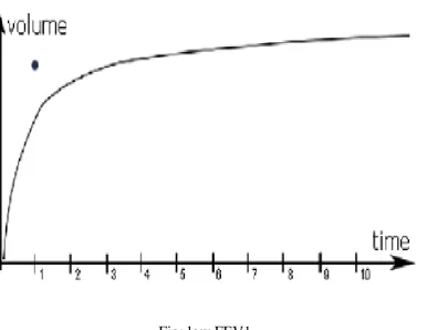
The quantity of air that can be forcefully exhaled in 1 second is
termed as Forced expiratory volume 1 (FEV1)
The maximum amount of air that can be forcefully exhaled is termed
as Forced vital capacity (FVC)
The standard volume of air that is exhaled during the mid portion of
FVC is termed as Mid expiratory flow (MEF25 - 75)
The peak flow rate during expiration is called as Peak expiratory flow
rate (PEFR) - normal range is 400-600 L/min
It is a consistent method of differentiating obstructive
airway disorders and restrictive lung diseases.
Precise spirometry can only be performed with proper training and
LIMITATIONS
1. It requests a well trained technician and patient support for
accuracy of test
2. There is a little variability in normal predictive value
3. It should be interpreted in the milieu of a proper history, physical
examination and added diagnostic tests.
ADVANTAGES
Simple & supportive in reaching diagnosis
Used as a first stride in detecting lung function abnormalities &
Answer important questions such as:
Is there airflow limitation? If so how severe is it?
Is there a response to bronchodilator therapy? If so how much?
Is there barrier present down the major airways?
PERFORMING
SPIROMETRY
Preparation
1. Equipment :
Checking for leaks
Fresh mouth piece
Checking recording equipment
Performing calibrations
2. Patient
The height and weight are measured
Any contraindications should be ruled out
The procedure is explained to the individual and should be
demonstrated
The person should sit erect and look straight ahead to avoid
stretching of trachea.
Basic
Components
Maximal inspiration
Continued expiration until maximal amount of air is exhaled up to
residual volume - at least a 6 seconds of exhalation in adults
Correct maneuver
The exhaled volume should be delivered from the level of
maximum inspiration with maximal exertion.
The maneuver is started immediately with a 'blast' . There
should be rapid rise to peak flow, and the attempt is sustained.
The exhalation should continue to the residual volume. ( exhalation
continued Up to vital capacity).
It should not be troubled by coughing or sneezing.
ATS/ERS recommendations are as follows:
FVC minimum duration 6 sec ( 3 sec for children) or plateau in the volume time
curve, subject cannot or could not continue to exhale
FVC end of test criteria Subject cannot or should not continue further
exhalation or the volume time curve shows an obvious
plateau or the forced exhalation is of reasonable
duration
FVC maximum number of maneuvers
8, both in adults and children
FVC maneuver
acceptability
Unsatisfactory start of expiration
Back extrapolated volume > 5% of FVC or 150 ml, Whichever is greater
Coughing which interferes with measurement of FEV1 and /or FVC.
Early termination of expiration Valsalva maneuver
A leak
An obstructed mouth piece Effort that is not maximal FVC and FEV1
reproducibility
the largest and second largest FVC and FEV1
NORMAL VALUES
The outcome of Spirometry are reported as absolute
(measured) values and as predicted percentage of normal values.
Normal values will depend on the individuals age, sex, ethnicity and
height. There is no universal standardization for normal values and the
values that differ from one laboratory to the other.
Thus normal reference values were obtained by performing tests
in thousands of people based on age, sex, ethnicity and height.
FACTORS DETERMINING LUNG VOLUMES
HEIGHT- shorter persons have lesser lung volumes
GENDER - females have lesser lung volumes than males
AGE – lung volume keeps on rising in children, steady in adults and as age advances the ERV decreases and RV,FRC increases .
Normal value = 80 - 120% of predicted
SPIROMETRY REPORT : typically consists of absolute numerical
values or graphical depiction of the same or a mixture of both.
Common Numerical Values
-75%
Graphical representation
- Time - Volume curve
FEV1 (Forced Expiratory Volume in the first second) is the volume
expired in the first second of the test .
FEV1%=FEV1/FVC X100.
roughly 80% of all the air is exhaled out in the first second by a healthy
individual out of their lungs during the FVC exercise.
FEV1% will be reduced in case of barrier in upper airways.
OBSTRUCTIVE PATTERN:
Either intrathoracic or extrathoracic.
FEV1 % = observed FEV1 / predicted FEV1 < 80%
FEV1 / FVC % = observed FEV1 / observed FVC < 75%
Severity of obstruction can be graded based on FEV1% based on
GOLD criteria as 80% mild, 80%-50% moderate,50%-30% severe,<30%
very severe
FEV1 /FVC may erroneously be normal due to air trapping which
cause markedly
Reduced FVC in persons with moderate to severe obstruction.
FEV1 / FVC will be less than 70-80% in elderly due to age related
decline without major obstruction. Early stages of neuromuscular
disorders can cause low
FEV1 /FVC due to reduction in FEV1. In obstructive disorders
CLASSIFICATION BASED ON FEV1 AND FVC VALUES
RESTRICTIVE PATTERN:
Pleural and parenchymal fibrotic diseases, chest wall
diseases
FVC % = observed FVC / predicted FVC < 75%
Reduced FVC is not sufficient for diagnosis but it can suggest the
probability of restrictive abnormality. It mainly depends on patient’s
severe obstruction . Restrictive diseases has to be confirmed by extent of
Residual volume (RV) and Total Lung Capacity (TLC) by Helium
dilution technique and Body Plethysmography. However interpretation of
spirometric data with clinical correlation is sufficient for all practical
INTERPRETATION OF SPIROGRAM
The spirometric proportions are recorded in a graphic representation
called Flow –Volume loop. It has volume in X axis and time in Y axis. At
point zero both flow and time are zero. Once the patient exhales it reaches
a crest within 150ms which is called Peak Expiratory Flow( PEF). This
peak flow represents the air exhaled from the proximal larger airways.
After the peak the curve rapidly descends and reaches 25% , which is
After 75% of the air exhaled it reaches FEF 75%.
The mean flow between 25% and 75% is called FEF 25-75% . This
gives a measure of airflow in the medium sized airways. This parameter
is the first to decline in most of the respiratory diseases. Almost 90% of
the air is exhaled in the first second . FVC is achieved if the flow
reaches zero.
Obstructive Airway Disease:
In obstructive airway diseases like COPD and Asthma the small
airways are narrowed and flow volume loop shows a concave pattern or
The PEF is normal since the air in larger airways is expelled easily.
Since smaller airways are narrowed the air is expelled slowly leading to a
low flow. This causes sharp fall in the flow volume loop. Both FEV1 and
FEF 25-75 are low.
[image:63.595.112.499.286.584.2]Obstructive airway disease:
Restrictive Lung Disease:
Restrictive lung diseases are characterized by low total lung volume .
spirometry can point out the probability of restriction and it has to be
established by other methods. Since there is no obstruction the curve of
flow volume is normal but the FVC is reduced. Peak Expiratory Flow can
be normal or low.
Fig: Low FVC with normal shape in restrictive lung disease:
Fig: Too low FEV1 in restrictive lung disease in volume - time curve.
UPPER AIRWAY OBSTRUCTION (UAO) :
The following are the characteristics of upper airway obstruction.
Midpoint maximal inspiratory flow is FIF 50% < 100 L/min
Ratio of midpoint maximal expiratory flow to midpoint maximal
inspiratory flow: FEF50% > 1/FIF50%
Ratio of forced expiratory volume in 1 second to peak expiratory
Ratio of forced expiratory volume in 1 second to forced expiratory
volume in 0.5 seconds FEV1/FEV0.5 > 1.5
Extra thoracic obstruction can be classified into following categories.
1. Extra thoracic Obstruction
-variable
The obstruction is more significant during inspiration and expiration
is normal. This occurs due to the fact that during expiration the obstruction
is overcome by the force of expiration. In flow –volume loop the
inspiratory part is flattened and expiratory part is normal.
2. Intra thoracic Obstruction -
variable
In intra thoracic obstruction, during inspiration the trachea is
sucked out and during expiration it causes partial obstruction.
Inspiratory part of the flow – volume loop is normal and the expiratory
part is flattened.
2. Fixed obstruction of the Large
Airways:
The obstruction can be both extra thoracic and intra thoracic. Both
inspiratory and expiratory part of the flow- volume loop are flattened.
TROUBLESHOOTING: Common cause for inconsistent spirometry is
patient technique. Other causes are as follows
1. Incomplete inspiration and sub optimal expiration
2. Delayed maximal effort that under estimates FEV1
3. Inadequate emptying of lungs occurs commonly in COPD
4. Incomplete sealing of lips around mouth piece underestimates FVC
and FEV1
5. Exhalation through nose
6. Coughing
7. Mouth piece obstruction by teeth
.
AIMS AND OBJECTIVE
1. Early detection of COPD in asymptomatic smokers by using
spirometer in south India.
2. To correlate BMI, smoking index and age of onset of first smoke
with FEVI/FVC Ratio.
3. To Study the relationship between SI and FEV1FVC ratio with
MATERIALS AND
MATERIALS AND METHODS
PLACE OF STUDY:
MAHATMA GANDHI MEMORIAL GOVERNMENT
HOSPITAL attached to KAP VISWANATHAM GOVERNMENT
MEDICAL COLLEGE, TIRUCHIRAPPALLI in southern region of
India
DEPARTMENT IN WHICH STUDY CONDUCTED:
Department of General Medicine
PERIOD OF STUDY: From JANUARY 2014 to August 2014
STUDY DESIGN : A prospective descriptive cross sectional study
ETHICAL COMMITTEE: Institutional ethical committee approval
obtained. The study population was well explained about the study and
its purpose in local language. Informed and written consent obtained
INCLUTION CRITERIA:
1.Subjects with history of smoking and age above 25 years
2.No significant respiratory symptoms
3. Regular smokers
4. Willing to undergo spirometry
5. Willing to give consent to participate in the study
EXCLUTION CRITERIA:
1. Subjects with smoking cessation
2. History of respiratory disease like tuberculosis, bronchial asthma or
occupational lung disease
3. On inhaled bronchodilators or corticosteroids
4. Diabetes mellitus, hypertension and coronary artery heart disease
5. Chest wall and vertebral deformities like pectus,kyphosis,scoliosis
6. Inadequate spirometry like air escape, failure to reach plateu are
excluded
STUDY GROUP :
Patients attending the out patient department of tertiary care
hospital ,MAHATMA GANDHI MEMORIAL GOVERNMENT
HOSPITAL attached to KAP VISWANATHAM GOVERNMENT
MEDICAL COLLEGE, TIRUCHIRAPPALLI in southern region of
SAMPLE SIZE: Calculated from prevalence in North Indian and
Madras study. Total number of subjects included was 174. All are men
with history of smoking
MATERIALS:
1.EASY ONE spirometer
2. Disposable mouth piece
3. Weighing scale
4. Stadiometer
5. Printed materials explaining ill effects of smoking and benefits of
smoking cessation regarding health and financial concerns
SPIROMETER
Easy one spirometer. It is a handy spirometer based on ultra sonic flow
sensor system. The benefit of this spirometer is disposable flow tube can
be inserted between transducers that prevents cross infectivity. in view of
the fact that the tube acts a transparent barrier sorting out the airflow and
transducer it does not require calibration. Also this spirometer is not
affected by the composition of gas is added advantage. This type of
compact, microprocessor integrated spirometer is recommended in various
studies for screening of smokers for COPD. In our study we used same
METHODOLOGY
After getting consent from subjects they were given short lecture
using printed modules in a language Tamil and demonstration of
spirometry is done by the technician who does the procedure. Quantum
of smoking was calculated using smoking index.
Smoking index: number of cigarettes or bidies per day mutiplied by
number of years of smoking. Smoking index selected because number of
appropriate than pack years.
Height ,weight and BMI was considered. They were subjected to
spirometry 15 mins after 400 mcg of salbutamol nebulisation as per
GOLD criteria. The predicted and measured values of FEV1, FVC,
FEV1/FVC, PEFR,FEF 25-75 for all the patients were recorded.
Minimum of three trials and maximum of eight trials done for each
subject based on guidelines from American Thoracic Society.
The data were analyzed as per GOLD criteria and the subjects
classified as mild, moderate, severe and very severe air flow obstruction
using statistical test.
RESULTS
FIGURE - 1 Age distribution of smokers in percentage
28-38 years 28.7% (n=50)
39-49 years 38.5% (n=68)
50-60 years 29.3% (n=51)
61-71years 2.8% (n=5)
PERCENTAGE OF SMOKERS
FIGURE - 2 BMI distribution of smokers in percentage
BMI – Body mass index
1. <18.5 5.17% (n=9)
2. 18.5- 24.9 76.43% (n=134)
3. 25.0- 29.9 13.21%(n=23)
4. 30.0-34.9 4.02% (n=8)
0.00% 10.00% 20.00% 30.00% 40.00% 50.00% 60.00% 70.00% 80.00%
<18.5 18.5-24.9 25-29.9 30-34.9
DISTRIBUTION OF BMI IN SMOKERS
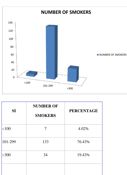
FIGURE - 3 Smoking index distribution in smokers
SI
NUMBER OF
SMOKERS
PERCENTAGE
<100 7 4.02%
101-299 133 76.43%
>300 34 19.43%
0 20 40 60 80 100 120 140
<100
101-299
>300
NUMBER OF SMOKERS
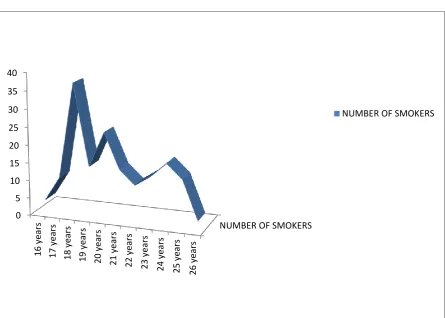
FIGURE - 4 Age of first smoke among smokers
1. Youngest age is 16 years
2. Maximum number observed is at 18 years
NUMBER OF SMOKERS 0 5 10 15 20 25 30 35 40 16 ye ar s 17 ye ar s 18 ye ar s 19 ye ar s 20 ye ar s 21 ye ar s 22 ye ar s 23 ye ar s 24 ye ar s 25 ye ar s 26 ye ar s
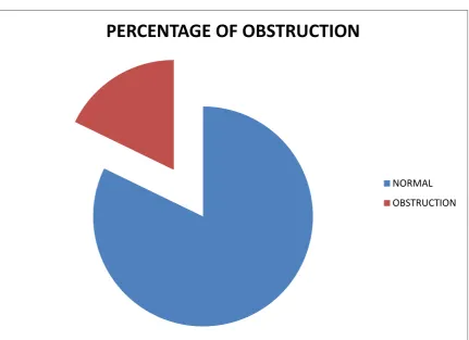
FIGURE- 5 Distribution of obstructive lung disease in spirometry
Normal spirometry – 82.18%(n=143)
Obstruction - 17.81%(n=31)
Among obstructive pattern
Mild obstruction - 61.29%(n=19)
Moderate obstruction- 38.7%(n=12)
PERCENTAGE OF OBSTRUCTION
TABLE 1
PARAMETERS
(N=174) MEAN(SD) MINIMUM MAXIMUM
AGE 44.9943 (8.794) 28 65
ONSET
20.65
(2.634) 16 26
BMI 22.6925 (3.101) 16.5 32
SI
226.821
(77.315) 80 450
FVCp
87.31
(58.383) 58 822
FEV1p
79.97
(16.248) 51 152
FEV1/FVCp
98.55
(17.961) 60 139
MEF 25-75
64.4
(31.458) 11 191
MEF 75
68.68
(23.448) 18 137
MEF 50
66.66
(24.668) 22 133
MEF 25
59.90
(32.588) 7 247
PEF
69.74
(22.198) 24 128
FVC m
3.2059
(.78459) 1.33 5.46
FEV1m
2.7059
(.68959) 1 4.53
FEV1/FVC m
.7755
Onset - age of first smoke
BMI -Body mass index
SI -Smoking index
FVCp - Forced vital capacity (Percent)
FEV1p – Forced expiratory volume in 1 second( percent)
MEF 25-75 – Mid expiratory flow
PEF - Peak expiratory flow
FVCm - Forced vital capacity (measured)
FEV1m – Forced expiratory volume in 1 second(measured)
TABLE 2
ONE SAMPLE T TEST
PARAMETERS
(N=174) MEAN(SD) p VALUE
AGE 44.9943 (8.794) 0.075
ONSET 20.65 (2.634) 0.001***
BMI 22.6925 (3.101) 0.101
SI 226.821 (77.315) 0.001***
FVCp 87.31 (58.383) 0.001***
FEV1p 79.97 (16.248) 0.002**
FEV1/FVCp 98.55 (17.961) 0.018*
MEF 25-75 64.4 (31.458) 0.065
MEF 75 68.68 (23.448) 0.651
MEF 50 66.66 (24.668) 0.062
MEF 25 59.90 (32.588) 0.035*
PEF 69.74 (22.198) 0.564
FVC m 3.2059 (.78459) 0.054
FEV1m 2.7059 (.68959) 0.322
FEV1/FVC m .7755 (.07193) 0.001***
*is p value <0.05
**is p value <0.01
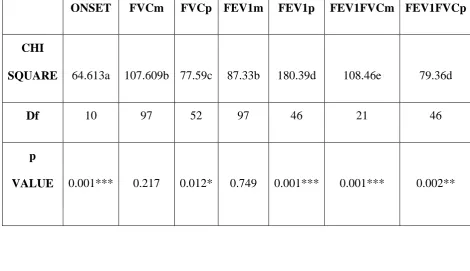
TABLE 3A
ONSET FVCm FVCp FEV1m FEV1p FEV1FVCm FEV1FVCp
CHI
SQUARE 64.613a 107.609b 77.59c 87.33b 180.39d 108.46e 79.36d
Df 10 97 52 97 46 21 46
p
[image:85.595.87.558.120.391.2]VALUE 0.001*** 0.217 0.012* 0.749 0.001*** 0.001*** 0.002**
TABLE 3B
PEF
MEF25-75 MEF75 MEF50 MEF25 BMI SI AGE
CHI
SQUARE 69.86f 61.55g 71.07h 85.03i 98.41j 108.89k 240.25l 57.72m
Df 67 80 68 71 78 83 39 35
p VALUE 0.382 0.938 0.376 0.122 0.059 0.03* .001*** .009**
*is p value <0.05
**is p value <0.01
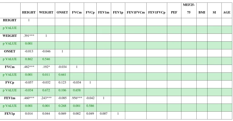
TABLE 4
HEIGHT WEIGHT ONSET FVCm FVCp FEV1m FEV1p FEV1FVCm FEV1FVCp PEF
MEF25-75 BMI SI AGE
HEIGHT 1
p VALUE
WEIGHT .391*** 1
p VALUE 0.001
ONSET -0.013 -0.046 1
p VALUE 0.862 0.546
FVCm .482*** .192* -0.034 1
p VALUE 0.001 0.011 0.661
FVCp -0.057 -0.032 0.123 -0.034 1
p VALUE -0.034 0.672 0.106 0.658
FEV1m .460*** .243*** -0.085 .956*** -0.042 1
p VALUE 0.001 0.001 0.268 0.001 0.586
p VALUE 0.859 0.567 0.369 0.979 0.521 0.923
FEV1FVCm 0.044 0.063 0.021 0.084 -0.125 0.126 .257*** 1
p VALUE 0.565 0.406 0.78 0.272 0.099 0.098 0.001
FEV1FVCp -0.077 -0.021 -0.042 -0.088
-.229** -0.088 .254*** .460*** 1
p VALUE 0.312 0.787 0.583 0.25 0.002 0.25 0.001 0.001
PEF 0.128 .193* 0.008 .154* -0.135 .167* .377*** .345*** .353*** 1
p VALUE 0.092 0.011 0.916 0.043 0.075 0.028 0.001 0.001 0.001
MEF25-75 0.078 -0.004 0.018 0.091 -.151* 0.089 .383*** .313*** .704*** .485*** 1
p VALUE 0.307 0.961 0.81 0.234 0.046 0.241 0.001 0.001 0.001 0.001
BMI -.259*** .756*** -0.037 -0.106 0.003 -0.039 0.05 0.038 0.039 .167* -0.047 1
p VALUE 0.001 0.001 0.633 0.163 0.973 0.606 0.511 0.616 0.606 0.028 0.535
SI -0.006 -0.036 -0.009 -.187* 0.133 -.225** -.158* -.476*** -.406***
-.256*** -.216**
-0.041 1
AGE -.197** -0.13 0.069 -.387** 0.114
-.478*** -0.058 -.300*** -0.04 -0.088 -0.046 0.017 .616*** 1
Pvalue 0.009 0.088 0.364 0.003 0.135 0.001 0.445 0.001 0.6 0.247 0.543 0.82 0.001
*is p value <0.05
**is p value <0.01
SUMMARY
FIGURE 1 Shows
Age distribution of smokers in percentage
39-49 years 38.5% (n=68) is commonly observed data
FIGURE 2 shows
BMI distribution of smokers in percentage
18.5- 24.9 76.43% (n=134)
FIGURE 3 shows
Smoking index distribution in smokers
SI 101-299 in 76.43% (n=133)
FIGURE 4 shows
Age of first smoke among smokers
1. Youngest age is 16 years
2. Maximum number observed is at 18 years
FIGURE 5 shows
Distribution of obstructive lung disease in spirometry
Obstruction - 17.81%(n=31)
Among obstructive pattern
Mild obstruction - 61.29% (n=19)
Moderate obstruction- 38.7% (n=12)
TABLE 1
Distribution of parameters in mean and standard deviation
Age group – 44.99(8.79) in years
Age of first smoke - 20.65 (2.634) in years
BMI – 22.69(3.10)
SI - 226.82 (77.31)
FVCp – 87.31( 58.38)
FEV1p -79.97(16.24)
FEV1FVCp – 98.55(17.96)
MEF 2575 – 64.4(31.45)
MEF75 – 68.68 (23.44)
MEF50 – 66.66(24.66)
FEV1m – 2.70(.071)
FEV1FVCm - 0.77(0.071)
FVCm – 3.2 (0.78)
TABLE 2
One sample T test
FEV1FVCp and MEF25 have p value < 0.05
FEV1p has p value <0.01
Age of first smoke, SI,FVCp and FEV1FVCm have p value <0.001
TABLE 3
Chi square test
BMI - 108.46 (p value <0.05 )and FVCp -77.59(p value <0.05)
Age - 57.724(p value < 0.01 ) and FEV1FVCp -79.386(p value
0.002)
Age of first smoke , SI -240.25, FEV1p- 180.39 and FEV1FVCm
-108.46
TABLE 4
Correlation coefficient
SI has significant negative correlation with FEV1m _
0.225(p 0.003) ,FEV1p _0.158 ( p 0.03), FEV1FVCm
_0.476(p0.001) , FEV1FVCp _0.406 ( p 0.001) , PEF _0.256 (p
0.001) ,MEF 25-75 _0.216(P 0.004)
FEV1FVCm has significant positive correlation with
FEV1p 0.257 (p 0.001)
FEV1FVCp has significant negative correlation with
_0.229 (p 0.002) and significant positive correlation with FEV1p
0.254 (p 0.001), FEV1FVCm 0.460 ( p0.001)
MEF 25-75 has significant positive correlation with FEV1p
0.383(p 0.001) , FEV1FVCm 0.313 (0.001) , FEV1FVCp 0.704
(p 0.001) ,PEF 0.485 ( p 0.001)
DISCUSSION
This study was conducted for earlier detection of COPD in
smokers even before the occurance of symptoms and signs of obstructive
lung disease. The diagnostic criteria used for case finding is as specified
in GOLD guidelines. The portable spirometer (Easy one)was used to
perform the spirometry. The population in the study group is
representative of patients attending the out patient department in the
tertiary care hospital Tiruchirappalli,South India. The prevalence of
COPD in this population is 10.2%. the prevalence of smoking has
increased and 50% of male smoke cigarette or bidis. Our aim is to detect
the presence of COPD earlier in asymptomatic smokers and to correlate
the severity of obstructive pattern with SI.
All the participants were male. Since female smoking is not
socially accepted in this region only a little proportion smoke. It was
ensured that all the participants continue to smoke and does not have
respiratory symptoms and signs during the study. Occupational history
suggestive of exposure to lung disease have been excluded from the
study. Most of them were sales representatives, vendors and auto/taxi
drivers.
All are subjected to 400 mcg salbutamol nebulisation fifteen
performed by single technician by demonstrating technique before study.
Participants were well informed with printed material regarding ill effects
of smoking and benefits of smoking cessation.
The parameters collected were age of first cigarette,
BMI,SI,FEV1,FEV1/FVC both measured and
percent,MEF25-75,MEF75,MEF50 and MEF 25. The results were statiscally analyzed.
AGE: The mean age was 44.994 (8.794). Most of them were
between 39-49 years(n=67) 38.5%. youngest age in this study is 28 years.
Age group 61-71 years is only 2.87% (n=5). significant negative
correlation with FVCm _0.387(p 0.003), FEV1m _0.478(p 0.001)
,FEV1FVCm _0.300(p 0.001).This correlates well with Natural history
pattern of COPD in which as the age advances the severity of disease
progresses as explained by Fletcher and Peto curve. Less number of
patients in >60 years is probably due to occurrence of symptoms as age
advances and Our study included only asymptomatic patients.
BMI: The mean BMI was 22.6925(3.1010). about 76.43(n=133)
were in 18.5-24.9 BMI range. Underweight noted only in 5.172%(n=9)
probably our study included only asymptomatic population of smokers.
Since most of the participants are middle aged working group they were
in BMI range of normal to overweight 89.64% (n=140). There is no
SMOKING INDEX: About 78.735% (n=137) participants had
moderate SI i.e 100-300. Severe SI observed in 21.26%(n=37). SI and
age has significant positive correlation coefficient 0.616(p 0.001) which
is consistent with previous studies. SI and FEV1/FVC ratio both m and p
has significant negative correlation coefficient (p 0.001) i.e SI increases
FEV1/FVC ratio decreases. Moderate obstruction had SI of 400 (88.25)
and mild obstruction had SI of 310(66.50). SI also has significant
negative correlation coefficient (p 0.001) with MEF 25-75 which is again
consistent with presence of small airway disease in smokers with COPD.
This parameter was less correlated in previous north Indian study .
FEV1m_ 0.225(p 0.003) , FVCm _0.187(0.01) alone had correlation with
SI for diagnosis of obstructive lung disease. When percentage is
considered they does not correlate. Thus FEV1/FVC ratio is gold
standard for screening presence of airflow limitation in smokers as
indicated in study Stralelis G, et al and Zielinski et al. is consistent with
our study.
Age of first smoke: Majority started smoking around 20 years.
About 1/5 th (n=37) 19.54 % started to smoke at 18 years. Age of first
smoke does not have any correlation with obstructive lung disease in our
correlates with severity of airflow limitation. Previous studies also given
the same report.
FVC: It has also got significant negative correlation coefficient (p
0.001) with FEV1/FVC ratio. These are well established in various
studies and reproduced in our study also.
FEV1: Significant positive correlation coefficient (p 0.001)
with,FEV1/FVC, MEF 25-75 which is consistent from previous studies.
According to GOLD criteria 19/174 had mild and 12/174 had moderate
obstruction pattern. All the participants with obstructive spirometry are
asymptomatic during study. But when only FEV1 value is considered
57.47% (n=100) had FEV1 >80. 42.52% (n=74)had FEV1 50-80%. So in
determining the obstructive pathology we need FEV1/FVC or
FEV1/FEV6. FEV1 alone is not useful as screening criteria for diagnosis
of COPD in asymptomatic smokers.
FEV1/FVC: Airflow limitation with FEV1/FVC ratio <70 was
noted in 17.816%(n=31). Out of which when applied to GOLD guidelines
mild obstruction noted in 61.2903%(n=19). Moderate obstruction seen in
38.7096%(n=12). None of them had severe or very severe obstruction.
FEV1/FVC has Significant positive correlation coefficient (p 0.001) with
FEV1and MEF25-75 . It has also got significant negative correlation
MEF25-75: has Significant positive correlation coefficient (p
0.001) with FEV1p and FEV1/FVCm , FEV1FVCp ratio . It has also
got significant negative correlation coefficient (p 0.001) with SI. This
parameter is less correlated in previous trials used for screening of
COPD. This parameter needs further large trials.