“CLINICAL STUDY OF FEVER WITH THROMBOCYTOPENIA WITH SPECIAL REFERENCE TO INFECTIVE ETIOLOGY AND
COMPLICATIONS ADMITTED TO GOVERNMENT ROYAPETTAH HOSPITAL, CHENNAI”
Dissertation submitted to
THE TAMILNADU DR. M.G.R MEDICAL UNIVERSITY
CHENNAI
In partial fulfillment of regulations
For award of the degree of
M.D (GENERAL MEDICINE)
BRANCH – 1
KILPAUK MEDICAL COLLEGE, CHENNAI-10
BONAFIDE CERTIFICATE
This is to certify that dissertation named “CLINICAL STUDY OF
FEVER WITH THROMBOCYTOPENIA WITH SPECIAL REFERENCE TO INFECTIVE ETIOLOGY AND COMPLICATIONS ADMITTED TO GOVERNMENT ROYAPETTAH HOSPITAL, CHENNAI” is a bonafide work performed by Dr. M.Manoj , post graduate student, Department of Medicine, Kilpauk Medical College, Chennai-10, under my guidance and supervision in fulfillment of regulations of the Tamilnadu Dr. M.G.R Medical University for the award of M.D. Degree Branch I (General Medicine) during the academic period from May 2011 to April 2014.
Prof. Dr. N. Gunasekaran M.D., DTCD Prof. Dr. S. Mayilvahanan M.D., The Director, Institute of Non Communicable Diseases, Professor and Unit Chief Superintendent, Prof. and Head, Department of Medicine Department of General Medicine, Government Royapettah Hospital
Government Royapettah Hospital Kilpauk Medical College,Chennai-10 Kilpauk Medical College,Chennai-10
Prof. P. Ramakrishnan M.D., D.L.O
The DEAN Government Kilpauk Medical College
DECLARATION
I solemnly declare that this dissertation “CLINICAL STUDY OF
FEVER WITH THROMBOCYTOPENIA WITH SPECIAL REFERENCE TO INFECTIVE ETIOLOGY AND COMPLICATIONS ADMITTED TO GOVERNMENT ROYAPETTAH HOSPITAL, CHENNAI” was prepared by me at Government Kilpauk Medical College and Hospital, Chennai, under the guidance and supervision of Dr. S. Mayilvahanan M.D., Professor, Department of Internal Medicine, Government Royapettah Hospital, Chennai.
This dissertation is submitted to The Tamil Nadu Dr. M.G.R. Medical University, Chennai in partial fulfillment of the University regulations for the award of the degree of M.D. Branch I (General Medicine).
Place: Chennai
ACKNOWLEDGEMENT
At the outset, I would like to thank my beloved Dean, Kilpauk Medical College Prof. Dr. P. Ramakrishnan, M.D., D.L.O., for his kind permission to conduct the study in Kilpauk Medical College. I would like to express my special thanks to The Director, Institute of Non Communicable Diseases,
Superintendent, Prof. and Head, Department of General medicine Dr. N. Gunasekeran M.D., DTCD., Govt. Royapettah Hospital for permitting me to conduct this study.
I would like to thank wholeheartedly, Prof. Dr. S.Mayilvahanan M.D.,
my unit Chief and Professor of Medicine for his encouragement and guidance during the study.
I also express my special thanks to Prof. Dr. K.T. Jayakumar M.D., Prof. Dr.R.Sabarathnavel M.D., I am extremely thankful to Assistant Professor of Medicine, Dr.P.Paranthaman M.D., Dr.S.Kalaichelvi M.D., and
Dr.G.Ranjani M.D., for their assistance and guidance.
I would always remember with extreme sense of thankfulness, the
co-operation and criticism shown by my fellow post graduate colleague and friends.
I would like to extend my gratitude to my parents and my sister for their unconditional support.
CONTENTS
1. INTRODUCTION 2. AIM OF STUDY
3. REVIEW OF LITERATURE 4. MATERIALS AND METHODS 5. RESULTS
6. DISCUSSION 7. CONCLUSIONS
APPENDIX
BIBLIOGRAPHY ABBREVIATIONS PROFORMA MASTER CHART
CLINICAL STUDY OF FEVER WITH THROMBOCYTOPENIA WITH SPECIAL REFERENCE TO INFECTIVE ETIOLOGY AND
COMPLICATIONS ADMITTED TO GOVERNMENT ROYAPETTAH HOSPITAL, CHENNAI
ABSTRACT
BACKGROUND:
Fever with thrombocytopenia is a common condition that is associated with an increased risk of morbidity and mortality.Infections like Malaria, Dengue, Leptospirosis, Typhoid, HIV & septicemia are some of the common causes of fever with thrombocytopenia. Therefore a well organized systemic approach that is carried out with an awareness of causes of fever with thrombocytopenia can shorten the duration of investigations and bring out diagnosis. Hence, a need for study to know the causes and complications of fever with thrombocytopenia.
AIMS AND OBJECTIVES
1) To study the etiology of short duration fever with thrombocytopenia 2) To study the clinical presentation and the laboratory profile of patients presenting with fever and thrombocytopenia.
MATERIALS AND METHODS :
This was a cross sectional study which included all new patients above 18 years with fever (temperature > 99.9 F) and thrombocytopenia (platelet count less than 1,50,000cells/ cu.mm ) admitted to Government Royapettah hospital. The data of each patient was collected on a proforma specially designed for this study which includes demographic details, clinical features, past medical history, clinical and Lab values which will be analysed for statistical significance and correlation.
RESULTS :
CONCLUSION AND INTERPRETATION:
1) Infections are one of the most common causes of thrombocytopenia. The leading cause of fever with thrombocytopenia in our study was Malaria. 2) In malarial infection, the most common species was Plasmodium vivax followed by Plasmodium falciparum. 3) Dengue fever was the second commonest cause of febrile thrombocytopenia. 4) There is no direct correlation between the severity of thrombocytopenia and the bleeding manifestation. 5) Bleeding manifestation was present only in 11% of patients in the study. So in majority of patients, the
thrombocytopenia was transient and asymptomatic. 6) Prophylactic platelet
transfusion may not be required in all cases of severe thrombocytopenia. It may be restricted to selected patients with bleeding manifestation or platelets
<10,000/cumm which may indicate bone marrow compromise.7) Most of the patients in our study did not require platelet or blood transfusion and the platelet count significantly increased after the treatment of the underlying infection.
KEYWORDS :
INTRODUCTION
There is an alarming increase in the incidence of fever with
thrombocytopenia. Routinely we come across many cases, both as inpatients and outpatients presenting with fever with thrombocytopenia. Infection is one of the common cause of thrombocytopenia. Thrombocytopenia in fever can predict the cause and thus helps in early diagnosis and treatment, preventing further fatal outcome associated with it.
Though patients can initially present with simple fever, in due
course it can lead to unpredictable outcomes including death at times, therefore the aim of the study is to analyse the clinical profile of fever with
thrombocytopenia, as early diagnosis and timely intervention prevents adverse outcomes and can save lives.
AIM OF THE STUDY
1) To study the etiology of short duration fever with thrombocytopenia 2) To study the clinical presentation and the laboratory profile of patients presenting with fever and thrombocytopenia.
REVIEW OF LITERATURE
THROMBOCYTOPENIA IN MALARIA
Thrombocytopenia is one of the most common complications of both Plasmodium vivax and Plasmodium falciparum malaria. It is pertinent that the finding of thrombocytopenia in patient, may be an indication for a thorough
lookout into the blood smear to rule out malaria as the cause. This fact is especially important in the workup for thrombocytopenia in febrile patients.
Malaria affects almost all blood components and is a true
haematological infectious disease. Anaemia and thrombocytopenia are the most frequent malaria-associated haematological complications (Wickramasinghe & Abdalla 2000)1 and have received more attention in the scientific literature due to their associated mortality.
In the current field of Travel Medicine, the rapid increase in the number of people travelling to tropical areas has added a great challenge for malaria diagnosis because the thick blood smear (the standard diagnosis in endemic areas) has high specificity but only when performed by experienced microscopists.
malaria diagnosis (D’Acremont et al. 2002)2
. Another study has reported 60% sensitivity and 88% specificity of thrombocytopenia for malaria diagnosis in acute febrile patients (Lathia & Joshi 2004)3.
The sensitivity of thrombocytopenia together with the acute febrile syndrome was 100% for malaria diagnosis, with a specificity of 70%, a positive predictive value of 86% and a negative predictive value of 100% (Patel et al.2004)4 In 2005, 138 of 684 (20.1%) malarial cases hospitalised in a tertiary care centre in Manaus had thrombocytopenia as the cause of admission, which corresponded to 6.8% of hospitalisations due to all causes in this reference
institution (unpublished observations). Hospitalisation, however, does not add any benefit to the patient and because there is no evidence for any intervention, this simply increases public health costs in underdeveloped and under-resourced areas.
Since the beginning of the 1970s, there have been reports proposing that malaria-associated thrombocytopenia is quite similar in Plasmodium vivax
and Plasmodium falciparum infections (Beale et al. 1972)5. However, more recent data in India has shown how thrombocytopenia exhibited a heightened frequency and severity among patients with P. vivax infection (Kochar et al. 2010)6.
malaria. Recent studies have shown that thrombocytopenia is equally or even more common in P. vivax malaria contrary to the popular belief that it may be observed in P. falciparum malaria. More recent data in India has shown how
thrombocytopenia exhibited a heightened frequency and severity among patients with P. vivax infection. Thrombocytopenia was seen in 40-90 percent of patients infected with P. falciparum infection inIndia. Maximum thrombocytopenia occurred on the fifth or sixth day of infection, and gradually returned to normal within 5-7 days after parasitemia ceased.
HOW LOW IS THE PLATELET COUNT IN MALARIA?
In a systematic review of literature, platelet counts less than 150,000/mm3 occurred in 24-94% in patients with acute malaria and this
frequency was not different between the two major species that affected humans.
MECHANISM OF THROMBOCYTOPENIA IN MALARIA :
The speculated mechanisms leading to thrombocytopenia are:
1) coagulation disturbances,
2) splenomegaly,
3) bone marrow alterations,
4) antibody-mediated platelet destruction,
5) oxidative stress and the role of platelets as cofactors in triggering severe malaria.
1 ) Coagulation disturbance - A study based on 31 American soldiers in Vietnam with chloroquine-resistant falciparum malaria noted the following changes in the acute phase of the disease using the same patients as their own controls during convalescence: decrease in the platelet count and prothrombim activation time, increase in the activated thromboplastin time, and reduction in factors V, VII and VIII with normal fibrinogen (Dennis et al. 1967)7.
2) Splenomegaly - The spleen in malaria has played a crucial role in the immune response against the parasite, as well as controlling parasitaemia due to the
phagocytosis of parasitised red blood cells (RBCs) (Engwerda et al. 2005)9. Some data suggested that platelets were sequestered in the spleen during the acute
infection (Skudowitz et al. 1973)10.
3) Bone marrow alterations - a “dysmegakaryopoiesis” was proposed, similar to
what happened in the human malarial anaemia model, where dyserythropoiesis was triggered by cytokines (Menendez et al. 2000)11. In the few studies that examined the bone marrow for this purpose, megakaryocytic lineage was apparently
4)Antibody-mediated platelet destruction - There is evidence that platelet-associated IgG (PAIgG) is increased in malaria and is platelet-associated with
thrombocytopenia. During acute malaria, thrombocytopenia is most probably associated with the binding of parasite antigens to the surface of platelets to which antimalarial antibodies also bind, leading to the in situ formation of immune
complexes (Kelton et al. 1983)16.
Because the generation of immune complexes is proportional to the amount of available antigen, the negative correlation between platelet count and peripheral parasitaemia reported in many studies (Lacerda 2007, Silva
2009)17,18 corroborates immune mechanisms as a potential mechanism of platelet destruction.
5) Oxidative stress - Free radicals may play an important role in the platelet destruction in malarial infection.
The Relationship Between Thrombocytopenia And Severe Malaria
Severe thrombocytopenia (platelet count under 50,000/mm3), despite not being considered severe malaria according to World Health Organization criteria (WHO 2010) due to the inability to cause death per se, has been
occasionally associated with severity (Gerardin et al. 2002, Rogier et al. 2004)20,21.
17 patients from Manaus affected by any of the WHO malaria severity criteria with confirmed P. vivax monoinfection, 14 presented with
thrombocytopenia, suggesting that this haematological complication can be explored as a marker of the severity for this species (Alexandre et al. 2010)22.
From the case reports available, the association between severe cases with thrombocytopenia is evident. However, that can be due to bias
publication, where prospective studies would be needed to validate this association. On the other hand, considering that many studies point to a clear
MANAGEMENT OF THROMBOCYTOPENIA IN MALARIA :
Data from experimental models are presented and, despite not being rare, there is no clear recommendation on the adequate management of this
haematological complication.
In most cases, a conservative approach is adopted and platelet counts usually revert to normal ranges a few days after efficacious antimalarial treatment.
More studies are needed to specifically clarify if thrombocytopenia is the cause or consequence of the clinical disease spectrum.
Platelet transfusion has been widely followed, but with no con-firmed efficacy. The indication of prophylactic platelet transfusion when platelet counts are under 10,000/mm3 probably applies only when the bone marrow is compromised and is not able to release efficacious platelets (Rebulla 2000). This does not seem to be the case in malaria. Keeping platelet counts between 50,000 and 100,000/mm3 is a formal indication only in patients undergoing surgical procedures (Rebulla 2001)25. In a tertiary care centre in the Western Brazilian Amazon over a 12-month period, 10.4% (20/191) of patients who received platelet transfusion were diagnosed with vivax or falciparum malaria (Lacerda et al.
transfusion were maintaining a platelet count below 10,000/mm3 and discrete bleeding. In a further 6% of patients, only a very low platelet count was described. In this group of 40% of patients, the alleged reason was minor bleeding despite having non-severe thrombocytopenia; in 33%, no indication was verified. These data point to the little existing evidence of the recommendations for platelet
transfusion in these patients. The corrected count increment to evaluate transfusion efficacy was not calculated for any patient. The low efficacy of platelet transfusion in general is well described for several acute infectious diseases (de Paula et al. 1993)28, probably due to peripheral immune mechanisms of destruction that do not spare the transfused platelets.
Indications for platelet transfusion in cases when DIC is suspected and diagnosed, the formal clinical indication persists, as recommended elsewhere (Franchini 2005)29. Due to the impossibility of using frozen platelets in routine clinical practice, other platelet substitutes and preparations are being investigated (Blajchman 2003)30. Except in atypical cases of ITP with severe bleeding, there is no evidence for the use of human intravenous immunoglobulin, even in cases of severe thrombocytopenia (Lacerda et al. 2004)31.
2007)32 and with the lack of robust evidence of immune-mediated destruction of platelets as a major mechanism. It was also found that in patients with cerebral falciparum malaria, dexamethasone exacerbated the neurological symptoms and increased the frequency of gastrointestinal bleeding (Warrell et al. 1982, Hoffman et al. 1988)33,34. However, in none of these studies was platelet recovery analysed as a secondary endpoint.
Immune modulators are also candidates in the adjuvant antimalarial therapy (Muniz-Junqueira et al. 2005, Mohanty et al. 2006)35,36, based on the drug-induced inhibition of adhesion molecules in RBCs and platelets (Muniz-Junqueira 2007)37. The exploration of drugs known by their anti-inflammatory effect,
modulating TNF, e.g., pentoxyfylline and thalidomide, upon severe malaria, could not only contribute to the understanding of the mechanisms of severity but also clarify the association between platelets and severe disease.
The frequency of thrombocytopenia (i.e., platelet count below 150,000/mm3) in malarial infection ranges from 24-94% in the literature, despite the low occurrence of severe bleeding, even in the case of severe malaria. It is still unclear whether this haematological complication is more frequent in P. vivax or
SYSTEMATIC REVIEW OF STUDIES,
ESTIMATING THROMBOCYTOPENIA IN MALARIAL PATIENTS IN INDIA (2002-2011)
REFERENCES TYPE OF
PATIENTS
AGE
RANGE
SPECIES N THROMBOCYTOPENIA
%
Mohapatra et al.
(2002)
Inpatients
and
outpatients
15-60 y P.v. 110 3.6 (< 100,000)
Jadhav et al. (2004) Inpatients
and
outpatients
All ages P.v. 973 65 (50,000-150,000)
Kumar and
Shashirekha (2006)
Inpatients
and
outpatients
All ages P.v. 27 88.8 (< 150,000)
Prasad et al. (2009) Inpatients < 5 y P.f. 40 85 (< 150,000)
Kochar et al. (2010) Inpatients
and
outpatients
All ages P.v./P.f.
and mixed
1,064 24.6 (< 150,000)
George and
Alexander (2010)
Inpatients 18-66 y P.v. 30 93.3 (< 150,000)
Srivastava et al.
(2011)
COLLATED CASE REPORTS OF PLASMODIUM VIVAX-ASSOCIATED THROMBOCYTOPENIA IN INDIA (2002-2011)
REFERENCES PLATELET
COUNT
(X 1,000/MM3)
BLEEDING PLATELET
TRANSFUSION
OBSERVATION
Makkar et al. (2002) 8 Gingival bleeding Yes -
Aggarwal et al. (2005) 6 Petecchiae Yes -
Katira and Shah (2006) 14-92 No Yes -
Kaur et al. (2007) 30 No No Acute renal failure
Kaur et al. (2007) 30 Petecchiae No Acute renal failure
Vij et al. (2008) NA Gingival bleeding No NA
Parakh et al. (2009) 5-42 Petecchiae No Cerebral malaria,
shock and acute renal
failure
Thapa et al. (2009) 11 Petecchiae and
mucosal bleeding
Yes Hepatitis and shock
Harish and Gupta
(2009)
1 Intracranial bleed No Seizures
Bhatia and Bhatia
(2010)
THROMBOCYTOPENIA IN DENGUE FEVER
Dengue Haemorrhagic Feveris a probable case of dengue and haemorrhagic tendency evidenced by one or more of the following:
1) Positive tourniquet test
2) Petechiae, ecchymosis or purpura
3) Bleeding from mucosa (mostly epistaxis or bleeding from gums), injection sites or other sites
4) Haematemesis or melena
5) Thrombocytopaenia (platelets 100,000/cu.mm or less) and
6) Evidence of plasma leakage due to increased capillary permeability manifested by one or moreof the following:
– A >20% rise in haemotocrit for age and sex
– A >20% drop in haemotocrit following treatment with fluids as compared to
baseline
– Signs of plasma leakage (pleural effusion, ascites or hypoproteinaemia).
CAUSE OF THROMBOCYTPENIA IN DENGUE:
Although not fully elucidated, recent evidence indicates that severe Dengue viral infections increase vas cular permea bility that leads to decreased intravascular fluid volume and consequen t
hemoconcentration and hypotension in infected patients. Another feature of Dengue viral infection is thrombocytopenia, which is common in both mild and severe diseases.
Dengue virus has been isolated from polymorphonuclear
leukocytes, monocyte / macrophages, dendritic cells and others38 .It has also been detected in megakaryocyte progenitors and circulating platelets 39,40,41 These findings suggest that Dengue virus may induce thrombocytopenia via direct
interactions with megakaryocytes and platelets. Dengue virus has also been shown to reduce circulating platelet counts independent of virus attachment or entry into platelets or their precursors. Thus, two mechanisms are probably involved in dengue-induced thrombocytopenia:
1)IMPAIRED THROMBOPOIESIS
Marrow suppression within 2–4 days of dengue viral infection can contribute to thrombocytopenia . Viral RNA has been isolated from bone marrow specimens of infected individuals, suggesting that dengue targets the marrow and hematopoietic system.42 Bone marrow studies also reveal diminished megakaryopoiesis during the onset of dengue infection and clinical recovery is associated with normal megakaryocyte topography and platelet counts 43
Suppression of megakaryopoiesis occurs either directly, due to infection and suppression of hematopoietic progenitor cells or indirectly, via impairment of stromal cells that function by altering the repertoire of cytokines in the bone
marrow microenvironment. In regard to direct effects, Nakao et al. 44 demonstrated that Dengue virus- 4 propagates in human bone marrow progenitors in vitro and alters their proliferative capacity. Dengue viral infection suppresses proliferation of human cord blood progenitors and Dengue Virus -2 inhibits the differentiation of CD34+ progenitors into megakaryocytes, presumably by inducing apoptosis in infected cells45,46
characterized the viral antigen-positive cells. This investigation demonstrated two types of stromal cells that were positive for viral antigens: adventitial reticular cells and bone marrow dendritic cells. Altered cytokine production by infected stroma is the most probable mechanism of marrow suppression during DV infection. The in vitro findings described above and the hematological findings of leukopenia in conjunction with thrombocytopenia in dengue patients are used as argument in favor of dengue globally suppressing bone marrow hematopoiesis. 48 However, emerging evidence indicates that dengue infection also has extramedullary effects on circulating platelets.
2) INCREASED PERIPHERAL DESTRUCTION
a) Autoimmune-induced platelet activation and clearance b) Platelet–leukocyte and platelet–endothelial cell interactions c) Platelet dysfunction
d) Direct infection e) Soluble mediators
Autoimmune-induced platelet activation and clearance
Several groups have put forth the autoimmune hypothesis, which postulates that host-generated anti-Dengue virus antibodies crossreact with
dengue patients can bind platelets and higher levels of antiplatelet IgM are observed in severe DV infections when compared to classical dengue fever50. Moreover, dengue patient serum or rabbit anti nonstructural protein-1 (NS1)
induce complementmediated lysis in platelets51,52 which may contribute to the loss of circulating platelets during dengue illness. Autoantibodies directed against NS1 target human platelets and fibrinogen and induce thrombocytopenia in mice 53
A molecular mimicry mechanism has been proposed in which the C-terminal region of NS1 shows sequence homology with integrins on the surface of platelets. In clinical settings, increased levels of platelet-associated immunoglobulin (PAIgM or PAIgG) and phagocytosis of platelets by macrophages correlates with thrombocytopenia during the acute phase of secondary dengue infection 54
Similarly, anti-NS1 autoantibodies or pooled sera from dengue patients enhance the engagement of immunoglobulinopsonized platelets by macrophages.
Platelet–leukocyte and platelet–endothelial cell interactions
Platelet dysfunction
There are a few studies examining platelet function in dengue disease. Among these, it has been shown that dengue serum abnormally activates platelets and inhibits platelet aggregation .
Direct infection
Recent studies indicate that dengue virus directly interacts and activates platelets. DV induces morphological changes in normal platelets typical of activation, including the presence of filopodia and degranulation . In parallel, Dengue virus increases the expression of surface P-selectin and fibrinogen binding . Dengue viral RNA and viral-like particles have also been detected in platelets of affected patients.
Soluble mediators
Key mediators that activate platelets and induce thrombocytopenia are often present in dengue infection. Monocytes from a donor infected with Dengue virus-1 respond to a second hit of dengue virus-2 by generating Platelet Activating Factor (PAF), a lipid mediator that augments platelet aggregation. This observation is in agreement with others demonstrating that thrombocytopenia and disease severity is reduced in mice lacking the PAF receptor (PAFr). Fibrin
increased, creating a milieu for enhanced platelet activation. An array of cytokines, such as tumor necrosis factor-a (TNF-a) and interleukin-1b (IL-1b) are also
produced during dengue infection. These cytokines have been linked to the onset and regulation of thrombosis and hemostasis and it has been demonstrated that increased TNF-a and IL-1 b in dengue patients correlates with thrombocytopenia .
TARGETING PLATELETS IN THE TREATMENT OF DENGUE INFECTION
Although thrombocytopenia is frequently observed in patients with dengue, severe bleeding is rare. When it occurs, however, excessive bleeding is associated with a high lethality. It is controversial as to whether the intensity of thrombocytopenia predicts bleeding risk in dengue patients; nonetheless, it is well accepted that severe thrombocytopenia associates with hemorrhagic
manifestations. In addition, it is probable that other factors, such as disseminated intravascular coagulation (DIC), hepatic impairment and/or vascular dysfunction, act in concert with thrombocytopenia to induce bleeding.
restrictions. Importantly, these recommendations are based solely on the opinions of experts or small observational studies rather than randomized clinical trials. Other treatments considered for dengue-induced thrombocytopenia are anti-D immune globulin (anti-D) and PAFr antagonists.56 Of these, anti-D has shown promise in the treatment of severe thrombocytopenia in DHF patients while a PAFr antagonist relieved thrombocytopenia in a mouse model of dengue
infection . By contrast, IVIG did not hasten the recovery of thrombocytopenia in dengue patients with secondary DV infection. Moving forward, it will be important to consider nontraditional roles of platelets in the treatment of dengue. This
includes their role in regulating viral infection and replication, inflammation and vascular integrity, which may identify new molecular targets for the treatment of dengue infection
THROMBOCYTOPENIA IN HIV INFECTION
Thrombocytopenia in HIV was first described in 1982. The prevalence is more or less 40%, depending on which literature is quoted. Thrombocytopenia is associated with increased morbidity and mortality,
accelerated deterioration in CD4 counts and accelerated progression to full-blown AIDS. In a meta-analysis of 5 trials involving > 3 000 patients, both treatment-naïve and treatment-experienced patients, thrombocytopenia was found to be one of 8 factors that correlated with a poorer prognosis and more rapid progression to full-blown AIDS in spite of antiretroviral treatment.
A recent study showed that platelets have the ability to ‘engulf ' the Human immunodeficiency virus and Staphylococcus aureus – perhaps another
reason why thrombocytopenia is prone to a more rapid acceleration of disease. Severe thrombocytopenia also limits one’s treatment options, as many drugs cause bone marrow suppression and peripheral platelet consumption. HIV enters the megakaryocytes and platelets via the CXCR4 receptors. Once the virus is in the megakaryocyte it starts to cause havoc, as shown by the change
Thrombocytopenia early on in HIV is mainly due to peripheral destruction, while later on in the advanced stage (AIDS) it is more likely to be due to decreased production. In fact, CD4 counts above 200 are associated with
increased peripheral destruction while thrombocytopenia in CD4 counts of < 200 is associated with decreased platelet production.
In one study the authors found a 3-fold increase in megakaryocytes in patients with HIV. However, there was no increase in the mean platelet mass, suggesting the presence of dysmegakaryopoiesis. This suggests that
thrombocytopenia in HIV is multifactorial because of:
• direct HIV infection of the megakaryocyte, causing apoptosis
• dysmegakaryopoiesis, abnormal and dysfunctional production of megakaryocytes
and platelets
• peripheral destruction of platelets due to cross-reactivity of HIV Abs.
Who to treat
There are no standardized guidelines in the treatment of HIV-induced thrombocytopenia.
The generally accepted guideline is to treat when there are < 30 000 platelets or < 50 000 if the patient is on warfarin or a haemophiliac. One must still remember that thrombocytopenia correlates with a poorer outcome and
to this, treatment of thrombocytopenia is immunosuppressive in its nature and therefore should be given in conjunction with antiretrovirals. A platelet count of 50000 post-treatment is quite acceptable for protection against bleeding, but still does predict a poorer outcome with regards to mortality and morbidity.
Therefore thrombocytopenia occurs as a result of: • increased peripheral destruction or
• increased peripheral sequestration or • decreased production or
• a combination of the above.
Approach to management
Do a bone marrow biopsy, to asses megakaryocyte numbers and morphology. One can then easily exclude granulomas, Kaposi’s sarcoma and lymphoma or even
fibrosis in the marrow. Once the decision is made to treat, then the difficulty is deciding how to treat.
Steroids
patients should be monitored carefully and be on antiretrovirals. Patient should not stay on high-dosage therapy for more than a month. If no response or a nominal response is seen, it should be considered as treatment failure and alternative treatment given. Prednisone does not stimulate viral replication but does however accelerate the course of Kaposi’s sarcoma.
Intravenous immunoglobulin
Intravenous immunoglobulin (IVIG) is effective in raising a
patient’s platelet counts. However, it is not cheap and results can be short lasting. It
is not a cost-effective way of maintaining a platelet count and should only be used in chronic cases as a last resort. In the acute setting it is good for raising counts prior to a splenectomy or if patients have severe or uncontrollable haemorrhage. This would then be used in conjunction with platelet transfusions. The exact mechanisms of action are not entirely understood – suffice to say that excessive immunoglobulin (Ig) overwhelms the immune system and stimulates the
suppressor B cells to suppress endogenous Ig production. The IVIG also blocks the Fc receptors in the spleen and macrophages, thereby limiting their
Anti-D
The anti-rhesus globulin can also be used with varying success. This should not be used in patients who are Rh+ as it may cause haemolysis. It can however be used if the patient has a normal haemoglobin. Its effects, if there is going to be a response, are said to be longer lasting than IVIG.
Splenectomy
Splenectomy is the usual 2nd-line of therapy with the HIV-negative cohort.There were concerns initially that it would accelerate the course of HIV. This was before antiretroviral therapy was commonly available. At the moment, besides the concerns of infections from capsulated organisms and malaria,
splenectomy may be a good alternative. The literature has shown splenectomy to attenuate immune reconstitution syndrome and has shown to produce patients with a higher CD4 and CD8 counts along with a slower progression to AIDS. Splenic irradiation is of no value in this situation.
Megestrol acetate
This drug was initially used to treat cachexia and anorexia in HIV. It is known to block the Fc receptors in macrophages. Trials have shown that
counts or viral loads. Danazol is still of value in idiopathic thrombocytopenic purpura, even in HIVinduced ITP.
Transfusions
Transfusions have a transient effect, they are expensive and should be limited to emergencies and during surgery. Normally they should be given with IVIG to have a longer lasting effect. Transfusions have other sideeffects e.g.
transfusion reactions, infectionsand transfusion-related acute lung injury (TRALI). Multiple transfusions are known to decrease immunity and stimulate HIV-1
expression.
Novel therapies
These are still in experimental phases but include entities such as recombinant thrombopoietin, alpha interferon and IL-6, IL-3, AND IL-11.
THROMBOCYTOPENIA IN LEPTOSPIROSIS
also with other organ involvement including myocarditis, aseptic meningitis, and hemorrhagic diathesis.
The hemorrhagic manifestations of leptospirosis occur in many ways. Pulmonary hemorrhage is a dreaded complication because it is associated with a high mortality rate. In 1984, an epidemic of pulmonary hemorrhagic fever occurred in Korea. Extensive investigations were undertaken to determine the cause, including clinical, pathological and epidemiological studies of all possible causative agents. The investigations finally came up with leptospirosis as the culprit.
In the year 1995, an outbreak of an acute febrile illness and pulmonary hemorrhage occurred during the period of October to November. More than 2000 persons were affected and at least 40 patients died from acute pulmonary
hemorrhage and respiratory insufficiency. The initial considerations were dengue hemorrhagic fever and the hantavirus pulmonary syndrome, but serologic tests detected anti-leptospiral antibodies and immunohistochemical staining of tissues from fatal cases demonstrated leptospiral antigens present in various organs. Together with myocarditis, pulmonary hemorrhage and GI bleeding were identified as common complications leading to death.
With the outbreaks of pulmonary hemorrhage in association with leptospirosis, our focus should shift to the mechanisms causing the bleeding diathesis in this
particular disease. Thrombocytopenia is thought to be fleeting, mild and rare. However, reviews reported increasing prevalence of thrombocytopenia. Some authors postulated that this could possibly be attributed to
1) disseminated intravascular coagulation (DIC) or a toxin or cytotoxin mediated mechanism;
2) as a direct complication of leptospiral vasculitis as a general phenomenon of septicemia, or due to an undetected platelet antibody.
Whether either of these mechanisms is operating alone or in combination is uncertain and merits extensive investigation.
It is important for clinicians to be aware and to recognize the various ways in which leptospirosis can present. Although classically occurring as an acute febrile illness with renal failure and jaundice, the other less common manifestations may predominate. The changing pattern of the virulence of the disease could have been better explained by identifying the serovars involved. It has been speculated that a new strain of leptospires may be responsible for the more severe presentation in some patients. Our poor yield in culture studies for Leptospira, and the
Thrombocytopenia can occur in mild to moderate to severe forms; majority of the platelet counts were between 20,000 and 80,000.Thrombocytopenia in leptospirosis was also associated with increased number of fatal complications. The presence of thrombocytopenia indicated a more severe form of leptospirosis.. Mortality rate was higher in the thrombocytopenic group compared to the
nonthrombocytopenic group
THROMBOCYTOPENIA IN TYPHOID FEVER
The hematological changes are common in typhoid fever and these include anemia, leucopoenia, eosinophilia, thrombocytopenia and sub clinical disseminated intravascular coagulation. Thrombocytopenia is a common finding amongst patients presenting with typhoid fever as it causes reversible bone marrow suppression. Bone marrow suppression and hemophagocytosis are considered to be an important mechanism in producing hematological changes.57
Many cases of typhoid fever have peripheral blood cytopenias that were not concurrent bone marrow suppression, suggesting a peripheral
the pancytopenia. The term haemophagocytosis describes the pathological finding of activated macrophages and engulfing erythrocytes, leucocytes, platelets, and their precursors cells.59
THROMBOCYTOPENIA IN SEPSIS :
Thrombocytopenia is a frequent finding in critical illness and is commonly employed in clinical trials of severe sepsis therapies as a marker of hematologic organ dysfunction.
In the ICU setting platelets <1,00,000/mm3 are identified in 20-40% of patients. In a study of ICU patients sepsis was identified as a major risk factor for thrombocytopenia.
Mechanism:
Sepsis induced thrombocytopenia is multifactorial in origin.
2) Inflammatory mediators and bacterial products such as endotoxins can
contribute to sepsis induced thrombocytopenia by enhancing platelet reactivity and adhesivity.
3) Phagocytosis of platelets by reticuloendothelial elements may also contribute to cytopenias in sepsis.
4) Immune mechanisms may contribute to sepsis-induced thrombocytopenia.
Nonspecific platelet-associated antibodies can be detected in up to 30% of ICU
patients. In these patients, nonpathogenic IgG presumably binds to bacterial
products on the surface of platelets, to an altered platelet surface, or as immune
complexes. A subset of patients with platelet-associated antibodies have
autoantibodies directed against glycoprotein IIb/IIIa. These antibodies have been
implicated in the pathogenesis of immune thrombocytopenic purpura and, although
not proved, may play a role in mediating sepsis-induced thrombocytopenia.
Microscopy of bone marrow in patients with sepsis often
The acute phase response is often characterized by increased
platelet counts (thrombocytosis). However, patients who are admitted to the ICU
with or without underlying sepsis are more commonly diagnosed as
thrombocytopenia. Thrombocytopenia occurs in up to 20% of medical ICU and
35% of surgical ICU admissions61,62,63. Sepsis is a clear risk factor for
thrombocytopenia, with an estimated incidence of 35% to 59%64,65. In addition, there is an inverse relationship between the severity of sepsis and the platelet
count.
In a prospective study of critically ill patients with
thrombocytopenia only 34% had a diagnosis of DIC. Secondary consumptive
thrombocytopenia and DIC represent an extreme in the continuum of hemostatic
abnormalities. In addition to sepsis-related mechanisms, other causes of
thrombocytopenia should be considered in the critically ill patient. For example,
thrombocytopenia may occur as a complication of heparin therapy. Other types of
drug induced thrombocytopenia are rare in the ICU setting. Dilutional
thrombocytopenia may occur in patients with trauma or those who have undergone
complicated surgery. Preexisting underlying disease, including cancer and immune
thrombocytopenic purpura, may also contribute to a low platelet count. Given the
inverse correlation between platelet count and mortality and the proposed
development of thrombocytopenia in the patient with sepsis is best regarded as
maladaptive.
Clinical Manifestations and Diagnosis
Thrombocytopenia is a common cause of bleeding in the ICU
setting. Patients with thrombocytopenia may have petechiae, purpura, bruising, or
bleeding. Thrombocytopenia is diagnosed on the basis of the complete blood cell
count. A peripheral smear may show evidence of platelet clumping. If that is the
case, the platelet count should be remeasured in blood withdrawn into a non-EDTA
containing tube. If the thrombocytopenia is associated with consumptive
coagulopathy, the DIC screen may be abnormal, and the peripheral smear may
show schistocytes. Although patients with sepsis may have increased
platelet-associated IgG, testing for this gives nonspecific results and does not help to guide
therapy.
Prognosis
Thrombocytopenia is a predictor of mortality in patients in the ICU
and in patients with severe sepsis65,66. The degree and duration of
thrombocytopenia, as well as the net change in the platelet count, are important
lower than 100 x109/L, mortality continues to increase, whereas the risk of bleeding does not increase.
Treatment
Patients with severe thrombocytopenia should be treated with
platelet transfusions. Although guidelines for prophylactic transfusions in patients
with chemotherapy-induced thrombocytopenia have been established, the threshold
for transfusions for the thrombocytopenic patient with sepsis is not as clear. In the
absence of confounding factors, patients should probably receive transfusions
when the platelet count is less than 10000 – 150000/cumm. If the patient has
concomitant coagulopathy (eg. liver disease), active bleeding, or platelet
MATERIALS AND METHODS: Study group :
All new patients above 18 years with fever (temperature > 99.9 F) and
thrombocytopenia (platelet count less than 1,50,000cells/ cu.mm ) admitted to Government Royapettah hospital
Study design : Cross-Sectional study
Place of study : Government Royapettah Hospital
Duration of study : 6 months (March-August 2013)
This study protocol was approved by the Ethical committee for research studies of Government Kilpauk Medical College Hospital, Chennai.
Inclusion criteria :
All new patients above 18 years with fever ( temperature > 99.9 F ) and thrombocytopenia ( platelet count less than 1,50,000 cells/ cu.mm )
Exclusion criteria :
Patients presenting with thrombocytopenia without fever
Patients with thrombocytopenia already diagnosed to have haematological disorder / malignancy , on treatment with chemotherapy and other immunosuppresants
Diagnosed cases of platelet disorders and dysfunction
Patients on treatment with antiplatelet drugs and other drugs causing thrombocytopenia
Patients with cirrhosis and chronic liver disease
METHODOLOGY:
STUDY
Questionnair
Case sheets of patients
Temperature charts
VARIABLES
Age group - four groups were made, below 20yrs, 20-40yrs ,40 – 60yrs
and above 60 yrs
Sex - male and female
Fever duration
Chills and rigor- if present or not
Joint pain- recorded from the history
Head ache- the duration and episodes of headache assessed, and considered present or absent
Myalgia-those having muscle pain for more than 2 days, without any relief even with rest are considered as myalgia present
Organomegaly – this includes both hepatomegaly and spleenomegaly, it is assessed by the clinical examination of the patient, the clinical records and the ultrasonogram results.
Bleeding – any bleeding manifestation like purpura, petechiae, hematuria, malena, hematemesis was considered as positive
Anemia – less than 10 gm hemoglobin were considered as having anemia
Thrombocytopenia- A count below 1,50,000/cumm was considered to have thrombocytopenia
Creatinine – a creatinine valve > 1 mg/dl was considered as renal involvement
ALT/AST – values more than 40 IU/L was considered elevated for each
Serum bilirubin – more than 1mg/dl was considered as hyperbilirubinemia
Peripheral smear study or QBC for malaria – was considered diagnostic for malaria
Widal test or blood culture for Salmonella typhi - was considered diagnostic for enteric fever
IgM for leptospirosis or MAT – was considered diagnostic for leptospirosis
ELISA/Western blot – was taken as positive for HIV
patients in whom the diagnosis could not be made even after all the above
mentioned investigations were categorised as having unspecified viral fever.
Once the specific diagnosis was reached, patients were treated specifically and symptomatically. Data was collected by using pre-tested proforma meeting the objectives of the study. The data collected would be transferred in to a Master Chart, which was then subjected for statistical analysis.
Data collection:
The data of each patient was collected on a proforma specially designed for this study and which includes demographic details, clinical features, past medical history, clinical and Lab values which was analysed for statistical significance and correlation.
STATISTICAL ANALYSIS
OBSERVATION AND RESULTS
1.DISTRIBUTION OF THE PLATELET COUNT IN THE STUDY POPULATION
Table 1. showing the distribution of platelet count in the study population
Among the 100 patients in the study group, the maximum number of patients (54%) had a platelet count between 50,000 to 100000.
[image:55.612.160.441.459.675.2]Platelet count less than 20000 were found in only three patients.
Fig.1. A pie diagram showing the distribution of platelet count in the study population
Platelet Count
100001-150000
50001- 100000 20001- 50000 < 20000 PLATELET COUNT FREQUENCY PERCENT
< 20000 3 3.0
20001-50000 10 10.0
50001-100000 54 54.0
2.DISTRIBUTION OF THE STUDY POPULATION ACCORDING TO DIAGNOSIS AND PLATELET COUNT
Table 2. showing the distribution of study population according to diagnosis and platelet count DIAGNOSIS
PLATELET COUNT
TOTAL
< 20000 20001-50000 50001-100000
100001-150000 Malaria Count
1 4 24 13 42
% within Diagnosis 2.4% 9.5% 57.1% 31.0% 100.0%
Dengue
Count 1 3 14 9 27
% within Diagnosis 3.7% 11.1% 51.9% 33.3% 100.0%
Typhoid
Count
0 0 5 1 6
% within Diagnosis .0% .0% 83.3% 16.7% 100.0%
Leptospirosis
Count 0 0 2 3 5
% within Diagnosis .0% .0% 40.0% 60.0% 100.0%
Sepsis
Count 0 2 1 0 3
% within Diagnosis
.0% 66.7% 33.3% .0% 100.0%
Unspecified viral fever
Count
0 1 8 7 16
% within Diagnosis .0% 6.3% 50.0% 43.8% 100.0%
HIV
Count 1 0 0 0 1
% within Diagnosis
Out of 100 patients, a definitive diagnosis was made in 84 patients.
Among them, the leading cause of febrile thrombocytopenia in this study was
MALARIA (42 cases).
The second most common cause was DENGUE FEVER accounting for 27 cases.
It was followed by
UNSPECIFIED VIRAL FEVER (16 cases), TYPHOID (6 cases),
LEPTOSPIROSIS (5 cases), SEPSIS (3 cases), and
HIV (1 case) in that order.
Only three patients were having platelet counts less than 20,000 in this study. They were caused by malaria, dengue fever and HIV each.
3.DISTRIBUTION OF THE STUDY POPULATION ACCORDING TO THE BLEEDING MANIFESTATION DIAGNOSIS BLEEDING
MANIFESTATION TOTAL
Yes No
Malaria
Count 5 37 42
% within Diagnosis 11.9% 88.1% 100.0%
Dengue
Count 1 26 27
% within Diagnosis 3.7% 96.3% 100.0%
Typhoid
Count 0 6 6
% within Diagnosis 0% 100.0% 100.0%
Leptospirosis
Count 1 4 5
% within Diagnosis 20.0% 80.0% 100.0%
Sepsis
Count 2 1 3
% within Diagnosis 66.7% 33.3% 100.0%
Unspecified viral fever
Count 2 14 16
% within Diagnosis 12.5% 87.5% 100.0%
HIV
Count 0 1 1
% within Diagnosis .0% 100.0% 100.0%
Total
Count 11 89 100
% within Diagnosis 11.0% 89.0% 100.0%
A total number of 11 patients had various bleeding manifestations out of the 100 study population.
5 out of 42 patients with malaria had shown bleeding manifestation (11.9%)
1 out of 27 patients with dengue fever had shown bleeding manifestation (3.7%)
2 out of 3 patients with sepsis had bleeding manifestations (66.7%)
1 out of 5 patients with leptospirosis had bleeding manifestation (20%)
[image:60.612.150.462.402.642.2]In the unspecified viral fever group, 2 out of 16 patients had bleeding manifestation (12.5%)
Fig.3.1. showing the distribution of study population according to bleeding manifestation
Bleeding Manifestation
No
Fig.3.2. showing the distribution of study population according to bleeding manifestation Diagnosis HI V Un spec ifie
4.AGEWISE DISTRIBUTION OF STUDY POPULATION
DIAGNOSIS
AGE GROUP IN YEARS
TOTAL
< 20 21-39 40-59 > 60
Malaria 8 15 12 7 42
Dengue 2 13 11 1 27
Typhoid 3 3 0 0 6
Leptospirosis 2 1 1 1 5
Sepsis 0 0 3 0 3
Unspecified viral fever
3 5 4 4 16
HIV 0 0 0 1 1
Total 18 37 31 14 100
Fig. 4. showing age wise distribution of study population Diagn osis HI V Un sp ecifie
d vira
l fe Se psis Le pto spiro sis Typh oid De ng ue M ala ria C ou nt 16 14 12 10 8 6 4 2 0
Age G roup in ye ars
< 20
21-39
40-59
5.DISTRIBUTION OF THE STUDY POPULATION ASSOCIATED WITH CHILLS AND RIGORS
Table 5. showing distribution of study population associated with chills and rigors
71 out of 100 patients had fever associated with chills and rigors.
32 out of this 71 patients were diagnosed with malaria.
16 patients with dengue fever, 4 patients with typhoid fever, 5 patients with leptospirosis and 14 patients with unspecified viral fever were having fever associated with chills and rigors.
DIAGNOSIS
CHILLS & RIGORS TOTAL
Yes No
Malaria Count 32 10 42
% within Diagnosis 76.2% 23.8% 100.0%
Dengu
Count 16 11 27
% within Diagnosis 59.3% 40.7% 100.0%
Typhoid
Count 4 2 6
% within Diagnosis 66.7% 33.3% 100.0%
Leptospirosis
Count 5 0 5
% within Diagnosis 100.0% .0% 100.0%
Sepsis
Count 0 3 3
% within Diagnosis .0% 100.0% 100.0%
Unspecified viral fever
Count 14 2 16
% within Diagnosis 87.5% 12.5% 100.0%
HIV
Count 0 1 1
% within Diagnosis .0% 100.0% 100.0%
Total Count 71 29 100
Fig.5. showing distribution of study population associated with chills and rigors 6.DISTRIBUTION OF THE STUDY POPULATION ASSOCIATED WITH HEADACHE
DIAGNOSIS
HEADACHE TOTAL
Yes No
Malaria Count 29 13 42
% within Diagnosis 69.0% 31.0% 100.0%
Dengue
Count 15 12 27
% within Diagnosis 55.6% 44.4% 100.0%
Typhoid
Count 4 2 6
% within Diagnosis 66.7% 33.3% 100.0%
Leptospirosis
Count 4 1 5
% within Diagnosis 80.0% 20.0% 100.0%
Sepsis
Count 3 0 3
% within Diagnosis 100.0% .0% 100.0%
Unspecified viral fever
Count 14 2 16
% within Diagnosis 87.5% 12.5% 100.0%
HIV
Count 1 0 1
% within Diagnosis 100.0% .0% 100.0%
Total Count 70 30 100
% within Diagnosis 70.0% 30.0% 100.0%
Table 6. showing distribution of study population associated with headache Diagnosis HI V Un spec ifie
d vira
l fe Se psis Le pto spiro sis Typh oid De ng ue M ala ria C o u n t 40 30 20 10 0
Chills & Rigors
Yes
70 out of 100 patients had fever associated with headache.
29 out of this 70 patients were diagnosed with malaria.
[image:66.612.171.477.354.564.2]15 patients with dengue fever, 4 patients with typhoid fever, 4 patients with leptospirosis, 3 patients with sepsis and 14 patients with unspecified viral fever were having fever associated with headache.
Fig.6. showing distribution of study population associated with headache Diagn osis
HIV Unspec if ied viral f e Sepsis
Leptosp irosis Typhoid
Dengue Malaria
C
ou
nt
40
30
20
10
0
Heada che
7.DISTRIBUTION OF THE STUDY POPULATION ASSOCIATED WITH GENERAL EXAMINATION FINDINGS
DIAGNOSIS
GENERAL EXAMINATION TOTAL
NORMAL ANEMIA JAUNDICE
Malaria
Count 34 4 4 42
% within Diagnosis 81.0% 9.5% 9.5% 100.0%
Dengue
Count 25 2 0 27
% within Diagnosis 92.6% 7.4% .0% 100.0%
Typhoid
Count 5 1 0 6
% within Diagnosis 83.3% 16.7% .0% 100.0%
Leptospirosis
Count 2 0 3 5
% within Diagnosis 40.0% .0% 60.0% 100.0%
Sepsis
Count 0 1 2 3
% within Diagnosis .0% 33.3% 66.7% 100.0%
Unspecified viral fever
Count 13 3 0 16
% within Diagnosis 81.3% 18.8% .0% 100.0%
HIV
Count 0 1 0 1
% within Diagnosis .0% 100.0% .0% 100.0%
Total Count 79 12 9 100
% within Diagnosis 79.0% 12.0% 9.0% 100.0%
Table 7. showing distribution of study population associated with general examination findings
12 % of the total patients had anemia and 9% of them had jaundice overall.
2 out of 27 patients with dengue fever were found to be anemic (7.4%). None had jaundice.
33% of patients with sepsis had anemia and 67 % had jaundice.
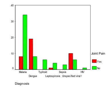
8.DISTRIBUTION OF THE STUDY POPULATION ASSOCIATED WITH JOINTPAIN
DIAGNOSIS
JOINT PAIN TOTAL
Yes No
Malaria Count 8 34 42
% within Diagnosis 19.0% 81.0% 100.0%
Dengue
Count 19 8 27
% within Diagnosis 70.4% 29.6% 100.0%
Typhoid
Count 0 6 6
% within Diagnosis .0% 100.0% 100.0%
Leptospirosis
Count 1 4 5
% within Diagnosis 20.0% 80.0% 100.0%
Sepsis
Count 0 3 3
% within Diagnosis .0% 100.0% 100.0%
Unspecified viral fever
Count 10 6 16
% within Diagnosis 62.5% 37.5% 100.0%
HIV
Count 0 1 1
% within Diagnosis .0% 100.0% 100.0%
Total
Count 38 62 100
[image:68.612.95.517.329.650.2]% within Diagnosis 38.0% 62.0% 100.0%
38% of the total study population presented with joint pain.
19 % of patients with malaria had joint pain whereas 70.4% of patients with dengue fever had joint pain.
[image:69.612.75.452.309.612.2]62.5% of patients with unspecified viral fever had joint pain.
Fig. 8. showing distribution of study population associated with joint pain
Diagnosis
HIV
Unspec ified viral fe Sepsis
Leptospirosis Typhoid
Dengue Malaria
C
o
u
n
t
40
30
20
10
0
Joint Pain
Yes
9.DISTRIBUTION OF THE STUDY POPULATION ASSOCIATED WITH NAUSEA AND VOMITING
Table 9. showing distribution of study population associated with nausea and vomiting
54 out of 100 patients had nausea and vomiting as one of the complaint in presenting illness.
27 patients with malaria and 11 patients with dengue fever had nausea and vomiting. DIAGNOSIS NAUSEA,VOMIT TOTAL
Yes No
Malaria
Count 27 15 42
% within Diagnosis 64.3% 35.7% 100.0%
Dengue
Count 11 16 27
% within Diagnosis 40.7% 59.3% 100.0%
Typhoid
Count 3 3 6
% within Diagnosis 50.0% 50.0% 100.0%
Leptospirosis
Count 4 1 5
% within Diagnosis 80.0% 20.0% 100.0%
Sepsis
Count 3 0 3
% within Diagnosis 100.0% .0% 100.0%
Unspecified viral fever
Count 6 10 16
% within Diagnosis 37.5% 62.5% 100.0%
HIV
Count 0 1 1
% within Diagnosis .0% 100.0% 100.0%
Total Count 54 46 100
Fig. 9. showing distribution of study population associated with nausea and vomiting
Diagn osis
HIV
Un spe
cifie
d vira
l fe Se
psis Lep
tos piro
sis
Typho
id De
ngue
Ma laria
C
ou
nt
30
20
10
0
Nause a,Vomit
10.DISTRIBUTION OF THE STUDY POPULATION ASSOCIATED WITH ALTERED SENSORIUM
DIAGNOSIS ALTERED
SENSORIUM
TOTAL
Yes No
Malaria Count 3 39 42
% within Diagnosis 7.1% 92.9% 100.0%
Dengue
Count 1 26 27
% within Diagnosis 3.7% 96.3% 100.0%
Typhoid
Count 0 6 6
% within Diagnosis .0% 100.0% 100.0%
Leptospirosis
Count 0 5 5
% within Diagnosis .0% 100.0% 100.0%
Sepsis
Count 2 1 3
% within Diagnosis 66.7% 33.3% 100.0%
Unspecified viral fever
Count 2 14 16
% within Diagnosis 12.5% 87.5% 100.0%
HIV
Count 0 1 1
% within Diagnosis .0% 100.0% 100.0%
Total
Count 8 92 100
% within Diagnosis 8.0% 92.0% 100.0%
Table 10. showing distribution of study population associated with altered sensorium
8 of the patients presented with altered sensorium in the study group.
Fig. 10. showing distribution of study population associated with altered sensorium
Diagn osis
HIV
Un spe
cifie
d viral f
e Sep
sis Lep
tospiro
sis Typ
hoid De
ngue
Ma laria
Co
un
t
50 40 30 20 10 0
Altere d Sensoriu m
11.DISTRIBUTION OF THE STUDY POPULATION ASSOCIATED WITH HEPATOMEGALY AND SPLENOMEGALY
DIAGNOSIS
HEPATOMEGALY TOTAL
Yes No
Malaria
Count 12 30 42
% within Diagnosis 28.6% 71.4% 100.0%
Dengue
Count 1 26 27
% within Diagnosis 3.7% 96.3% 100.0%
Typhoid
Count 4 2 6
% within Diagnosis 66.7% 33.3% 100.0%
Leptospirosis
Count 5 0 5
% within Diagnosis 100.0% .0% 100.0%
Sepsis
Count 1 2 3
% within Diagnosis 33.3% 66.7% 100.0%
Unspecified viral fever
Count 2 14 16
% within Diagnosis 12.5% 87.5% 100.0%
HIV
Count 1 0 1
% within Diagnosis 100.0% .0% 100.0%
Total
Count 26 74 100
[image:74.612.105.515.142.450.2]% within Diagnosis 26.0% 74.0% 100.0%
Table 11. showing distribution of study population associated with hepatomegaly
26 patients in the total study group had hepatomegaly.
Fig. 11. showing distribution of study population associated with hepatomegaly
45 out of 100 study population had splenomegaly.
29 out of 49 patients with splenomegaly had malaria.
12.DISTRIBUTION OF THE STUDY POPULATION ASSOCIATED WITH ASCITES DIAGNOSIS ASCITES TOTAL
Yes No
Malaria
Count 3 39 42
% within Diagnosis 7.1% 92.9% 100.0%
Dengue
Count 0 27 27
% within Diagnosis .0% 100.0% 100.0%
Typhoid
Count 0 6 6
% within Diagnosis .0% 100.0% 100.0%
Leptospirosis
Count 2 3 5
% within Diagnosis 40.0% 60.0% 100.0%
Sepsis
Count 2 1 3
% within Diagnosis 66.7% 33.3% 100.0%
Unspecified viral fever
Count 0 16 16
% within Diagnosis .0% 100.0% 100.0%
HIV
Count 0 1 1
% within Diagnosis .0% 100.0% 100.0%
Total
Count 7 93 100
[image:76.612.91.522.165.459.2]% within Diagnosis 7.0% 93.0% 100.0%
Table 12. showing distribution of study population associated with ascites
Fig. 12. showing distribution of study population associated with ascites
Diagnosis
HIV
Unspec ified viral fe Sepsis
Leptospirosis Typhoid
Dengue Malaria
C
o
u
n
t
50
40
30
20
10
0
Ascites
Yes
13 .DISTRIBUTION OF THE STUDY POPULATION ASSOCIATED WITH ANEMIA DIAGNOSIS HB (G%) TOTAL
<= 10 > 10
Malaria
Count 11 31 42
% within Diagnosis 26.2% 73.8% 100.0%
Dengue
Count 6 21 27
% within Diagnosis 22.2% 77.8% 100.0%
Typhoid
Count 1 5 6
% within Diagnosis 16.7% 83.3% 100.0%
Leptospirosis
Count 3 2 5
% within Diagnosis 60.0% 40.0% 100.0%
Sepsis
Count 2 1 3
% within Diagnosis 66.7% 33.3% 100.0% Unspecified
viral fever
Count 4 12 16
% within Diagnosis 25.0% 75.0% 100.0%
HIV
Count 1 0 1
% within Diagnosis 100.0% .0% 100.0% Total
Count 28 72 100
[image:78.612.70.478.143.447.2]% within Diagnosis 28.0% 72.0% 100.0% Table 13. showing distribution of study population associated with anemia
28 patients out of the entire study group presented with anemia.
Out of the 28 people, 11 had malaria as their diagnosis
6 patients with dengue fever presented with anemia.
Fig. 13. showing distribution of study population associated with anemia Diagn osis
HIV
Unspec if ied viral f e Sepsis
Leptosp irosis Typhoid
Dengue Malaria
C
ou
nt
40
30
20
10
0
Hb (G %)
<= 10
14 .DISTRIBUTION OF THE STUDY POPULATION ASSOCIATED WITH LEUKOCYTOSIS
DIAGNOSIS
TLC TOTAL
< 11000 > 11000
Malaria
Count 35 7 42
% within Diagnosis 83.3% 16.7% 100.0%
Dengue
Count 27 0 27
% within Diagnosis 100.0% .0% 100.0%
Typhoid
Count 3 3 6
% within Diagnosis 50.0% 50.0% 100.0%
Leptospirosis
Count 2 3 5
% within Diagnosis 40.0% 60.0% 100.0%
Sepsis
Count 0 3 3
% within Diagnosis .0% 100.0% 100.0%
Unspecified viral fever
Count 15 1 16
% within Diagnosis 93.8% 6.3% 100.0%
HIV
Count 1 0 1
% within Diagnosis 100.0% .0% 100.0%
Total
Count 83 17 100
% within Diagnosis 83.0% 17.0% 100.0%
Table 14. showing distribution of study population associated with leukocytosis