15
Review
Noninvasive positive pressure ventilation as treatment for acute
respiratory failure in critically ill patients
Massimo Antonelli and Giorgio Conti
Università Cattolica del Sacro Cuore, Rome, Italy
Abstract
Our current state of knowledge on noninvasive positive pressure ventilation (NPPV) and technical aspects are discussed in the present review. In patients with chronic obstructive pulmonary disease, NPPV can be considered a valid therapeutic option to prevent endotracheal intubation. Evidence suggests that, before eventual endotracheal intubation, NPPV should be considered as first-line intervention in the early phases of acute exacerbation of chronic obstructive pulmonary disease. Small randomized and non-randomized studies on the application of NPPV in patients with acute hypoxaemic respiratory failure showed promising results, with reduction in complications such as sinusitis and ventilator-associated pneumonia, and in the duration of intensive care unit stay. The conventional use of NPPV in hypoxaemic acute respiratory failure still remains controversial, however. Large randomized studies are still needed before extensive clinical application in this condition.
Keywords:acute respiratory failure, bronchoscopy, chronic obstructive pulmonary disease, facial mask, hypercapnia, hypoxaemia, noninvasive ventilation, nasal mask
Received: 21 July 1999
Revisions requested: 16 December 1999 Revisions received: 18 December 1999 Accepted: 18 December 1999 Published: 24 January 2000
Crit Care2000, 4:15–22
The electronic version of this article can be found online at http://ccforum.com/content/4/1/015
© Current Science Ltd
ARF = acute respiratory failure; BiPAP = bilevel positive airway pressure; COPD = chronic obstructive pulmonary disease; FiO2, fractional inspired oxygen; NPPV = noninvasive positive pressure ventilation; PaO2, arterial partial oxygen tension; PaCO2, arterial partial carbon dioxide tension; PEEP = positive end-expiratory pressure; PSV = pressure support ventilation.
Introduction
Mechanical ventilation through an endotracheal tube is a well established, accepted and life-saving procedure for patients with acute respiratory failure (ARF). In mechani-cally ventilated patients, however, endotracheal intubation is the single most important predisposing factor for devel-oping nosocomial bacterial pneumonia and infections [1,2] and increases the risk for sinusitis. Placement and maintenance of endotracheal tube increases patient’s dis-comfort and stress, and often necessitates administration of sedative agents. Endotracheal intubation may also cause injuries and ulcerations of the tracheal mucosa that is in contact with tube’s cuff, inducing inflammation,
oedema and submucosal haemorrhage. These conditions represent the pathological basis of other complications, such as airway stenosis [3,4].
16
and could reduced mortality [12]. Similarly, noninvasive continuous positive airway pressure was effective in patients with cardiogenic pulmonary oedema, particularly in those with hypercapnia [14–16]. This therapy also decreased the rates of intubation and complications [5] and improved survival in patients with various forms of acute hypoxaemic respiratory failure (pneumonia, conges-tive hearth failure, chest wall impairment, etc) [17,18].
In patients with acute hypoxaemic respiratory failure, NPPV is as effective as conventional ventilation, delivered through an endotracheal tube, in improving gas exchange [5]. The technique of NPPV is flexible and can be applied both continuously and intermittently, allows speech and swallowing, and is accepted well by patients.
Materials and techniques
An alert and cooperative patient is essential for initiating NPPV or mask continuous positive airway pressure. Table 1 summarizes the characteristics of appropriate patients. During NPPV, patients must be able to synchro-nize respiratory efforts voluntarily with those of the ventila-tor. COPD hypercapnic patients with narcosis may represent an exception. Alertness in the majority of these patients is improved within 15–30 min. During NPPV, patients can achieve a level of control and independence that is totally different from when they are intubated, and sedation is infrequently required. However, it should be avoided in patients with severe hypotension or life-threaten-ing arrhythmia, and in those who require an endotracheal tube to protect the airways (coma, impaired swallowing, etc; Table 1). Patients who have refractory hypoxaemia [arterial partial oxygen tension (PaO2)/fractional inspired oxygen (FiO2) ≤60], morbid obesity (> 200% of ideal body weight) or with unstable angina or acute myocardial infarc-tion should be closely managed by experienced personnel [5,19]. Criteria for NPPV discontinuation and endotracheal intubation must be thoroughly taken into account in order to avoid dangerous delays (Table 2).
Interface
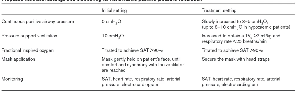
NPPV can be administered both with nasal and full-face masks. The nasal mask is usually well tolerated because it causes less claustrophobia and discomfort. It allows eating, drinking and expectorating. Conversely, a facial mask is preferable in severe respiratory failure, because dyspneic patients breath through the mouth in order to bypass resistance of the nasal passages, and mouth opening during nasal mask ventilation results in air leakage and decreased effectiveness [20,21]. Masks are firmly secured with elastic straps (Fig. 1) to the face in order to avoid air leaks and consequent malfunction. The dead space volumes of a facial and a nasal mask are 250 ml and 105 ml, respectively [22]. Dead space volume from the mask and the oropharynx may affect the effectiveness of ventilation.
In mild forms of ARF, a nasal mask could be tried first, switching to a facial mask if necessary. Among 12 patients (11 with COPD and one with pneumonia) with ARF who failed NPPV with a conventional facial or nasal mask (due to leaks or discomfort), switching to the new full-face mask improved gas exchange and avoided endotracheal intuba-tion in 84% of the patients [23]. A mask with transparent dome is preferred because it allows visual monitoring of the oral airway for the presence of secretions (Fig. 1). The mask should be lightweight to aid in its application and have a soft, pliable, adjustable seal to reduce trauma and leaking [24]. The mask is secured with head straps (Fig. 1). In our experience, skin necrosis occurs in 7% of patients treated with NPPV for periods exceeding 72 h; after discontinuation of NPPV, however, rapid healing of the dermal lesions is observed, usually in 7–10 days. The correct approach to limit these lesions is to place adhe-sive dressings to the points of major pressure (usually the bridge of the nose) to increase the pressure dissipation surface and reduce the depth of skin necrosis.
Table 1
Criteria for selection of patients for noninvasive positive pressure ventilation
Conscious and cooperative patient (chronic obstructive pulmonary disease patients may be an exception)
No need for urgent endotracheal intubation to protect the airways or remove copious secretions
No acute facial trauma
No recent gastroesophageal surgery No active gastrointestinal bleeding No impaired swallowing
Haemodynamic and rhythm stability Face mask adequately fitted
Table 2
Criteria for noninvasive positive pressure ventilation discontinuation and endotracheal intubation
Mask intolerance due to pain, discomfort or claustrophobia Inability to improve gas exchanges and/or dyspnea
Haemodynamic instability or evidence of cardiac ischaemia or ventricular dysarrhythmia
Need for urgent endotracheal intubation to manage secretions or protect the airways
17 When a full-face mask is used and the opening pressure
of the upper oesophageal sphincter (25–30 cmH2O) [25] is overcome, gastric distension may occur, but this is not a common event. Some masks allow the passage of a naso-gastric tube, protecting from the risk of aerophagia, even at pressures above 25 cmH2O.
The mask is connected to the ventilator, in the same way as an endotracheal tube. To prevent drying of the nasal passages and oropharynx, a humidifier should be con-nected, but the heater should be turned off because the upper airways that naturally warm inspired gas are not bypassed. As an alternative to the humidifier, we prefer to add a heat–moisture exchanger to the ventilatory circuit in order to ensure ‘natural’ humidification and heating, and to reduce the risk of bacterial colonization (Fig. 1).
With NPPV, tidal volume, gas exchange, respiratory rate and diaphragmatic activity are improved in proportion to the amount of pressure applied [20,26]. Most NPPV studies used pressure-limited ventilation delivered by a broad range of ventilators. Pressure-limited ventilation improves the efficacy of spontaneous breathing by allowing an optimal synchrony between patient effort and delivered assistance. Inspiration is initiated by the patient’s activation of the inspiratory muscles and of the inspiratory glottic abductors, with consequent widening of the glottis. During pressure support ventilation (PSV), the patient’s effort determines volume and duration of inspiration. Gas flow begins after the patient’s inspiratory effort reduces pres-sure in the inspiratory circuit of the ventilator by a predeter-mined value, usually 1–2 cmH2O. Pressure-control ventilation has a preset inspiratory time and respiratory
rate. It may ventilate patients with low ventilatory drive more effectively. In comparison with volume-cycled ventilation, pressure-limited ventilation minimizes peak inspiratory mask pressure and air leakage. Although tidal volume may vary as a function of the change in airway resistance and com-pliance, this variance has been an uncommon problem in our experience. In three comparison studies of assist-con-trolled ventilation and PSV in patients with hypercapnic ARF, PSV was as effective as assist-controlled ventilation in reducing the work of breathing and improving gas exchange [27], but was better tolerated [27,28] and asso-ciated with fewer complications [27]. During NPPV of stable COPD patients, flow triggering reduces the respira-tory effort and intrinsic positive end-expirarespira-tory pressure (PEEP) during both PSV and assist-controlled ventilation when compared with pressure triggering [29]. No differ-ences were found between 1 and 5 l/min flow triggers [29]. Two reports [30,31] found nasal ventilation with assist-controlled ventilation to be ineffective and time con-suming in end-stage obstructive lung disease. The present trend is to favour PSV, which has the advantage of better patient comfort and fewer complications [27].
The initial ventilator settings are continuous positive airway pressure 0 cmH2O and PSV 10 cmH2O; the mask is then gently held on patient’s face until the patient is comfort-able and in synchrony with the ventilator. FiO2is titrated to achieve an oxygen saturation over 90%. After the mask is secured, continuous positive airway pressure is slowly increased to 3–5 cmH2O, and PSV is increased to obtain the largest (> 7 ml/kg) exhaled tidal volume, a respiratory rate below 25 breaths/min and patient comfort. These objectives may not be achieved in patients with severe lung disease or with a leaky interface. It is important to recognize that excessive PSV levels can cause excessive inflation, with consequent patient–ventilator asynchrony and activation of expiratory muscles during inspiration (Table 3) [32].
During the early phase of milder respiratory failure and after an initial period of continuous administration (3–6 h in our experience) NPPV can be intermittently applied, with periods of 10–20 min interruption when it is not used. For sicker patients, NPPV application has to be continu-ous for at least 12–24 h [5,18]. Discontinuation is allowed for short periods only when the clinical situation improves. Aggressive physiotherapy is crucial during the periods of NPPV discontinuation. Endotracheal intubation must be rapidly available, when indicated (Table 2).
Applications of noninvasive positive pressure
ventilation in patients with chronic obstructive
pulmonary disease
Physiologic response
[image:3.612.56.297.91.278.2]NPPV is a valid method to treat COPD patients and to avoid endotracheal intubation [6,9,26,33,34]. In COPD
Figure 1
Patient undergoing noninvasive positive pressure ventilation. The face mask (FM), with its soft inflatable cushion (IC), is connected to the mechanical ventilator (not shown) through a catheter mount (CM), with a heat–moisture exchanger (HME) included in the respiratory circuit. The face mask is secured with elastic straps (ES).
ES FM
HME
CM
patients with acute exacerbation, the increased flow resis-tance and the inability to complete the expiration before inspiration results in high levels of dynamic hyperinflation. Dynamic hyperinflation alters diaphragm geometry, and reduces its strength and endurance. Also, minor increases in air flow resistance (as caused by airway secretions or bronchospasm) or an augmented ventilatory demand (as in case of fever or infection) in this context can cause res-piratory muscle fatigue, with rapid shallow breathing, wasted ventilation, hypercapnia and respiratory acidosis. The work of breathing is increased to overcome the inspi-ratory threshold load due to auto-PEEP and to drive the tidal volume against increased airway resistances.
The classical approach to management of exacerbated COPD is a combination of pharmacological interventions (bronchodilators, steroids, antibiotics and inotropes) and low-rate oxygen. When this conservative approach is not successful, patients are intubated and mechanically ventilated.
NPPV is a tool to correct the increased work of breathing and avoid intubation. The combination of PEEP and posi-tive pressure ventilation or PSV offsets the auto-PEEP level (eliminating the additional inspiratory load) and reduces the work of breathing that the inspiratory muscles must generate to produce the tidal volume. When appro-priate levels of inspiratory pressure are delivered, tidal volume increases and respiratory rate decreases. Under these conditions NPPV significantly reduces arterial partial carbon dioxide tension (PaCO2), restoring normal pH, and induces a rapid and progressive decrease in diaphrag-matic activity as shown by electromyography [26].
It must be remembered that the level of applied PEEP must never exceed the amount of auto-PEEP, in order to avoid iatrogenic increases of hyperinflation. No change in end-expiratory volume has been reported if PEEP applied
by face-mask ventilation does not exceed the 80–90% of the auto-PEEP [35].
Clinical results
In one of the first applications of NPPV in a small sample of COPD patients [33], the improvements in gas exchanges suggested the possibility of avoiding endotra-cheal intubation altogether. In a case–control study, Brochard et al [26] showed that this approach could reduce both the need for endotracheal intubation and the duration of hospital stay, with obvious economic implica-tions. In the first randomized, prospective study on 60 COPD patients, Bott et al [6] compared NPPV adminis-tered through nasal mask with conventional therapy as a treatment for ARF. Patients receiving NPPV had a signifi-cant reduction in PaCO2 and dyspnea score, and improved survival (90 versus 70%; P< 0.01).
The efficacy of NPPV in acute exacerbation of COPD was also evaluated in a European prospective randomized mul-ticentre study [9]. In that trial, 85 COPD patients without cardiogenic pulmonary oedema, pneumonia or postopera-tive ARF were randomly assigned to receive conventional treatment (oxygen therapy plus drugs) or NPPV in addition to conventional treatment. After 1 h, NPPV achieved a sig-nificant improvement in gas exchange. The group of patients randomly assigned to NPPV had a significantly lower intubation rate (26 versus 74%; P< 0.001), lower complication rate (14 versus 45%; P< 0.01), lower duration of hospital stay (23 ± 17 versus 35 ± 33 days; P< 0.02) and lower mortality rate (9 versus 29%; P< 0.02).
[image:4.612.54.557.112.268.2]In another randomized study on 26 COPD patients that compared NPPV delivered through nasal mask with con-ventional treatment [11], the authors reported a reduction in intubation rate, with a significant improvement in PaO2, heart rate and respiratory rate in the NPPV group, even though PaCO2 did not significantly decrease. A further 18
Table 3
Proposed ventilator settings and monitoring for noninvasive positive pressure ventilation
Initial setting Treatment setting
Continuous positive airway pressure 0 cmH2O Slowly increased to 3–5 cmH2O,
(up to 8–10 cmH2O in hypoxaemic patients) Pressure support ventilation 10 cmH2O Increased to obtain a TVe>7 ml/kg and
respiratory rate <25 breaths/min Fractional inspired oxygen Titrated to achieve SAT >90% Titrated to achieve SAT >90% Mask application Mask gently held on patient’s face, until Secure the mask with head straps
comfort and synchrony with the ventilator are reached
Monitoring SAT, heart rate, respiratory rate, arterial SAT, heart rate, respiratory rate, arterial pressure, electrocardiogram pressure, electrocardiogram
randomized study on hypercapnic and hypoxaemic ARF [36] suggested that early application of NPPV facilitates improvement, decreases the need for invasive mechanical ventilation, and decreases the duration of hospitalization. Thirty patients were randomized to receive standard treat-ment, or NPPV in addition to standard treatment. With standard treatment, there was significant improvement only in respiratory rate (P< 0.05). With NPPV, PaO2 (P< 0.001), PaCO2(P< 0.001), pH (P< 0.001) and res-piratory rate (P< 0.001) improved significantly compared with baseline. Duration of hospital stay for the NPPV group was shorter (P< 0.05) than that in the standard treatment group. One patient in the NPPV group required invasive mechanical ventilation.
Lofaso et al [37] recently reported the risk of carbon dioxide rebreathing with bilevel positive airway pressure (BiPAP) ventilators when PEEP was not applied or when the expiratory time was too short; this might partly explain the limited PaCO2modification noted by Kramer et al[11]
In a randomized trial in 50 patients with acute exacerba-tion of COPD, noninvasive PSV during weaning reduced weaning time (P= 0.021), shortened the duration of stay in the intensive care unit (P= 0.005), decreased the inci-dence of nosocomial pneumonia, and improved 60-day survival rates (P= 0.009) [34].
Noninvasive positive pressure ventilation in
patients with acute respiratory failure
Physiologic response
Following the good results obtained in patients with acute exacerbations of COPD and the promising data from a few pilot studies (retrospective or nonrandomized) [33,38,39] in patients with ARF, NPPV is now currently under clinical evaluation as a possible alternative to con-ventional ventilation with endotracheal intubation and as a means to reduce the intubation rate during ARF. NPPV is adopted in ARF that is not related to COPD, with the aim of decreasing the amount of spontaneous work of breath-ing and correctbreath-ing the rapid shallow breathbreath-ing that is always present in acute conditions. NPPV can prevent res-piratory muscle fatigue and endotracheal intubation.
NPPV can be administrated via a nasal or a full-face mask. Selection criteria and criteria for NPPV discontinuation and endotracheal intubation are similar to those described for COPD patients (Tables 1 and 2). In our experience and that of others [5,25], face masks seem more appropriate for patients affected by severe hypoxaemia, who are usually tachypneic and breathe through the mouth.
Clinical results
Nonrandomized, noncontrolled studies
Meduri et al[33] in 1989 reported one of the first clinical applications of NPPV in patients with ARF. In that study
PSV and pressure control ventilation were used through face masks in four patients affected by cardiogenic and noncardiogenic pulmonary oedema, with good results in three. Subsequently, Pennock et al [39] reported suc-cessful treatment in 50% of a large group of patients with ARF of different aetiologies; promising results were obtained in the subgroup of patients affected by postoper-ative ARF. Similar results were achieved using noninvasive ventilation (always through a nasal mask) in a second study [40]. Wysocki et al [41] reported a 47% success rate in the treatment of ARF patients.
In a study conducted on 64 patients admitted to the emer-gency department for ARF due to cardiogenic pulmonary oedema and pneumonia [42] there was a significant improvement in arterial blood gases after 1 h of continu-ous positive airway pressure ventilation. In the cardiogenic pulmonary oedema group, PaO2 surpassed 100 mmHg
with a clear-cut improvement in PaCO2 and pH
(P< 0.0001, for both parameters). In the pneumonia group, oxygenation was also improved, but with the per-sistence of a significant shunt. Fifty-four patients (84%) were successfully ventilated under continuous positive airway pressure, avoided intubation and had a favourable prognosis, mainly in the cardiogenic pulmonary oedema group, without side effects.
In a pilot study conducted in patients with haematologi-cal malignancies complicated by ARF [43] 15 out of 16 individuals were successfully treated with NPPV deliv-ered via nasal mask by means of a BiPAP ventilator (Respironics, Pittsburgh, USA). PaO2/FiO2 and arterial oxygen saturation significantly improved after 1 h of treatment (P< 0.01).
NPPV delivered by simplified ventilators via facial or nasal mask can be effective for routine care, after adequate per-sonnel training [44]. In 40 patients with hypercapnic ARF compared with 30 matched historical patients under con-ventional treatment, NPPV was associated with a reduc-tion in negative events, such as endotracheal intubareduc-tion, and mortality together (17 versus 60%; P= 0.0002), but not mortality alone (5 versus 13.5%; not significant). Sig-nificant and rapid improvements in PaCO2 and pH between baseline and subsequent evaluations (P= 0.066) were obtained.
The utility of NPPV to prevent nosocomial pneumonia in patients who need assisted ventilation was recently reported in a prospective epidemiological survey on a cohort of 320 consecutive patients, 75% of whom had ARF not related to COPD [45]. Twenty-seven patients had 28 episodes of ventilator-associated pneumonia, but the incidence of ventilator-associated pneumonia was 0.85/100 days of tracheal intubation and 0.16/100 days
In a recent meta-analysis [46], Keenan and Brake analyzed more than 200 published and unpublished studies and concluded that the addition of NPPV to standard therapy in patients with ARF improves survival and decreases the need for endotracheal intubation. This effect was more evident in patients whose cause of ARF was an exacerba-tion of COPD, however.
To assess the efficacy of NPPV in routine use, 80 patients (aged 71 ± 1.3 years and Acute Physiology and Chronic Health Evaluation II score of 17.2 ± 0.6) received BiPAP for ARF [47]. Thirty-one of them (39%) had hypoxaemic ARF and 25 (31%) had hypercapnic ARF; the other 24 patients (30%) suffered from ARF of varied origin. BiPAP was successful in 80% of the patients with hypercapnic respiratory failure, but only in 15 of the 31(48%) patients with hypoxaemic ARF. BiPAP success was marked, with increased PaO2/FiO2 in the hypoxaemic group, but the risk failure was significantly greater (risk ratio 2.6, 95% confidence interval 1.1–6.1) for patients with hypoxaemic ARF than for those with hypercapnic respiratory failure.
Randomized studies
The first prospective randomized study dedicated to this topic was reported by Wysocki et al[18], who randomized 41 non COPD patients with ARF to receive face-mask mechanical ventilation versus conventional medical therapy. NPPV reduced the need for endotracheal intubation (36 versus 100%, P= 0.02), the duration of intensive care unit stay (13 ± 15 days versus 32 ± 30 days; P= 0.04) and mor-tality rate (9 versus 66%; P= 0.06), but only in those patients with hypercapnia (PaCO2> 45 mmHg). No signifi-cant differences in the hypoxaemic group without hypercap-nia were seen. On the basis of these results, the authors concluded that NPPV was not beneficial when used sys-tematically in all forms of ARF not related to COPD. Kramer et al[11] randomized 31 patients to receive nasal BiPAP or conventional therapy for the treatment of ARF. This study showed significant improvement in vital signs as well as reduction in endotracheal intubation rates in the group treated with NPPV compared with the conventionally treated group (31 versus 73% endotracheal intubation rates, respectively; P< 0.05). Evidence from this study sup-ported the use of NPPV in order to avoid intubation and possibly reduce mortality, but this conclusion again applied mainly to the subgroup of 23 (74%) patients with acute exacerbation of COPD. Only eight patients (36%) had ARF due to pneumonia (four patients), congestive heart failure (two patients), asthma (one patient) or pulmonary embolism (one patient). These studies were specifically dedicated to evaluate NPPV as a preventive tool against endotracheal intubation, and not as an alternative treatment for ARF.
We randomized 64 patients with hypoxaemic ARF who had not improved with aggressive medical therapy to face-mask NPPV or endotracheal intubation with conventional
mechanical ventilation [5]. Both groups had improvement in the PaO2:FiO2ratio after 1 h of ventilatory support. Ten out of the 32 patients in the NPPV group required endotracheal intubation. The 32 patients in the conventional ventilation group had more frequent and serious complications (38 versus 66%; P= 0.02), and pneumonia and sinusitis related to the endotracheal tube (3 versus 31%; P= 0.003). Among the survivors, the patients in the NPPV group had a shorter duration of mechanical ventilation (3 ± 3 versus 6 ± 5; P= 0.006) and a shorter duration of intensive care unit stay (6.6 ± 5 versus 14 ± 13 days; P= 0.002). Our trial suggested that, for patients with severe respiratory distress, NPPV may lead to more favourable outcomes than conven-tional ventilation, in the hands of experienced staff and in a setting in which this technology can be rapidly and safely administered. Furthermore a post hoc subgroup analysis was performed for patients with a simplified acute physio-logic score lower than 16, or 16 or greater. The 19 patients with a simplified acute physiologic score of 16 or greater, irrespective of group, had similar outcomes, whereas in the 45 patients with a simplified acute physiologic score lower than 16 NPPV was superior to conventional ventilation.
Conversely, Wood et al [48] had a substantially negative evaluation of the use of NPPV when applied in patients with hypoxaemic ARF. These authors randomized 27 patients in the emergency department to receive conven-tional medical therapy or NPPV for the treatment of acute hypoxaemic respiratory failure. The 16 patients who were randomized to the noninvasive ventilation group had a intu-bation rate (44%) and duration of intensive care unit stay similar to those of the 11 patients who received medical treatment alone, but there was a trend toward a greater rate of hospital mortality among the patients in the NPPV group compared with patents in the conventional medical therapy group [four patients (25%) versus none; P= 0.123]. Several factors may have influenced these neg-ative results and may have represented biases in patient selection. First, case mix showed an unbalanced distribu-tion of patients with pneumonia (44% in the NPPV group versus 12% in the conventional medical therapy group). Second, 14% of patients had COPD. Finally, patients in the NPPV group had a baseline PaO2that was significantly lower than that in the conventional medical treatment group (59.8 versus 71.3 mmHg, respectively). Moreover, even though the PaCO2levels were similar in the two groups, the range was clearly hypercapnic (around 56 mmHg in the two groups). These differences may represent increased severity of ARF in the NPPV group, and objective conclu-sions are therefore difficult to make.
Noninvasive positive pressure
ventilation-assisted bronchoscopy in severely
hypoxaemic patients
affected by haematological malignancies, transplantations or other conditions [49]. In these individuals, early diagno-sis of the aetiological agent is paramount. Unfortunately, severe hypoxaemia in nonintubated patients represents a major contraindication to fiberoptic bronchoscopy and/or bronchoalveolar lavage [50], often meaning that treatment must be initiated on an empirical basis.
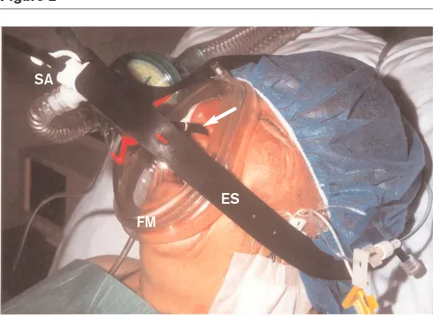
We proposed a new technique to perform fiberoptic bron-choscopy and/or bronchoalveolar lavage in severely hypoxaemic, nonintubated patients by means of PSV administered through a face mask (Fig. 2) [51]. All patients were administered NPPV for 10 min before start-ing bronchoscopic manoeuvres and NPPV was main-tained for at least 90 min after the procedure. In all patients it was possible to identify the agent that caused pneumonia, and to start an early and specific treatment. None of the patients needed endotracheal intubation. PaO2:FiO2 ratio and oxygen saturation increased signifi-cantly after the application of NPPV and remained high throughout the study.
In a recently concluded randomized study (unpublished data) on the use of brochoalveolar lavage with or without NPPV for the diagnosis of nosocomial pneumonia in 26 hypoxaemic patients (PaO2:FiO2< 200), we were able to demonstrate that NPPV is more efficient than Venturi mask in correcting hypoxaemia during brochoalveolar lavage, but 1 h after treatment PaO2:FiO2 was not differ-ent in the two groups.
Bronchoscopy with NPPV seems to be a feasible, safe and effective technique to allow an early and accurate diagnosis of pneumonia in nonintubated, severely
hypox-aemic patients. Even though good cooperation of the patient and thorough monitoring of vital functions are essential, the technique appears very promising for appli-cation on a large scale in immunocompromized patients.
Conclusion
In COPD patients, NPPV can be considered as a thera-peutic option to prevent endotracheal intubation (reducing the additive morbidity and mortality) and to deliver artificial ventilatory support [6,9]. When no contraindication exists, a trial of NPPV should be always considered if a COPD patient is observed to be in the early phase of respiratory failure.
Some evidence suggests the utility of NPPV as a first-line intervention in hypoxaemic ARF and for NPPV-assisted bronchoscopy in severely hypoxaemic patients. The appli-cation of NPPV in patients suffering from ARF not related to COPD, despite some interesting and very promising preliminary results [5,11], still remains controversial. Large, prospective, randomized, multicentre studies are therefore needed.
Acknowledgement
The authors are grateful to Dr GU Meduri, because some of the contents of the present review are inspired by his clinical and scientific work.
References
1. Meduri GU: Noninvasive ventilation.In: Physiological Basis of Venti-latory Support. Edited by Marini J, Slustky A. Series on Lung Biology in Health and Disease. Marcel Dekker, Inc: New York, 1998:921–998. 2. Nourdine K, Combes P, Carton MJ, Beuret P, Cannamela A, Ducreux
JC: Does noninvasive ventilation reduce the ICU nosocomial infection risk?: a prospective clinical survey. Intens Care Med
1999, 25:567–573.
3. Torres A, Aznar R, Gatell JM, et al: Incidence, risk and prognosis factors of nosocomial pneumonia in mechanically ventilated patients.Am Rev Respir Dis1990, 142:523–528.
4. Burns HP, Dayal VS, Scott A, Van Nostran ANP, Bryce DP: Laryngo-tracheal trauma: observation on its pathogenesis and its preven-tion following prolonged OT intubapreven-tion in the adult.Laryngoscope
1979, 89:1316–1325.
5. Antonelli M, Conti G, Rocco M, et al: A comparison of noninvasive positive-pressure ventilation and conventional mechanical ventila-tion in patients with acute respiratory failure.N Engl J Med1998,
339:429–435.
6. Bott J, Carroll MP, Conway JH, et al: Randomized controlled trial of nasal ventilation in acute ventilatory failure due to chronic obstructive airways disease.Lancet1993, 341:1555–1558. 7. Ahmed AH, Fenwick L, Angus RM, Peacock AJ: Nasal ventilation v
doxapram in the treatment of type II respiratory failure complicat-ing chronic airflow obstruction [abstract].Thorax 1992, 1:858. 8. Daskalopoulou E, Tsara V, Fekete K, Koutsantas V, Christaki P:
Treat-ment of acute respiratory failure in COPD patients with positive airway pressure via nasal mask (NPPV) [abstract].Chest1993,
103:271S.
9. Brochard L, Mancebo J, Wysocki M, et al: Noninvasive ventilation for acute exacerbations of chronic obstructive pulmonary disease.N Engl J Med1995, 333:817–822.
10. Martin TJ, Sanders MH, Bierman MI, Hovis JD: Non-invasive applica-tion of bi-level positive airway pressure to prevent endotracheal intubation in acute respiratory failure [abstract]. Crit Care Med
1994, 23:A129.
11. Kramer N, Meyer TJ, Meharg J, Cece RD, Hill NS: Randomized, prospective trial of noninvasive positive pressure ventilation in acute respiratory failure.Am J Respir Crit Care Med1995, 151:
[image:7.612.57.298.94.270.2]1799–1806. 21
Figure 2
Fiberoptic bronchoscopy during noninvasive positive pressure ventilation. The patient is connected to the ventilator via a face mask (FM) secured with elastic straps (ES). The bronchoscope is passed through a seal adapter (SA), in order to allow mechanical ventilation. The arrow indicates the optical instruments advanced into the nose.
ES FM
12. Keenan SP, Kernerman PD, Cook DJ, et al: The effect of noninvasive positive pressure ventilation on mortality in patients admitted with acute respiratory failure: a meta-analysis.Crit Care Med1997, 25: 1685–1692.
13. Meduri GU, Turner RE, Abou-Shala N, Tolley E, Wunderink RG: Non-invasive positive pressure ventilation via face mask: first-line intervention in patients with acute hypercapnic and hypoxemic respiratory failure.Chest1996, 109:179–193.
14. Rasanen J, Heikkila J, Downs J, et al: Continuous positive airway pressure by face mask in acute cardiogenic pulmonary edema. Am J Cardiol1985, 55:296–300.
15. Bersten AD, Holt AW, Vedic AE, Skowronski GA, Baggoley CJ: Treat-ment of severe cardiogenic pulmonary edema with continuous positive airway pressure delivered by face mask.N Engl J Med
1991, 325:1825–1830.
16. Lin M, Yang Y, Chiany H, et al: Reappraisal of continuous positive airway pressure therapy in acute cardiogenic pulmonary edema: short-term results and long-term follow-up. Chest 1995, 107: 1379-86
17. Martin TJ, Sanders MH, Bierman MI, Hovis JD: Non-invasive applica-tion of bi-level positive airway pressure to prevent endotracheal intubation in acute respiratory failure [abstract].Crit Care Med
1994, 23:A129.
18. Wysocki M, Tric L, Wolff MA, Millet H, Herman B: Noninvasive pres-sure support ventilation in patients with acute respiratory failure: a randomized comparison with conventional therapy.Chest1995,
107:761–768.
19. Pankow W, Hijjeh N, Schuttler F, et al: Influence of noninvasive pos-itive pressure ventilation on inspiratory muscle activity in obese subjects.Eur Respir J1997, 10:2847–2855.
20. Carrey Z, Gottfried SB, Levy RD: Ventilatory muscle support in res-piratory failure with nasal positive pressure ventilation. Chest
1990, 97:150–158.
21. Soo Hoo GW, Santiago S, Williams AJ: Nasal mechanical ventila-tion for hypercapnic respiratory failure in chronic obstructive pul-monary disease: determinants of success and failure.Crit Care Med1994, 22:1253–1261.
22. Criner GJ, Travaline JM, Brennan KJ, Kreimer DT: Efficacy of a new full face mask for noninvasive positive pressure ventilation.Chest
1994, 106:1109–1115.
23. Roy B, Kreimer DT, Mullarkey J, Dantzler E, Criner GJ: Noninvasive positive pressure ventilation with a total face mask for acute respi-ratory failure [abstract].Am J Respir Crit Care Med1996, 153:A608. 24. Branson RD, Hurst JM, DeHaven CB: Mask CPAP: state of the art.
Respir Care1985, 30:846–857.
25. Meduri GU: Non invasive positive-pressure ventilation in patients with chronic obstructive pulmonary disease and acute respiratory failure.Curr Opin Crit Care1996, 2:35–46.
26. Brochard L, Isabey D, Piquet J, et al: Reversal of acute exacerba-tions of chronic obstructive lung disease by inspiratory assistance with a face-mask.N Engl J Med1990, 323:1523–1530.
27. Vitacca M, Rubini F, Foglio K, et al: Noninvasive modalities of posi-tive pressure ventilation improve the outcome of acute exacerba-tions in COLD patients.Intens Care Med1993, 19:450–455. 28. Richard JC, Molano C, Tengang B, et al: Nasal intermittent positive
pressure ventilation versus bilevel pressure ventilation during acute respiratory failure in patients with COPD [abstract].Am J Respir Crit Care Med1996, 153:A609.
29. Nava S, Bruschi C, Ambrosino N, Paturno V, Confalonieri M: Inspira-tory effort during noninvasive mechanical ventilation with flow and pressure triggers in COPD patients.Intens Care Med1995, 21: S120.
30. Chevrolet JC, Jolliet P, Abajo B, Toussi A, Luis M: Nasal positive pressure ventilation in patients with acute respiratory failure. Diffi-cult and time-consuming procedure for nurses.Chest1991, 100: 775–782.
31. Soo Hoo GW, Williams AJ: Noninvasive face-mask ventilation in patients with acute hypercapnic respiratory failure [abstract]. Chest1993, 103:1304.
32. Jubran A, Van De Graaff WB, Tobin MJ: Variability of patient-ventila-tor interaction with pressure support ventilation in patients with chronic obstructive pulmonary disease.Am J Respir Crit Care Med
1995, 152:129–136.
33. Meduri GU, Conoscenti CC, Menashe P, Nair S: Non-invasive face mask ventilation in patients with acute respiratory failure.Chest
1989, 95:865–870.
34. Nava S, Ambrosino N, Clini E, et al: Noninvasive mechanical ventila-tion in the weaning of patients with respiratory failure due to chronic obstructive pulmonary disease. A randomized, controlled trial.Ann Intern Med1998, 128:721–728.
35. Appendini L, Patessio A, Zanaboni S, et al: Physiologic effects of positive end-expiratory pressure and mask pressure support during exaacerbations of chronic obstructive pulmonary disease. Am J Repir Crit Care Med1994, 149:1069–1076.
36. Celikel T, Sungur M, Ceyhan B, Karakurt S: Comparison of noninva-sive positive pressure ventilation with standard medical therapy in hypercapnic acute respiratory failure. Chest 1998, 114: 1636–1642.
37. Lofaso R, Brochard L, Touchard D, et al: Evaluation of carbon dioxide rebreathing during pressure support ventilation with airway management system (BiPAP) devices. Chest1995, 108: 772–778.
38. Meduri GU, Fox RC, Abou-Shala N, et al: Noninvasive mechanical ventilation via face mask in patients with acute respiratory failure who refused endotracheal intubation. Crit Care Med1994, 22: 1584–1590.
39. Pennock BE, Crawshan L, Kaplan PD: Noninvasive nasal mask ven-tilation for acute respiratory failure.Chest1994, 105:441–444. 40. Lapinsky SE, Mount DB, Mackey D, et al: Management of acute
res-piratory failure due to edema with nasal positive pressure support.Chest1994, 105:229–231.
41. Wysocki M, Tric L, Wolff MA, et al: Noninvasive pressure support ventilation in patients with acute respiratory failure.Chest1993,
103:907–913.
42. L’Her E, Moriconi M, Texier F, et al: Non-invasive continuous posi-tive airway pressure in acute hypoxaemic respiratory failure: experience of an emergency department.Eur J Emerg Med1998,
5:313–318.
43. Conti G, Marino P, Cogliati A, et al: Noninvasive ventilation for the treatment of acute respiratory failure in patients with hematologic malignancies: a pilot study.Intens Care Med1998, 24:1283–1288. 44. Corbetta L, Ballerin L, Putinati S, Potena A: Efficacy of noninvasive
positive pressure ventilation by facial and nasal mask in hyper-capnic acute respiratory failure: experience in a respiratory ward under usual care.Monaldi Arch Chest Dis1997, 52:421–428. 45. Guerin C, Girard R, Chemorin C, De Varax R, Fournier G: Facial mask
noninvasive mechanical ventilation reduces the incidence of nosocomial pneumonia. A prospective epidemiological survey from a single ICU.Intens Care Med1997, 23:1024–1032. 46. Keenan SP, Brake D: An evidence-based approach to noninvasive
ventilation in acute respiratory failure. Crit Care Clin 1998, 14: 359–372.
47. Alsous F, Amoanteg-Adjepong Y, Manthous CA: Noninvasive ventila-tion: experience at a community teaching hospital. Intens Care Med1999, 25:458–463.
48. Wood KA, Lewis L, Von Harz B, Kollef MH: The use of noninvasive positive pressure ventilation in the emergency department: results of a randomized clinical trial.Chest1998, 113:1339–1346. 49. Gentile G, Micozzi A, Girmenia C, et al: Pneumonia in allogenic and autologous bone marrow recopients.Chest1993, 104:371–375. 50. American Thoracic Society: Clinical role of bronchoalveolar lavage
in adults with pulmonary disease.Am Rev Respir Dis1990, 142: 481–486.
51. Antonelli M, Conti G, Riccioni L, Meduri GU: Non-invasive positive pressure ventilation via face mask during bronchoscopy with bronchoalveolar lavage in high risk hypoxemic patients. Chest
1996, 110:724–728.
Authors’ affiliation:Department of Anesthesiology and Intensive Care, Università Cattolica del Sacro Cuore, Rome, Italy
Correspondence:Massimo Antonelli, MD, Department of Anesthesiology and Intensive Care, Università Cattolica del Sacro Cuore, Largo F.Vito 1, 00168 Rome, Italy. Tel: +39 06 3015 4386; fax: +39 06 3013 450; e-mail: max.antonelli@flashnet.it