R E S E A R C H A R T I C L E
Open Access
Factors associated with the development of
Congenital Zika Syndrome: a case-control
study
Giulia P. Lima
†, Daniel Rozenbaum
†, Clarisse Pimentel, Ana Cristina Cisne Frota, Daniela Vivacqua,
Elizabeth S. Machado, Fernanda Sztajnbok, Thalita Abreu, Raquel A. Soares and Cristina B. Hofer
*Abstract
Background:We aim to investigate possible maternal- and pregnancy-related factors associated with the development of Congenital Zika Syndrome (CZS) in children of mothers with probable gestational infection.
Methods:This case-control study, we recruited mother-infant pairs between May 2015 and October 2017 in a pediatric infectious disease clinic in Rio de Janeiro. Inclusion criteria required either that the mother reported Zika infection symptoms during pregnancy or that the infant presented with clinical or imaging features of the CZS. Exclusion criteria included detection of an alternative cause for the patient’s presentation or negative polymerase chain reaction assays for Zika in all specimens tested within 12 days from the beginning of maternal symptoms. Infants with CZS (CDC definition) were selected as cases and infants without CZS, but with probable maternal Zika virus infection during pregnancy, were selected as controls. Maternal and pregnancy-related informations were collected and their relationship to the presence of congenital anomalies due to CZS was assessed by Fisher exact or Mann-Whitney test.
Results:Out of the 42 included neonates, 24 (57.1%) were diagnosed with CZS (cases). The mean maternal age at the birth was 21 years old. The early occurrence of maternal symptoms during pregnancy was the only variable associated with CZS (odds ratio = 0.87, 95% CI: 0.78–0.97).
Case’s mothers presented symptoms until the 25th week of gestational age (GA), while control’s mothers presented until 36th weeks of GA. Income; illicit drug, alcohol, or tobacco use during pregnancy; other infections during pregnancy (including previous dengue infection) were not associated with CZS.
Conclusions:Our study corroborates the hypothesis that Zika virus infection earlier in pregnancy is a risk factor to the occurrence of congenital anomalies in their fetuses.
Keywords:Congenital Zika Syndrome, Rio de Janeiro, Case-control, Gestational age
Background
On March 31, 2016, the World Health Organization published a statement on the evidence for a causal link between Zika virus infection during pregnancy and the occurrence of congenital brain abnormalities, including microcephaly [1]. With further observational studies, a whole set of congenital abnormalities was identified and
linked to in utero Zika virus infection. This set of abnor-malities was termed the Congenital Zika Syndrome (CZS) and may include, in addition to microcephaly, craniofacial disproportion, irritability, spasticity, seizures, feeding difficulties, ocular abnormalities, and hearing loss on examination, as well as calcifications, cortical disorders, and ventriculomegaly on neuroimaging. Up until October 2017, there were 3689 confirmed cases of the congenital syndrome associated with Zika virus infection in the Americas, most of them in developing countries and affecting the most socially vulnerable groups [2–4]. Despite the substantial reduction in the
© The Author(s). 2019Open AccessThis article is distributed under the terms of the Creative Commons Attribution 4.0 International License (http://creativecommons.org/licenses/by/4.0/), which permits unrestricted use, distribution, and reproduction in any medium, provided you give appropriate credit to the original author(s) and the source, provide a link to the Creative Commons license, and indicate if changes were made. The Creative Commons Public Domain Dedication waiver (http://creativecommons.org/publicdomain/zero/1.0/) applies to the data made available in this article, unless otherwise stated.
* Correspondence:cbhofer@hucff.ufrj.br
†Giulia P Lima and Daniel Rozenbaum had the same participation on the
manuscript.
number of new cases of Zika virus infection, it is import-ant that preventive measures be drawn due to the high prevalence of Aedes aegypti mosquitoes in several areas and the possibility of recurrent waves of infection, as has been observed with other arboviruses in other locations.
It has been estimated that 5 to 42% of neonates from infected pregnant women develop congenital abnormal-ities [2, 5–8]. Nevertheless, there is only a paucity of studies specifically designed to elucidate which mater-nal- and pregnancy-related factors may be associated with the development of congenital abnormalities among infected pregnant women [9–11]. The identification of such factors might allow the development of interven-tions aiming to prevent the Congenital Zika Syndrome (CZS). We, therefore, conducted a case-control study to investigate potential risk factors for the development of CZS in neonates from infected pregnant women. We hypothesized that factors associated with the prenatal period (co-infections, trimester of Zika virus infection, and maternal health variables) would be associated with development of CZS.
Methods Study design
This is a nested case-control study. Cases were defined as infants with CZS and controls as infants without CZS. Both possibly exposed to Zika virus during antenatal period.
Study population
Recruitment occurred between April 2015 and October 2017 at the Instituto de Puericultura e Pediatria Marta-gão Gesteira’s (IPPMG) pediatric infectious disease clinic, Rio de Janeiro, Brazil. We enrolled mother-infant pairs if they satisfied at least one of two criteria: 1) the mother had a history of gestational symptoms compat-ible with Zika virus infection, defined as a skin rash with or without other characteristic findings (e.g., fever, rash, conjunctivitis, and/or arthralgia), or 2) the infant was referred to our clinic due to clinical and/or radiologic findings of CZS. The CZS was defined as: severe micro-cephaly (> 3 SD below the mean), brain abnormalities, ocular findings, congenital contractures, neurological impairments. Patients were excluded if they failed to provide informed consent or if their clinical manifesta-tions could be better explained by an etiology other than Zika virus infection (all infants were screened by a trained geneticist and serologic tests were performed for several other congenital infections). We also excluded mother-infant pairs when the mother had negative poly-merase chain reaction assays for Zika in all specimens tested within 12 days from the beginning of her symp-toms. Among the remaining patients, infants with CZS were selected as cases and infants without CZS, but with
probable maternal Zika virus infection during pregnancy, were selected as controls.
Data collection
Every recruited pregnant mother was evaluated by an obstetrician and an infectious disease physician. During this evaluation, we filled out a standardized questionnaire including the following demographic and pregnancy-re-lated variables: maternal age (in years), income (in Brazilian minimum wages units), smoking status, alcohol and illicit drug consumption (if present or not), number of previous gestations, gestational week in which prenatal care had begun, and gestational age at symptom onset (based on an obstetric ultrasound at the first trimester). Pregnant women were serologically evaluated for HIV, syphilis, toxoplasmosis, rubella, and dengue. Serologic tests for Zika infection were not performed because they were either unavailable or unreliable at the time patients were recruited [12]. A real-time reverse transcription polymerase chain reaction (RT-PCR) for Zika was also performed for women referred to our clinic with recent symptoms (up to 12 days of symptoms).
Infants were evaluated by an infectious disease phys-ician, a neurologist, and a geneticist. Every infant should have been assessed with:1) Zika virus RT-PCR on blood specimens, 2) laboratory evaluation for other congenital infectious etiologies including toxoplasmosis, syphilis, cytomegalovirus and rubella, and HIV, 3) a neuroimaging study, 4) an abdominal ultrasonography, 5) a brainstem evoked response audiometry (BERA), 6) an echocardio-gram, and 7) a fundoscopy performed by an ophthalmolo-gist. However, since some infants were referred at a later age, it was not always feasible to perform the complete evaluation.
In order to exclude other causes for the congenital abnormalities, all children were evaluated by an experi-enced geneticist to rule out genetic abnormalities. Other infections were ruled out as described; cytomegalovirus: a negative urine PCR in the first 21 days of life and/or the IgG clearance during the concept infancy period (with-out IgM reactive). Toxoplasmosis, rubella, and dengue infections: IgG clearance during the concept infancy period (without IgM reactive). Syphilis: non-treponemic test (VDRL) negative. HIV: serology test (ELISA) non-reactive.
Statistical analysis
not performed, since the epidemic finished in Brazil, therefore we would not be able to enroll new patients.
STATA, v13.0 (Texas, USA) was used for all the statistical analysis.
Ethical statement
This project and its informed consent were approved by the Ethics Committee of the IPPMG (approval number: 1.546.045). Written informed consent was obtained from all participants guardians. The guardians were approached during the paticipant’s first visit at the Infectious Diseases Clinic, if they accepted to participate, data were systemat-ically collected during their clinical appointments.
Results
Sample characteristics
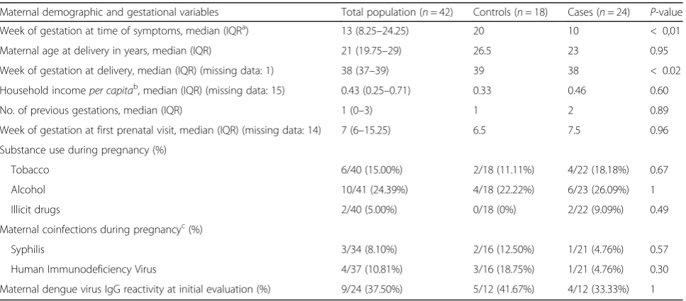
A total of 42 infants were included in our analysis, 18 controls and 24 cases. Infants were born between 12/11/ 2015 and 07/08/2017. Ten of them were born to mothers who had received antenatal care at our clinic, providing prospective information. The 32 remaining children were recruited either for being born with con-genital abnormalities compatible with CZS or because their mothers had a history of symptoms compatible with Zika infection during pregnancy. The demographic characteristics of the sample are shown in Table1.
Laboratory confirmation of maternal infection
PCR analyses for Zika in blood, urine or both were per-formed in 19 of the 36 mothers that reported a rash dur-ing pregnancy, and 11 (57.9%) tested positive in at least one specimen. Eight mothers with negative PCR results
were not excluded either because they only had one specimen tested or because the PCR test was performed more than 12 days after symptoms onset. Among symp-tomatic women without a laboratory confirmation, the diagnosis of probable Zika infection was made based on clinical and epidemiologic information, as well as with the exclusion of other known causes (drug allergies or other infections).
Infant assessment - evaluation of congenital anomalies Twenty-one infants had microcephaly. One of them was born with a cephalic perimeter of 32 cm, but the head circumference stopped growing afterwards, and there-fore was also considered as having microcephaly. Thirty-six children performed a neuroimaging exam. Among 6 children that were not evaluated with any neuroimaging exam, all of them were born with normal cephalic perimeter and were classified as controls since there were no other clinical or imaging abnormalities. Other components of the evaluation (fundoscopy, BERA, abdominal US and echocardiography) were performed in most infants. Among the children with CZS, fundoscopy was abnormal in 9 infants from a total of 21 who performed the exam; BERA was altered in 3 from a total of 17; 5 infants presented with patent foramen ovale, although at least 3 resolved in a subsequent echocardi-ography; 2 were born with clubfoot and 2 with cryptorchidism.
Laboratory confirmation of CZS
[image:3.595.57.541.495.707.2]PCR analyses for Zika in the blood, liquor or both were performed in only 11 (26%) of our infants. All of them
Table 1Maternal demographics between cases and controls
Maternal demographic and gestational variables Total population (n= 42) Controls (n= 18) Cases (n= 24) P-value Week of gestation at time of symptoms, median (IQRa) 13 (8.25–24.25) 20 10 < 0,01
Maternal age at delivery in years, median (IQR) 21 (19.75–29) 26.5 23 0.95 Week of gestation at delivery, median (IQR) (missing data: 1) 38 (37–39) 39 38 < 0.02 Household incomeper capitab, median (IQR) (missing data: 15) 0.43 (0.25–0.71) 0.33 0.46 0.60
No. of previous gestations, median (IQR) 1 (0–3) 1 2 0.89 Week of gestation at first prenatal visit, median (IQR) (missing data: 14) 7 (6–15.25) 6.5 7.5 0.96 Substance use during pregnancy (%)
Tobacco 6/40 (15.00%) 2/18 (11.11%) 4/22 (18.18%) 0.67 Alcohol 10/41 (24.39%) 4/18 (22.22%) 6/23 (26.09%) 1 Illicit drugs 2/40 (5.00%) 0/18 (0%) 2/22 (9.09%) 0.49 Maternal coinfections during pregnancyc(%)
Syphilis 3/34 (8.10%) 2/16 (12.50%) 1/21 (4.76%) 0.57 Human Immunodeficiency Virus 4/37 (10.81%) 3/16 (18.75%) 1/21 (4.76%) 0.30 Maternal dengue virus IgG reactivity at initial evaluation (%) 9/24 (37.50%) 5/12 (41.67%) 4/12 (33.33%) 1
a
IQR, interquartile range b
Household income was presented in monthly Brazilian minimum wages, which is R$937 Brazilian reals (approximately $290 U.S. dollars) c
yielded negative results. The median age at which the test was performed was 4 days.
Association between occurrence of CZS and maternal variables
We tested the association between the presence of CZS and the following variables: maternal age, gestational age in which symptoms occurred, gestational age in which prenatal care had started, occurrence of maternal coin-fections, history of previous dengue infection (IgG reactivity in the first maternal evaluation), and use of alcohol, tobacco, or illegal drugs during pregnancy (Table 1). Gestational age in which infection occurred was the only variable that showed a statistically signifi-cant association with the presence of the CZS. Earlier in-fection was positively associated with the development of CZS (odds ratio = 0.87, 95% CI: 0.78–0.97; p< 0.01). The Fig. 1 demonstrated the gestational age at the mother’s infection for cases and controls. Case’s mothers presented symptoms earlier than controls.
Discussion
In our study, we were able to evaluate several maternal factors that would be associated with CZS among
exposed infants. Lower gestational age at the maternal Zika infection was the main factor associated with CZS. Indeed, all mothers from infants classified as cases presented symptoms before the 25th week of gestational age, while controls presented them at any point during gestation. We also found a negative association between gestational age at delivery and the presence of CZS. This last finding is likely a result, rather than a cause, of the development of the CZS.
Evidence that earlier maternal infection during preg-nancy is associated with a higher likelihood of develop-ment of congenital abnormalities in the infant has already been provided by case-control, cohort and modelling studies as well as by reports of national sur-veillance systems [6–8, 10, 11, 13–17]. The biological plausibility of this finding is supported by the knowledge that other congenital infections also show the same asso-ciation between early gestational age during infection and incidence of congenital abnormalities.
The literature has a relative paucity of data on which other maternal factors may be associated with the devel-opment of CZS among exposed fetuses. Three studies have already tried to answer this question using a differ-ent design than ours. Vdiffer-entura et al. conducted a
[image:4.595.59.541.384.715.2]case-control study to look for factors associated with the development of ophthalmologic abnormalities among infants deemed to have CZS with microcephaly. The only prenatal/maternal factor associated with ophthal-mologic findings among children with microcephaly was the timing of gestational infection [11]. Halai et al. ana-lyzed the outcomes of 123 gestations with laboratory evi-dence of Zika infection. They tested the association of congenital abnormalities with the maternal viral load, the severity of maternal symptoms at acute infection, as well as with the presence of maternal dengue virus IgG. No significant associations were found [13]. The third study, conducted by Santa Rita et al., compared mothers of neonates with and without microcephaly to assess which factors were associated with congenital anomalies [14]. The presence of a probable Zika infection during pregnancy was a study variable, not an inclusion criteria as it was in our study. The authors found a trend toward a higher prevalence of positive maternal dengue IgG and of maternal homemaker occupation among mothers of infants with CZS. The positive dengue IgG could repre-sent a marker of increased exposition to Aedes aegypti. The association with the homemaker occupation may also be related to an increased exposition to mosquitoes, due to the known deposition of Aedes eggs in domestic water or poorer socioeconomical status, since the mother did not have an income [2,3,11].
Our study has two major limitations. First, it was an exploratory analysis of our clinical database. Since our sample size was not calculated to detect clinically signifi-cant differences in the variables evaluated, we could have missed small effect sizes if they existed. Secondly, due to logistical reasons, our study was limited by a significant amount of missing data, especially for the confirmation of infant and maternal Zika infection. The lack of laboratory confirmation of CZS in our cases does not seem so important given the epidemiological context and the exclusion of other known causes for the con-genital anomalies. Likewise, the absence of a Zika virus RT-PCR test in 57.9% of the symptomatic mothers could lead to a misclassification bias. However, other causes of exanthematic disease were ruled out in our cohort. Dur-ing our study period, Brasil et al. investigated 345 preg-nant women with a cutaneous eruption in Rio de Janeiro, finding a positive Zika RT-PCR in 53% of tested women [10]. Thus, it is likely that most of the patients included in our study without laboratory confirmation were, indeed, infected by the Zika virus, considering the clinical context and the epidemiological situation in Rio de Janeiro at our study period.
Despite these limitations, our study brings two import-ant contributions. First, it corroborates to the growing body of evidence indicating that the early moment of in-fection during pregnancy is a risk factor for the
development of the CZS in the fetus. Secondly, we dem-onstrated that all cases of CZS occurred up to 25 weeks of gestational age, which should be confirmed or refuted in larger studies.
Conclusion
Efforts and public health measures to decrease the consequences of Zika virus infection among pregnant women must be emphasized in the beginning of preg-nancy, when those measures although more useful are more difficult to implement. Our study highlights the need for larger studies designed to identify risk factors for the occurrence of congenital abnormalities among infants born to infected pregnant women. The identifica-tion of reversible or avoidable risk factors could be of great public health importance.
Abbreviations
BERA:Brainstem evoked response audiometry; CZS: Congenital Zika Syndrome; ELISA: Enzyme-linked immunosorbent assay; GA: Gestational age; HIV: Human immunodeficiency virus; IPPMG: Instituto de Puericultura e Pediatria Martagão Gesteira; PCR: Polymerase chain reaction; RT-PCR: Real-time reverse transcription polymerase chain reaction; SD: Standard deviation; US: Ultrasound; VDRL: Venereal disease research laboratory test
Acknowledgements N/A
Funding
Faperj (Chamada de Arboviroses, rede 6, sub-rede A, CBH), CNPQ (PQ-2, for CBH). The funding body did not have any participation in the design of the study and collection, analysis, and interpretation of data, and in writing the manuscript.
Availability of data and materials
The datasets used and/or analyzed during the current study are available from the corresponding author on reasonable request.
Authors’contributions
GPL, DR, CP, DV, FS, and CBH designed the study, followed the participants, collected the data, analyzed the data, wrote the manuscript. ACCF, ESM, TA, RAS followed the participants, collected the data, and approved the final version of the manuscript. All authors read and approved the final manuscript.
Ethics approval and consent to participate
This project and its informed consent were approved by the Ethics Committee of the IPPMG, Written informed consent was obtained from all participants guardians.
Consent for publication N/A
Competing interests
The authors declare that they have no competing interests.
Publisher’s Note
Springer Nature remains neutral with regard to jurisdictional claims in published maps and institutional affiliations.
Received: 22 March 2018 Accepted: 15 March 2019
References
1. WHO. Zika Situation report. 31 March 2016. On:http://www.who.int/ emergencies/zika-virus/situation-report/31-march-2016/en/
2. Pan American Health Organization. Zika cumulative cases [Internet]. 2017 [cited 2017 Oct 26] Available from:http://www.paho.org/hq/index. php?option=com_content&view=article&id=12390&Itemid=42090&lang=en/
Accessed 5 Dec 2018.
3. Costello A, Dua T, Duran P, Gülmezoglu M, Oladapo OT, Perea W, et al. Defining the syndrome associated with congenital Zika virus infection. Bull World Health Organ. 2016;94(6):406–406A [cited 2018 Jan 03].
4. Miranda-Filho DB, Martelli CM, Ximenes RA, Araújo TV, Rocha MA, Ramos RC, et al. Initial description of the presumed congenital Zika syndrome. Am J Public Health. 2016;106(4):598–600 [cited 2018 Jan 03].
5. United Nations Development Programme. A Socio-economic Impact Assessment of the Zika Virus in Latin America and the Caribbean: with a focus on Brazil, Colombia and Suriname. 2017 Apr [cited 2017 Dec 04] Available from: http://www.undp.org/content/undp/en/home/librarypage/hiv-aids/a-socio-economic-impact-assessment-of-the-zika-virus-in-latin-am.html
6. Shapiro-Mendoza CK, Rice ME, Galang RR, Fulton AC, VanMaldeghem K, Prado MV, et al. Pregnancy outcomes after maternal Zika virus infection during pregnancy—U.S. territories, January 1, 2016-April 25, 2017. MMWR Morb Mortal Wkly Rep. 2017;66(23):615–21 [cited 2017 Dec 04]. 7. Pomar L, Malinger G, Benoist G, Carles G, Ville Y, Rousset D, et al.
Association between Zika virus and fetopathy: a prospective cohort study in French Guiana. Ultrasound Obstet Gynecol. 2017;49(6):729–36.
8. Reynolds MR, Jones AM, Petersen EE, Lee EH, Rice ME, Bingham A, et al. Vital signs : update on Zika virus–associated birth defects and evaluation of all U . S . Infants with congenital Zika virus exposure—U.S. Zika pregnancy registry, 2016. MMWR Morb Mortal Wkly Rep. 2017;66(13):366–73 [cited 2017 Dec 04].
9. Bhadelia N. Prospective cohort study of pregnant Brazilian women elucidates link between Zika virus infection and fetal abnormalities. Evid Based Med. 2016;0(0):1–1.
10. Patrícia Brasil, José P. Pereira, Jr.,M. ElisabethMoreira, Rita M. Ribeiro Nogueira, Luana Damasceno, Mayumi Wakimoto, et al. Zika virus infection in pregnant women in Rio de Janeiro—preliminary report. NEJM. 2016; 375:2321–2334 [cited 2017 Dec 04].
11. Ventura CV, Maia M, Travassos SB, Martins TT, Patriota F, Nunes ME, et al. Risk factors associated with the Ophthalmoscopic findings identified in infants with presumed Zika virus congenital infection. JAMA Ophthalmol. 2016;134(8):912–8 [cited 2017 Dec 04].
12. Priyamvada L, Quicke KM, Hudson WH, Onlamoon N, Sewatanon J, Edupuganti S, Pattanapanyasat K, Chokephaibulkit K, Mulligan MJ, Wilson PC, Ahmed R, Suthar MS, Wrammert J. Human antibody responses after dengue virus infection are highly cross-reactive to Zika virus. Proc Natl Acad Sci U S A. 2016 Jul 12;113(28):7852–7.
13. Halai U, Nielsen-Saines K, Moreira M, Sequeira P, Pereira Junior J, Zin A, et al. Maternal Zika virus disease severity, virus load, prior dengue antibodies and their relationship to birth outcomes. Clin Infect Dis. 2017;65(6):877–83 [cited 2017 Dec 04].
14. Santa Rita TH, Barra RB, Peixoto GP, Mesquita PG, Barra GB. Association between suspected Zika virus disease during pregnancy and giving birth to a newborn with congenital microcephaly: a matched case–control study. BMC Res Notes. 2017;10:457 [cited 2017 Dec 04].
15. Cuevas EL, Tong VT, Rozo N, Valencia D, Pacheco O, Gilboa SM, et al. Preliminary Report of Microcephaly Potentially Associated with Zika Virus Infection During Pregnancy—Colombia, January–November 2016. MMWR Morb Mortal Wkly Rep. 2016;65(49):1409–13 [cited 2017 Dec 04]. 16. Schuler-Faccini L, Ribeiro EM, Feitosa IML, Horovitz DDG, Cavalcanti DP,
Pessoa A, et al. Possible association between Zika virus infection and microcephaly - Brazil, 2015. Morb Mortal Wkly Rep. 2016;65(3):59–62 [cited 2017 Dec 04].