Cancers: Analysis of 3,775 Cases Using RNA-Seq
Joseph D. Khoury,aNizar M. Tannir,bMichelle D. Williams,cYunxin Chen,bHui Yao,dJianping Zhang,dErika J. Thompson,e
the TCGA Network, Funda Meric-Bernstam,f,gL. Jeffrey Medeiros,aJohn N. Weinstein,dXiaoping Sud Departments of Hematopathology,a
Genitourinary Medical Oncology,b
Pathology,c
Bioinformatics and Computational Biology,d Genetics,e
Investigational Cancer Therapeutics,f
and Surgical Oncology,g
MD Anderson Cancer Center, Houston, Texas, USA
Elucidation of tumor-DNA virus associations in many cancer types has enhanced our knowledge of fundamental oncogenesis
mechanisms and provided a basis for cancer prevention initiatives. RNA-Seq is a novel tool to comprehensively assess such
asso-ciations. We interrogated RNA-Seq data from 3,775 malignant neoplasms in The Cancer Genome Atlas database for the presence
of viral sequences. Viral integration sites were also detected in expressed transcripts using a novel approach. The detection
ca-pacity of RNA-Seq was compared to available clinical laboratory data. Human papillomavirus (HPV) transcripts were detected
using RNA-Seq analysis in head-and-neck squamous cell carcinoma, uterine endometrioid carcinoma, and squamous cell
carci-noma of the lung. Detection of HPV by RNA-Seq correlated with detection by
in situ
hybridization and immunohistochemistry
in squamous cell carcinoma tumors of the head and neck. Hepatitis B virus and Epstein-Barr virus (EBV) were detected using
RNA-Seq in hepatocellular carcinoma and gastric carcinoma tumors, respectively. Integration sites of viral genes and oncogenes
were detected in cancers harboring HPV or hepatitis B virus but not in EBV-positive gastric carcinoma. Integration sites of
ex-pressed viral transcripts frequently involved known coding areas of the host genome. No DNA virus transcripts were detected in
acute myeloid leukemia, cutaneous melanoma, low- and high-grade gliomas of the brain, and adenocarcinomas of the breast,
colon and rectum, lung, prostate, ovary, kidney, and thyroid. In conclusion, this study provides a large-scale overview of the
landscape of DNA viruses in human malignant cancers. While further validation is necessary for specific cancer types, our
find-ings highlight the utility of RNA-Seq in detecting tumor-associated DNA viruses and identifying viral integration sites that may
unravel novel mechanisms of cancer pathogenesis.
T
he association between infection with DNA viruses and
neo-plasia is well established in a variety of cancer types (
1
). The
oncogenic potential of DNA viruses is variable, and their role in
cancer pathogenesis is mediated through various mechanisms,
in-cluding, for example, mutagenic integration into the host genome
and expression of oncogenic viral proteins (
2
,
3
). The elucidation
of such mechanisms has played a key role in enhancing our
un-derstanding of cancer pathogenesis even as novel aspects of DNA
virus biology continue to be unraveled (
4
). Furthermore, since
eradication of viral infections through prevention and vaccination
initiatives could have a drastic epidemiologic impact on the
inci-dence of virus-associated cancers, there has been a vigorous search
for tumor-associated viruses in neoplasms where such an
associ-ation has remained elusive.
One of the best understood causal relationships is between
human papillomavirus (HPV) infection and squamous neoplasia
of the anogenital and head-and-neck regions. HPV is a small,
50-to 55-nm-diameter, nonenveloped, double-stranded DNA virus
that carries out its life cycle in either mucosal or cutaneous
strat-ified squamous epithelia (
5
). The viral genome (8 kb in size) is
amplified initially as extrachromosomal circular elements
(epi-somes) but may eventually integrate into the host genome. Over
120 types of HPV have been identified, of which those capable of
infecting humans are designated high risk or low risk on the basis
of their association with human neoplasms and oncogenic
poten-tial. The oncoproteins E5, E6, and E7 are the primary agents
re-sponsible for initiation and progression of HPV-associated
can-cers, and they operate primarily by abrogating negative growth
regulators and inducing genomic instability. The integration of
HPV DNA into the host cell genome is considered an important
step in malignant progression and is commonly identified in
non-invasive and non-invasive carcinomas associated with high-risk types
HPV16 and HPV18 (
6–8
). HPV integration sites, with a
predilec-tion for sites of known genomic fragility, have been found to be
distributed randomly over the whole genome in one study (
9
), and
the majority of integrated HPV genomes appear to be actively
transcribed (
10
,
11
).
Hepatocellular carcinoma (HCC) is one of the leading causes
of cancer-related mortality in the world and is strongly associated
with chronic hepatitis B virus (HBV) infection. The HBV virion
consists of partially double-stranded DNA packaged with a core
protein (HBcAg) and DNA polymerase within envelope proteins
(HBsAg) (
12
). Integration of viral DNA into the genome of HCC
cells has been demonstrated in several studies, and insertional
mutagenesis has been identified as a critical step in HBV-mediated
HCC pathogenesis (
13
,
14
). Integration sites were initially
thought to be distributed randomly throughout the host genome,
but data supporting a more deliberate process that preferentially
involves transcribed regions of critical genes have been
report-ed.(
15–19
).
The Epstein-Barr virus (EBV) (also known as human
herpes-Received2 February 2013Accepted28 May 2013
Published ahead of print5 June 2013
Address correspondence to Xiaoping Su, xsu1@mdanderson.org, or Joseph D. Khoury, jkhoury@mdanderson.org.
Copyright © 2013, American Society for Microbiology. All Rights Reserved.
doi:10.1128/JVI.00340-13
on November 7, 2019 by guest
http://jvi.asm.org/
virus 5; HHV5) is a DNA virus that infects over 90% of the world’s
population before adolescence. It has been associated with a wide
variety of human malignancies of epithelial, hematolymphoid,
and mesenchymal derivation (
2
,
20
,
21
). Gastric carcinoma
asso-ciated with EBV appears to comprise a distinct entity that is
pre-dominant in younger male individuals (
22
,
23
). This subset of
gastric carcinoma, 8 to 10% of cases, is more prevalent in
Cauca-sian and Hispanic patients than ACauca-sians, and it shows no
associa-tion with
Helicobacter pylori
infection (
22
). In these cases, EBV
appears to play a direct oncogenic role through genome-wide
al-teration of promoter methylation (
24
), microRNA (miRNA)
ex-pression, and expression of genes involved in cell motility and
transformation pathways (
25
). The integration status of EBV in
gastric carcinoma remains poorly understood.
The discovery of novel cancer-associated viruses and the
genomic effects of known viruses on the cancer cell genome
re-mains incomplete, technically complex (
26
), and an arena in
which significant resources have been and continue to be
con-sumed. To provide an overview of the landscape of DNA
virus-tumor associations in malignant cancers, we developed a method
to conduct a comprehensive survey of The Cancer Genome Atlas
(TCGA) RNA-Seq database for viral transcripts and, where
appli-cable, their integration sites within the host genome.
MATERIALS AND METHODS
The Cancer Genome Atlas data.Annotated RNA-Seq data were downloaded from the Cancer Genomics Hub (CGHub;https://cghub.ucsc.edu) and the TCGA Data Portal (https://tcga-data.nci.nih.gov/tcga/) at various time points between 18 May and 3 August 2012. Patient enrollment and utilization of data were conducted in accordance with TCGA human subjects protection and data access policies (http://cancergenome.nih.gov/PublishedContent/Files /pdfs/6.3.1_TCGA_Human_Subjects_and_Data_Access_policies_FINAL_0 11211.pdf).
RNA sequencing.Total RNA for each sample was converted into a template molecule library for sequencing on an Illumina HiSeq 2000 ac-cording to the Illumina TruSeq sample preparation kit protocol. Briefly, poly(A) mRNA was purified from total RNA (1g) using poly(T)
oligo-nucleotide-attached magnetic beads. The mRNA then was fragmented, and the first strand of cDNA was synthesized from the cleaved RNA frag-ments using reverse transcriptase and random primers. Following the synthesis of the second strand of cDNA, end repair was performed on overhangs using T4 DNA polymerase and Klenow DNA polymerase, fol-lowed by adenylation of the 3=ends and ligation of sequencing adapters to the ends of the cDNA fragments. The purified cDNA templates were en-riched by 15 cycles of PCR amplification and validated using BioAnalyzer (Agilent, Santa Clara, CA) to assess size, purity, and concentration of the purified cDNA libraries.
The cDNA libraries were placed on an Illumina c-Bot for paired-end (PE) cluster generation according to the protocol outlined in the Illumina HiSeq analysis user guide (part 11251649, RevA). The template cDNA libraries (1.5g) were hybridized to a flow cell, amplified, linearized, and denatured to create a flow cell with single-stranded DNA (ssDNA) ready for sequencing. Each flow cell was sequenced on an Illumina HiSeq Ge-nome Analyzer. Each sample underwent one lane of PE sequencing ac-cording to the protocol outlined in the Illumina HiSeq user guide (part 11251649, RevA). After completion of the 50-cycle PE sequencing run, bases and quality values (FASTQ files) were generated for each read with the current Illumina pipeline.
Mapping/alignment.We first performed quality checks on sequencing data using the HTSeq package (http://www-huber.embl.de/users/anders /HTSeq/doc/count.html). The raw paired-end (PE) reads in FASTQ format were then aligned to the human reference genome, GRCh37/hg19 (hg19), using MOSAIK open-source alignment software. MOSAIK works with PE reads and uses both a hashing scheme and the Smith-Waterman algorithm to produce gapped optimal alignments and to map exon junction-spanning reads with a local alignment option for RNA-Seq data.
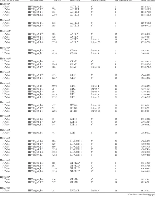
[image:2.585.40.547.78.297.2]Virus detection from RNA-Seq.Our approach (39) started by com-putationally subtracting human sequences, followed by generating a set of nonhuman sequences (e.g., viruses) on RNA-Seq. Once raw PE reads from RNA-Seq were aligned to the human genome reference, any read with more than half a read length mapped to the human reference genome is removed along with its paired mate in this subtraction step. Thus, a set of nonhuman sequences was generated after human sequence subtrac-tion. In the second step, our algorithm determined whether the nonhu-man sequences match any known viral sequences by searching a compre-hensive database that includes all known viral sequences (Genome TABLE 1Neoplasms in The Cancer Genome Atlas Database surveyed for tumor-associated DNA viruses
Tumor type
No. of samples analyzed
No. of reads per sample
Freeze date (mo/day/yr)
Min Median Max
Breast carcinoma 750 61000,388 109861,408 217446,562 8/2/2012
Clear cell renal cell carcinoma 460 30478,126 110937,134 242361,532 8/3/2012
Ovarian serous cystadenocarcinoma 419 102128,078 197051,552 452149,776 8/3/2012
Uterine corpus endometrioid carcinomaa 254 9988,571 31894,092 38324,550 8/3/2012
Head-and-neck squamous cell carcinoma 239 78123,066 152296,210 243733,050 8/2/2012
Lung adenocarcinoma 225 52466,630 103258,332 270583,684 8/3/2012
Lung squamous cell carcinoma 219 54253,346 127784,296 191438,728 8/2/2012
Cutaneous melanoma 214 91516,350 158684,346 269997,828 8/3/2012
Acute myeloid leukemia 179 105463,130 147722,794 190844,514 5/18/2012
Glioblastoma 168 78576,316 128354,626 240986,684 8/3/2012
Thyroid carcinoma 157 96982,636 159965,288 310309,740 8/2/2012
Colon adenocarcinomaa 138 9923,980 26751,110 37682,149 8/2/2012
Gastric adenocarcinoma 71 114694,894 184402,162 237944,836 8/2/2012
Rectal adenocarcinomaa 66 19527,419 27027,473 33162,004 8/2/2012
Prostate adenocarcinoma 53 65688,652 132183,938 214386,560 7/31/2012
Papillary renal cell carcinoma 47 91281,498 168971,506 243228,666 7/24/2012
Lower-grade glioma 47 120564,192 160524,446 205076,246 8/2/2012
Hepatocellular carcinoma 69 68607,776 140488,184 188335,198 8/3/2012
aNon-paired-end data in TCGA database.
on November 7, 2019 by guest
http://jvi.asm.org/
Information Broker for Viruses [GIB-V];http://gib-v.genes.nig.ac.jp/) and quantified virus representation by a measure of the virus genome coverage (or overall count of mapped reads) to determine the existence of viruses in human samples. The algorithm subsequently excluded non-transcribed viral genome elements to eliminate/reduce the potential of nonsense reads or inclusion of nontranscribed viral genomic elements. The expression level of each viral transcript was measured by the normal-ized depth of coverage within each viral transcript. The cutoff of viral gene expression detection was empirically determined by profiling the distri-bution of viral gene expression levels across multiple cancer-associated viruses (e.g., HPV16, HPV33, and EBV) and multiple patient samples. Any viral expression level below the cutoff was treated as no expression.
Identification of virus integration sites.The genomes of viruses with expression levels detected in the previous steps were concatenated into a single genome, named chrVirus, with related annotation of each virus in refFlat format. A new hybrid reference genome, named hg19Virus, was built by combining hg19 and chrVirus. All PE reads without computa-tional subtraction were again mapped to this reference (hg19Virus). If the PE reads were uniquely mapped with one end to hg19 and the other to chrVirus, the read pair was reported as a discordant read pair. All discor-dant reads were annotated by using the genes and viruses defined in the
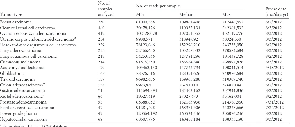
curated refFlat file. Our algorithm subsequently employed BLAT (27) to align the discordant reads against the hybrid reference genome (hg19Virus) and discards any discordant read pair if both discordant reads are aligned to the same gene or virus. It then clustered the remaining discordant read pairs that support the same integration (fusion) event (e.g., HBV-MLL4) and selected them as fusion candidates. A dynamic clustering procedure was implemented to accurately determine the exact fusion junction between a human gene and a virus. Specifically, the boundary for each discordant read cluster of a candidate fusion was esti-mated on the basis of discordant read mapping locations and orientations with fragment length distribution as a constraint of cluster size, which was measured by using the reads’ genomic locations, excluding intronic sizes if mapped reads were located across adjacent exons in a candidate fusion. For the forward-aligned discordant reads in a fusion candidate, the clus-tering process started with the right-most read, and the genomic coordi-nate for the right-most read was used to define thein silicofusion junction, excluding outliers within the discordant read cluster. In order to remove outliers within a cluster, our algorithm implemented the robust “extreme studentized deviate” multiple-outlier procedure. If the outliers came from the right end of the cluster, the outliers were removed and the clustering process restarts with a newin silicofusion junction. If the outliers come FIG 1Visualization of HPV16 integration breakpoints in the HPV16 genome. The frequency of integration breakpoints at different loci in the HPV16 genome is shown as a blue histogram. The scale bar indicates the number of tumors. The locations of the genes encoding HPV16 E6 (red), E7 (dark green), E1 (orange), E2 (purple), E4 (green), E5 (gray), L2 (light orange), and L1 (yellow) proteins are shown. Genomic positions are numbered.
on November 7, 2019 by guest
http://jvi.asm.org/
[image:3.585.97.490.68.461.2]from the left-most end of the cluster, the cluster size was reset with thein silicofusion junction intact by excluding the outlier reads. For the reverse-aligned discordant reads, the clustering process started with the left-most read, and the genomic coordinate for the left-most read was used to define thein silicofusion junction with the same outlier detection/removal pro-cessing step. For either side of the candidate fusion partner (gene or virus), this clustering process was performed independently. This dynamic clus-tering procedure accurately determined the exact fusion junction between a human gene and a virus. Meanwhile, anin silicosequence was generated using the consensus of reads within discordant read clusters for each fu-sion candidate to help the PCR primer design, which facilitates quick PCR validation.
RESULTS
RNA sequencing data from 3,775 malignant neoplasms in the
TCGA database were interrogated for the presence and, as
appli-cable, integration of site of viral transcripts. The overall median
read number per sample was 132,183,938 (
Table 1
). None of the
virus-positive cancers in this analysis harbored more than one
type/strain of virus.
Human papillomavirus detection in malignant cancers.
We
analyzed 239 squamous cell carcinomas of the head-and-neck
re-gion (HNSCC) available in the TCGA database. We detected HPV
transcripts in 36 tumors as the following: 30 tumors with HPV16,
5 tumors with HPV33, and 1 tumor with HPV35. We also detected
EBV in 1 tumor (discussed below). Among all cases with HPV
transcripts, E7 was expressed in 22, E6 in 20, E1 in 17, and E4 in 8
tumors. In 24 tumors, HPV transcripts encoding key viral
pro-teins/oncoproteins were integrated in the tumor genome, with the
majority in association with known genes (
Fig. 1
and
2
). Tumors
with HPV integration harbored the following types: 19 HPV16
⫹, 4
HPV33
⫹, and 1 HPV35
⫹(
Table 2
). Of the tumors with HPV
integration, 18 have both E6 and E7 integration sites, 4 have only
E7 integration sites, and 2 have only E6 integration sites.
The clinicopathologic characteristics of HNSCC patients with
available data (
n
⫽
35) are summarized in
Tables 3
and
4
. HPV
status detected by our method correlated with perfect sensitivity
and specificity with known clinicopathologic variables and with
established methods for HPV detection (colorimetric
in situ
hy-bridization and/or p16
ink4aexpression) (
28
). For this HNSCC
data set, the sensitivity for HPV16 detection was 100%, with 95%
confidence intervals (CI) of 67.6 to 100%, and specificity for
HPV16 detection was 100%, with 95% CI of 90.4 to 100%, as
reported previously (
39
).
We also detected HPV16 in two tumors of histologic types
rarely associated with this virus. From a group of 219 lung
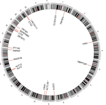
squa-FIG 2Integration sites of HPV16 in head-and-neck squamous cell carcinoma tumors in the human genome (hg19). Chromosome numbers are shown (*, detected in two cases).
on November 7, 2019 by guest
http://jvi.asm.org/
[image:4.585.116.475.67.422.2]TABLE 2Head-and-neck squamous cell carcinoma tumors with integrated HPV transcripts
TCGA ID and virus Viral transcript Virus site Host gene Integration site Chromosome no. Gene position
H736801A
HPV16 HPV16gp1_E6 70 ACTL7B 3= 9 111259747 HPV16 HPV16gp2_E7 559 ACTL7B 3= 9 111241132 HPV16 HPV16gp3_E1 903 ACTL7B 3= 9 111237008 HPV16 HPV16gp4_E2 2769 ACTL7B 3= 9 111541176
H740601A
HPV16 HPV16gp1_E6 106 ACTL7B 3= 9 110495875 HPV16 HPV16gp2_E7 565 ACTL7B 3= 9 110497920
H648101Aa
HPV16 HPV16gp2_E7 812 ANPEP 5= 15 90358960 HPV16 HPV16gp3_E1 978 ANPEP 5= 15 90359954 HPV16 HPV16gp2_E7 649 ANPEP Intron 1 15 90350002 HPV16 HPV16gp5_E4 3355 ANPEP Intron 20 15 90333738
H544301A
HPV16 HPV16gp2_E7 561 CD274 Intron 4 9 5462885 HPV16 HPV16gp8_L1 6758 CD274 Intron 4 9 5464598
H648701Aa
HPV16 HPV16gp1_E6 43 CRAT 3= 9 131856423 HPV16 HPV16gp3_E1 2249 CRAT 3= 9 131856542 HPV16 HPV16gp2_E7 670 CRAT Intron 14 9 131857745
H775401A
HPV16 HPV16gp2_E7 663 CTIF 5= 18 45664322 HPV16 HPV16gp3_E1 1227 CTIF 5= 18 45664232
H696101A
HPV16 HPV16gp8_L1 5573 ETS2 Intron 4 21 40186953 HPV16 HPV16gp1_E6 75 ETS2 Intron 7 21 40191504 HPV16 HPV16gp2_E7 561 ETS2 Intron 7 21 40191445 HPV16 HPV16gp5_E4 3385 ETS2 Intron 8 21 40193588 HPV16 HPV16gp3_E1 2352 ETS2 Intron 7 21 40190767
H647201A
HPV16 HPV16gp1_E6 407 IFT140 Intron 18 16 1612024 HPV16 HPV16gp2_E7 561 IFT140 Intron 18 16 1612023 HPV16 HPV16gp3_E1 2206 IFT140 Intron 18 16 1612004
H736901A
HPV16 HPV16gp1_E6 60 KLF12 3= 13 73910872 HPV16 HPV16gp2_E7 570 KLF12 3= 13 73952914 HPV16 HPV16gp3_E1 944 KLF12 3= 13 73910982
H474101A
HPV16 HPV16gp1_E6 447 KLF5 5= 13 73620072
H738501A
HPV16 HPV16gp1_E6 119 LINC00111 5= 21 43089331 HPV16 HPV16gp2_E7 620 LINC00111 5= 21 43089341 HPV16 HPV16gp3_E1 889 LINC00111 5= 21 43084796 HPV16 HPV16gp4_E2 3655 LINC00111 5= 21 43087464 HPV16 HPV16gp5_E4 3355 LINC00111 5= 21 43090560 HPV16 HPV16gp7_L2 4262 LINC00111 5= 21 43059265
H597101A
HPV16 HPV16gp1_E6 115 NBPF22P 5= 5 84616300 HPV16 HPV16gp2_E7 567 NBPF22P 5= 5 84624716 HPV16 HPV16gp3_E1 2056 NBPF22P 5= 5 84619865 HPV16 HPV16gp5_E4 3355 NBPF22P 5= 5 84629561
H544201A
HPV16 HPV16gp1_E6 106 OR1M1 5= 19 9113164 HPV16 HPV16gp2_E7 567 OR1M1 5= 19 9113071
H407701B
HPV16 HPV16gp1_E6 73 RAD51B Intron 7 14 68758607
(Continued on following page)
on November 7, 2019 by guest
http://jvi.asm.org/
mous cell carcinoma tumors, one case harbored HPV16, where
E1, E6, and E7 transcripts were highly expressed and integrated in
NROB1
(also known as
DAX1
), a gene involved in steroidogenesis
and cell cycle regulation. In this case, no E2, E4, E5, L1, or L2
transcripts were detected. Additionally, 1 of 253 endometrial
car-cinoma tumors harbored HPV16, where E1, E2, E4, E5, E6, and E7
transcripts were highly expressed, and no L1 or L2 transcripts were
detected. The integration site of the latter could not be
deter-mined, because the TCGA endometrial carcinoma RNA-Seq data
were not paired end.
Epstein-Barr virus detection in malignant cancers.
We
ana-lyzed 71 cases of gastric carcinoma in the TCGA database and
detected EBV transcripts in 4 (5.6%) tumors. Of the four tumors
with unequivocal EBV association, all harbored transcripts
encod-TABLE 2(Continued)
TCGA ID and virus Viral transcript Virus site Host gene Integration site Chromosome no. Gene position
HPV16 HPV16gp3_E1 858 RAD51B Intron 7 14 68699906 HPV16 HPV16gp4_E2 3625 RAD51B Intron 7 14 68741641 HPV16 HPV16gp6_E5 3855 RAD51B Intron 7 14 68683627 HPV16 HPV16gp2_E7 565 RAD51B Intron 7 14 68704019 HPV16 HPV16gp5_E4 3434 RAD51B Intron 7 14 68741498
H532301A
HPV16 HPV16gp3_E1 903 SERPINB3 3= 18 61317170 HPV16 HPV16gp8_L1 5701 SERPINB3 3= 18 61317182 HPV16 HPV16gp1_E6 130 SERPINB3 Exon 8 18 61322624 HPV16 HPV16gp2_E7 561 SERPINB3 Exon 8 18 61322588
H555901A
HPV16 HPV16gp5_E4 3355 TCTEX1D1 Intron 1 1 67220366 HPV16 HPV16gp2_E7 568 TCTEX1D1 Intron 2 1 67236076 HPV16 HPV16gp4_E2 3717 TCTEX1D1 Intron 2 1 67229043 HPV16 HPV16gp6_E5 3853 TCTEX1D1 Intron 2 1 67229161 HPV16 HPV16gp3_E1 2567 TCTEX1D1 Intron 2 1 67229895 HPV16 HPV16gp1_E6 124 TCTEX1D1 Intron 3 1 67239538
H647301A
HPV16 HPV16gp1_E6 106 TNFSF4 5UTR 1 173176246 HPV16 HV16gp2_E7 577 TNFSF4 5UTR 1 173176043
H474101A
HPV16 HPV16gp1_E6 117 TP63 5= 3 189226255 HPV16 HPV16gp2_E7 578 TP63 5= 3 189239853
H775401A
HPV16 HPV16gp1_E6 139 ZBTB7C Intron 1 18 45567460 HPV16 HPV16gp2_E7 567 ZBTB7C Intron 1 18 45567416
H710001A
HPV33 HPV33gp1_E6 509 EPSTI1 Intron 5 13 43537483 HPV33 HPV33gp5_E4 3356 EPSTI1 Intron 6 13 43528079 HPV33 HPV33gp8_L1 5597 EPSTI1 Intron 6 13 43528256
H647101A
HPV33 HPV33gp1_E6 93 TEAD1 Intron 5 11 12897532 HPV33 HPV33gp2_E7 559 TEAD1 Intron 5 11 12897531 HPV33 HPV33gp3_E1 858 TEAD1 Intron 5 11 12897627
H783201A
HPV33 HPV33gp1_E6 116 MIR802 5= 21 36470118 HPV33 HPV33gp2_E7 560 MIR802 5= 21 36470162 HPV33 HPV33gp3_E1 858 MIR802 5= 21 36470118
H710001A
HPV33 HPV33gp5_E4 3347 PHLDB2 Intron 1 3 111451446 HPV33 HPV33gp3_E1 1905 PHLDB2 Intron 1 3 111499049 HPV33 HPV33gp2_E7 671 PHLDB2 Intron 1 3 111496513 HPV33 HPV33gp3_E1 858 PLCXD2 Intron 1 3 111413679 HPV33 HPV33gp2_E7 747 PLCXD2 Intron 1 3 111431547
H759101A
HPV35 HPV35gp2_E7 698 ACCN1 Intron 1 17 32128921 HPV35 HPV35gp3_E1 863 ACCN1 Intron 1 17 32128931 HPV35 HPV35gp1_E6 128 ACCN1 Intron 1 17 32143974
aPositive for HPV DNA by colorimetricin situhybridization and/or for p16 overexpression by immunohistochemistry (data unavailable for the other cases).
on November 7, 2019 by guest
http://jvi.asm.org/
[image:6.585.42.541.82.617.2]ing A73, RPMS1, BARF0, BALF3, BALF4, BALF5, LF1, LF2, and
BILF1. One tumor additionally had significant levels of transcripts
encoding BNLF2a/b, LMP1, and LMP2A (
Table 5
). Of note,
un-like others who demonstrated EBNA1 expression
in vitro
in gastric
[image:7.585.40.545.79.634.2]carcinoma cell lines (
29
), we did not identify EBNA1 in any of the
gastric carcinoma tumor samples with EBV transcripts. This
might be due to biological variation between primary tumor
sam-ples and cell lines or low EBNA1 mRNA copy numbers. In the
TABLE 3Clinicopathologic characteristics of patients with head-and-neck squamous cell carcinomaa
Parameter
HPV positive HPV negative All HNSCC
Pvalueb
n % n % n %
Gender
Female 4 11.4 59 29.2 63 26.6
Male 31 88.6 143 70.8 174 73.4
All 35 100 202 100 237 100 0.04
Tumor site
Alveolar ridge 2 5.7 4 2 6 2.5
Base of tongue 5 14.3 4 2 9 3.8
Buccal mucosa 0 0 7 3.5 7 3
Floor of mouth 1 2.9 21 10.4 22 9.3
Hard palate 1 2.9 2 1 3 1.3
Hypopharynx 1 2.9 0 0 1 0.4
Larynx 1 2.9 70 34.8 71 30.1
Lip 0 0 1 0.5 1 0.4
Oral cavity 4 11.4 31 15.4 35 14.8
Oral tongue 3 8.6 56 27.9 59 25
Oropharynx 1 2.9 2 1 3 1.3
Tonsil 16 45.7 3 1.5 19 8.1
All 35 100 201 100 236 100 ⬍0.0001
Tumor stagec
I 1 3 12 6.1 13 5.7
II 5 15.2 46 23.5 51 22.3
III 5 15.2 40 20.4 45 19.6
IVA 22 66.7 92 46.9 114 49.8
IVB 0 0 4 2 4 1.8
IVC 0 0 2 1 2 0.9
All 33 100 196 100 229 100 0.56
Lymph node status
N0 12 40 77 38.5 89 38.7
N1 5 16.7 32 16 37 16.1
N2 1 3.3 9 4.5 10 4.3
N2a 1 3.3 4 2 5 2.2
N2b 9 30 38 19 47 20.4
N2c 2 6.7 27 13.5 29 12.6
N3 0 0 3 1.5 3 1.3
NX 0 0 10 5 10 4.3
All 30 100 200 100 230 100 0.73
Metastasis
M0 35 100 199 99 234 99.2
M1 0 0 2 1 2 0.8
All 35 100 201 100 236 100 1
Histologic grade
G1 1 2.9 10 5 11 4.6
G2 14 40 138 68.3 152 64.1
G3 17 48.6 49 24.3 66 27.9
G4 1 2.9 0 0 1 0.4
GX 2 5.7 5 2.5 7 3
All 35 100 202 100 237 100 0.0022
vHPVin situhybridization
Negative 0 0 35 100 35 85.4
Positive 6 100 0 0 6 14.6
All 6 100 35 100 41 100 ⬍0.0001
HPV status by p16 IHC
Negative 0 0 36 100 36 83.7
Positive 7 100 0 0 7 16.3
All 7 100 36 100 43 100 ⬍0.0001
aNominal variables grouped by HPV status (integrated and nonintegrated) detected by RNA-Seq data. IHC, immunohistochemistry; IQR, interquartile range.
bDetermined by Fischer’s exact test.
cAccording to AJCC staging criteria.
on November 7, 2019 by guest
http://jvi.asm.org/
single head-and-neck squamous cell carcinoma tumor associated
with EBV, the most abundant transcripts included those that
en-code BARF1/2, BdRF1, BMRF2, BLF1, BNLF2b, BBLF1, BMRF1,
BLRF2, and BNLF2a. None of the tumors analyzed in this study
had evidence of EBV integration into the host genome.
Hepatitis B virus detection in malignant cancers.
We
ana-lyzed 69 HCC tumors available in the TCGA database and
de-tected HBV transcripts in 16 (23%) tumors. Eight of these patients
had serologic evidence of HBV infection, and one patient was
HBV (and hepatitis C virus) seronegative. Serologic data on the
remaining patients were either negative or not available. Virus
integration was identified in 15 (94%) of these tumors. Our data
demonstrate frequent HBV integration within previously
identi-fied genes, namely,
TERT
(5 tumors) and
MLL4
(3 tumors),
sug-gesting that these sites are particularly susceptible to HBV
inser-tion. None of the tumors had both
MLL4
and
TERT
involvement.
Other insertion sites were restricted to single cases (
Table 6
). Of
the tumors with HBV integration, 11 have X protein integration
sites, 8 have S protein integration sites, 6 have core/E antigen
in-tegration sites, 4 have pre-S protein inin-tegration sites, 4 have
poly-merase 1 integration sites, and 3 have polypoly-merase 2 integration
sites. Integration of two or more HBV genes was detected in 8
tumors, whereas in the remaining 7 tumors integration of only
one HBV gene was detected. Interestingly, the latter group
in-cluded the 3 tumors with
MLL4
involvement and 2 with
TERT
involvement.
We additionally identified HBV transcripts in one case among
460 clear-cell renal cell carcinoma tumors analyzed. In this tumor,
as well as in one HCC tumor, we detected HBV S protein
tran-scripts integrated into
GLI2
, generally considered a marker of
ac-tivation of the sonic hedgehog signaling pathway, which has been
shown to play a role in aggressive types of renal cell carcinoma
(
30–32
) and in HCC (
33
,
34
).
Malignant cancers without detectable DNA viruses.
After
confirming the accuracy of our tool (
39
) in malignancies that are
known to be associated with DNA viruses, we turned our attention
to screening all remaining TCGA tumors on which RNA-Seq data
were available. Our analysis showed no evidence of transcribed
viral elements in any of the following neoplasms: acute myeloid
leukemia, breast carcinoma (ductal and lobular), colonic and
rectal adenocarcinoma, lung adenocarcinoma, prostate
adenocar-cinoma, cutaneous melanoma, ovarian serous
cystadenocarci-noma, papillary renal cell carcicystadenocarci-noma, thyroid carcicystadenocarci-noma,
lower-grade glioma, and glioblastoma multiforme.
DISCUSSION
The ability to identify viral integration points within the host
ge-nome using RNA-Seq provides a useful tool to study DNA viruses
in cancer. For instance, our findings are consistent with other
reports that have demonstrated preferential HBV insertion into
MLL4
and
TERT
in HCC (
18
,
19
,
35
). Although such a finding
might result from an inherent susceptibility for insertional
mu-tagenesis at these loci, the fact that these two genes are more
re-currently involved in HCC might reflect a selection bias, whereby
mutation of
MLL4
or
TERT
confers a selective advantage that
leads to tumor development. Support for the latter possibility is
suggested by our data indicating that among 7 HCC tumors with
HBV insertion at a single gene locus, 5 tumors (71%) had
involve-ment of
MLL4
or
TERT
(3 with
MLL4
involvement, 2 with
TERT
involvement). This suggests that the threshold for malignant
transformation is lower when these genes, particularly
MLL4
, are
disrupted through HBV insertional mutagenesis.
[image:8.585.40.546.77.199.2]The interplay between viral infections and the miRNA
ma-chinery is the subject of intensive investigation. In addition to
the modulating effect of host miRNAs on viral gene expression,
mounting evidence suggests the existence of viral mechanisms
leading to reprogramming of host miRNA machinery (
36
). In a
TABLE 4Continuous variables of clinicopathologic characteristics of patients with head-and-neck squamous cell carcinoma
Parameter n
Value for HPV status and smoking historya
Pvaluec
Min Mean Max IQR No. NAb
HPV status
Positive 35 35 58 82 13.5 0
Negative 202 19 62 87 14.5 0
All 237 19 62 87 15 0 0.05
Smoking history
Positive 18 0 30.5 102.5 28.8 17
Negative 114 0.7 48 180 37.1 88
All 132 0 45 180 32.2 105 0.0084
a
For HPV status, minimum, mean, and maximum values are given as age in years. For smoking history, minimum, mean, and maximum values are given as pack-years. bNumber of cases for which no data were available.
c
Determined by Mann-Whitney test.
TABLE 5EBV transcripts detected in four gastric carcinoma tumors
Gene symbol
Expression level for TCGA ID no.a:
S425301A S427101A S429801A S437601A
EBV_RPMS1 66 73 2 65
EBV_BILF1 45 48 1 36
EBV_LF1 57 62 2 47
EBV_LF2 105 118 4 88
EBV_BALF5 100 112 4 101
EBV_A73 206 229 7 203
EBV_BALF3 162 174 5 157
EBV_BALF4 160 156 4 138
EBV_BARF0 197 244 8 222
EBV_LMP-2A-1 10 0 0 0
EBV_LMP-1 22 0 0 1
EBV_BNLF2a 98 0 0 1
EBV_BNLF2b 106 0 0 1
a
Data refer to the expression level of viral transcripts estimated by the normalized depth of coverage within each viral transcript.
on November 7, 2019 by guest
http://jvi.asm.org/
[image:8.585.41.286.543.706.2]recent study by Wang and colleagues, the HBV X protein was
demonstrated to trigger selective miRNA downregulation (
37
).
Among the tumors with HBV insertion in this study, we
iden-tified two with involvement of miRNA genes (
MIR4457
and
MIR4424
). The function of these miRNAs in HCC is unknown.
Interestingly, both genes have X protein insertion sites,
sug-gesting that insertional mutagenesis comprises another
mech-anism by HBV which modulates the host miRNA repertoire.
The pathogenetic role of DNA viruses has been well established
in some cancers but remains unsettled in others. One of the main
[image:9.585.42.544.77.586.2]findings in our study is the absence of transcribed DNA viral
tran-scripts in some of the most prevalent human cancers, including
acute myeloid leukemia, cutaneous melanoma, low- and
high-grade gliomas of the brain, and adenocarcinomas of the breast,
colon and rectum, lung, prostate, ovary, kidney, and thyroid. In
squamous cell carcinoma of the head and neck, our analysis
achieved 100% sensitivity and specificity compared to established
methods (
28
) for HPV detection. Furthermore, the vast majority
of positive results across all cancers assessed were in line with
expected outcomes (e.g., HPV in squamous cell carcinoma of the
TABLE 6Hepatocellular carcinoma tumors with integrated HBV transcripts
TCGA ID Viral transcript Virus site Host gene Integration site Chromosome no. Gene position
L525801A HBVgp3_X protein 1742 MLL4 Intron 5 19 36214027
L526201A HBVgp1_polymerase-1 1245 FOXI2 5= 10 129391432
L526201A HBVgp3_X protein 1419 FOXI2 5= 10 129391427
L526401A HBVgp2_S protein 282 ITGAD 5= 16 31402241
L526401A HBVgp4_core/E antigen 1991 ITGAD Intron 5 16 31413434
L526401A HBVgp4_precore/core protein 1883 ITGAD Intron 5 16 31413431
L526401A HBVgp3_X protein 1748 TEAD1 Intron 2 11 12711205
L526401A HBVgp1_polymerase-1 1359 TECRL 3= 4 63651241
L526401A HBVgp2_S protein 372 TECRL 3= 4 63647552
L526401A HBVgp3_X protein 1373 TECRL 3= 4 63651170
LA10W01A HBVgp2_S protein 211 CLDN10 Intron 3 13 96216582
LA10W01A HBVgp2_pre-S1/pre-S2/S-2 2943 CLDN10 Intron 3 13 96229482
LA10W01A HBVgp2_pre-S2/S-1 42 CLDN10 Intron 3 13 96216538
LA10W01A HBVgp4_core/E antigen 2290 CLDN10 Intron 3 13 96229477
LA10W01A HBVgp3_X protein 1540 DPY19L2P1 3= 7 35111018
LA10W01A HBVgp2_S protein 341 KCNH1 Intron 7 1 210977324
LA10W01A HBVgp3_X protein 1800 KCNH1 Intron 7 1 211058920
LA11601A HBVgp2_S protein 337 C19orf55 Intron 6 19 36255648
LA11601A HBVgp2_S protein 420 MLL4 Intron 5 19 36213891
LA11901A HBVgp4_core/E antigen 2097 MLL4 Exon 3 19 36212301
LA1EA01A HBVgp1_polymerase-2 2403 GLI2 Intron 1 2 121585830
LA1EA01A HBVgp2_S protein 412 GLI2 Intron 1 2 121585623
LA1EA01A HBVgp2_pre-S2/S-1 23 GLI2 Intron 1 2 121585885
LA1EA01A HBVgp4_core/E antigen 1966 GLI2 Intron 1 2 121585602
LA1EA01A HBVgp2_pre-S2/S-1 25 TERT Intron 2 5 1291950
LA1EA01A HBVgp3_X protein 1675 VRK2 5= 2 57459913
LA1EI01A HBVgp2_S protein 386 FN1 Intron 12 2 216279398
LA1EI01A HBVgp4_core/E antigen 2006 GALNTL6 Intron 2 4 173136585
LA1EL01A HBVgp3_X protein 1659 LOC282980 3= 10 1863160
LA1EL01A HBVgp3_X protein 1684 NCOR2 Intron 12 12 124912988
LA1HT01A HBVgp2_S protein 406 TERT Intron 1 5 1294653
LA25U01A HBVgp1_polymerase-1 1369 MIR4457 3= 5 1295462
LA25U01A HBVgp3_X protein 1373 MIR4457 3= 5 1295516
LA25U01A HBVgp2_S protein 433 OBFC2A 5= 2 192364684
LA25U01A HBVgp3_X protein 1730 OBFC2A 5= 2 192367406
LA25U01A HBVgp3_X protein 1569 TERT Intron 1 5 1294676
LA25Z01A HBVgp3_X protein 1562 NDUFA4 3= 7 10697705
LA3CJ01A HBVgp3_X protein 1782 IL-6 5= 7 22735275
LA3CJ01A HBVgp3_X protein 1711 TERT Intron 1 5 1295037
LA3CK01A HBVgp1_polymerase-1 1330 DDX18 5= 2 118543485
LA3CK01A HBVgp3_X protein 1584 DDX18 5= 2 118543232
LA3CK01A HBVgp1_polymerase-2 2735 RNU6ATAC 3= 9 137028935
LA3CK01A HBVgp3_X protein 1635 RNU6ATAC 3= 9 137029164
LA3MB01A HBVgp1_polymerase-2 2391 TERT Intron 6 5 1272107
LA3MB01A HBVgp2_S protein 406 TERT Intron 6 5 1272167
LA3MB01A HBVgp3_X protein 1577 TERT Intron 6 5 1272106
LA3MB01A HBVgp4_core/E antigen 2035 TERT Intron 6 5 1272250
LA3MB01A HBVgp3_X protein 1503 CDK15 Intron 8 2 202709075
LA3MB01A HBVgp3_X protein 1660 MIR4424 5= 1 178593988
on November 7, 2019 by guest
http://jvi.asm.org/
head and neck, HBV in hepatocellular carcinoma, etc.). On the
basis of these results, the probability of false-negative results in
this analysis is believed to be low. Indeed, Bexfield and Kellam (
38
)
performed an elegant simulation to estimate the probability of
detecting viral sequences based on the viral genome transcript
sequence frequency and the number of sequence reads generated.
On the basis of their model, 10 million sequence reads gives a
⬎
99.99% probability of detecting at least one viral sequence if
every cell is infected and the viral genome transcript sequence
frequency is 0.0001%. However, 10 million sequence reads gives a
⬎
60% probability of detecting at least one viral sequence if only 1
in 10 cells is infected. For our TCGA cohort, the median number
of sequence reads is more than 100 million (
Table 1
). Thus, we
have a
⬎
99.9% probability of detecting at least one viral sequence
in the TCGA cohort even if only 1 in 10 cells is infected with
0.0001% viral genome transcript sequence frequency. However,
the probabilities of detecting viral sequence decrease to
approxi-mately 84% with 20 million reads. Accordingly, and unless
yet-unknown pathogenic DNA viruses are discovered in the future,
the yield of future searches for DNA viruses in these types of
can-cers is likely to be limited.
One of the limitations of this study is the lack of available
corresponding tissue samples for direct validation. For instance,
further evaluation of the single case of clear cell renal cell
carci-noma with HBV transcripts would have been warranted. In
addi-tion, it could be argued that RNA-Seq might not detect integration
events that are biologically deleterious but do not result in
expres-sion of viral transcripts. The latter point, however, does not seem
to be a common event (
13–15
,
18
,
19
,
26
). However, while further
validation of specific findings identified in this study is warranted,
the overall outcome of the analysis highlights the utility of
RNA-Seq in detecting tumor-associated DNA viruses and identifying
viral integration sites that may unravel novel mechanisms of
can-cer pathogenesis.s
ACKNOWLEDGMENTS
This work was supported in part by TCGA grant U24CA143883, National Center for Research Resources grant UL1TR000371 (F.M.-B., J.N.W., X.S.), and The University of Texas MD Anderson Cancer Center support grant P30 CA016672. None of the funding sources had any role in study design, data collection, data analysis, data interpretation, or writing of the manuscript.
J.D.K. and X.S. conceived and designed the study and prepared the manuscript. X.S. designed and developed the bioinformatics algorithm. X.S., Y.C., H.Y., J.Z., and E.J.T. performed sequencing data analysis and statistical data analysis. N.M.T., M.D.W., F.M.-B., and L.J.M. reviewed findings for specific cancer types and also wrote and/or edited the manu-script. J.N.W. reviewed bioinformatics design and results and also wrote and/or edited the manuscript.
None of the authors has conflicts of interest pertaining to this study to declare.
REFERENCES
1.Martin D, Gutkind JS.2008. Human tumor-associated viruses and new insights into the molecular mechanisms of cancer. Oncogene27(Suppl. 2):S31–S42.
2.Ng SB, Khoury JD.2009. Epstein-Barr virus in lymphoproliferative pro-cesses: an update for the diagnostic pathologist. Adv. Anat. Pathol.16:40 –55. 3. Poreba E, Broniarczyk JK, Gozdzicka-Jozefiak A. 2011. Epigenetic mechanisms in virus-induced tumorigenesis. Clin. Epigenet.2:233–247. 4.Boss IW, Plaisance KB, Renne R.2009. Role of virus-encoded
micro-RNAs in herpesvirus biology. Trends Microbiol.17:544 –553.
5.Moody CA, Laimins LA.2010. Human papillomavirus oncoproteins: pathways to transformation. Nat. Rev. Cancer10:550 –560.
6.Alazawi W, Pett M, Arch B, Scott L, Freeman T, Stanley MA, Coleman N.2002. Changes in cervical keratinocyte gene expression associated with integration of human papillomavirus 16. Cancer Res.62:6959 – 6965. 7.Pett MR, Herdman MT, Palmer RD, Yeo GS, Shivji MK, Stanley MA,
Coleman N.2006. Selection of cervical keratinocytes containing inte-grated HPV16 associates with episome loss and an endogenous antiviral response. Proc. Natl. Acad. Sci. U. S. A.103:3822–3827.
8.Dall KL, Scarpini CG, Roberts I, Winder DM, Stanley MA, Muralidhar B, Herdman MT, Pett MR, Coleman N.2008. Characterization of nat-urally occurring HPV16 integration sites isolated from cervical keratino-cytes under noncompetitive conditions. Cancer Res.68:8249 – 8259. 9.Wentzensen N, Vinokurova S, von Knebel Doeberitz M.2004.
System-atic review of genomic integration sites of human papillomavirus ge-nomes in epithelial dysplasia and invasive cancer of the female lower gen-ital tract. Cancer Res.64:3878 –3884.
10. Ziegert C, Wentzensen N, Vinokurova S, Kisseljov F, Einenkel J, Hoe-ckel M, von Knebel Doeberitz M.2003. A comprehensive analysis of HPV integration loci in anogenital lesions combining transcript and ge-nome-based amplification techniques. Oncogene22:3977–3984. 11. Smith PP, Friedman CL, Bryant EM, McDougall JK.1992. Viral
inte-gration and fragile sites in human papillomavirus-immortalized human keratinocyte cell lines. Genes Chromosomes Cancer5:150 –157. 12. Dandri M, Locarnini S.2012. New insight in the pathobiology of hepatitis
B virus infection. Gut61(Suppl. 1):i6 –i17.
13. Fujimoto A, Totoki Y, Abe T, Boroevich KA, Hosoda F, Nguyen HH, Aoki M, Hosono N, Kubo M, Miya F, Arai Y, Takahashi H, Shirakihara T, Nagasaki M, Shibuya T, Nakano K, Watanabe-Makino K, Tanaka H, Nakamura H, Kusuda J, Ojima H, Shimada K, Okusaka T, Ueno M, Shigekawa Y, Kawakami Y, Arihiro K, Ohdan H, Gotoh K, Ishikawa O, Ariizumi S, Yamamoto M, Yamada T, Chayama K, Kosuge T, Yamaue H, Kamatani N, Miyano S, Nakagama H, Nakamura Y, Tsunoda T, Shibata T, Nakagawa H.2012. Whole-genome sequencing of liver can-cers identifies etiological influences on mutation patterns and recurrent mutations in chromatin regulators. Nat. Genet.44:760 –764.
14. Sung WK, Zheng H, Li S, Chen R, Liu X, Li Y, Lee NP, Lee WH, Ariyaratne PN, Tennakoon C, Mulawadi FH, Wong KF, Liu AM, Poon RT, Fan ST, Chan KL, Gong Z, Hu Y, Lin Z, Wang G, Zhang Q, Barber TD, Chou WC, Aggarwal A, Hao K, Zhou W, Zhang C, Hardwick J, Buser C, Xu J, Kan Z, Dai H, Mao M, Reinhard C, Wang J, Luk JM. 2012. Genome-wide survey of recurrent HBV integration in hepatocellu-lar carcinoma. Nat. Genet.44:765–769.
15. Murakami Y, Saigo K, Takashima H, Minami M, Okanoue T, Brechot C, Paterlini-Brechot P.2005. Large scaled analysis of hepatitis B virus (HBV) DNA integration in HBV related hepatocellular carcinomas. Gut 54:1162–1168.
16. Dejean A, Bougueleret L, Grzeschik KH, Tiollais P.1986. Hepatitis B virus DNA integration in a sequence homologous to v-erb-A and steroid receptor genes in a hepatocellular carcinoma. Nature322:70 –72. 17. Wang J, Chenivesse X, Henglein B, Brechot C.1990. Hepatitis B virus
integration in a cyclin A gene in a hepatocellular carcinoma. Nature343: 555–557.
18. Paterlini-Brechot P, Saigo K, Murakami Y, Chami M, Gozuacik D, Mugnier C, Lagorce D, Brechot C.2003. Hepatitis B virus-related inser-tional mutagenesis occurs frequently in human liver cancers and recur-rently targets human telomerase gene. Oncogene22:3911–3916. 19. Ferber MJ, Montoya DP, Yu C, Aderca I, McGee A, Thorland EC,
Nagorney DM, Gostout BS, Burgart LJ, Boix L, Bruix J, McMahon BJ, Cheung TH, Chung TK, Wong YF, Smith DI, Roberts LR. 2003. Integrations of the hepatitis B virus (HBV) and human papillomavirus (HPV) into the human telomerase reverse transcriptase (hTERT) gene in liver and cervical cancers. Oncogene22:3813–3820.
20. Deyrup AT.2008. Epstein-Barr virus-associated epithelial and mesenchy-mal neoplasms. Hum. Pathol.39:473– 483.
21. Thompson MP, Kurzrock R.2004. Epstein-Barr virus and cancer. Clin. Cancer Res.10:803– 821.
22. Lee JH, Kim SH, Han SH, An JS, Lee ES, Kim YS.2009. Clinicopath-ological and molecular characteristics of Epstein-Barr virus-associated gastric carcinoma: a meta-analysis. J. Gastroenterol. Hepatol.24:354 –365. 23. Takada K.2000. Epstein-Barr virus and gastric carcinoma. Mol. Pathol.
53:255–261.
24. Zhao J, Liang Q, Cheung KF, Kang W, Lung RW, Tong JH, To KF,
on November 7, 2019 by guest
http://jvi.asm.org/
Sung JJ, Yu J.2012. Genome-wide identification of Epstein-Barr virus-driven promoter methylation profiles of human genes in gastric cancer cells. Cancer119:304 –312.
25. Marquitz AR, Mathur A, Shair KH, Raab-Traub N.2012. Infection of EpsteBarr virus in a gastric carcinoma cell line induces anchorage in-dependence and global changes in gene expression. Proc. Natl. Acad. Sci. U. S. A.109:9593–9598.
26. Rozenblatt-Rosen O, Deo RC, Padi M, Adelmant G, Calderwood MA, Rolland T, Grace M, Dricot A, Askenazi M, Tavares M, Pevzner SJ, Abderazzaq F, Byrdsong D, Carvunis AR, Chen AA, Cheng J, Correll M, Duarte M, Fan C, Feltkamp MC, Ficarro SB, Franchi R, Garg BK, Gulbahce N, Hao T, Holthaus AM, James R, Korkhin A, Litovchick L, Mar JC, Pak TR, Rabello S, Rubio R, Shen Y, Singh S, Spangle JM, Tasan M, Wanamaker S, Webber JT, Roecklein-Canfield J, Johannsen E, Barabasi AL, Beroukhim R, Kieff E, Cusick ME, Hill DE, Munger K, Marto JA, Quackenbush J, Roth FP, DeCaprio JA, Vidal M. 2012. Interpreting cancer genomes using systematic host network perturbations by tumour virus proteins. Nature487:491– 495.
27. Kent WJ.2002. BLAT–the BLAST-like alignment tool. Genome Res.12: 656 – 664.
28. Liang C, Marsit CJ, McClean MD, Nelson HH, Christensen BC, Had-dad RI, Clark JR, Wein RO, Grillone GA, Houseman EA, Halec G, Waterboer T, Pawlita M, Krane JF, Kelsey KT.2012. Biomarkers of HPV in head and neck squamous cell carcinoma. Cancer Res.72:5004 –5013. 29. Malik-Soni N, Frappier L.2012. Proteomic profiling of EBNA1-host
protein interactions in latent and lytic Epstein-Barr virus infections. J. Virol.86:6999 –7002.
30. Furler RL, Uittenbogaart CH.2012. GLI2 regulates TGF-beta1 in human CD4(⫹) T cells: implications in cancer and HIV pathogenesis. PLoS One 7:e40874. doi:10.1371/journal.pone.0040874.
31. Swartling FJ, Savov V, Persson AI, Chen J, Hackett CS, Northcott PA, Grimmer MR, Lau J, Chesler L, Perry A, Phillips JJ, Taylor MD,
Weiss WA.2012. Distinct neural stem cell populations give rise to disparate brain tumors in response to N-MYC. Cancer Cell21:601– 613.
32. Cao Y, Wang L, Nandy D, Zhang Y, Basu A, Radisky D, Mukhopadhyay D.2008. Neuropilin-1 upholds dedifferentiation and propagation pheno-types of renal cell carcinoma cells by activating Akt and sonic hedgehog axes. Cancer Res.68:8667– 8672.
33. Kim Y, Yoon JW, Xiao X, Dean NM, Monia BP, Marcusson EG.2007. Selective down-regulation of glioma-associated oncogene 2 inhibits the proliferation of hepatocellular carcinoma cells. Cancer Res.67:3583– 3593.
34. Wang Y, Han C, Lu L, Magliato S, Wu T.2013. Hedgehog signaling pathway regulates autophagy in human hepatocellular carcinoma cells. Hepatology [Epub ahead of print.] doi:10.1002/hep.26394.
35. Saigo K, Yoshida K, Ikeda R, Sakamoto Y, Murakami Y, Urashima T, Asano T, Kenmochi T, Inoue I.2008. Integration of hepatitis B virus DNA into the myeloid/lymphoid or mixed-lineage leukemia (MLL4) gene and rearrangements of MLL4 in human hepatocellular carcinoma. Hum. Mutat.29:703–708.
36. Skalsky RL, Cullen BR.2010. Viruses, microRNAs, and host interactions. Annu. Rev. Microbiol.64:123–141.
37. Wang Y, Jiang L, Ji X, Yang B, Zhang Y, Fu XD.2013. Hepatitis B viral RNA directly mediates down-regulation of the tumor suppressor microRNA miR-15a/miR-16-1 in hepatocytes. J. Biol. Chem [Epub ahead of print.] doi:10.1074/jbc.M113.458158.
38. Bexfield N, Kellam P.2011. Metagenomics and the molecular identifica-tion of novel viruses. Vet. J.190:191–198.
39. Chen Y, Yao H, Thompson EJ, Tannir NM, Weinstein JN, Su X.2013. VirusSeq: software to identify viruses and their integration sites using next-generation sequencing of human cancer tissue. Bioinformatics29: 226 –267.
on November 7, 2019 by guest
http://jvi.asm.org/