Graziele Pereira Oliveira,aJuliana Reis Cortines,bBetânia Paiva Drumond,aJônatas Santos Abrahãoa
aLaboratório de Vírus, Departamento de Microbiologia, Instituto de Ciências Biológicas, Universidade Federal
de Minas Gerais, Belo Horizonte, MG, Brazil
bInstituto de Microbiologia Paulo de Góes, Departamento de Virologia–Centro de Ciências da Saúde, Cidade
Universitária, Ilha do Fundão, Rio de Janeiro, RJ, Brazil
ABSTRACT The inclusion ofMimiviridae members in the putative monophyletic nu-cleocytoplasmic large DNA virus (NCLDV) group is based on genomic and phylog-enomic patterns. This shows that, along with other viral families, they share a set of genes known as core or “hallmark genes,” including the gene for the major capsid protein (MCP). Although previous studies have suggested that the maturation of mimivirus MCP transcripts is dependent on splicing, there is little information about the processing of this transcript in other mimivirus isolates. Here we report the char-acterization of a new mimivirus isolate, called Kroon virus (KV) mimivirus. Analysis of the structure, synteny, and phylogenetic relationships of the MCP genes in many mimivirus isolates revealed a remarkable variation at position and types of intronic and exonic regions, even for mimiviruses belonging to the same lineage. In addition, sequencing of KV and Acanthamoeba polyphaga mimivirus (APMV) MCP transcripts has shown that inside the family, even related giant viruses may present different ways to process the MCP mRNA. These results contribute to the understanding of the genetic organization and evolution of the MCP gene in mimiviruses.
IMPORTANCE Mimivirus isolates have been obtained by prospecting studies since 2003. Based on genomic and phylogenomic studies of conserved genes, these viruses have been clustered together with members of six other viral families. Although the ma-jor capsid protein (MCP) gene is an important member of the so-called “hallmark genes,” there is little information about the processing and structure of this gene in many mimi-virus isolates. In this work, we have analyzed the structure, synteny, and phylogenetic re-lationships of the MCP genes in many mimivirus isolates; these genes showed remark-able variation at position and types of intronic and exonic regions, even for mimiviruses belonging to the same lineage. These results contribute to the understanding of the ge-netic organization and evolution of the MCP gene in mimiviruses.
KEYWORDS KV, mimivirus,Megavirales, giant virus, isolation, capsid, splicing
T
he term virus is used to define a group of biological entities united under a set of generic and polythetic features (1). Among these features, the viruses can be linked together primarily by their dependence on the host biosynthetic machinery and also by their intracellular parasitic character. The fact that there is no a single gene uniting all of the viruses in a separated phylogenetic clade means that these organisms are considered a polyphyletic group, in which the different viral species emerged from distinct ancestors (2–4). However, there are some specific subgroups of viruses in which a set of genes, known as “core/hallmark genes,” are present in all the members, suggesting a probable common ancestry between them (4). The nucleocytoplasmicReceived10 October 2017Accepted30
October 2017
Accepted manuscript posted online8
November 2017
CitationBoratto PVM, Dornas FP, da Silva LCF,
Rodrigues RAL, Oliveira GP, Cortines JR, Drumond BP, Abrahão JS. 2018. Analyses of the Kroon virus major capsid gene and its transcript highlight a distinct pattern of gene evolution and splicing among mimiviruses. J Virol 92:e01782-17.https://doi.org/10.1128/JVI .01782-17.
EditorJulie K. Pfeiffer, University of Texas
Southwestern Medical Center
Copyright© 2018 American Society for
Microbiology.All Rights Reserved. Address correspondence to Jônatas Santos Abrahão, [email protected].
on November 6, 2019 by guest
large DNA viruses (NCLDVs) comprise one of those subgroups and include seven different viral families:Ascoviridae,Asfarviridae,Iridoviridae,Marseilleviridae,Mimiviridae, Phycodnaviridae, andPoxviridae(5, 6). These viral families share a set of core genes that are generally related to the replication of the viral genome and also to the composition of the viral particle structure, encoding the major capsid protein, a primase-helicase, a family B DNA polymerase, a packaging ATPase, and a transcription factor (5, 6).
Amoeba giant viruses belonging to theMimivirusgenus (Mimiviridaefamily) were first described in 2003, during a characterization study of a pathogenic amoeba-associated microorganism that was related to outbreaks of nosocomial pneumonia in a hospital in Bradford, England (7). Since then, a large diversity of related viruses has been isolated from many different places (8–13). As new giant viruses were being described, these organisms started to be phylogenetically classified based on the sequence of the gene related to DNA polymerase B. This gene is conserved between different viral members but presents sufficient differences to separate them in three different lineages (A, B, and C) (14). Other genes that form part of the group of hallmark genes have not been as well described in the literature when it comes to the phylogenetic relationships between mimiviruses. One example is that related to the synthesis of the major capsid protein (MCP). This gene plays a key role in the formation of the viral particle structure, representing the most abundant glycoprotein in Acan-thamoeba polyphaga mimivirus (APMV) (15). Interestingly, in a previous study, Azza and colleagues observed that the APMV MCP gene is structured in five different regions, with three exons separated by two intronic sites (16). The idea of a gene undergoing splicing in giant viruses has also been shown in another study, which confirmed that the transcript of the APMV capsid gene is composed of three exonic regions (17). As observed for DNA polymerase B, the MCP gene also presents high homology rates in other members of the genusMimivirus. However, there is not much information on how this gene is structured in differentMimivirusmembers.
Here we report the characterization of Kroon virus (KV), a new mimivirus belonging to lineage A. In this study, we analyzed the structure, synteny, and phylogenetic relationships of the major capsid gene (a hypothetically conserved gene shared by the Mimiviridae members) in different giant virus isolates, including the mimivirus KV. By using phylogenetic analyses and comparing the order of the intronic and exonic regions that compose the MCP genes of these giant viruses, we observed a distinct pattern of genetic organization and gene evolution involving KV and other mimiviruses, even among members of lineage A, to which KV belongs. These differences are also reflected by the way in which the MCP mRNA is spliced. By sequencing the mature transcripts of the MCP gene in both APMV (type species among mimiviruses and a member of lineage A) and KV, we have observed many differences in terms of content and organization, suggesting that this gene is processed in a different form in KV. Taken together, our results enabled us to highlight the MCP gene as a new marker that could also help in future studies involving the characterization of mimiviruses.
RESULTS
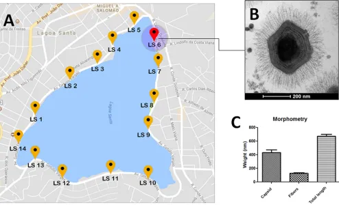
Virus isolation.In 2012, in an attempt to isolate novel strains of giant viruses, we decided to collect water samples from several urban lakes located in different cities of Brazil. A total of 14 water samples were collected at equidistant points around an urban lagoon located in the city of Lagoa Santa, Brazil (Fig. 1A). Inoculation of these samples in Acanthamoeba castellanii monolayers prompted us to isolate a new amoeba-associated microorganism, initially characterized by the observation of cytopathic effects (CPEs) in these cell cultures 3 to 4 days after infection. The CPEs were charac-teristic of the isolation of other giant viruses, including cell rounding, the absence of vacuoles, and cell lysis at later times of viral infection.
Virus characterization.With this new microorganism, we started to investigate any resemblance with other mimivirus isolates by using transmission electron microscopy (TEM), morphometric analysis, and analysis of genomic amplification by quantitative
on November 6, 2019 by guest
http://jvi.asm.org/
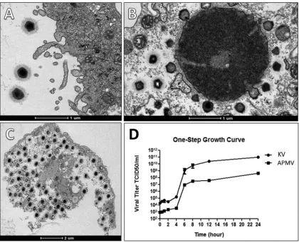
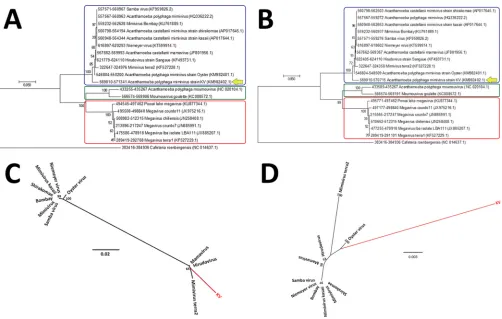
PCR (qPCR). First, we have linked our new isolated virus to members of the family Mimiviridaesince an amplification of the conserved RNA helicase gene was detected. Electron microscopy and morphometric assays enabled us to observe a great number of viral particles, with a pseudoicosahedral symmetry, fibrils surrounding the capsid, and a size that is comparable to that which has been observed in other studies involving mimivirus isolation (7, 8, 11–13). These mimivirus-like particles presented an average size of 673.43 nm in total, fibers of about 128.9 nm, and a capsid size of 433.2 nm, spread along the acanthamoebal cytoplasm (Fig. 1B and C). KV also presents several characteristics that resemble steps of mimivirus replication, as we could observe (i) the initial process of viral penetration, in which the viral particles start to be surrounded byAcanthamoebapseudopods; (ii) the presence of electron-dense regions in the amoebal cytoplasm called viral factories, where the giant virus particles start to be assembled; and (iii) the release of mature particles by cell lysis (Fig. 2A to C). By performing one-step growth curve assays, we observed that our isolate reached the plateau for viral production at around 8 to 10 h postinfection (the same as described for other mimiviruses), but with viral titers exceeding the production of APMV particles by about 2.5 logs (Fig. 2D). Finally, the phylogenetic characterization of our isolate, for both the RNA helicase and DNA polymerase genes, has clustered it with members of lineage A fromMimiviridae, which includesAcanthamoeba castellaniimamavirus, mimi-virus Terra2, Niemeyer mimi-virus, Samba mimi-virus, and oyster mimi-virus, among others (8, 9, 13, 18) (Fig. 3).
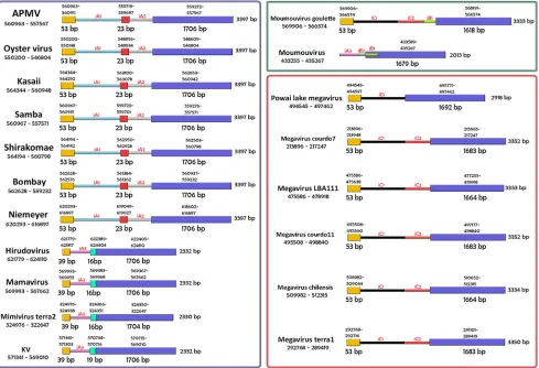
MCP gene layout.We then started to become more interested in a specific gene that is present among members that compose the family Mimiviridae. This gene encodes the major capsid protein (MCP). In APMV, it has been demonstrated that the MCP gene is responsible for producing a spliced mRNA, with a structure that is composed of three exons separated by two untranscribed regions (16, 17). In another
FIG 1KV mimivirus collection site, electron microscopy, and morphometric analysis. (A) A map of the Central Lagoon, from where the samples were collected. (B) An image of KV virus particle, observed from the transmission electron microscopy assays. (C) Sizes of different components of the KV particle: only the
capsid, only the fibers, and the particle as a whole.
on November 6, 2019 by guest
[image:3.585.47.541.73.373.2]giant virus, Acanthamoeba castellanii mamavirus, this gene was found to be lacking the two intronic regions but presented its own type of untranscribed sequence (10). As far as we know, no other studies have investigated how the capsid gene is arranged and phylogenetically related in members of the familyMimiviridae. After extensive analysis of the mimivirus MCP gene, we have seen that the MCP gene of these viruses can present different layouts. The first layout is slightly more complex than the others, being present in most members of lineage A. It is composed of three exons (called here e1, e2, and e3), separated by two intronic regions that are specific for members of this lineage (called here iA1 and iA2) (Fig. 4). Interestingly, not all the members from lineage A share this same structure. KV and three other analyzed members of lineage A (hirudovirus, mamavirus, and mimivirus Terra2) have a different layout for their MCP genes. In these viruses, the MCP gene has the described structure: (i) a fraction of the region corresponding to exon e1 in APMV, followed by (ii) a specific untranscribed region (called iA3 here) and, finally, by (iii) another specific region (called exon eX here), which is followed by a sequence homologue to exon e3 in APMV (Fig. 4). The third layout is most closely related to members of lineage C. In these viruses, the same transcribed regions composed of exons e1 and e3 are separated by two adjacent and specific intronic regions called iC1 and iC2 (the exception is Powai lake megavirus, which has the e1 and e3 regions separated only by iC1). As for the mimiviruses belonging to lineage B, the small number of isolate sequences deposited in the NCBI
FIG 2KV replication cycle. The major steps of the KV life cycle are highlighted by transmission electron microscopy and one-step growth curve assays. (A) Giant viral particles initiate the process of viral penetration by being phagocytosed by Acanthamoeba castellaniicells. (B) A later step, in which a KV viral factory, located at the cytoplasm of an infected amoebal host, starts to produce and bud several particles. (C) The last step of KV replication, in which the produced mature particles start to be released by lysis of the amoebal cell. (D) The general replication cycle of KV in comparison with that of APMV by one-step growth curve assays. After 24 h of infection, KV exceeds the production of APMV particles by about 2.5 logs.
on November 6, 2019 by guest
http://jvi.asm.org/
[image:4.585.42.462.70.411.2]database have imposed some difficulties to better analyze the group. From what we can see, two members from this lineage, Moumouvirus goulette and Moumouvirus moumouvirus, have considerably diverged when we consider how their MCP genes are structured, with the first one resembling the structure of mimiviruses from lineage C
FIG 3Phylogenetic analyses for many representatives of the differentMimiviridaelineages (A, B, and C), including KV, based on the RNA helicase (A) and DNA polymerase B amino acid sequences (B). For these analysis, we used MEGA 7 software (maximum-likelihood method and 1,000 bootstrap replicates). The differentMimiviridaelineages are highlighted using differently colored boxes (lineage A, blue; lineage B, green; lineage C, red), and KV is shown by a yellow arrow, being clustered with other mimivirus isolates belonging to lineage A. For each sequence, the gene identification numbers are indicated.
FIG 4General scheme showing the structural layout of the MCP genes belonging to several members of theMimiviridaefamily. The differentMimiviridae
lineages are highlighted using differently colored boxes (lineage A, blue; lineage B, green; lineage C, red). Representatives from these lineages seem to present distinct genetic structures for the gene. However, the general layout for this conserved gene may also suffer variations inside members of the same lineage, as observed for KV, mamavirus, hirudovirus, and mimivirus Terra2 compared to other members of lineage A.
on November 6, 2019 by guest
[image:5.585.42.545.71.221.2] [image:5.585.44.533.362.696.2]and the second resembling the layout observed for KV, with some differences related to the absence of exons e1 and eX, in addition to the inclusion of two short intronic regions (iB1 and iB2) (Fig. 4).
Major capsid gene phylogenetic analysis. Structural differences involving the capsid gene, as described above for several mimiviruses, may reflect the way in which evolution has affected the diverse relationships between members of the familyMimiviridaefor such a conserved gene. To get an idea of these relationships, we have constructed phylogenetic trees that covered either the entire sequence of the MCP gene or just the sequence related to exon e3 (the largest and most conserved of the exons described above). Interestingly, the constructed phyloge-netic trees almost reflected the profile which was previously described in the structural composition of MCP for the different giant viruses. When the capsid genes of these viruses were analyzed, considering either the whole gene or just the e3 fragment, we observed a clear separation that divided the viral members into three groups corresponding to lineages A, B, and C, as expected based on other hallmark genes (Fig. 5A and B). The construction of phylogenetic trees containing members of lineage A alone also matched the disposition of groups from trees that considered the three lineages together (Fig. 5C and D). This is relevant because in those trees, KV and the other three viruses that share a distinct pattern of MCP organization (hirudovirus, mamavirus, and mimivirus Terra2) are represented as the viruses that were most phylogenetically separated from the rest of the members in
FIG 5Phylogeny of the MCP genes of differentMimiviridaemembers. Here a phylogenetic analysis for many representatives of the differentMimiviridaelineages is shown (A, B, and C), including for the new Brazilian mimivirus isolate KV strain, based on the nucleotide sequences of the whole MCP gene or for just the sequences corresponding to exon 3. In panel A, the rectangular tree layout represents an analysis of the whole gene, considering members of the three different lineages, highlighted by differently colored boxes (lineage A, blue; lineage B, green; lineage C, red). In panel B, the same members are considered, but the phylogenetic relationships were based on the nucleotide sequences of just exon 3. In panel C, the image represents a radial tree, corresponding to analysis of the whole MCP gene for members of lineage A alone. Finally, in panel D, the phylogenetic relationships of members of lineage A are represented by another radial tree, but this time considering only sequences belonging to exon 3. For these analysis, we have used MEGA 7 software (maximum-likelihood method and 1,000 bootstrap replicates). For each sequence, the gene identification numbers are indicated.
on November 6, 2019 by guest
http://jvi.asm.org/
[image:6.585.43.545.68.387.2]lineage A (the exception is oyster virus, which is also located on a separate branch in trees corresponding to the analysis of exon 3) (Fig. 5D). The same occurred for Powai lake megavirus, occupying a different branch from the members belonging to lineage C (Fig. 5A and B). As forMoumouvirus goulette, even with this MCP gene structure resembling the organization of the capsid gene in viruses of lineage C, the phylogenetic trees have demonstrated that this virus has been grouped together with Moumouvirus moumouvirus, the other member belonging to lineage B of mimiviruses (Fig. 5A and B).
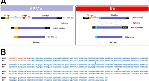
Analysis of the MCP gene transcript. As structural differences in the MCP gene have been shown to influence viral grouping in the phylogenetic trees described above, we chose to investigate whether these differences could also determine major changes along the sequence of the mature transcript related to this gene. For this test, we sequenced and analyzed the mRNAs encoding the MCPs belonging to APMV and KV. For APMV, the sequences corresponding to the three exons of MCP gene are conserved in the final transcript (Fig. 6). The gene, initially organized in a sequence of 3,496 nucleotides, is reduced after mRNA maturation in a sequence of 1,782 nucleo-tides, with a query cover of 51% and identity of 100% to the original gene. As observed in another study, the transcript is processed as a sequence that excludes the two intronic regions present originally in this gene. For KV, however, the virus seems to use as its mature mRNA only the region corresponding to the larger exon e3 and the adjacent exon X, excluding from the processed transcript both the region homologous to the exon e1 in APMV and the intronic region iA3 (Fig. 6). Curiously, analysis of the nucleotide sequence from exon eX has shown this region as being a small fragment from the 53-bp region belonging to exon e1 in the APMV-like viruses (data not shown). In addition, during the transcription and formation of the mature mRNA, the regions corresponding to the 5=untranslated region (UTR) and 3=UTR seem to be inverted from the observed in the original sequence. In KV, the gene is initially present as a sequence
FIG 6General scheme representing the process of splicing for the MCP gene in APMV and KV isolates. (A) The APMV capsid transcript suffers a process of splicing in which its processed form is composed by the sequences of exon 1, exon 2, and exon 3. KV, however, beyond its different MCP gene layout, uses as mature mRNA only the regions corresponding to exons eX and e3. aa, amino acids. (B) Alignment for the amino acid sequence produced by APMV and KV after translation of their respective MCP genes. The arrows indicate mismatched amino acids.
on November 6, 2019 by guest
[image:7.585.43.541.79.352.2]of 2,332 nucleotides, being reduced after mRNA maturation to a sequence of 1,716 nucleotides. The transcript has a query cover of 73% and an identity of 100% with the original gene.
DISCUSSION
Members of the familyMimiviridaehave been isolated worldwide from an increasing variety of samples (7, 8, 10, 12, 13). As demonstrated in other studies, environments where there are high concentrations of organic matter seem to work as hot spots, marking the presence of a great diversity of mimiviruses, which is probably related to the suitable habitat for their amoebal host to live (8, 19). In this study, we have managed to obtain another mimivirus isolate belonging to lineage A of the family Mimiviridae. To date, around a hundred mimivirus (and related viruses) isolates have been obtained by many different strategies that use the culture of amoebal cells, especially species that are included in theAcanthamoebagenus (12, 13, 20, 21). Despite the considerable number of isolated giant viruses, the vast majority belong to lineage A (19). This could be related to the fact that most of the platforms for mimivirus isolation employAcanthamoebaorganisms as the main type of cellular culture. The use of this narrow host spectrum as cell support for mimivirus isolation may be filtering away the discovery of other species of giant viruses that would potentially be present in the samples.
Transmission electron microscopy has shown us that KV has a viral cycle typical of other mimiviruses. Nevertheless, by undertaking a more detailed analysis of the cycle, one-step growth curve assays have demonstrated that this new isolate, under the same conditions, produces a much larger number of mature viral particles than APMV, highlighting that even phylogenetically related, different mimivirus isolates may pres-ent distinct replication profiles inAcanthamoeba.
By analyzing the structural composition of the major capsid protein gene in different members of the familyMimiviridae, we have reinforced a previous obser-vation which suggested that for some genes, there is a certain level of dichotomy separating mimiviruses inside lineage A (8). In this study, we have observed that for the capsid gene, KV, mamavirus, hirudovirus, and mimivirus Terra2 present a different genetic structure (considering the intronic and exonic regions of the gene) from homologous regions in other members of the same lineage. This creates inside lineage A an apparent separation with two existent groups, the one corresponding to the four viruses mentioned above and the second group composed of other viruses analyzed in this work: APMV, mimivirus Shirakomae, oyster virus, mimivirus Bombay, mimivirus Kasaii, Niemeyer virus, and Samba virus. This clear dichotomy gives us an important clue, enabling the MCP gene to be considered to follow distinct evolutionary pathways for each group of mimivirus isolates. This has also shown that these many giant viruses harbor different structural variations for such a conserved gene as the MCP gene.
In addition to the structural results, the dichotomic event was also supported when we considered phylogenetic trees associated with both the whole MCP gene and just for the region of exon 3 in the different mimiviruses. In those trees, the giant viruses composed by KV, mamavirus, hirudovirus, and mimivirus Terra2 have remained at positions separate from those of others belonging to lineage A. The fact that this pattern has been the same considering the two situations implies that if we take into account the phylogenetic trees made for the MCP gene, the use of the region associated with e3 alone gives us an interesting biomarker that also serves to separate mimiviruses in the current classification involving the three lineages. As stated before, interestingly, this dichotomic phenomenon has also already been seen for many other genes encoding different aminoacyl-tRNA synthetases, indicating that KV could repre-sent a divergent lineage A mimivirus (8).
Another interesting factor linked to the phylogenetic analysis that comprehends the MCP gene is related to the position of oyster virus in the evolutionary trees. As observed, even with a gene structure that differs from that observed for KV, mamavirus,
on November 6, 2019 by guest
http://jvi.asm.org/
relevant differences that are related not only to the structural part of the gene but also to how this element is processed after transcription.
MATERIALS AND METHODS
Collection of environmental samples.Water samples were collected at Central Lagoon, Lagoa Santa, Minas Gerais, Brazil (Fig. 1A). The lagoon has a total area of 1.31 km2, an approximate perimeter
of 6.5 km, and a maximum depth of between 6 and 7 m (22). It normally receives household wastewater. A total of 14 water samples, 1 ml each, were collected along all its extension, at equidistant places, and stored at 4°C until isolation procedures were performed. The samples were collected on the surface of the water, near aquatic plants and areas that indicated the formation of biofilms.
Amoebal culture procedures. Acanthamoeba castellanii(ATCC 30010) amoebas were grown in 7-cm2cell culture flasks (Nunc, USA), in peptone-yeast extract-glucose (PYG) medium supplemented with
7% fetal calf serum (FCS; Cultilab, Brazil), 25 mg/ml of amphotericin B (Fungizone; Cristalia, São Paulo, Brazil), 500 U/ml of penicillin, and 50 mg/ml of gentamicin (Schering-Plough, Brazil).
Enrichment protocol and isolation procedures.Samples were initially submitted to an enrichment protocol adapted from the works of Arslan and collaborators and Arbraho et al. (12, 23). Briefly, 500l of each sample was added to 4.5 ml of water-rice medium (40 grains of rice per liter of water) and kept in the dark at room temperature for 20 days. Following this incubation, an input of 5,000 amoebas was performed and the samples were incubated under the same conditions for more than 10 days. The samples were then filtered through 1.2-m membranes (to retain impurities) and then through 0.2-m membranes (to retain potential giant viruses isolated in these samples). The membranes were eluted in 500l of phosphate-buffered saline (PBS), and 100l of each eluate was inoculated into amoebal monolayers, contained in 96-well plates in a total series of three passages. Cytopathic effects (CPEs) were evaluated daily.
DNA extraction and PCR assays.Samples presenting CPEs in amoebal monolayers were tested for mimivirus by a qPCR targeting the conserved helicase gene (primers 5=-ACCTGATCCACATCCCATAA CTAAA-3=and 5=-GGCCTCATCAACAAATGGTTTCT-3=). The DNA was extracted by phenol-chloroform-isoamyl alcohol (PCI) and quantified by NanoDrop before the PCR assays. The qPCR was performed with a commercial mix (Power SYBR green; Applied Biosystems, USA), primers (4 mM each), and 1 ml of sample in a reaction mixture with a 10-ml final volume. All reactions were performed in a StepOne thermocycler with the following program: 95°C for 10 min and 40 cycles of 95°C for 15 s and 60°C for 15 s, followed by a dissociation step (specific midpoint temperature [Tm]⫽73°C).
The whole genome of KV was obtained and partially analyzed as described in another study (GenBank accession numberKM982402.1) (24).
Virus purification procedures.The KV particles were isolated and purified from infected amoebas as previously described (13). Briefly, after reaching confluence, the amoebas were infected with the new isolated giant virus and incubated at 37°C until the appearance of CPEs. Virus-rich supernatants from the infected amoebas were collected and filtered through a 0.8-m (Millipore, USA) filter to remove amoeba debris. The viruses were then purified using a sucrose cushion suspended in PBS, stored at⫺80°C, and titrated using 50% tissue culture infective dose (TCID50) methodology (25).
TEM analyses.For transmission electron microscopy (TEM),A. castellaniicells were infected with KV at a multiplicity of infection (MOI) of 0.01. Uninfected amoebas were used as controls. When⬃70% of the cells presented cytopathic effects, the amoeba monolayer was fixed with 2.5% glutaraldehyde (Merck, Germany) for 1 h at room temperature in 0.1 M sodium phosphate buffer. Amoebas were then postfixed with 2% osmium tetroxide and embedded in Epon resin, and ultrathin sections were examined under a transmission electron microscope (TECNAI G2-20; SuperTwin FEI; 120 kV) at the Center of Microscopy, Universidade Federal de Minas Gerais (UFMG), Brazil.
One-step growth curve.To evaluate the replication profile of the new giant virus isolate, six-well plates containing 1⫻107amoebas/well were infected with KV at an MOI of 10 and incubated at 32°C
for 0, 0.5, 1, 2, 4, 6, 8, 12, and 24 h. Infected cells were collected and centrifuged, and the pellet was used for titration in amoebas. The viral titer was determined using the TCID50method, calculated with the
Reed-Muench method (25).
Synteny analysis of the MCP gene.In this work, we have also searched for differences in the layout of the major capsid protein genes of several mimivirus samples. Given that APMV, as the type species of
on November 6, 2019 by guest
the familyMimiviridae, has the best-characterized genome among mimiviruses, its nucleotide sequence was used as a reference for MCP analyses. For each gene feature belonging to APMV (exons 1, 2, and 3 and introns 1 and 2), we searched for similar sequences in other viruses using the BLAST tool from the NCBI database (E value of 10⫺3). Sequences that were found to be homologous to APMV were separated,
and then the final gene layouts were constructed based on the position and orientation occupied by these different sequences. Specific MCP gene features found in some viruses were also searched for in other mimivirus samples. A scheme of the MCP gene was prepared, including isolates from all mimivirus lineages (Fig. 4).
MCP mRNA sequencing.Twenty-four-well plates containing 1⫻105amoebas/well were infected
with KV or APMV at an MOI of 5 and incubated at 32°C. After 30 min and 6 h, cells were collected and centrifuged, and the pellet was homogenized and used for total RNA extraction, reverse transcription, and conventional PCR for the MCP gene amplification. The amplicons were examined by electrophoresis in a 1% agarose gel with Tris-borate-EDTA (TBE) buffer and run at 150 V. The amplicon band was purified and sequenced in both orientations, in triplicate (3730 DNA analyzer; Thermo Fisher Scientific, Waltham, MA). The sequences were analyzed using the alignment tool available in the software MEGA 7.0.
Phylogenetic analyses.For the phylogenetic analyses, separated alignments for the major capsid protein, DNA polymerase B subunit, RNA helicase genes, and the exon 3 region of the major capsid gene were used. Strains of several viruses of the three mimivirus lineages (A, B, and C) were selected to assemble the data set. The predicted nucleotide sequences were obtained from GenBank and aligned using ClustalW in MEGA 7.0 software. Trees were constructed using maximum-likelihood method and bootstrap of 1,000.
ACKNOWLEDGMENTS
We thank colleagues from Gepvig, Laboratório de Vírus, Centro de Microsopia da UFMG, and Aix Marseille Université for their excellent support. Also, we thank Pro-reitoria de pesquisa da Universidade Federal de Minas Gerais, CAPES, FAPEMIG, CNPq.
J.S.A. is a CNPq researcher. We declare no conflict of interest.
REFERENCES
1. Lwoff A. 1957. The concept of virus. J Gen Microbiol 17:239 –253. 2. Koonin EV, Yutin N. 2010. Origin and evolution of eukaryotic large
nucleo-cytoplasmic DNA viruses. Intervirology 53:284 –292.https://doi .org/10.1159/000312913.
3. Moreira D, Lopez-Garcia P. 2009. Ten reasons to exclude viruses from the tree of life. Nat Rev Microbiol 7:306 –311.
4. Koonin EV, Senkevich TG, Dolja VV. 2006. The ancient virus world and evolution of cells. Biol Direct 1:29.https://doi.org/10.1186/1745-6150 -1-29.
5. Iyer LM, Aravind L, Koonin EV. 2001. Common origin of four diverse families of large eukaryotic DNA viruses. J Virol 75:11720 –11734.https:// doi.org/10.1128/JVI.75.23.11720-11734.2001.
6. Iyer LM, Balaji S, Koonin EV, Aravind L. 2006. Evolutionary genomics of nucleo-cytoplasmic large DNA viruses. Virus Res 117:156 –184.https:// doi.org/10.1016/j.virusres.2006.01.009.
7. La Scola B, Audic S, Robert C, Jungang L, de Lamballerie X, Drancourt M, Birtles R, Claverie JM, Raoult D. 2003. A giant virus in amoebae. Science 299:2033.https://doi.org/10.1126/science.1081867.
8. Boratto PV, Arantes TS, Silva LC, Assis FL, Kroon EG, La Scola B, Abrahao JS. 2015. Niemeyer virus: a new mimivirus group A isolate harboring a set of duplicated aminoacyl-tRNA synthetase genes. Front Microbiol 6:1256.https://doi.org/10.3389/fmicb.2015.01256.
9. Andrade KR, Boratto PP, Rodrigues FP, Silva LC, Dornas FP, Pilotto MR, La Scola B, Almeida GM, Kroon EG, Abrahao JS. 2015. Oysters as hot spots for mimivirus isolation. Arch Virol 160:477– 482.https://doi.org/10.1007/ s00705-014-2257-2.
10. Colson P, Yutin N, Shabalina SA, Robert C, Fournous G, La Scola B, Raoult D, Koonin EV. 2011. Viruses with more than 1,000 genes: Mamavirus, a new Acanthamoeba polyphaga mimivirus strain, and reannotation of Mimivirus genes. Genome Biol Evol 3:737–742.https://doi.org/10.1093/ gbe/evr048.
11. Yoosuf N, Yutin N, Colson P, Shabalina SA, Pagnier I, Robert C, Azza S, Klose T, Wong J, Rossmann MG, La Scola B, Raoult D, Koonin EV. 2012. Related giant viruses in distant locations and different habitats: Acan-thamoeba polyphaga moumouvirus represents a third lineage of the Mimiviridae that is close to the megavirus lineage. Genome Biol Evol 4:1324 –1330.https://doi.org/10.1093/gbe/evs109.
12. Arslan D, Legendre M, Seltzer V, Abergel C, Claverie JM. 2011. Distant
Mimivirus relative with a larger genome highlights the fundamental features of Megaviridae. Proc Natl Acad Sci U S A 108:17486 –17491. https://doi.org/10.1073/pnas.1110889108.
13. Campos RK, Boratto PV, Assis FL, Aguiar ER, Silva LC, Albarnaz JD, Dornas FP, Trindade GS, Ferreira PP, Marques JT, Robert C, Raoult D, Kroon EG, La Scola B, Abrahao JS. 2014. Samba virus: a novel mimivirus from a giant rain forest, the Brazilian Amazon. Virol J 11:95.https://doi.org/10 .1186/1743-422X-11-95.
14. Yutin N, Colson P, Raoult D, Koonin EV. 2013. Mimiviridae: clusters of orthologous genes, reconstruction of gene repertoire evolution and proposed expansion of the giant virus family. Virol J 10:106.https://doi .org/10.1186/1743-422X-10-106.
15. Renesto P, Abergel C, Decloquement P, Moinier D, Azza S, Ogata H, Fourquet P, Gorvel JP, Claverie JM. 2006. Mimivirus giant particles incorporate a large fraction of anonymous and unique gene products. J Virol 80:11678 –11685.https://doi.org/10.1128/JVI.00940-06.
16. Azza S, Cambillau C, Raoult D, Suzan-Monti M. 2009. Revised Mimivirus major capsid protein sequence reveals intron-containing gene structure and extra domain. BMC Mol Biol 10:39.https://doi.org/10.1186/1471 -2199-10-39.
17. Legendre M, Audic S, Poirot O, Hingamp P, Seltzer V, Byrne D, Lartigue A, Lescot M, Bernadac A, Poulain J, Abergel C, Claverie JM. 2010. mRNA deep sequencing reveals 75 new genes and a complex transcriptional landscape in Mimivirus. Genome Res 20:664 – 674. https://doi.org/10 .1101/gr.102582.109.
18. Yoosuf N, Pagnier I, Fournous G, Robert C, Raoult D, La Scola B, Colson P. 2014. Draft genome sequences of Terra1 and Terra2 viruses, new members of the family Mimiviridae isolated from soil. Virology 452-453:125–132. 19. Dornas FP, Khalil JY, Pagnier I, Raoult D, Abrahao J, La Scola B. 2015.
Isolation of new Brazilian giant viruses from environmental samples using a panel of protozoa. Front Microbiol 6:1086.https://doi.org/10 .3389/fmicb.2015.01086.
20. Khalil JY, Robert S, Reteno DG, Andreani J, Raoult D, La Scola B. 2016. High-throughput isolation of giant viruses in liquid medium using au-tomated flow cytometry and fluorescence staining. Front Microbiol 7:26. https://doi.org/10.3389/fmicb.2016.00026.
21. Legendre M, Bartoli J, Shmakova L, Jeudy S, Labadie K, Adrait A, Lescot M, Poirot O, Bertaux L, Bruley C, Coute Y, Rivkina E, Abergel C, Claverie