HSP90 Chaperoning in Addition to Phosphoprotein Required for
Folding but Not for Supporting Enzymatic Activities of Measles and
Nipah Virus L Polymerases
Louis-Marie Bloyet,a,b,c,d,eJérémy Welsch,a,b,c,d,e,fFrançois Enchery,a,b,c,d,eCyrille Mathieu,a,b,c,d,e,fSylvain de Breyne,a,b,c,d,e Branka Horvat,a,b,c,d,e,fBoyan Grigorov,a,b,c,d,e,g* Denis Gerliera,b,c,d,e,f
CIRI, International Center for Infectiology Research, Université de Lyon, Lyon, Francea; INSERM, U1111, Lyon, Franceb; CNRS, UMR5308, Lyon, Francec; Université Claude Bernard Lyon 1; Centre International de Recherche en Infectiologie, Lyon, Franced; ENS Lyon, Lyon, Francee; Laboratoire d’Excellence Ecofect, Lyon, Francef; Cancer Research Center of Lyon (CRCL), INSERM, U1052, Université Claude Bernard Lyon 1, Lyon, Franceg
ABSTRACT
Nonsegmented negative-stranded RNA viruses, or members of the order
Mononegavirales
, share a conserved gene order and the
use of elaborate transcription and replication machinery made up of at least four molecular partners. These partners have
co-evolved with the acquisition of the permanent encapsidation of the entire genome by the nucleoprotein (N) and the use of this
N-RNA complex as a template for the viral polymerase composed of the phosphoprotein (P) and the large enzymatic protein (L).
Not only is P required for polymerase function, but it also stabilizes the L protein through an unknown underlying molecular
mechanism. By using NVP-AUY922 and/or 17-dimethylaminoethylamino-17-demethoxygeldanamycin as specific inhibitors of
cellular heat shock protein 90 (HSP90), we found that efficient chaperoning of L by HSP90 requires P in the measles, Nipah, and
vesicular stomatitis viruses. While the production of P remains unchanged in the presence of HSP90 inhibitors, the production
of soluble and functional L requires both P and HSP90 activity. Measles virus P can bind the N terminus of L in the absence of
HSP90 activity. Both HSP90 and P are required for the folding of L, as evidenced by a luciferase reporter insert fused within
mea-sles virus L. HSP90 acts as a true chaperon; its activity is transient and dispensable for the activity of meamea-sles and Nipah virus
polymerases of virion origin. That the cellular chaperoning of a viral polymerase into a soluble functional enzyme requires the
assistance of another viral protein constitutes a new paradigm that seems to be conserved within the
Mononegavirales
order.
IMPORTANCE
Viruses are obligate intracellular parasites that require a cellular environment for their replication. Some viruses particularly
depend on the cellular chaperoning apparatus. We report here that for measles virus, successful chaperoning of the viral L
poly-merase mediated by heat shock protein 90 (HSP90) requires the presence of the viral phosphoprotein (P). Indeed, while P
pro-tein binds to the N terminus of L independently of HSP90 activity, both HSP90 and P are required to produce stable, soluble,
folded, and functional L proteins. Once formed, the mature P
ⴙ
L complex no longer requires HSP90 to exert its polymerase
functions. Such a new paradigm for the maturation of a viral polymerase appears to be conserved in several members of the
Mononegavirales
order, including the Nipah and vesicular stomatitis viruses.
V
iruses with a nonsegmented negative-stranded RNA genome,
or members of the order
Mononegavirales
, share a common
and highly conserved genomic organization and replication
ma-chinery that are unique in the living world. Indeed, the L protein,
or polymerase, which is endowed with all of the enzymatic
activ-ities required for the synthesis of RNA and the capping and
poly-adenylation of viral transcripts, cannot use naked viral genomic
RNA in a processive way (
1
). Instead, L associates with the
phos-phoprotein (P) to dynamically anchor the polymerase complex to
the nucleocapsid and/or to enable its processivity. This
nucleocap-sid is made up of a regular homopolymer of nucleoproteins, N,
that entirely covers the viral genome (see references
2
to
5
for
reviews). The L polymerase processively synthesizes RNA
tran-scripts only when associated with its P cofactor (
1
,
6–9
). Virus
replication also requires the association of N and P to form soluble
N
0P complexes (
10
) that are used as encapsidation substrates (
7
).
N
0P possibly associates with the P
⫹
L polymerase to form larger
complexes (
11
,
12
). Thus, the successful execution of viral
tran-scription and replication relies on the coordinated interaction of
the L, P, and N proteins.
The polymerase ensures both transcription from the
negative-strand genome and replication by copying the negative-negative-strand
RNA genome into positive-strand mRNAs and an antigenome,
respectively (
13
). According to the current model (
14
,
15
),
poly-merases that are packaged within the viral particles start primary
transcription upon delivery of the nucleocapsid into the
cyto-plasm. This results in the linear accumulation of viral mRNAs. As
Received31 March 2016Accepted3 May 2016
Accepted manuscript posted online11 May 2016
CitationBloyet L-M, Welsch J, Enchery F, Mathieu C, de Breyne S, Horvat B, Grigorov B, Gerlier D. 2016. HSP90 chaperoning in addition to phosphoprotein required for folding but not for supporting enzymatic activities of measles and Nipah virus L polymerases. J Virol 90:6642–6656.doi:10.1128/JVI.00602-16.
Editor:D. S. Lyles, Wake Forest School of Medicine
Address correspondence to Boyan Grigorov, [email protected], or Denis Gerlier, [email protected].
*Present address: Boyan Grigorov, Centre de Recherche en Cancérologie de Lyon, CRCL, INSERM U1052, Lyon, France.
B.G. and D.G. contributed equally to this work.
Copyright © 2016, American Society for Microbiology. All Rights Reserved.
on November 7, 2019 by guest
http://jvi.asm.org/
the resident nucleocapsid continues to recruit an increasing
num-ber of neopolymerases translated from these viral transcripts, the
rate of viral transcript accumulation switches from linear to
expo-nential. A few hours later, the polymerases start replicating the
genome into a faithful antigenome copy that itself is used as a
template for new genomes.
How P and L from members of the order
Mononegavirales
associate into a functional polymerase is not well understood. On
the one hand, L is stabilized by P, as shown for vesicular stomatitis
virus (VSV) (
16
,
17
), Sendai virus (
7
), respiratory syncytial virus
(RSV) (
18
), Ebola virus (EboV) (
19
), and measles virus (MeV)
(
20
). In the case of VSV, P binding induces prominent
conforma-tional changes in L (
21
), the atomic structure of which, in complex
with a P fragment, has been very recently solved by cryoelectron
microscopy (
22
). On the other hand, the replication of VSV,
para-influenza virus 5 (PIV5), human PIV2 (HPIV2), HPIV3, simian
virus 41 (
23
), EboV (
24
), and RSV (
25
) strictly requires active heat
shock protein 90 (HSP90). Indeed, HSP90 seems to be necessary
to ensure the stability of L, although its requirement for
polymer-ase function in cells remains unknown. In this study, we
deci-phered the concerted action of P and HSP90 to give rise to a
soluble, folded, and functional polymerase in the case of MeV and
obtained similar findings on Nipah virus (NiV) and VSV.
MATERIALS AND METHODS
Plasmids, viruses, cells, and drugs.L and P expression vectors were un-der the control of the T7 promoter. The L and P proteins of MeV (Gen-Bank accession no.AF266287) were subcloned under the control of the T7 promoter and the encephalomyocarditis virus internal ribosome entry site (IRES) for protein expression with a pEMC plasmid backbone (26). We found that L expression was increased when L and P were both ex-pressed from the same plasmid with a homemade single plasmid contain-ing a duplicated expression cassette (each gene havcontain-ing its own T7 pro-moter and IRES). Plasmids pSC6-N and pSC6-M were used to express the MeV N and M proteins (26). Flag/L1-362 was cloned into the pEMC vector. All plasmids were made by PCR and InFusion-mediated recombi-nation (Clontech). BSR-T7 (27) cells that stably express the T7 polymer-ase were used to express proteins from the T7 promoter. BSR-T7-Slam cells are BSR-T7 cells transduced by a LentiVector to stably express the human Slam receptor. HeLa cells (28), Vero/hSLAM cells (29), or BSR-T7-hSlam and brain slices from 9-day-old SLAM⫻IFNARkomice (30) were infected with the MeV Schwarz vaccine strain (31), recombinant MeV Mor-Flag/L expressing an L protein tagged with a Flag peptide at its N terminus and built according to reference26, MeV-IC323-GFPm[1], MeV Mor-Flag/L[Gaussia], NiV-enhanced green fluorescent protein (EGFP) (32), or VSV-GFP (33). MeV-IC323-GFPm[1] was built to ex-press GFP in an additional transcription unit located in the first position before the N gene. MeV Mor-Flag/L[Gaussia] was engineered to express L protein tagged with a Flag peptide at its N terminus andGaussiacoding sequence fused in frame between amino acids (aa) L1709 and L1710. Recombinant MeVs were rescued as previously described (26,34). Viruses were titrated by the 50% tissue culture infective dose (TCID50) titration method (31). Cells were treated as indicated by MG132 (Peptide Interna-tional, Inc.), 17-dimethylaminoethylamino-17-demethoxygeldanamycin (17-DMAG), NVP-AUY922 (InvivoGen), cycloheximide, or 3-methylad-enine (3-MA; Sigma). Stock solutions of MG132, cycloheximide, 17-DMAG, NVP-AUY922, and 3-MA were prepared in either dimethyl sul-foxide (the former two) or water (the latter three). Small interfering RNAs (siRNAs) targeting human HSP90␣/and a control siRNA (sc-35608) from Santa Cruz Biotech were transfected 1 day before MeV infection with Oligofectamine transfection reagent (Thermo Fisher Scientific).
Ex vivoorganotypic brain explants.Brain slices were prepared from SLAM⫻IFNARkomice (35) and maintained in culture as detailed
previ-ously (30). Briefly, hippocampi were isolated from the brains of 9-day-old mice and cut with a McIlwain tissue chopper (WPI-Europe) to obtain 350-m-thick progressive slices. The brain slices were then dissociated in cold Hanks balanced salt solution buffer and laid out on Millipore cell culture insert membranes (Millicell cell culture insert, 30 mm, hydro-philic polytetrafluoroethylene). Slices were subsequently cultured in Glu-taMAX minimal essential medium supplemented with 25% horse serum, 5 g/liter glucose, 1% HEPES, and 1 mg/ml human recombinant insulin at 37°C in 5% CO2in a humidified atmosphere. The medium was changed 24 h later and then every 2 days. Infection of organotypic brain explants was performed after 2 days of culture by placing 104PFU of MeV IC323-EGFP in the center of the hippocampal slices. Medium containing 17-DMAG was added at the concentrations indicated either 4 h before infec-tion or 10 or 24 hpi. The progression of infecinfec-tion was monitored for 72 hpi, and pictures were obtained with an inverted fluorescence microscope (Zeiss) and an AxioCam camera (Zeiss).
RT-qPCR analysis.For quantification of viral RNA accumulation, 105 Vero/hSLAM cells were infected with MeV IC323 or NiV at a multiplicity of infection (MOI) of 1 for 30 min at 4°C in fetal calf serum-free medium. The cells were then incubated at 37°C in 10% fetal calf serum-containing medium with or without 20g/ml cycloheximide or 2M 17-DMAG and harvested every 2 h. RNA was extracted with a column purification kit (NucleoSpin RNA; Macherey-Nagel) and measured by reverse transcrip-tion-quantitative PCR (RT-qPCR) as described previously for MeV (36) and NiV (37) with the following modifications to selected viral genomes. The negative-strand genome was reverse transcribed with sense 5=-tagged
M primers 5=-GCAGGGCAATCTCACAATCAGGAAAACTGGTGTTC
TACAACAA-3=for MeV and 5=-GTGATCAAAGTGAGAATGAGCTAA
CGGCTGTTTGCTCAAATGGG-3=for NiV. The cDNAs were diluted (1: 10) and PCR quantified with sense tagged primer 5=-GCAGGGCAATCT
CACAATCAGG-3= and antisense M primer 5=-TGAAGGCCACTGC
ATT-3=for MeV and tagged primer 5=-GTGATCAAAGTGAGAATGAG
CT-3=and antisense M primer 5=-GCTGCTACTCGGCTGATCTCAC-3= for NiV.
Flow cytometry. Cells infected with MeV IC323-EGFP were trypsinized, washed, and fixed in 1% paraformaldehyde for 10 min at room temperature. Fluorescence signals were quantified with Accuri C6 (BD Biosciences).
Western blotting.Monolayers of Vero/hSLAM, BSR-T7, or
BSR-T7-hSlam cells were either infected with MeV at the MOI indicated or trans-fected with plasmids coding for N, P, M, or Flag/L with JetPrime (Polyplus). Depending on the experiment, cells were treated with drugs or not treated and lysed in either urea-free buffer (50 mM Tris HCl [pH 7.5], 100 mM NaCl, 1 mM EDTA, 0.1% NP-40, 5% glycerol, 1 mM dithiothre-itol [DTT], cOmplete protease inhibitor [Roche]) or 8 M urea-containing buffer (50 mM Tris HCl [pH 7.5], 100 mM NaCl, 1 mM EDTA, 0.1% NP-40, 5% glycerol, 1 mM DTT, 8 M urea, cOmplete protease inhibitor). The proteins were separated by sodium dodecyl sulfate-polyacrylamide gel electrophoresis (SDS-PAGE) and electrotransferred onto polyvi-nylidene difluoride membranes (Hybond-P; Amersham Biosciences). To optimize the efficiency of electroblotting transfer onto the membranes, SDS was omitted from both the running and stacking gels. The mem-branes were then incubated for 2 h with primary antibodies specific for N (cl25 antibody) (38), P (49.21 antibody) (39), M (anti-M antibody, MAB8910; Chemicon), Flag/L (anti-Flag antibody; Sigma), cellular glyc-eraldehyde 3-phosphate dehydrogenase (GAPDH; MAB374 antibody; Chemicon), or cellular HSP90 (H114; Santa Cruz); washed five times with Tris-buffered saline containing 0.05% Tween 20; and incubated with per-oxidase-conjugated sheep anti-mouse or anti-rabbit IgG antibody (Pro-mega) for 1 h at room temperature. The membranes were then washed five times with Tris-buffered saline– 0.05% Tween 20 and incubated in Covalight reagent (Covalab), and chemiluminescent signals were mea-sured with the VersaDoc Imaging System (Bio-Rad). To separate the sol-uble and insolsol-uble fractions, cells were lysed in urea-free lysis buffer at 100
l/well (for a six-well plate) for 20 min on ice and centrifuged for 15 min
on November 7, 2019 by guest
http://jvi.asm.org/
at 15,000⫻gand 4°C, and the supernatants were collected (soluble frac-tion). To allow efficient electrophoretic migration of the L protein, urea was added to the supernatants to obtain 4 M urea. The pellets (insoluble fraction) were washed twice in urea-free lysis buffer before being dissolved in 100l of the urea-containing buffer for 20 min on ice and clarified by centrifugation for 15 min at 15,000⫻gand 4°C. Proteins were detected by SDS-PAGE and Western blotting.
Coimmunoprecipitation from infected cells.Vero/hSLAM cells (7⫻ 106) were seeded into a 150-mm dish and infected with MeV-Flag/L 1 day later. At 28 h postinfection (hpi), the cells were lysed in 1 ml of NaMo-containing buffer (50 mM Tris HCl [pH 7.4], 150 mM NaCl, 1 mM EDTA, 0.1% NP-40, 5% glycerol, 1 mM DTT, cOmplete protease inhibi-tor, 20 mM NaMo), incubated for 20 min on ice, and centrifuged for 15 min at 15,000⫻gand 4°C. The supernatant was incubated with anti-Flag antibody-coated magnetic beads (Sigma) for 2 h. The beads were then washed four times in washing buffer (50 mM Tris HCl [pH 7.4], 150 mM NaCl, 0.05% NP-40, 5% glycerol, 1 mM DTT, 20 mM NaMo), and Flag/L proteins were eluted for 1 h with 100l of washing buffer containing 3⫻Flag peptide (Sigma) at 300 ng/l. To 100l of each protein sample, 90
l of 10 M urea and 40l of 6⫻loading buffer (0.35 M Tris-HCl [pH 6.8], 10.3% SDS, 36% glycerol, 0.6 M DTT, 0.012% bromophenol blue) were added prior to SDS-PAGE and Western blot analysis.
Coimmunoprecipitation from transfected cells.BSR-T7 cells (1.2.⫻ 106) were seeded into a 60-mm dish and transfected with P, Flag/L-362, or Flag/N 1 day later. At 7 h posttransfection, the cells were treated with 2M 17-DMAG and 5M MG132 or not treated. At 24 h posttransfection, the cells were lysed in 300l of NaMo-containing buffer (50 mM Tris HCl [pH 7.4], 150 mM NaCl, 1 mM EDTA, 0.1% NP-40, 5% glycerol, cOm-plete protease inhibitor, 20 mM NaMo), incubated for 20 min on ice, and centrifuged for 15 min at 4°C at 15,000⫻g. The supernatant was incu-bated with anti-Flag antibody-coated magnetic beads (Sigma) for 2 h. The beads were then washed four times in washing buffer (50 mM Tris HCl [pH 7.4], 150 mM NaCl, 0.05% NP-40, 5% glycerol, 20 mM NaMo), and Flag-tagged proteins were eluted for 1 h with 50l of washing buffer containing 3⫻Flag peptide (Sigma) at 300 ng/l. To 100l of each pro-tein sample, 20l of 6⫻loading buffer (0.35 M Tris HCl [pH 6.8], 10.3% SDS, 36% glycerol, 0.6 M DTT, 0.012% bromophenol blue) was added, and the samples were boiled for 3 min at 100°C prior to SDS-PAGE and Western blot analysis. Anti-HSP90 antibody immunoprecipitation was performed with an anti-HSP90 monoclonal antibody (MAb; H114; Santa Cruz) and protein G coupled to Sepharose beads as detailed previously (39). For protein labeling, transfected BSR-T7 cells were starved for 30 min in Dulbecco’s modified Eagle’s medium minus methionine and cys-teine (Gibco). This was then replaced with the same medium containing EasyTag EXPRESS35S Protein Labeling Mix (PerkinElmer) at 100Ci/ml for the times indicated (pulse). After one washing with phosphate-buff-ered saline (PBS), cells were chased with complete culture medium. 17-DMAG at 2M was added as indicated. Cells harvested at various times after the labeling pulse were lysed with NaMo lysis buffer. Supernatants obtained after centrifugation at 15,000⫻gwere subsequently immuno-precipitated with anti-Flag antibody-coated magnetic beads, and the re-sulting Flag-depleted supernatants were immunoprecipitated with anti-MeV P MAb 49.21 and protein G coupled to Sepharose beads (GE Healthcare) as described previously (39). Total35S-labeled cellular pro-teins and immunoprecipitated viral propro-teins were resolved by SDS-PAGE and quantified with a Molecular Dynamics PhosphorImager. Protein half-life was estimated from the best exponential fit forrvalues of⬎0.97 whenever possible.
6ⴛHis-tagged protein precipitation.BSR-T7 cells (2.5⫻106) were seeded into 100-mm culture dishes and cotransfected with 9g of an equal mixture of three plasmids coding for hemagglutinin-tagged ubiqui-tin (HA-Ub) (39), MeV P, and either a Flag-tagged or a Flag- and 6⫻ His-tagged MeV L protein. Seven hours later, 2M 17-DMAG and 5M MG132 were added or not added. At 24 h posttransfection, the cells were lysed in 1 ml of DNPI-10 denaturing buffer (8 M urea, 50 mM NaH2PO4,
300 mM NaCl, 10 mM imidazole, pH 8.0), incubated for 20 min on ice, sonicated twice for 15 s each time, and centrifuged for 15 min at 15,000⫻g
and 4°C. The supernatant was incubated on Ni-nitrilotriacetic acid (NTA) agarose beads (Protino Ni-NTA Agarose; Macherey-Nagel) for 6 h. The beads were washed and eluted as recommended by the man-ufacturer. Loading buffer was added prior to SDS-PAGE and Western blot analysis.
Immunostaining.Vero/hSLAM cells were seeded into Lab-Tek (0.5⫻ 105cells/well; Iwaki) and infected 1 day later with MeV-Flag/L at an MOI of 0.1. At 24 hpi, the cells were fixed in 1% formaldehyde for 30 min, saturated, and permeabilized in PBS containing 4% fetal bovine serum (FBS) and 0.3% Triton X-100 (PBS-FBS-Triton buffer). Primary antibod-ies were diluted in PBS-FBS-Triton buffer and incubated on the cells overnight at 4°C. The Flag/L protein was stained with a murine anti-Flag MAb (1:200 dilution; Sigma), cellular HSP90 was stained with a rabbit anti-HSP90 polyclonal antibody (H114; 1:100; Santa Cruz), and P was stained with either a murine anti-P MAb 49.21 at 1:100 or a rabbit poly-clonal antibody (39) at 1:500. The cells were then washed four times and incubated with 4=,6-diamidino-2-phenylindole (DAPI; 1:1,000) and ei-ther anti-mouse IgG Alexa Fluor 555 (1:750; Invitrogen) or anti-rabbit IgG Alexa Fluor 488 (1:750; Invitrogen). After four washes, slides were mounted with Fluoprep mounting medium (bioMérieux catalog no.75521).
Cell viability assays.For each experiment with drug-treated cell lines, cell viability was assessed with the alamarBlue assay (Life Technologies) according to the manufacturer’s protocol. Fluorescence emission was read with a Tecan Infinite 200 PRO series microplate reader.
Statistical analyses.All statistical analyses were done with the Student
ttest.
Ethics statements and compliance with institutional rules and na-tional laws.All experiments were performed according to the French national charter on the ethics of animals and animal protocols approved by the CECCAPP animal ethical committee (CECCAPP_ENS_2014_ 033); class 2, 3, and 4 OGM authorization number 5854; and dual-use research MOT authorization no. ADE-021072012-7 and AMO-026352012-5 from the Agence Nationale de la Santé et du Médicament.
RESULTS
HSP90 activity is required for MeV growth at a postentry step.
HSP90 belongs to the essential cellular proteins, since several
hun-dred client proteins, including major cellular kinases, rely on its
chaperon activity (
40–42
). The involvement of HSP90 activity in
an intracellular process is best studied with geldanamycin and its
derivatives that compete with ATP for binding to HSP90’s ATPase
site (
43
). Inhibition of HSP90 ATPase activity with geldanamycin
and its derivative 17-DMAG (
44
,
45
) prevented the growth of
MeV IC323-EGFP (
Fig. 1A
to
D
), as shown by the impairment of
both the expression of a GFP reporter gene from MeV (
Fig. 1A
and
C
) and MeV production (
Fig. 1B
and
D
). NVP-AUY922, another
known inhibitor of HSP90 belonging to another class of chemical
compounds (
46
), also inhibited MeV infection in a
dose-depen-dent manner (
Fig. 1E
). Neither 17-DMAG nor NVP-AUY922
sig-nificantly affected cellular reductase activities in the alamarBlue
assay, a measurement that correlates with cell viability (
47
; see also
reference
48
for a discussion of the use of the activity of a cellular
enzyme to assess cell viability and/or growth) (
Fig. 1F
and
G
). The
inhibition of HSP90 by 17-DMAG also prevented the growth of
VSV and NiV (data not shown), in agreement with a previous
report on VSV (
23
). Since HSP90 is very abundant and essential,
its silencing with a specific siRNA is rather difficult. Yet,
attenua-tion of the level of HSP90 expression with a specific siRNA in
either Vero or HeLa cells resulted in less expression of MeV N
protein (
Fig. 1H
, top) and infectious virus production (
Fig. 1H
,
on November 7, 2019 by guest
http://jvi.asm.org/
bottom) than in cells treated with a control siRNA. Considering
the similar effects of the two different approaches (interfering
RNA and drug treatment in different cells), we are confident that
the apparent common antiviral effect is the result of the specific
inhibition of cellular HSP90 activity. Indeed, the two chemically
distinct HSP90 inhibitors we used decrease the chance of
observ-ing a biological effect related to any “off-target” activity, as
re-ported in the case of 17-DMAG (
49
,
50
). Furthermore, the
mech-anism of action of both drugs is well characterized, as shown by
their perfect docking into the ATPase site of HSP90 (
44
,
51
).
As HSP90 chaperoning mechanisms can be altered in cancer
cells (
40
) and therefore in cell lines, the role of HSP90 in MeV
replication was also assessed in a more relevant system based on
MeV infection of primary slices of murine hippocampus
conserv-ing the three-dimensional architecture of the brain (
30
). HSP90
activity was also found to be critical in an
ex vivo
model of MeV
infection of the mouse brain (
Fig. 2A
, compare the right and left
top parts). Moreover, viral propagation throughout the brain
slices was also blocked upon the late addition of 17-DMAG (
Fig.
2A
, bottom), while the brain tissues supported the drug with no
visible side effect.
In cell culture, the addition of 17-DMAG at 4 hpi, i.e., after
the virus entry step (
14
), still prevented the expression of the
viral N, P, and L proteins (
Fig. 2B
to
D
). The expression of viral
proteins was inhibited upon the addition of the drug at 6, 8, 10,
or even 12 hpi. Interestingly L could be detected in significant
amounts only in nontreated infected cells. Since the virus
mostly undergoes transcription and replication at these time
points (
14
,
34
,
52
), this suggests an inhibitory effect at the level
of viral RNA synthesis.
L protein accumulation in the presence of P requires HSP90.
Since HSP90 is involved in the folding and/or stabilization of
nu-merous client proteins (see reference
53
for a review), we
evalu-ated the requirement of HSP90 activity for the production of the
individual components involved in the synthesis of the viral RNA
or its regulation (
Fig. 3A
shows the experimental setup).
Surpris-ingly, whole-cell extracts solubilized in 8 M urea contained similar
levels of the MeV N, P, M, and Flag/L proteins synthesized in the
presence or absence of 17-DMAG (
Fig. 3B
). Accordingly, in
in
vitro
translation assays with rabbit reticulocyte lysate, the addition
of 17-DMAG did not affect translation from mRNAs with either a
cap or an IRES as the recruiting signal for ribosomes (data not
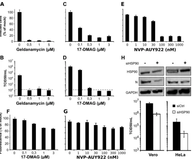
FIG 1HSP90 activity is required for efficient growth of MeV. (A to E) Inhibition of HSP90 activity with geldanamycin, 17-DMAG, or NVP-AUY922 abolishes MeV growth. Vero cells were infected with a recombinant MeV coding for GFP as a viral reporter at an MOI of 0.5 and treated with geldanamycin (A, B), 17-DMAG (C, D), or NVP-AUY922 (E) or not treated. Viral infection was assessed at 24 hpi by measuring GFP expression by flow cytometry (A, C, E) or by measuring MeV production (B, D). 17-DMAG or NVP-AUY922 treatment did not kill cells during 24 h of treatment, as shown by the limited reduction of cellular reductase activity observed at high concentrations with alamarBlue reagent (F, G). (H) Reduction of HSP90 expression by siRNA reduces MeV growth in simian Vero and human HeLa cells. Vero and HeLa cells were transfected with a siRNA targeting the HSP90 mRNA (siHSP90) or a control siRNA (siCtrl) and infected 1 day later with MeV (Schwarz strain) at an MOI of 1. The expression of HSP90 and MeV N protein was assessed by Western blotting (top) at 1 day postinfection, and the impact on MeV production was assessed by the TCID50titration method (bottom).on November 7, 2019 by guest
http://jvi.asm.org/
[image:4.585.96.494.64.392.2]shown). This indicates that HSP90 and 17-DMAG are not
in-volved in protein synthesis
per se
, in agreement with the
HSP90-independent
in vitro
translation of p53 (
54
).
When MeV Flag/L was coexpressed with P, the amount of
de-tectable proteins reached levels similar to those observed after
infection with recombinant MeV Flag/L (
Fig. 3C
) and higher than
the level of Flag/L protein that accumulated in the absence of P
(
Fig. 3D
, compare the L
⫹
ctrl and L
⫹
P parts on the left side). Note
that an N-terminal
⬃
110-kDa fragment of Flag/L was detected
only after transfection, suggesting artifactual generation linked to
this particular mode of expression. Upon 17-DMAG treatment,
MeV L expressed alone was poorly affected, while in the presence
of P only a residual amount of L protein could be detected (
Fig.
3D
, compare the L
⫹
ctrl and L
⫹
P parts; see also panels B and F).
The expression of P remained mostly unchanged (
Fig. 3D
, L
⫹
P
parts) as observed when P was expressed alone (
Fig. 3B
). Similar
phenotypes were observed with L proteins of VSV and NiV, which
were found to disappear upon 17-DMAG treatment whether they
were coexpressed with their homologous P partner or not (
Fig.
3D
). Moreover, the disappearance of MeV L when it was
coex-pressed with P was also observed upon treatment with 100 or
1,000 nM NVP-AUY922 (
Fig. 3E
), i.e., a dose that readily inhibits
MeV growth (
Fig. 1E
). The 17-DMAG-mediated loss of MeV L
protein detection was alleviated in the presence of the proteasome
inhibitor MG132 (
Fig. 3F
, L
⫹
P parts and quantification in the
histogram at the bottom), but not in the presence of 3-MA, an
inhibitor of the autophagy pathway (
Fig. 3F
). In addition, in the
presence of P, MeV L protein was found to be ubiquitinated after
HSP90 inhibition in the presence of the proteasome inhibitor
MG132, as shown by the anti-HA immunolabeling of Flag/
6
⫻
His/L produced in the presence of HA-Ub and pulled down
with Ni beads (
Fig. 3G
). Importantly, the variations in L protein
levels observed upon drug treatment could not be explained by
variations in cell viability assessed by measuring cellular reductase
activities (
Fig. 3H
). All of these results suggest that when it is
coexpressed with P in the absence of HSP90 activity, L is
ubiquiti-nated and efficiently targeted to the proteasomal protein
degrada-tion pathway. This feature is not so prominent in the absence of P,
where the L produced seems rather stable, an observed feature of
MeV L (see the statistical analysis in the legend to
Fig. 3
) but
neither NiV L nor VSV L.
Accumulation of a soluble form of L protein requires both P
and HSP90.
In the previous experiments, an 8 M urea-containing
lysis buffer was used to detect all of the MeV L proteins present in
cells. Since misfolded proteins can form insoluble aggregates,
cells were alternatively lysed in urea-free lysis buffer and both
soluble and insoluble fractions were separated by
centrifuga-tion at 15,000
⫻
g
before treatment with urea and analysis by
Western blotting (
Fig. 4A
).
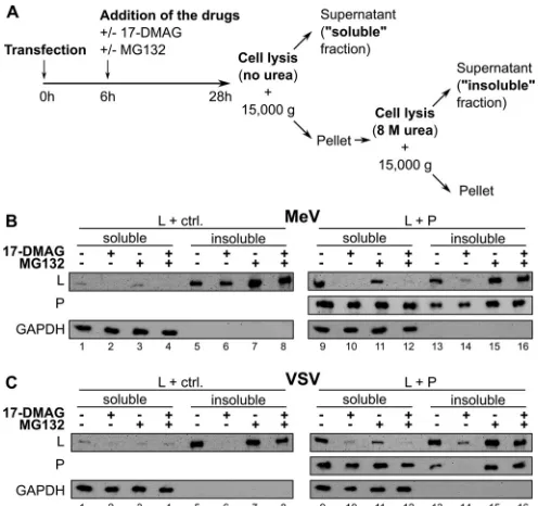
In the absence of P, most of the L protein was found in the
insoluble fraction, with only trace amounts of soluble L (
Fig. 4B
,
compare lanes 1 and 5). In the presence of MG132, the amount of
insoluble L increased, whereas that of its soluble counterpart was
stable (
Fig. 4B
, compare lanes 1 and 5 with lanes 3 and 7,
respec-tively). Very similar data were obtained upon the addition of
17-DMAG in the absence or presence of MG132 (
Fig. 4B
, compare
lanes 5 and 7 with lanes 6 and 8, respectively). The loss of only a
small amount of soluble L was observed (
Fig. 4B
, compare lanes 1
and 3 with lanes 2 and 4, respectively). As a control, GAPDH was
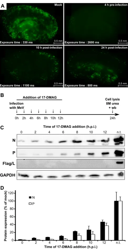
FIG 2HSP90 activity is required at a postentry step and for viral propagation.(A) Inhibition of HSP90 activity with 17-DMAG blocks ongoing MeV propa-gation inex vivomouse brain explants. Brain slices in organotypic cultures were infected with a recombinant MeV IC323-GFP strain (104PFU) and treated at different time points with 1M 17-DMAG. Observations were made at 3 days postinfection, and viral infection was assessed by measuring GFP expression by fluorescence microscopy (A) (note the increased exposure times for earlier treatment with 17-DMAG so as to visualize various levels of fluo-rescence intensity). (B to D) Addition of 17-DMAG at different times postin-fection blocks the ongoing viral replication cycle. As schematized in panel B, 2
M 17-DMAG was added at various times after the infection of Vero/hSLAM cells with Mor-Flag/L at an MOI of 1 and the production of MeV N, P, and Flag/L and cellular GAPDH from 8 M urea-treated whole-cell extracts was monitored by Western blotting (C) with quantification after normalization for GAPDH content (D). Data obtained in the presence of the solvent are indi-cated as nontreated (n.t.).
on November 7, 2019 by guest
http://jvi.asm.org/
[image:5.585.42.285.63.534.2]only recovered in the soluble fractions under all of the conditions
tested (
Fig. 4B
, lanes 1 to 8, two bottom parts, and lanes 9 to 16).
When L was coexpressed with P, its distribution in both the
supernatant and pellet of the cell lysate changed radically, with
approximately equal proportions of soluble and insoluble L (
Fig.
4B
, compare lanes 9 and 13). P was also found in both the soluble
and insoluble fractions. While treatment with the proteasome
in-hibitor did not strongly affect the soluble and insoluble L fractions
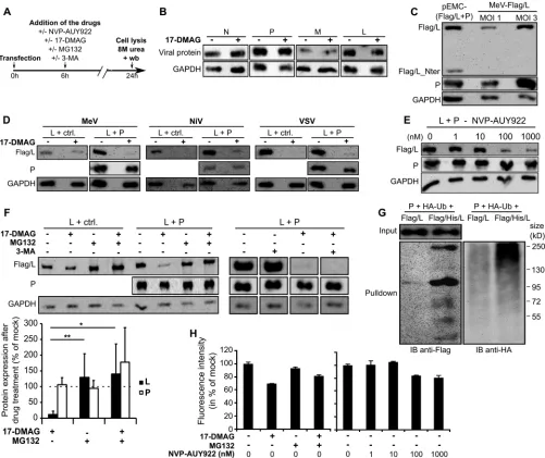
FIG 3Requirement of HSP90 activity for the production of stable P⫹L polymerase complexes. (A) Schematic view of the experiment. BSR-T7 cells were transfected with plasmids and 6 h later treated with 2M 17-DMAG and/or 5M MG132, 5 mM 3-MA, or 1 to 1,000 nM NVP-AUY922 or not treated. The total protein content of cells was analyzed after cell lysis in an 8 M urea-containing lysis buffer at 28 h posttransfection by SDS-PAGE and Western blotting. (B) HSP90 activity is not required for individual production and stabilization of MeV N, P, M, and L proteins in BSR-T7 cells, as shown by the total protein levels in the absence or presence of 2M 17-DMAG added at 6 h posttransfection. (C) Similar levels of MeV Flag/L protein expressed from a pEMC-(Flag/L⫹P) expression vector and MeV-Flag/L in BSR-T7-hSlam cells as measured at 24 h posttransfection or postinfection. (D) Inhibition of HSP90 activity by 17-DMAG hampers the stability of MeV L protein when it is coexpressed with P and not with the controlRenillaluciferase (ctrl., left side). Quantification of Western blotting signals from multiple analyses, expressed as percent inhibition after normalization to GAPDH protein levels, showed significant P-dependent L degradation upon 17-DMAG treatment (n⫽5;P⫽0.0054 [Studentttest]). Inhibition of HSP90 also hampers the stability of both the NiV and VSV L proteins independently of the coexpression of their homologous P partner (D, middle and right parts). (E) Inhibition of HSP90 activity by NVP-AUY922 induces L loss. (F) The loss of L coexpressed with P in the absence of HSP90 activity is alleviated by 5M MG132 (F, L⫹P) but not by the autophagy inhibitor 3-MA, as shown by statistical analysis (F, histogram) by Studentttest (17-DMAG versus MG132,P⫽0.0083 [**]; 17-DMAG versus 17-DMAG⫹MG132,P⫽0.0175 [*] [n⫽4]), despite relatively large standard deviations because of intrinsic limitations of quantification by Western blotting and/or experiment-to-experiment variations. (G) MeV L protein is ubiquitinated upon HSP90 inhibition. Shown are the results of the pulling down on Ni beads of MeV Flag/L and Flag/6⫻His/L covalently bound to HA-Ub from BSR-T7 cells coexpressing Flag/L or Flag/6⫻His/L, P, and HA-Ub in the presence of HSP90 and proteasome inhibitors (2M 17-DMAG and 5M MG132, respectively). Eluates were analyzed by SDS-PAGE and Western immunoblotting (IB) with anti-Flag (left) and anti-HA (right) antibodies. (H) Cell viability was not strongly affected by drug treatment under the experimental conditions used. BSR-T7 cells were treated with drugs under the same conditions (time and concentra-tions) as in panels D to F. For each condition, 1 h before lysis time, 1⫻alamarBlue solution was added and 2 h later, fluorescence levels were measured.on November 7, 2019 by guest
http://jvi.asm.org/
[image:6.585.42.543.67.488.2](
Fig. 4B
, lanes 11 and 15), treatment with 17-DMAG resulted in
the disappearance of both soluble and insoluble L protein (
Fig. 4B
,
compare lanes 9 and 13 with lanes 10 and 14, respectively).
Com-bined treatment with both 17-DMAG and MG132 resulted in
en-hanced recovery of L almost exclusively in the insoluble fraction
(
Fig. 4B
, lanes 12 and 16). Treatment with MG132 and/or
17-DMAG hardly changed the soluble/insoluble P ratio. Similar
re-sults were obtained with VSV L (
Fig. 4C
), with the major
differ-ence that VSV L made in the absdiffer-ence of P accumulated mostly as
insoluble material, as observed for MeV L, and was readily
de-graded upon treatment with 17-DMAG (
Fig. 4
, compare lane 6 in
panel C for VSV and lane 6 in panel B for MeV), as detected upon
coexpression with VSV P (
Fig. 4C
, compare lanes 6 and 14).
Therefore, both P and HSP90 activity are required for
accumula-tion of the L protein in a soluble form.
Association of HSP90 with L and P complexes.
In order to
perform its chaperon activity, HSP90 must, at least transiently,
form a complex with its client protein. Therefore, we searched for
evidence of any colocalization and/or complex formation between
HSP90 and the P and L proteins. MeV infection is characterized by
the accumulation of growing cytoplasmic inclusion bodies made
up of all of the components of the viral transcription and
replica-tion machinery, the N, P, and L proteins (
55–59
). HSP90 was
found to be diffused within the cytoplasm and enriched in the viral
inclusions, as observed by colocalization with individually labeled
P and L proteins in cells infected with recombinant MeV-Flag/L
and imaged by confocal microscopy (
Fig. 5
), while in noninfected
cells, HSP90 only displayed a diffused cytoplasmic distribution
(
Fig. 5
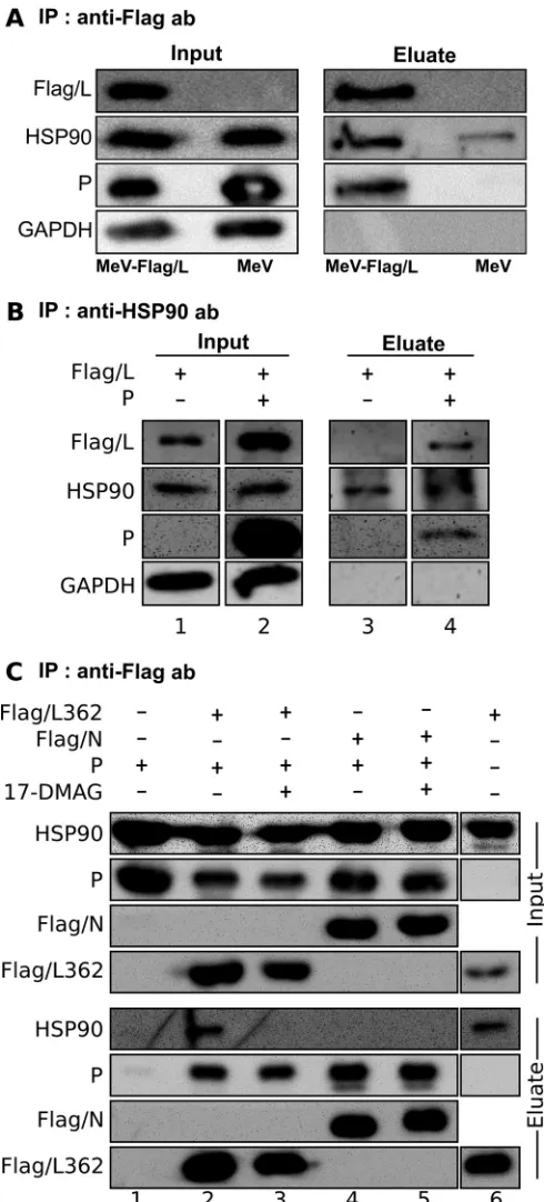
, mock). In addition, both HSP90 and P could be
coimmu-noisolated with Flag/L protein upon gentle elution from anti-Flag
antibody-coated beads with a Flag peptide (
Fig. 6A
), with HSP90
being recovered in greater amounts than those found in control
eluates. In a converse experiment, both Flag/L and P were
recov-ered in the eluate from anti-HSP90 beads from cells coexpressing
Flag/L and P in the absence of 17-DMAG (
Fig. 6B
, lane 4). When
produced in the absence of P, Flag/L was not found in the eluate
from anti-HSP90 beads, most likely because of the low solubility
of L without P (
Fig. 6B
, lane 3). Thus, both colocalization imaging
and coimmunoprecipitation experiments indicate that L, P, and
HSP90 can associate in protein complexes within infected cells.
P binding to the N terminus of L does not require HSP90
activity.
We then asked whether P binding to L can occur without
HSP90 activity. Within the MeV L protein, the P binding site has
been reported to map to the L1-408 fragment (
20
,
60
). In fact, the
L protein consists of six conserved regions (CRI to CRVI)
throughout the
Mononegavirales
order, except for the
Bornaviri-dae
family, which lacks CRVI (
61
). According to the alignment of
L from
Mononegavirales
with the recently solved VSV-L structures
(
22
), the L-362 fragment (which covers most of CRI) can be
de-signed as a possible independent folding unit. Indeed, the
struc-FIG 4L and P proteins associate into soluble complexes in the presence ofHSP90 activity. (A) Schematic view of the experiment. BSR-T7 cells were transfected with plasmids, and 6 h later, cells were treated with 2M 17-DMAG and/or 5M MG132 or not treated. Cells were lysed in urea-free lysis buffer at 28 h posttransfection, and soluble proteins were separated by centrif-ugation at 15,000⫻gfor 15 min (soluble fraction). The pellet was then washed twice in urea-free lysis buffer and incubated in 8 M urea-containing buffer. Denatured protein aggregates were finally separated after 15 min of centrifu-gation at 15,000⫻g(insoluble fraction). (B, C) Western blot analysis of sol-uble and insolsol-uble fractions from BSR-T7 cells cotransfected with L and a control (ctrl.) plasmid (left) or a P plasmid (right). Data for MeV are in panel B, and those for VSV are in panel C.
FIG 5MeV L, P, and HSP90 colocalize within virus-induced inclusion bodies. (A) Vero/hSLAM cells were infected at an MOI of 0.1 with a recombinant MeV expressing a Flag-tagged L protein or not infected. At 24 hpi, cells were fixed, the L or P (left side) and P or HSP90 (middle) proteins were immunolabeled, and images were overlaid for colocalization (right side). White arrows indicate noninfected cells. White bars represent 10m.
on November 7, 2019 by guest
http://jvi.asm.org/
[image:7.585.39.287.66.299.2] [image:7.585.301.544.67.380.2]ture of the corresponding crystalized VSV L N-terminal domain
(Protein Data Bank [PDB] code
5CHS
) (
62
) superimposes well
with that solved within the entire L protein (PDB code
5a22
) (
22
).
In contrast to full-length L, this fragment accumulated well in
soluble form in cells, and its accumulation level remained
un-changed upon the inhibition of HSP90 by 17-DMAG even when it
was coexpressed with P (
Fig. 6C
, input, compare lanes 2 and 3). P
was efficiently coimmunoprecipitated with Flag/L-362 even when
they were coexpressed in the presence of 17-DMAG, as it was with
the Flag/N protein, another P-binding protein (
Fig. 6C
, eluate,
compare lanes 2 and 3). Small but detectable amounts of HSP90
were also isolated with Flag/L-362 produced with or without P but
not with Flag/N (
Fig. 6C
, eluate, compare lanes 2 and 6 with lane
4). Upon HSP90 inhibition, no HSP90 could be detected after
Flag/L-362 precipitation (
Fig. 6C
, eluate, compare lanes 2 and 3).
Thus, P and HSP90 may bind to the N terminus of L
indepen-dently of each other and the interaction between L and HSP90
requires HSP90 activity.
Chimeric L/luciferase proteins allow intracellular evaluation
of L protein folding upon HSP90 assistance in the presence of P.
Taken together, the above data suggest that HSP90 can act as a
chaperon of L to allow proper folding. To monitor the folding
state of accumulated MeV L,
Gaussia
luciferase was fused between
residues 1709 and 1710 of L (L[
Gaussia
]), i.e., within the flexible
linker preceding the methyltransferase domain that encompasses
CRVI (
22
) (
Fig. 7A
, top). This position tolerates a foreign protein
graft, as shown by the rescue and growth of recombinant MeV
L[
Gaussia
] (
Fig. 7B
), in agreement with similar observations made
with other members of the order
Mononegavirales
(
55
,
63–65
). We
hypothesize that misfolding of the nascent L protein will induce
the misfolding of the downstream fused luciferase and
conse-quently loss of luciferase activity. According to Western blot
anal-ysis, chimeric MeV L[
Gaussia
] coexpressed with P exhibited the
same biochemical phenotype as that observed with the L protein
(compared
Fig. 7C
and
3F
). While the activity of
Gaussia
luciferase
per se
was insensitive to 17-DMAG in the absence or presence of
MG132 (
Fig. 7D
), that of L[
Gaussia
] was strongly inhibited by the
addition of 17-DMAG in the presence of P (
Fig. 7E
). However,
enzymatic activity could not be recovered upon simultaneous
in-hibition of the proteasome, suggesting that, in this case,
L[
Gaussia
] remains misfolded when its degradation by the
protea-some is prevented (compare
Fig. 7E
and
C
). P seems also to be
required for the proper folding of L, since L[
Gaussia
] expressed in
the absence of P displayed only marginal luciferase activity that
was not affected by any treatment (
Fig. 7F
).
When the
Gaussia
protein was appended at the C terminus of L
via a short flexible linker (L/
Gaussia
) (
Fig. 7A
, bottom), the loss of
luciferase activity induced by 17-DMAG (likely because of the
P-dependent induced degradation of L/
Gaussia
) was fully
pre-FIG 6HSP90 is coimmunoprecipitated with P and L from infected and trans-fected cells but is dispensable for the stability of P-L362 complexes. (A) Coim-munoprecipitation of HSP90 and P with L from infected cells. Vero/hSLAM cells were infected at an MOI of 1 with MeV strain Moraten expressing L protein tagged with the Flag peptide or not tagged (MeV-Flag/L and MeV, respectively). At 28 hpi, immunoprecipitation (IP) from the supernatant of infected cells lysed with urea-free buffer was performed with Flag anti-body (ab)-coated magnetic beads. Flag/L, HSP90, P, and GAPDH were ana-lyzed by SDS-PAGE and Western blotting as detailed in Materials and Meth-ods. Note the faint nonspecific immunoprecipitation of HSP90 from extracts of cells infected with MeV expressing L protein without the Flag tag. (B) Both P and Flag/L are coimmunoprecipitated with HSP90 on immobilized
anti-HSP90 antibodies. BSRT-7 cells were cotransfected with protein constructs at 24 h posttransfection, and proteins were immunoprecipitated by anti-HSP90 antibodies and protein G beads. Proteins in the cell extracts (input) and in the eluate from the Flag-tagged beads were detected by Western blotting. (C) The coimmunoprecipitation of P and L362 is not affected by inhibition of HSP90 activity. BSR-T7 cells were transfected to express P, Flag/N, and Flag/L362 with or without P. At 17 h posttransfection, cells were treated with 2M 17-DMAG or not treated. At 24 h posttransfection, Flag-tagged proteins were immuno-precipitated with anti-Flag antibody-coated beads, and the cell extracts (input) and the eluate from the Flag-tagged beads were subjected to Western blotting.
on November 7, 2019 by guest
http://jvi.asm.org/
[image:8.585.40.285.68.609.2]vented upon the addition of MG132 (
Fig. 7G
). This suggests that
when appended to the C terminus of L instead of being embedded
within the L structure, the
Gaussia
protein retains a functional fold
independently of the upstream L fold status. Alternatively, the two
last methyltransferase domain and C-terminal domain (CTD) of
L may fold somehow independently of the upstream domains.
Indeed, the enzymatically active methyltransferase domain of the
human metapneumovirus L protein (aa 1599 to 2005) has been
produced and its crystal structure has been solved (
66
) and active
MeV polymerase has been reconstituted by the reassembly via an
unrelated dimerization domain of two L split parts that
encom-passed the CRI-to-CRV and methyltransferase-CTD regions,
re-spectively (
67
).
In the presence of P, HSP90 acts as a
bona fide
chaperon of
the viral polymerase.
HSP90 client proteins require its activity
either for the folding step that occurs during their synthesis and/or
maturation (
68
) or to support the active conformation of their
enzymatic activity, as is the case for many kinases and ubiquitin E3
ligases (
69
). In the first case, once the clients have achieved their
mature folding, their stability and/or function usually no longer
rely on HSP90 (
68
). The type of HSP90 client the L polymerase is
was evaluated by two complementary approaches.
In a first set of experiments, the impact of 17-DMAG treatment
on the fate of neosynthesized Flag/L protein was investigated after
metabolic labeling with
35S-labeled cysteine and methionine,
fol-lowed by immunoprecipitation of soluble Flag/L by anti-Flag
an-tibody-coated beads. As expected, the coexpression of P and L
slows down the decay of
35S-labeled Flag/L (
Fig. 8B
and
C
,
com-pare white circles and triangles), as shown by a roughly 2.5-fold
increase in the estimated half-life (
⬃
14.8 h with P versus
⬃
5.9 h
without P). Furthermore, the addition of 2
M 17-DMAG on cells
after a 2-h chase delay (
Fig. 8A
shows the experimental scheme)
resulted in an
⬃
4-fold decrease in the rate of
35S-labeled Flag/L
produced without P (
Fig. 8B
and
C
, black triangles, half-life of
⬃
1.4 h). In contrast, the decay of
35S-labeled Flag/L produced in
the presence of P was mostly unaffected by this treatment (
Fig. 8B
and
C
, black circles, half-life of
⬃
12.8 h). When 17-DMAG was
applied before the metabolic labeling and maintained thereafter
(
Fig. 8A
shows the experimental scheme),
35S-labeled Flag/L
quickly disappeared after the end of the pulse, independently of
whether or not it was coexpressed with P (
Fig. 8D
and
E
, half-life
of
⬃
1.2 h in both cases), and in agreement with
Fig. 3D
and
F
. The
decay of P protein was also determined after
immunoprecipita-tion with a P-specific antibody and found not to be sensitive to
treatment with 17-DMAG (
Fig. 8F
to
I
), exhibiting
⬃
14.4-h and
⬃
13.9-h half-lives, respectively. In both pulse-chase experiments
done in the presence of P, amounts of
35S-labeled P
coimmuno-precipitated with anti-Flag/L beads (
Fig. 8B
and
D
) and those of
35
S-labeled Flag/L coimmunoprecipitated with anti-P beads (
Fig.
8F
and
H
) nicely parallel the fate of
35S-labeled Flag/L and that of
35S-labeled P, respectively. As mentioned earlier, an
⬃
110-kDa
35
S-labeled Flag/L N-terminal fragment that could correspond to
the RNA-dependent RNA polymerase domain (
22
) was clearly
identified in anti-Flag eluates (
Fig. 8B
and
D
). Since this fragment
was detected only in the transfection system and not in the anti-P
eluate (
Fig. 8F
and
H
), it likely represents an intermediate
degra-dation product of Flag/L not bound to P. Of note, the level of
35S
labeling of all cell proteins did not differ between samples by more
than 13% over the first 8 h, as determined by SDS-PAGE of labeled
cell extracts, thus excluding any bias during metabolic labeling of
the cells (data not shown). These results confirm that L is very
sensitive to the lack of HSP90 activity during its biosynthesis and
show that L proteins having matured for 2 h in the presence of P
and HSP90 activity become stable without any further
chaperon-ing by HSP90.
In a second set of experiments, we studied the fate of
steady-state L and P that have accumulated in the presence of active
HSP90 for 18 h. To ensure that we studied the fate of only the
preexisting pool of stabilized L proteins, the observation started
after an initial 2-h window of blocking of cellular translation with
cycloheximide (see the scheme in
Fig. 8J
). MeV Flag/L premade in
the presence of P and HSP90 activity displayed a similar decay upon
treatment with cycloheximide, 17-DMAG, or both (
Fig. 8K
), with an
FIG 7Enzymatic activity from MeV L[Gaussia] and L/Gaussiahybridmole-cules confirms that stable L folding is dependent on HSP90 activity. (A) Sche-matic view of MeV L[Gaussia] chimeric protein and L/Gaussiawith luciferase coding sequence inserted between residues 1709 and 1710 (top) and appended to the C terminus of L (bottom), respectively. (B) Luciferase activity in cells infected with recombinant MeV-L[Gaussia] virus (MOI of 1) at 24 hpi illus-trating tolerance of the internal graft of the luciferase domain. (C) L[Gaussia] chimeric protein recapitulates the detection phenotype of wild-type L protein observed in the presence of P with inhibition by 2M 17-DMAG treatment that is relieved by treatment with 5M MG132 (see the legend toFig. 3for experimental details). (D)Gaussialuciferase activity is insensitive to 17-DMAG treatment. Cells were cotransfected with glu1-P and glu2-P constructs that reconstitute theGaussialuciferase thanks to the P tetramerization domain (34). Note the similar slight enhancement of the luciferase signal upon the inhibition of the proteasome by MG132 in the absence or presence of 17-DMAG. (E, F) Luciferase activity from the L[Gaussia] chimeric protein in the presence of P is inhibited by 2M 17-DMAG but not restored upon simulta-neous treatment with MG132. MG132 versus 17-DMAG⫹MG132,P⫽0.0002 (***; Studentttest;n⫽6) (E). (F) In the absence of P, luciferase activity from L[Gaussia] is marginal (L⫹P versus L in the presence of MG132,P⫽0.0168 (*; Studentttest;n⫽4). (G)Gaussialuciferase appended to the C terminus of L remains active upon simultaneous HSP90 and proteasome inhibition, while it is lost upon the sole inhibition of HSP90.
on November 7, 2019 by guest
http://jvi.asm.org/
[image:9.585.41.285.67.353.2]FIG 8Impact of HSP90 activity on the fate of MeV L polymerase. (A to H) Monitoring of soluble MeV L and P after metabolic35S labeling performed in the absence or presence of 17-DMAG. (A) Schematic view of the experiment. BSR-T7 cells were transfected with P and/or L plasmid(s) and the day after were pulse-labeled with35S-labeled methionine and cysteine for either 1 h (top) or 0.5 h (bottom). At the times indicated, cells were lysed and soluble supernatants
on November 7, 2019 by guest
http://jvi.asm.org/
[image:10.585.42.542.68.697.2]apparent half-life of
⬃
13 to 14 h, i.e., in a range similar to that
observed in the pulse-chase study. We conclude that HSP90 is
dispensable for the maintenance of stable MeV P
⫹
L polymerase
complexes once they have been fully assembled and/or folded.
Similar results were obtained by studying the fate of VSV Flag/L
produced in the presence of P that remained stable for 12 h (
Fig.
8L
), as well as VSV P (data not shown). In contrast, VSV Flag/L
produced without P decayed much more quickly in the presence
of 17-DMAG (
Fig. 8M
, compare black and white symbols), with
an apparent
⬃
2- to 3-h half-life, while premade Flag/L remained
stable for almost 12 h in the absence of 17-DMAG.
Functions of MeV polymerase do not rely on HSP90 activity.
We then logically evaluated whether, once made, biochemically
stable L (in complex with P) can be functional in the absence of
any HSP90 activity. We took advantage of the active polymerases
that are resident in virions and ready to start primary viral
tran-scription upon nucleocapsid delivery into the cytoplasm of the
host cell (
14
). Being mediated by the fixed number of polymerases
found within the virion, this primary transcription is
character-ized by a linear mRNA accumulation phase for several hours until
newly made polymerases are recruited to the incoming
nucleo-capsid, resulting in a shift from a linear to an exponential viral
transcript accumulation rate, thus constituting a secondary
tran-scription phase. During these two trantran-scription phases that
pre-cede the onset of replication, every polymerase is very efficiently
recycled and/or recruited on the incoming nucleocapsid template,
as experimentally illustrated once more in
Fig. 9
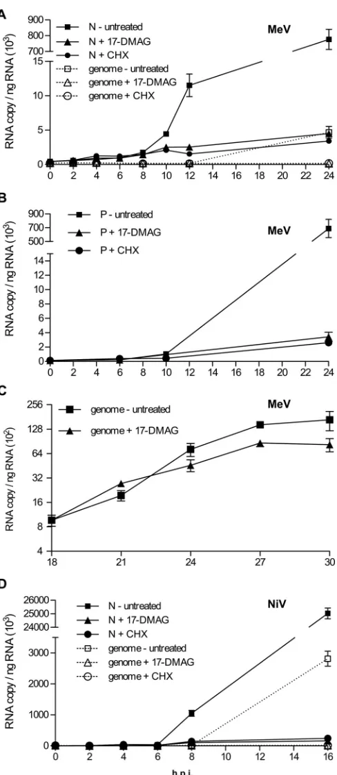
. In the absence of
any treatment, the N and P mRNAs quantified by RT-qPCR
accu-mulated linearly for the first
⬃
8 h and then exponentially further
on (
Fig. 9A
and
B
). When MeV-infected cells were treated with
cycloheximide at 30 min postinfection to block the neosynthesis
of any newly active viral polymerase, the linear phase of N mRNA
accumulation proceeded without switching to the exponential
phase, in agreement with our previous report (
14
). Interestingly,
the addition of 17-DMAG mimicked the protein translation
inhi-bition, with a similar rate of linear mRNA accumulation (
Fig. 9C
).
This suggests that preformed polymerases brought by infecting
virions do not depend on HSP90 activity to synthesize viral
mRNA. Furthermore, the absence of a switch of the mRNA
accu-mulation rate to the exponential phase indicates that HSP90
ac-tivity is required for the production of new polymerases, in
agree-ment with our previous results showing the misfolding and
degradation of L proteins under these conditions. The similar rate
of N mRNA accumulation upon treatment with cycloheximide
and 17-DMAG is in agreement with the biochemical stability of
fully mature L protein in complex with P shown in
Fig. 8
. The
reinitiation of viral transcription at the next gene start
signal/se-quence was also found to be unaffected upon treatment with
ei-ther cycloheximide or 17-DMAG, since similar linear
accumula-tion of P mRNA occurred (
Fig. 9B
). Finally, MeV genome
replication did not require HSP90 activity since genomes were still
accumulating after a late addition of 17-DMAG (
Fig. 9C
). Note,
however, that upon the early addition of 17-DMAG, the onset of
the replication phase was fully prevented (
Fig. 9A
, dotted lines),
likely because of the inhibition of the exponential transcription
phase. Similarly, secondary but not primary transcription of NiV
was also blocked in the presence of 17-DMAG, as observed in the
presence of cycloheximide (
Fig. 9D
). These data support the
hy-potheses that inhibition of HSP90 activity prevents the
produc-tion of new funcproduc-tional polymerases and that stable and mature
P
⫹
L polymerases are no longer sensitive to this inhibition.
DISCUSSION
Our overall data offer strong support for the idea that HSP90 acts
as a transient chaperon for
Mononegavirales
L polymerase that
needs to bind to its viral P partner in order to be stable and
func-tional. Since we used three known inhibitors of HSP90,
geldana-mycin, 17-DMAG, and NVP-AUY922, and HSP90-targeted
si-lencing, it is unlikely that our results are related to off-target
activities (
46
,
49
,
50
). Moreover, the phenotype observed here
perfectly fits what has been reported so far for hundreds of HSP90
clients (
69
).
Our results reveal an intricate interplay between HSP90 and P
for the production of a stable and soluble L polymerase. On the
one hand, in the absence of P, MeV L is unstable and undergoes
degradation by the proteasome for a small part and aggregation
into insoluble forms for the majority. In the absence of P, HSP90
is not able to properly fold L according to the data obtained with
our engineered luciferase-based folding assay. On the other hand,
in the presence of P, blocking of HSP90 at an early stage after
transfection of the expression vectors induces the degradation of
nearly all L proteins by the proteasome. When the latter is
inhib-ited, L accumulates in likely misfolded aggregates. Furthermore,
the stable recruitment of P by binding to the N-terminal region of
MeV L polymerase, possibly during protein synthesis (
20
,
60
),
occurs without the need for any HSP90 activity. As HSP90 is not
able to fold L into a stable form without P, we propose that P binds
and stabilizes L protein possibly during its biosynthesis, thanks to
a P binding site residing in the N-terminal part of MeV L (L1-362).
Indeed, in the absence of P, MeV L levels quickly fall off, mostly by
aggregation into insoluble complexes. P thus appears to act as a
prop that prevents the collapse of folded L until it reaches its final
form as part of an active L-P polymerase. In support of this
hy-pothesis, stable P
⫹
L complexes that are no longer sensitive to
HSP90 inhibition were observed only when 17-DMAG treatment
was delayed by 2 h after
35S metabolic labeling (
Fig. 8B
and
D
) and
were analyzed by immunoprecipitation (IP) with anti-Flag antibody-coated beads and anti-P antibody plus protein G beads. In some cases, 2M 17-DMAG was added after (top) or before (bottom) the pulse-labeling. Shown are autoradiograms (B, D) and quantification (C, E) of material eluted from anti-Flag antibody-coated beads incubated with the soluble cell extracts harvested at the times indicated after 1 h (B, C) or 0.5 h (D, E) of pulse-labeling according to top and bottom schemes in panel A, respectively. Panels F and G and panels H and I show data on the immunoprecipitation (with anti-P antibodies) of the same extracts as in panels B and C and panels D and E, respectively. Note the comparable stability of L and P in panels C and G when the two proteins were coexpressed. (J) Schematic view of experiments shown in panels K to M. Decay of premade MeV (K) and VSV (L, M) Flag/L proteins coexpressed (K, L) or not coexpressed (M) with P that have been allowed to be synthesized and stabilized for 18 h. After a protein synthesis block with 20g/ml cycloheximide for 2 h, cycloheximide was replaced with fresh medium supplemented with 2M 17-DMAG, 20g/ml cycloheximide (CHX), or both or not supplemented. Protein decay was measured from Western blotting signals (mean values and standard deviations from two independent samples) and expressed as a percentage of the Western blotting signal determined at the end of the cycloheximide block. Note that the stability of MeV L produced without P could not be technically studied because the level was too low for detection (seeFig. 4B).
on November 7, 2019 by guest
http://jvi.asm.org/
not when it was immediately applied just after the labeling pulse
(data not shown). Whether, as expected from a prop, MeV L that
has undergone proper folding can dissociate from and reassociate
with P to form a functional enzyme, as experimentally
docu-mented for VSV (
8
,
70
) and Newcastle disease virus (
9
), remains
to be investigated.
Once folded and stabilized by P, the L polymerase appears to be
long-lived, with an estimated half-life of 13 to 14 h, from the fate of
both
35S pulse-labeled and steady-state accumulated proteins.
This half-life was similar to that of MeV P, indicating that binding
to P drives L toward a similar long-lived state. This nicely fits the
observation of primary transcription at a constant rate by
virion-derived polymerases observed over 24 hpi in Vero cells when
ei-ther protein synthesis or L protein folding is prevented by
cyclo-heximide or an HSP90 inhibitor, respectively.
We anticipate the conservation of these mechanisms for the
polymerase of other members of the
Mononegavirales
order.
How-ever, the maturation of the polymerase complex from other
mem-bers of the
Mononegavirales
order likely differs in at least some
mechanical details. Indeed, without their P partner, the L proteins
of RSV (
71
), VSV, and NiV are more efficiently targeted for
pro-tease degradation upon HSP90 inhibition than their MeV
coun-terpart. Such a divergent behavior of HSP90 clients has also been
reported for cellular kinases, some of them preferentially ending
in intracellular aggregates and others being quickly degraded by
the proteasome (
6
). Rapid aggregation of MeV L in the absence of
P likely occurs and diverts most of it from degradation. That some
properly folded L proteins can be produced without P binding is
further supported by purified L proteins endowed with some
en-zymatic activities (RNA synthesis and capping) (
1
,
72
,
73
) and by
the successful association into processive transcriptases of P and L
proteins separately overproduced in insect cells in the case of VSV
(
16
) and SeV (
74
) and also in mammalian cells in the case of VSV
(
16
). However, P and L from PIV5 (
75
) and SeV (
7
) individually
produced in mammalian cells cannot associate. Likewise, the RSV
L protein cannot be detected in the absence of P even when
over-produced in insect cells (
18
). Finally, even VSV L produced alone
is rather unstable (
16
,
17
) and largely stabilized by P with a similar
transient requirement for HSP90 activity (
Fig. 8L
and
M
).
An analysis of the polymerase complex of the influenza virus, a
segmented negative-stranded RNA virus, suggested that HSP90
would be required for its polymerase activity (
76
). In contrast, in
the cases of MeV and NiV, HSP90 is clearly dispensable for the
various enzymatic activities and function of the viral polymerase.
The linear accumulation of MeV N and P transcripts upon
infec-tion in the presence of 17-DMAG over 24 h (i.e., well over the
estimated 2 h needed to consecutively transcribe the six viral genes
[
14
]) indicates that the polymerases recycle and are efficiently
switched on upon recycling to the 3=
leader region of the genome
containing the transcription promoter. Although the other
poly-merase activities (RNA capping and polyadenylation) were not
directly investigated, the fact that N and P transcripts accumulate
at similar rates after the inhibition of either HSP90 or protein
FIG 9Once made, MeV and NiV polymerases remain functional in theab-sence of HSP90 activity. (A and B) MeV polymerases of virion origin do not require HSP90 activity for their RNA synthesis activity. Shown are rates of accumulation of MeV N transcripts and genome RNA (A) and MeV P tran-scripts (B). (C) MeV polymerases can function in the replicase mode in the absence of HSP90 activity. The same experiment as in panels A and B was performed with addition of the drug at 18 hpi, i.e., during the ongoing repli-cation phase. (D) Virion-resident NiV polymerases do not require HSP90 activity for their RNA synthesis activity. Shown are rates of accumulation of N transcripts and genome RNA. Vero/hSLAM cells were infected with MeV
IC323 or NiV at an MOI of 1 and treated 30 min later with either 2M 17-DMAG or 20g/ml cycloheximide (CHX) or not treated. RNA were ex-tracted every 2 h, and viral RNAs were quantified by RT-qPCR. The mean values and standard deviations shown are from three independent infection samples.
on November 7, 2019 by guest
http://jvi.asm.org/
[image:12.585.43.287.66.627.2]translation suggests that the viral mRNAs have similar half-lives
under these conditions and therefore that mRNA processing
re-mains correctly fulfilled. Virus replication
per se
was also found
not to rely on HSP90 activity.
In conclusion, HSP90 appears to be indispensable for MeV,
NiV, and VSV replication by acting as a faithful chaperon of the L
polymerase of
Mononegavirales
. We propose a prop-like activity
for P behind the HSP90 chaperoning of L polymerase as a new
paradigm. Future work will address how and when P may prop up
the nascent L protein during its assistance with folding by the
HSP90 chaperoning machinery and to what extent this
mecha-nism is conserved throughout the
Mononegavirales
order. To this
end, we will benefit from the new intracellular luciferase-based
folding assay we have set up to probe the interactions between P
and L polymerase components. Finally the strong antiviral activity
of HSP90 inhibitors raises the possibility of their use as antiviral
drugs from which the emergence of an escape mutant virus is
unlikely to occur, as recently reported for RSV (
25
). Such a lack of
an escape mutant is not unexpected, owing to the fact that
anti-HSP90 inhibitors do not exert a direct antiviral activity by
target-ing a viral enzymatic activity. Instead, they prevent the
indispens-able chaperoning of any newly made viral L polymerase by the
cellular HSP90 machinery.
ACKNOWLEDGMENTS
We thank M. Billeter, R. Cattaneo, P. Pothier, M. Takeda, J. Perrault, K. Conzelmann, J. K. Rose, Y. Jacob, Y. Yanagi, and L. Naldini for useful reagents, A. Bouchardon for technical help, J. Brunel for having derived the BSR-T7-Slam cell line, and P. Lawrence for English language revision. This work has benefited from the Imaging (CIQLE) and Cytometry and Genetic Analysis (ANIRA) facilities of SFR BioSciences Gerland—Lyon Sud (CNRS UMS3444 INSERM US8) and SFR Santé Lyon Est (CNRS UMS3453, INSERM US7).
The research reported here was supported by ANR CARDINNATE (ANR-12-BSV3-010-01) and NITRODEP (ANR-13-PDOC-0010-01) grants (http://www.agence-nationale-recherche.fr/), and B.G.’s salary was supported by a Sanofi-Pasteur grant to D.G. L.M.B.’s salary was supported by the Fondation pour la Recherche Médicale (FRM). The funders had no role in study design, data collection and analysis, the decision to publish, or preparation of the manuscript.
L.M.B., B.G., S.D.B., and D.G. designed the research. L.M.B., J.W., B.H., and C.M. designed and/or contributed new reagents. L.M.B., F.E., B.G., J.W., S.D.B., and C.M. performed the research. L.M.B., F.E., J.W., C.M., B.G., S.D.B., and D.G. analyzed the data. All of us drafted or revised the manuscript and approved the final version.
FUNDING INFORMATION
This work, including the efforts of Denis Gerlier, was funded by Agence Nationale de la Recherche (ANR) (CARDINNATE ANR-12-BSV3-010-01). This work, including the efforts of Cyrille Mathieu, was funded by Agence Nationale de la Recherche (ANR) (NITRODEP ANR-13-PDOC-0010-01). This work, including the efforts of Louis-Marie Bloyet, was funded by Fondation pour la Recherche Médicale (FRM) (2015 postdoc-toral fellowship).
The funders had no role in study design, data collection and analysis, decision to publish, or preparation of the manuscript.
REFERENCES
1.Morin B, Rahmeh AA, Whelan SP.2012. Mechanism of RNA synthesis initiation by the vesicular stomatitis virus polymerase. EMBO J31:1320 – 1329.http://dx.doi.org/10.1038/emboj.2011.483.
2.Morin B, Kranzusch PJ, Rahmeh AA, Whelan SP.2013. The polymerase
of negative-stranded RNA viruses. Curr Opin Virol3:103–110.http://dx .doi.org/10.1016/j.coviro.2013.03.008.
3.Blocquel D, Bourhis J-M, Eléouët J-F, Gerlier D, Habchi J, Jamin M, Longhi S, Yabukarski F.2012. The original mechanics of Mononegavi-rales transcription and replication. Virologie16:225–257.
4.Gerlier D, Lyles DS.2011. Interplay between innate immunity and neg-ative-strand RNA viruses: towards a rational model. Microbiol Mol Biol Rev75:468 – 490.http://dx.doi.org/10.1128/MMBR.00007-11.
5.Kolakofsky D, Le Mercier P, Iseni F, Garcin D.2004. Viral RNA poly-merase scanning and the gymnastics of Sendai virus RNA synthesis. Vi-rology318:463– 473.http://dx.doi.org/10.1016/j.virol.2003.10.031. 6.De BP, Banerjee AK.1984. Specific interactions of vesicular stomatitis
virus L and NS proteins with heterologous genome ribonucleoprotein template lead to mRNA synthesis in vitro. J Virol51:628 – 634. 7.Horikami SM, Curran J, Kolakofsky D, Moyer SA.1992. Complexes of
Sendai virus NP-P and P-L proteins are required for defective interfering particle genome replication in vitro. J Virol66:4901– 4908.
8.Emerson SU, Yu Y.1975. Both NS and L proteins are required for in vitro RNA synthesis by vesicular stomatitis virus. J Virol15:1348 –1356. 9.Hamaguchi M, Yoshida T, Nishikawa K, Naruse H, Nagai Y. 1983.
Transcriptive complex of Newcastle disease virus. I. Both L and P proteins are required to constitute an active complex Virology128:105–117. 10. Guryanov SG, Liljeroos L, Kasaragod P, Kajander T, Butcher SJ.2015.
Crystal structure of the measles virus nucleoprotein core in complex with an N-terminal region of phosphoprotein. J Virol90:2849 –2857.http://dx .doi.org/10.1128/JVI.02865-15.
11. Qanungo KR, Shaji D, Mathur M, Banerjee AK.2004. Two RNA poly-merase complexes from vesicular stomatitis virus-infec
![FIG 7 Enzymatic activity from MeV L[DMAG. (E, F) Luciferase activity from the L[inhibition of the proteasome by MG132 in the absence or presence of 17-Gaussia] and L/Gaussia hybrid mole-cules confirms that stable L folding is dependent on HSP90 activity](https://thumb-us.123doks.com/thumbv2/123dok_us/141914.19058/9.585.41.285.67.353/enzymatic-activity-luciferase-inhibition-proteasome-conrms-dependent-activity.webp)