CXCL10/CXCR3-Dependent Mobilization
of Herpes Simplex Virus-Specific CD8
ⴙ
T
EM
and CD8
ⴙ
T
RM
Cells within Infected
Tissues Allows Efficient Protection
against Recurrent Herpesvirus Infection
and Disease
Ruchi Srivastava,aArif A. Khan,aSravya Chilukuri,aSabrina A. Syed,aTien T. Tran,a
Julie Furness,aElmostafa Bahraoui,b,cLbachir BenMohameda,d,e
Laboratory of Cellular and Molecular Immunology, Gavin Herbert Eye Institute, University of California Irvine,
School of Medicine, Irvine, California, USAa; INSERM, U1043, and CNRS, U5282, Toulouse, Franceb; Université
Paul Sabatier Toulouse, Toulouse, Francec; Department of Molecular Biology and Biochemistry, University of
California Irvine, Irvine, California, USAd; Institute for Immunology, University of California Irvine, School of
Medicine, Irvine, California, USAe
ABSTRACT Herpes simplex virus 1 (HSV-1) establishes latency within the sensory neurons of the trigeminal ganglia (TG). HSV-specific memory CD8⫹T cells play a
crit-ical role in preventing HSV-1 reactivation from TG and subsequent virus shedding in tears that trigger recurrent corneal herpetic disease. The CXC chemokine ligand 10 (CXCL10)/CXC chemokine receptor 3 (CXCR3) chemokine pathway promotes T cell immunity to many viral pathogens, but its importance in CD8⫹T cell immunity to recurrent herpes has been poorly elucidated. In this study, we determined how the CXCL10/CXCR3 pathway affects TG- and cornea-resident CD8⫹T cell responses to re-current ocular herpesvirus infection and disease using a well-established murine model in which HSV-1 reactivation was induced from latently infected TG by UV-B light. Following UV-B-induced HSV-1 reactivation, a significant increase in both the number and function of HSV-specific CXCR3⫹CD8⫹T cells was detected in TG and
corneas of protected C57BL/6 (B6) mice, but not in TG and corneas of nonprotected CXCL10⫺/⫺or CXCR3⫺/⫺ deficient mice. This increase was associated with a
signifi-cant reduction in both virus shedding and recurrent corneal herpetic disease. Fur-thermore, delivery of exogenous CXCL10 chemokine in TG of CXCL10⫺/⫺mice, using
the neurotropic adeno-associated virus type 8 (AAV8) vector, boosted the number and function of effector memory CD8⫹ T cells (TEM) and tissue-resident memory
CD8⫹T cells (T
RM), but not of central memory CD8⫹T cells (TCM), locally within TG,
and improved protection against recurrent herpesvirus infection and disease in CXCL10⫺/⫺ deficient mice. These findings demonstrate that the CXCL10/CXCR3
chemokine pathway is critical in shaping CD8⫹ T cell immunity, locally within
la-tently infected tissues, which protects against recurrent herpesvirus infection and disease.
IMPORTANCE We determined how the CXCL10/CXCR3 pathway affects CD8⫹ T cell
responses to recurrent ocular herpesvirus infection and disease. Using a well-established murine model, in which HSV-1 reactivation in latently infected trigeminal ganglia was induced by UV-B light, we demonstrated that lack of either CXCL10 chemokine or its CXCR3 receptor compromised the mobilization of functional CD8⫹ TEMand CD8⫹TRMcells within latently infected trigeminal ganglia following virus
re-activation. This lack of T cell mobilization was associated with an increase in recur-rent ocular herpesvirus infection and disease. Inversely, augmenting the amount of
Received16 February 2017Accepted25 April
2017
Accepted manuscript posted online3 May
2017
CitationSrivastava R, Khan AA, Chilukuri S,
Syed SA, Tran TT, Furness J, Bahraoui E, BenMohamed L. 2017. CXCL10/CXCR3-dependent mobilization of herpes simplex virus-specific CD8+T
EMand CD8+TRMcells
within infected tissues allows efficient protection against recurrent herpesvirus infection and disease. J Virol 91:e00278-17.
https://doi.org/10.1128/JVI.00278-17.
EditorJae U. Jung, University of Southern
California
Copyright© 2017 American Society for
Microbiology.All Rights Reserved. Address correspondence to Lbachir BenMohamed, Lbenmoha@uci.edu.
crossm
on November 7, 2019 by guest
http://jvi.asm.org/
CXCL10 in trigeminal ganglia of latently infected CXCL10-deficient mice significantly restored the number of local antiviral CD8⫹T
EMand CD8⫹TRMcells associated with
protection against recurrent ocular herpes. Based on these findings, a novel “prime/ pull” therapeutic ocular herpes vaccine strategy is proposed and discussed.
KEYWORDS CD8⫹T cells, CXCR3, CXCL10, herpes simplex virus, trigeminal ganglia,
CD8, chemokine receptors, chemokines, immunization
A
staggering 3.72 billion individuals worldwide (i.e., more than 52% of the world population) are currently infected with herpes simplex virus 1 (HSV-1), which causes a wide range of recurrent diseases throughout their lifetime (1–3). After the primary ocular HSV-1 infection, the virus establishes latency in sensory neurons of human trigeminal ganglia (TG), a state that lasts for the rest of the host’s life (1, 4–6). Sporadic reactivation of the virus from latently infected sensory neurons of TG produces virus shedding in tears that can be either relatively asymptomatic or can cause significant corneal scarring and potentially blinding herpetic stromal keratitis (HSK) in symptomatic individuals (5–7). There are about 50,000 symptomatic recurrent cases of HSK every year in the United States. Current antiviral drug therapies (e.g., acyclovir and derivatives) reduce recurrent corneal disease by ⬃45% but do not eliminate virus reactivation from latently infected neurons of TG, which are the root of the disease (8, 9). An effective immunotherapeutic vaccine able to prevent HSV-1 reactivation from TG would be a powerful and cost-effective means to prevent viral shedding in tears and reduce recurrent corneal disease and blindness (reviewed in reference 10).Following resolution of the primary infection, long-lived HSV-specific memory CD8⫹
T cell populations are generated and provide protection against secondary infections (11, 12). These memory CD8⫹T cell populations are heterogeneous, but they can be
divided into three major subpopulations: (i) the effector memory CD8⫹T cells (CD8⫹
TEMcells), (ii) the central memory CD8⫹T cells (CD8⫹TCMcells), and (iii) the
tissue-resident memory CD8⫹T cells (CD8⫹T
RMcells) (13, 14). The TG-resident HSV-specific
CD8⫹T cells play a crucial role in controlling HSV-1 reactivation from latently infected
sensory neurons of TG (7, 15, 16). Chronic infiltrates of CD8⫹ T cells surrounding
HSV-1-infected neurons are present in human TG (17–22). An important goal in the development of an immunotherapeutic herpes vaccine is to increase the numbers of functional HSV-specific CD8⫹T cells in latently infected TG (23–26). This would prevent
HSV-1 reactivation, stop or reduce virus shedding in the cornea, and cure recurrent herpetic disease. This task remains a major challenge, however, mainly because the TG tissue appears to be an “immunological closed compartment” inaccessible to circulat-ing conventional CD8⫹ T
CM cells and CD8⫹ TEM cells (27). Moreover, it is unclear
whether a therapeutic vaccine would be able to boost TG-resident memory CD8⫹T RM
cells (11, 14, 28, 29).
The chemokines CXCL9 (CXC chemokine ligand 9), CXCL10, and CXCL11, which exert their action via CXC chemokine receptor 3 (CXCR3), are considered crucial in directing CD8⫹T cells to sites of infection, such as the TG (13, 30, 31). Although these
chemo-kines are known to potentiate T cell responses in the context of several viral infections (reviewed in reference 32), how they regulate herpes T cell immunity to herpesvirus reactivation from latency remains to be determined. Given the prominent expression of CXCL10 and CXCR3 in human TG (19), we investigated the role of the CXCL10/CXCR3 chemokine axis in shaping CD8⫹T
CM, CD8⫹TEM, and CD8⫹TRMcell immunity against
recurrent herpes. We report for the first time that UV-B induced HSV-1 reactivation from TG of latently infected wild-type C57BL/6 (B6) mice, but not from TG of mice deficient for CXCR3 or for CXCL10 (i.e., CXCR3⫺/⫺and CXCL10⫺/⫺mice), resulted in the
follow-ing: (i) an increase in the number of HSV-specific CD8⫹T
EMcells and CD8⫹TRMcells in
the TG; (ii) an accumulation of polyfunctional cytotoxic and gamma interferon (IFN-␥ )-producing CD8⫹T
EMcells and CD8⫹TRMcells within latently infected TG; and (iii) a
reduction in virus shedding in tears and a decrease in recurrent corneal herpetic disease. Furthermore, changing the immunological microenvironment of the latently
Srivastava et al. Journal of Virology
on November 7, 2019 by guest
http://jvi.asm.org/
infected TG of CXCL10⫺/⫺ deficient mice, following local delivery of neurotropic
adeno-associated virus type 8 (AAV8) vector expressing CXCL10 chemokine, promoted the number and effector function of CD8⫹TEMcells and CD8⫹TRMcells in TG. This
promotion was associated with a significant reduction in virus shedding and recurrent herpetic disease. These findings strongly suggest that the CXCL10/CXCR3 chemokine pathway plays a critical role in shaping antiviral CD8⫹TEMand CD8⫹TRMcell immunity
against recurrent herpes. Based on these findings, a novel “prime/pull” therapeutic vaccine strategy to increase the number of functional CD8⫹T cells in latently infected TG is discussed.
RESULTS
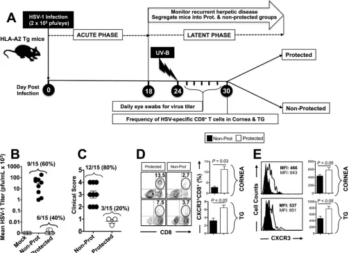
Frequent HSV-specific CXCR3ⴙCD3ⴙCD8ⴙT cells in trigeminal ganglia and cornea is associated with protection against virus shedding and recurrent ocular herpes disease. A group of 20 age- and sex-matched HLA-A*0201 transgenic mice (HLA Tg mice) were infected ocularly with 2⫻105PFU of HSV-1 (strain McKrae) without
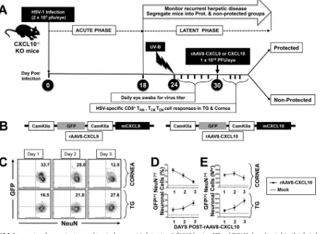
corneal scarification as described in Materials and Methods. On day 24 (i.e., during the latent phase), the eyes of 15 infected animals that survived acute infection were exposed to UV-B irradiation in order to induce reactivation of HSV-1 from latently infected trigeminal ganglia (TG). UV-B irradiation led to virus shedding in tears and recurrent corneal herpetic disease, as we recently described (5, 6). The timeline of HSV-1 infection, UV-B irradiation, and subsequent immunological and virological assays is illustrated in Fig. 1A. During the 10 days following UV-B irradiation, virus shedding in tears was detected in ⬃60% of latently infected susceptible HLA Tg mice (BALB/c genetic background) (Fig. 1B), and⬃80% developed apparent recurrent corneal her-petic disease (Fig. 1A and C).
Subsequently, mice were segregated into two groups: protected mice with no recurrent corneal herpes disease (with the severity of disease scored below 1 on a scale of 0 to 4) and nonprotected mice with severe recurrent corneal herpetic disease (score of 2 to 4 on a scale of 0 to 4). On day 30 postinfection, protected and nonprotected HLA Tg mice were euthanized, and their corneas and TG were harvested for immunological assays. The percentages of cornea- and TG-resident CXCR3⫹CD8⫹T cells specific to the human HLA-A*0201-restricted HSV-1 epitope, VP11/12220-228, were then compared for
protected and nonprotected mice. As shown in Fig. 1D, significantly higher percentages of CXCR3⫹CD8⫹T cells specific to the immunodominant HSV-1 VP11/12220-228epitope
were detected in the corneas and TG of protected mice compared to the corneas and TG of nonprotected mice (13.5% versus 2.7% and 7.5% versus 3.7%, respectively;P⬍ 0.05). Moreover, increased levels of CXCR3 expression were detected on CD8⫹T cells in the corneas and TG of protected mice compared to nonprotected mice (Fig. 1E).
Altogether, these results indicate that frequent HSV-specific CXCR3⫹CD8⫹T cells present in the corneas and TG of protected mice compared to nonprotected mice are associated with less virus shedding in tears and less recurrent corneal herpetic disease. This suggests a potential role of the CXCR3 signaling pathway in mobilizing protective HSV-specific CD8⫹T cells into infected tissues.
CXCR3-dependent accumulation of functional CD103ⴙ CD8ⴙ T cells in the cornea and trigeminal ganglia is associated with a reduction of virus shedding in tears and prevention of recurrent corneal herpetic disease.We next determined whether the CXCR3 signaling pathway might also be crucial in the effector function of HSV-specific CD8⫹T cells in the cornea and TG. Since HLA Tg mice deficient for CXCR3
(i.e., CXCR3⫺/⫺) are not available, we turned to the CXCR3-deficient mice that are
available on the B6 genetic background and their wild-type (WT) B6 control littermates (33, 34). Both WT and CXCR3⫺/⫺deficient mice develop H2b-restricted CD8⫹T cells
specific to the immunodominant HSV-1 gB epitope (gB498-505) following ocular
infec-tion with HSV-1.
A group of 20 age- and sex-matched WT B6 mice and 20 CXCR3⫺/⫺deficient mice
were ocularly infected with 2⫻105PFU of HSV-1 as described above and shown in Fig.
1A. On day 24 (i.e., during the latent phase), UV-B irradiation was used to induce virus
on November 7, 2019 by guest
http://jvi.asm.org/
reactivation and recurrent corneal herpetic disease in both WT and CXCR3⫺/⫺mice that
survive acute infection. We then compared the frequency of HSV-specific CD8⫹T cells
that accumulate in the corneas and TG of 10 WT B6 and CXCR3⫺/⫺ mice 30 days
following UV-B-induced reactivation. We also correlated the cytotoxic and IFN-␥ pro-ductive functions of CD8⫹T cells in TG and corneas of WT and CXCR3⫺/⫺mice with
virus shedding and recurrent corneal herpetic disease following UV-B-induced reacti-vation.
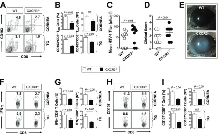
Significantly higher percentages (P⬍0.05) and numbers (P⫽0.04) of CD103high
CD8⫹T cells were detected in the TG of WT B6 mice compared to the TG of CXCR3⫺/⫺
deficient mice (Fig. 2A and B). In contrast, there were slight, but nonsignificant, differences in the numbers of CD8⫹T cells that were detected in the corneas of WT B6
mice compared to the corneas of CXCR3⫺/⫺deficient mice (P⬎0.05; Fig. 2A and B).
As shown in Fig. 2C, UV-B-induced reactivation led to significantly more virus shedding in the eyes of CXCR3⫺/⫺deficient mice compared to WT B6 mice (P⫽0.03).
FIG 1Trigeminal ganglia- and cornea-resident HSV-specific CXCR3⫹CD8⫹T cells are associated with protection against recurrent ocular herpesvirus infection
and disease. A group of 20 HLA Tg mice (6 to 8 weeks old) were ocularly infected on day 0 with 2⫻105PFU of HSV-1 (strain McKrae), as described in Materials and Methods. After establishment of latency (24 days postinfection), reactivation of latent virus was induced following irradiation with UV-B light. (A) Schematic representation of the time line of immunological and virological analyses in the UV-B mouse model of induced recurrent herpes. (B) Tears were collected daily for 10 days, and the presence of infectious virus in the tears was determined by plaque assay. The titer of virus detected in tear samples 6 days after UV-B irradiation was expressed as the mean virus load (in PFU per milliliter). (C) Recurrent corneal herpetic disease was monitored for up to 30 days after UV-B irradiation. Mice were observed daily for eye disease and clinically scored on a scale from 0 to 4. (D) Ten mice were euthanized on day 30 after UV-B exposure, and single-cell suspensions from corneas and TG were obtained after collagenase treatment, stained for markers of CXCR3⫹CD3⫹CD8⫹T cells, and analyzed
by FACS. Representative FACS plot of the frequency of VP11/12220-228-specific CXCR3⫹CD3⫹CD8⫹T cells detected in the corneas and TG of protected mice versus nonprotected (Non-Prot) mice. (E) Level of expression of CXCR3 receptor on CD8⫹T cells from corneas and TG of protected mice versus nonprotected
mice. The data are representative of two independent experiments. MFI, mean fluorescence intensity.
Srivastava et al. Journal of Virology
on November 7, 2019 by guest
http://jvi.asm.org/
[image:4.585.42.545.74.439.2]Moreover, during the 30 days following UV-B irradiation, 6 out of 10 CXCR3⫺/⫺deficient
mice compared to only 2 out of 10 WT B6 mice developed severe corneal lesions with high scores of 3 and 4 (P⫽0.04; Fig. 2D and E). On the other hand, the corneas and TG of CXCR3⫺/⫺deficient mice had significantly fewer IFN-␥-producing CD8⫹T cells
compared to the corneas and TG of WT B6 mice (P⬍ 0.05; Fig. 2F and G). Similarly, fewer CD107⫹CD8⫹T cells were detected in the corneas and TG of CXCR3⫺/⫺deficient
mice compared to the corneas and TG of WT B6 mice (P⬍0.05; Fig. 2H and I). Altogether, these results demonstrate a crucial role of the CXCR3 signaling pathway in the mobilization and/or accumulation of IFN-␥-producing cytotoxic CD107⫹CD8⫹T
cells in the cornea and TG. This suggests that higher mobilization and/or accumulation of IFN-␥-producing cytotoxic CD107⫹ CD8⫹ T cells is associated with control of
UV-B-induced reactivation, less virus shedding, and less recurrent herpetic disease. Increased recurrent ocular herpes is associated with reduced numbers of HSV-specific CD8ⴙT
RMand TEMcells in TG and corneas of latently infected CXCL10ⴚ/ⴚ
deficient mice following UV-B-induced HSV-1 reactivation.We next determined the role of the CXCR3 signaling pathway in the mobilization and/or accumulation of the three major CD8⫹T cell subpopulations (i.e., CD8⫹T
EMcells, CD8⫹TCMcells, and CD8⫹
TRMcells) in the cornea and TG, in addition to their relative contribution in the observed
protection against virus shedding and recurrent herpetic disease. Of the three T cell-attracting ligands of CXCR3 (i.e., CXCL9, CXCL10, and CXCL11), CXCL10 is the only chemokine that is predominantly expressed in human TG (19, 35). We therefore
FIG 2CXCR3⫺/⫺deficient mice developed less functional CD8⫹T cells in trigeminal ganglia and more recurrent corneal herpetic disease
compared to wild-type (WT) B6 mice. A group of CXCR3⫺/⫺and WT mice (6 to 8 weeks old) were ocularly infected on day 0 with 2⫻105 PFU of HSV-1 (strain McKrae). After establishment of latency (24 days postinfection), reactivation of latent virus was induced after irradiation with UV-B. Tears were collected daily for 6 days after UV-B irradiation, and mice were euthanized on day 6 after UV-B exposure. Single-cell suspensions of corneas and TG were obtained after collagenase treatment and stained for markers of CD8⫹T
RMcells, IFN-␥, and CD107, and analyzed by FACS. (A) Representative FACS plot of the frequency of HSV-specific CD8⫹T
RMcells detected in the corneas and TG of WT and CXCR3⫺/⫺mice. (B) Average frequencies (left) and absolute numbers (right) of HSV-specific CD8⫹T
RMcells detected in the corneas and TG of WT and CXCR3⫺/⫺mice. (C) Presence of infectious virus in the tears of WT and CXCR3⫺/⫺mice after UV-B
treatment. The data are expressed as mean of virus load (in PFU per milliliter). (D) Mice were observed daily for eye disease and clinically scored from 0 to 4. (E) Representative slit lamp images of WT and CXCR3⫺/⫺mice corneas. (F and H) Representative FACS data of the
frequencies of IFN-␥⫹CD8⫹T cells and CD107⫹CD8⫹T cells detected in the corneas and TG of WT and CXCR3⫺/⫺mice 6 days after UV-B
exposure. (G and I) Average frequencies and absolute numbers of IFN-␥⫹CD8⫹T cells and CD107⫹CD8⫹T cells in the corneas and TG
of WT and CXCR3⫺/⫺mice. The results are representative of two independent experiments. The indicatedPvalues, calculated using
unpairedttest, show statistical significance between WT and CXCR3⫺/⫺deficient mice. NS, not significant.
on November 7, 2019 by guest
http://jvi.asm.org/
[image:5.585.42.474.70.336.2]investigated whether lack of the CXCL10 chemokine would affect the mobilization and/or accumulation and effector function of CD8⫹T
EMcells, CD8⫹TCMcells, and CD8⫹
TRMcells in the cornea and TG following UV-B-induced reactivation.
A group of 20 sex- and age-matched CXCL10-deficient mice (CXCL10⫺/⫺) and 20 WT
B6 control littermates were infected ocularly on day 0 with 2 ⫻ 105 PFU of HSV-1
without corneal scarification as described in Materials and Methods. On day 24 (i.e., during the latent phase), UV-B irradiation was used to induce virus reactivation in surviving latently infected animals as described above.
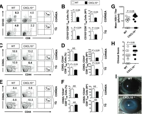
Following UV-B reactivation, significantly lower percentages (Fig. 3A and B, left) and numbers (Fig. 3B, right) of CD103highCCR7lowCD11ahighCD69highCD8⫹T
RMcells were
detected in the corneas and TG of CXCL10⫺/⫺mice than in the corneas and TG of WT
mice (P⫽0.05 for the cornea andP⫽0.04 for the TG). Similarly, a significantly lower percentage (Fig. 3C and D, left; P ⫽0.03) and number (Fig. 3D, right; P ⫽ 0.04) of CD44highCD62LlowCD8⫹T
EMcells were detected in TG of CXCL10⫺/⫺mice compared FIG 3CXCL10⫺/⫺deficient mice have fewer HSV-specific CD8⫹T
RMand TEMcells in the corneas and trigeminal ganglia and more virus shedding and more recurrent ocular herpetic disease compared to wild-type B6 mice. A group of CXCL10⫺/⫺and WT mice (6 to 8 weeks old) were ocularly
infected on day 0 with 2⫻105PFU of HSV-1 (strain McKrae) as described earlier. After establishment of latency (24 days postinfection), reactivation of latent virus was induced after irradiation with UV-B light. Ten mice were euthanized on day 30 after UV-B exposure, and single-cell suspensions from the corneas and TG were obtained after collagenase treatment at 37°C for an hour and stained for markers of CD8⫹T
RM, TEM, and TCMcells and analyzed by FACS. (A, C, and E) Representative FACS plots of the frequency of HSV-specific CD8⫹TRMcells (A), CD8⫹TEMcells
(C), and CD8⫹T
CMcells (E) detected in the corneas and TG of WT and CXCL10⫺/⫺mice. (B, D, and F) Average percentages (left) and absolute numbers (right) of HSV-specific CD8⫹TRMcells (B), CD8⫹TEMcells (D), and CD8⫹TCMcells (F) detected in the corneas and TG of WT and CXCL10⫺/⫺
mice. (G) Viral titer estimation detected in tear samples 6 days after UV-B irradiation expressed as mean of virus load (in PFU per milliliter). (H and I) Recurrent corneal herpetic disease was monitored for up to 30 days after UV-B irradiation. Mice were observed daily for eye disease and clinically scored on a scale of 0 to 4. The results are representative of two independent experiments. The indicatedPvalues, calculated using unpairedt
test, show statistical significance between WT and CXCL10⫺/⫺deficient mice. NS, not significant.
Srivastava et al. Journal of Virology
on November 7, 2019 by guest
http://jvi.asm.org/
[image:6.585.44.496.72.435.2]to TG of WT mice. However, no significant differences in the frequencies of CD44high
CD62LhighCD8⫹T
CMcells were detected in the corneas and TG of CXCL10⫺/⫺mice
compared to the corneas and TG of WT mice (Fig. 3E and F, left;P⬎0.05).
Moreover, as shown in Fig. 3G, UV-B-induced reactivation led to significantly more virus shedding in the eyes of CXCL10⫺/⫺deficient mice compared to WT B6 mice (P⬍
0.05). During the 30 days following UV-B irradiation, 7 out of 10 CXCL10⫺/⫺deficient
mice compared to only 3 out of 10 WT B6 mice developed severe corneal lesions with high scores of 3 and 4 (P⫽0.04; Fig. 3H and I).
These results point to a pivotal role of the CXCR3/CXCL10 signaling pathway in the mobilization and/or accumulation of antiviral CD8⫹TEMcells and CD8⫹TRMcells, but
not CD8⫹TCMcells, in the cornea and TG following UV-B-induced reactivation. These
results indicate that the failure to contain recurrent corneal herpetic infection and disease in CXCL10⫺/⫺deficient mice positively correlated with the loss of cornea- and
TG-resident CD8⫹TEMcells and CD8⫹TRMcells, but not CD8⫹TCMcells. These results
suggest a crucial role of CD8⫹TEMcells and CD8⫹TRMcells, but not CD8⫹TCMcells, in
protection against virus shedding and recurrent herpetic disease.
More dysfunctional VISTAⴙ PD-1ⴙ CD8ⴙ T cells are present in the TG and corneas of latently infected CXCL10ⴚ/ⴚdeficient mice than in wild-type B6 mice. A group of 20 sex- and age-matched CXCL10-deficient mice (CXCL10⫺/⫺) and 20 WT B6
control littermates were infected ocularly on day 0 with 2 ⫻ 105 PFU of HSV-1 as
described above. Ten days after UV-B-induced reactivation, the function and dysfunc-tion of CD8⫹T cells in the corneas and TG of 10 randomly picked CXCL10⫺/⫺deficient
mice and 10 WT mice were analyzed using several markers of immunological function and dysfunction. These markers included the production of antiviral IFN-␥, CD107a/b
cytotoxic degranulation, and expression of markers of exhaustion, including pro-grammed cell death protein 1 (also known as PD-1), T cell immunoreceptor with Ig and ITIM domains (also known as TIGIT), the V-domain Ig suppressor of T cell activation (also known as VISTA, 2B4, and Tim-3) (36–38).
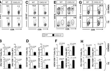
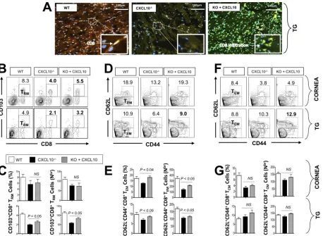
As shown in Fig. 4A and B, following UV-B-induced reactivation, significantly low numbers of IFN-␥-producing CD8⫹ T cells were detected in the corneas and TG of CXCL10⫺/⫺ deficient mice compared to B6 mice (P ⬍ 0.05). The percentage and number of IFN-␥producing CD8⫹T cells were reduced approximately twofold in TG of CXCL10⫺/⫺mice compared to TG of WT B6 mice. There is also a significant reduction of cytotoxic CD107⫹CD8⫹T cells in the corneas and TG of CXCL10⫺/⫺deficient mice
compared to the corneas and TG of B6 mice (P⬍0.05; Fig. 4C and D).
As shown in Fig. S1 in the supplemental material, following UV-B-induced reactiva-tion, significantly less functional TNF-␣-producing CD8⫹T cells were detected in both
the corneas and TG of CXCL10⫺/⫺deficient mice compared to the corneas and TG of
B6 mice (P⬍0.05). Similarly, significantly lower expression of GzmB and EOMES were detected on CD8⫹T cells from both the corneas and TG of CXCL10⫺/⫺deficient mice
compared to the corneas and TG of B6 mice (P⬍0.05). In contrast, significantly lower T-bet expression was detected only on CD8⫹T cells from TG of CXCL10⫺/⫺deficient
mice compared to TG of B6 mice (P⬍0.05).
Furthermore, as shown in Fig. 4E and F, there was an approximately twofold increase in the percentage and number of VISTA⫹ CD8⫹ T cells in the corneas and TG of
CXCL10⫺/⫺mice compared to the corneas and TG of WT B6 mice. A significant increase
in the percentage of PD-1⫹CD8⫹T cells was also detected in the corneas and TG of
CXCL10⫺/⫺mice compared to the corneas and TG of WT B6 mice (P⬍0.05; Fig. 4G and
H). CXCL10⫺/⫺mice also exhibited higher frequencies of Tim-3⫹CD8⫹T cells, 2B4⫹
CD8⫹T cells, and TIGIT⫹CD8⫹T cells in corneas and TG compared to WT mice (Fig. S2;
P⬍0.05).
Altogether, these results suggest that the lack of CXCL10 chemokine is associated with the following: (i) a significant impairment in the effector functions of CD8⫹T cells
(antiviral IFN-␥and CD107a/bcytotoxic degranulation) in both the corneas and TG of
CXCL10⫺/⫺ deficient mice; and (ii) a significant increase in exhausted PD-1⫹ CD8⫹,
TIGIT⫹CD8⫹, Tim-3⫹CD8⫹, 2B4⫹CD8⫹, and VISTA⫹CD8⫹T cells in the corneas and
on November 7, 2019 by guest
http://jvi.asm.org/
TG of CXCL10⫺/⫺deficient mice. Beside the reduced number of antiviral CD8⫹T EMand
TRMcells in the corneas and TG of CXCL10⫺/⫺deficient mice, the reduced function and
increased dysfunction of these CD8⫹T cells may further contribute to the enhanced
susceptibility to recurrent ocular herpetic infection and disease observed in CXCL10⫺/⫺
deficient mice following virus reactivation from latency. The results confirm a pivotal role of the CXCR3/CXCL10 signaling pathway in the antiviral CD8⫹T cell immunity in
the cornea and TG following virus reactivation.
Topical ocular application of neurotropic AAV8 vectors expressing CXCL10, but not CXCL9, chemokine promotes the protective function of antiviral CD8ⴙT
RM
cells and CD8ⴙTEM cells in the trigeminal ganglia of CXCL10ⴚ/ⴚmice. We next
determined whether correcting the apparent deficiency of CXCL10 chemokine in the TG of CXCL10⫺/⫺deficient mice would increase the number of CD8⫹T cells in TG,
reverse their dysfunction, and rescue their protective function. We used the neurotropic recombinant adeno-associated virus type 8 (rAAV8) vector that expresses CXCL10 chemokine locally in latently infected TG under the neurotropic calcium/calmodulin-dependent protein kinase type II subunit alpha (CamKII-␣) promoter.
As illustrated in Fig. 5, CXCL10⫺/⫺mice (n⫽20) were infected with HSV-1 on day
0, and the eyes of all animals were exposed on day 24 to UV-B irradiation in order to induce virus reactivation as described above. On day 30, half of the animals received topical application of 1⫻107PFU/eye of a neurotropic recombinant rAAV8 expressing
either CXCL10 or CXCL9 mouse chemokine together with green fluorescent protein (GFP), both under the neurotropic CamKII-␣ promoter (i.e., CXCL9 or rAAV8-CXCL10 [Fig. 5B]). Following topical application of rAAV8-rAAV8-CXCL10, the GFP protein
FIG 4More dysfunctional CD8⫹T cells are present in the corneas and trigeminal ganglia of CXCL10⫺/⫺deficient mice compared to the corneas
and trigeminal ganglia of wild-type B6 mice. HSV-1 latently infected WT and CXCL10⫺/⫺mice (n⫽10) were euthanized 6 days after UV-B exposure. Single-cell suspensions from corneas and TG were obtained after collagenase treatment at 37°C and stained for IFN-␥, CD107, VISTA, and PD-1 and analyzed by FACS. (A, C, E, and G) Representative FACS plots of the frequency of HSV-specific IFN-␥⫹CD8⫹T cells (A), CD107⫹CD8⫹
T cells (C), VISTA⫹CD8⫹T cells (E), and PD-1⫹CD8⫹T cells (G) detected in the corneas and TG of WT and CXCL10⫺/⫺mice. (B, D, F, and H) Average
percentages (left) and absolute numbers (right) of HSV-specific IFN-␥⫹CD8⫹T cells (B), CD107⫹CD8⫹T cells (D), VISTA⫹CD8⫹T cells (F), and PD-1⫹CD8⫹T cells (H) detected in the corneas and TG of WT and CXCL10⫺/⫺ mice. The results are representative of two independent
experiments. The indicatedPvalues, calculated using unpairedttest, show statistical significance between WT and CXCL10⫺/⫺deficient mice.
Srivastava et al. Journal of Virology
on November 7, 2019 by guest
http://jvi.asm.org/
[image:8.585.46.501.71.360.2]expression was detected by fluorescence-activated cell sorting (FACS) in NeuN⫹
neu-ronal cell suspensions from both the corneas and TG of CXCL10⫺/⫺deficient mice
within 1 day (Fig. 5C to E). While GFP expression decreased in the cornea over 3 days, its expression gradually increased in TG over time.
Moreover, a significant increase in CD8⫹T cells was detected by immunostaining in
TG of CXCL10⫺/⫺mice following UV-B-induced reactivation and topical ocular
appli-cation of rAAV8-CXCL10 compared to TG of untreated CXCL10⫺/⫺mice (Fig. 6A). We
next determined whether treatment with rAAV8-CXCL10 resulted in the mobilization and/or accumulation of the three major CD8⫹T cell subpopulations (i.e., CD8⫹ T
EM
cells, CD8⫹T
CMcells, and CD8⫹TRMcells) in TG and their relative contribution in the
protection against recurrent herpetic disease following UV-B-induced reactivation. We used FACS analysis to quantify the three CD8⫹T cell subpopulations in TG of
rAAV8-CXCL10-treated and untreated CXCL10⫺/⫺mice.
Topical ocular application of rAAV8-CXCL10 was associated with a significant in-crease in the percentage and number of CD103⫹ CD8⫹ T
RM cells (Fig. 6B and C)
FIG 5Coexpression of a neurotropic recombinant adeno-associated virus type 8 (fAAV8) bearing GFP and CXCL10 chemokine in latently infected trigeminal ganglia of CXCL10⫺/⫺mice following topical ocular application. CXCL10⫺/⫺mice (n⫽20) were ocularly infected using 2⫻105 wild-type McKrae (HSV-1) strain. Twenty-four days postinfection, when latency was fully established, animals were exposed to UV-B irradiation to induce virus reactivation. On day 30, half of the animals (n⫽10) received topical application of 1⫻107PFU/eye of a neurotropic recombinant adeno-associated virus type 8 expressing either CXCL9 or CXCL10 mouse chemokine together with GFP, both under the neurotropic CamKII-␣ promoter. The other half of the animals (n⫽10) received topical application of 1⫻107PFU/eye of an AAV8 empty vector (Mock). Mice were euthanized on day 1, day 2, and day 3 after rAAV8-CXCL9 or rAAV8-CXCL10 treatment. (A) Schematic representation of the time line of HSV-1 infection and rAAV8-CXCL9 and rAAV8-CXCL10 treatment in the UV-B-CXCL10⫺/⫺mouse model. KO, knockout. (B) Structure of recombinant AAV8
vector bearing the T cell-attracting chemokine CXCL9 or CXCL10 tagged with GFP with both chemokines expressed under the CamKII-␣promoter. (C) Representative FACS plot of the frequency of NeuN⫹neuronal cells expressing GFP in cornea and TG, detected on days 1, 2, and 3 after
rAAV8-CXCL10 treatment, in corneas and TG of CXC10⫺/⫺mice. (D) Average frequencies (left) and absolute number (right) of NeuN⫹neuronal
cells expressing GFP in cornea and TG, detected on days 1, 2, and 3 after rAAV8-CXCL10 treatment, in the corneas and TG of CXC10⫺/⫺mice. The
data are representative of two independent experiments. The indicatedPvalues, calculated using unpairedttest, show statistical significance between CXCL10-treated and untreated mice.
on November 7, 2019 by guest
http://jvi.asm.org/
[image:9.585.44.500.76.406.2]detected in TG of rAAV8-CXCL10-treated CXCL10⫺/⫺mice compared to TG of untreated
CXCL10⫺/⫺ mice (P ⬍ 0.05 for the cornea and P ⫽ 0.04 for the TG). Similarly, a
significantly lower percentage (Fig. 6D and E, left;P⫽0.04) and number (Fig. 6E, right; P⬍0.05) of CD44highCD62LlowCD8⫹T
RMcells were detected in TG of
rAAV8-CXCL10-treated CXCL10⫺/⫺mice compared to TG of untreated CXCL10⫺/⫺mice. However, no
significant differences in the frequencies of CD44highCD62LhighCD8⫹T
CMcells were
detected in the corneas and TG of rAAV8-CXCL10-treated CXCL10⫺/⫺mice compared
to the corneas and TG of untreated CXCL10⫺/⫺mice (Fig. 6F and G;P⬎0.05).
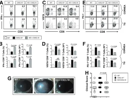
The IFN-␥production by CD8⫹T cells within both the cornea and TG of CXCL10⫺/⫺
deficient mice treated with rAAV8-CXCL10 was improved to a significantly higher level compared to untreated CXCL10⫺/⫺ deficient mice (P ⫽ 0.05; Fig. 7A and B). The FIG 6Topical ocular application of neurotropic AAV8 vector expressing CXCL10 increased the number of CD8⫹T
RMand TEMcells in the corneas and trigeminal ganglia of latently infected CXCL10⫺/⫺mice. Wild-type B6 mice (n⫽10) and CXCL10⫺/⫺mice (n⫽20) were ocularly infected using
2⫻105wild-type McKrae (HSV-1) strain. At 24 days postinfection, when latency was fully established, reactivation of latent HSV-1 infection was induced after UV-B irradiation. Six days after UV-B irradiation, half of CXCL10⫺/⫺mice (n⫽10) received topical application of 1⫻107PFU/eye of a neurotropic rAAV8 expressing either CXCL10 chemokine. The other half of the animals (n⫽10) received topical application of 1⫻107 PFU/eye of an AAV8 empty vector. (A) TG sections from WT and CXCL10⫺/⫺mice were costained using a MAb specific to CD8⫹T cells or to
neurons, together with GFP. For the wild-type B6 mice, PE-labeled CXCL10 (red), 4=,6=-diamidino-2-phenylindole (DAPI) (DNA stain) (blue), and CD8 infiltration orange color (yellow) are indicated. For the CXCL10⫺/⫺mice, PE-labeled CXCL10 (red), DAPI (blue), and CD8⫹T cells (yellow) are
indicated. For the CXCL10⫺/⫺mice 3 days after rAAV8-CXCL10 treatment, CXCL10 (red), DAPI (blue), CD8⫹cells (yellow) and GFP (green) are
indicated. Single-cell suspensions from corneas and TG were obtained after collagenase treatment at 37°C for an hour and stained for markers of CD8⫹T
RMcells, IFN-␥, and CD107a/band analyzed by FACS. CXCL10⫺/⫺mice were euthanized on day 30 after UV-B exposure, and single-cell suspensions from the corneas and TG were obtained after collagenase treatment at 37°C for an hour and stained for markers of CD8⫹TRM, TEM,
and TCMcells and analyzed by FACS. (B, D, and F) Representative FACS plots of the frequencies of HSV-specific CD8⫹TRMcells (B), CD8⫹TEMcells (D), and CD8⫹TCMcells (F) detected in the cornesa and TG of WT mice (left), untreated CXCL10⫺/⫺mice (middle), and CXCL10⫺/⫺mice treated
with rAAV8-CXCL10 (right). (C, E, and G) Average percentages (left) and absolute numbers (right) of HSV-specificCD8⫹T
RMcells (C), CD8⫹TEMcells (E), and CD8⫹TCMcells (G) detected in the corneas and TG of WT and CXCL10⫺/⫺mice untreated and treated with rAAV8-CXCL10. The data are
representative of two independent experiments. The indicated P values were calculated using one-way ANOVA and pairwise multiple comparisons were performed stratified by experiment. Bonferroni correction was utilized to adjust the multiple comparisons. SAS v.9.4 (Statistical Analysis System, Cary, NC) was used. Graphs were prepared with GraphPad Prism software (San Diego, CA). Data are expressed as the mean⫹ SD. Error bars show SEM.
Srivastava et al. Journal of Virology
on November 7, 2019 by guest
http://jvi.asm.org/
[image:10.585.44.502.70.405.2]exhaustion of cornea- and TG-resident CD8⫹T cells appeared to be reversed following
CXCL10 local treatment, as fewer VISTA⫹CD8⫹T cells (Fig. 7C and D) and fewer PD-1⫹
CD8⫹ T cells (Fig. 7E and F) were detected in TG and corneas of CXCL10⫺/⫺ mice
following treatment with rAAV8-CXCL10 compared to untreated mice. Finally, signifi-cantly less severe (Fig. 7G) and less frequent (Fig. 7H) recurrent corneal herpetic disease was detected in CXCL10⫺/⫺ deficient mice that were treated with rAAV8-CXCL10
compared to untreated CXCL10⫺/⫺mice. In contrast, no effect on corneal herpetic
disease was seen in CXCL10⫺/⫺mice treated with rAAV8-CXCL9 or an empty rAAV8
vector (data not shown).
Altogether, these results demonstrate for the first time a successful coexpression of the neurotropic recombinant adeno-associated virus type 8 bearing GFP and CXCL10 chemokine in latently infected trigeminal ganglia of CXCL10⫺/⫺mice following topical
ocular application. These results also demonstrate for the first time that topical ocular application of neurotropic rAAV8 vector expressing CXCL10 chemokine increased the number of CD8⫹T
EMand TRMcells in latently infected TG, reversed their dysfunction,
and rescued their protective function against recurrent herpetic disease following reactivation.
FIG 7Topical ocular application of neurotropic AAV8 vector expressing CXCL10 promotes the function of antiviral CD8⫹T cells in the
trigeminal ganglia and corneas of CXCL10⫺/⫺mice. WT B6 and CXCL10⫺/⫺mice were ocularly infected using 2⫻105wild-type McKrae (HSV-1) strain. At 24 days postinfection, when latency was fully established, reactivation of latent HSV-1 infection was induced after UV-B irradiation. Six days after UV-B irradiation, the CXCL10⫺/⫺mice were treated with rAAV8-CXCL10 using 1⫻107PFU in each eye. Single-cell suspensions from corneas and TG were obtained after collagenase treatment at 37°C for an hour and stained for CD8⫹T cells, IFN-␥, VISTA,
and PD-1 and analyzed by FACS. (A, C, and E) Representative FACS plots of the frequencies of HSV-specific IFN-␥⫹CD8⫹T cells (A), VISTA⫹
CD8⫹T cells (C), and PD-1⫹CD8⫹T cells (E) detected in the corneas and TG of WT and CXCL10⫺/⫺mice untreated and treated with
rAAV8-CXCL10. (B, D, and F) Average percentages (left) and absolute numbers (right) of HSV-specific IFN-␥⫹CD8⫹T cells (B), VISTA⫹CD8⫹
T cells (D), and PD-1⫹CD8⫹T cells (F) detected in the corneas and TG of WT and CXCL10⫺/⫺mice untreated and treated with
rAAV8-CXCL10. (G and H) Recurrent corneal herpetic disease was monitored for up to 30 days after UV-B irradiation and rAAV8-CXCL10 treatment. CXCL10⫺/⫺mice were observed daily for eye disease and clinically scored on a scale of 0 to 4. The results are representative
of two independent experiments. The indicatedPvalues, calculated using unpairedttest, show statistical significance between WT and CXCL10⫺/⫺deficient mice.
on November 7, 2019 by guest
http://jvi.asm.org/
[image:11.585.44.472.70.395.2]DISCUSSION
In the present study, we demonstrate that the lack of CXCL10 chemokine or its receptor CXCR3 compromises the mobilization of functional CD8⫹TEMand CD8⫹TRM
cells within TG of latently infected mice following UV-B-induced reactivation. This is associated with an increase in recurrent ocular herpesvirus infection and disease. Inversely, increasing the amount of CXCL10 in TG of latently infected CXCL10-deficient mice rescued local CD8⫹TEMand CD8⫹TRMcell responses and improved protection
against recurrent ocular herpes. Based on these findings, a novel “prime/pull” thera-peutic ocular herpes vaccine strategy that would increase the number of functional CD8⫹ TEM and CD8⫹ TRM cells within HSV-1 latently infected TG is proposed and
discussed.
Work from our laboratory and others has shown that the CD8⫹T cells are critical for
protection against recurrent ocular herpesvirus infection and disease (23, 24, 39). However, the relative contribution of each of the CD8⫹ T cell subpopulations in protection against recurrent herpes has not been reported. The three major subpopu-lations of memory T cells (i.e., CD8⫹TEMcells, CD8⫹TCMcells, and CD8⫹TRMcells) differ
in their phenotype, function, and anatomic distribution (12, 14, 40). TCM cells are
CD103lowCD62LhighCCR7high. T
EMcells are CD103lowCD62LlowCCR7low. TRMcells are
CD103highCD62LlowCCR7lowCD11ahighCD69high(12, 14, 40). Compared to T CMcells
which reside mostly in lymphoid tissues, TEMcells, which reside mostly in nonlymphoid
tissues, have a decreased ability to traffic to lymphoid tissues and lack CD62L and CCR7 (molecules directing lymph node entry) and express chemokine receptors such as CCR5 that are associated with homing to inflammatory sites (41, 42). Compared to TCMand
TEMcells, the TRMcells are generated locally within peripheral tissues, do not reenter
the circulation system, and appear therefore to play an essential role in locally guarding these peripheral tissues from secondary infections. To the best of our knowledge, this is the first report that demonstrates a crucial role for both the CD8⫹TEMand CD8⫹TRM
cell subpopulations, but not the CD8⫹ T
CM subpopulation, in protection against
recurrent herpesvirus infection and disease. Thus, based on these new findings, a worthy goal of a herpes immunotherapeutic vaccine should be to develop a vaccine strategy that would generate and maintain a sufficient number of antiviral memory CD8⫹TEMand CD8⫹TRMcells within peripheral tissues, such as latently infected TG, the
root of virus reactivation that leads to virus shedding and recurrent ocular herpetic disease (23–26). However, this goal remains unattainable because the TG appear to be a “closed immunological compartment” to accepting homing of conventional circulat-ing memory CD8⫹T cell subpopulations such as the CD8⫹ TEM cells that could be
migrating into latently infected TG from the neighboring draining lymph nodes through the circulation system (14, 43). To overcome this challenge, our study dem-onstrates for the first time that changing the microenvironment of latently infected TG in CXCL10⫺/⫺deficient mice by increasing the amount of local CXCL10 chemokine had the following effects: (i) rescued the number of both CD8⫹T
EMand CD8⫹TRM cell
subpopulations in TG, (ii) reversed the exhaustion of CD8⫹TEMand CD8⫹TRMcells in
TG, and (iii) reduced recurrent corneal herpetic disease. It is likely that accumulation of a sufficient number of CD8⫹T
EMand CD8⫹TRMcells within latently infected TG, by a
yet-to-be determined CXCL10/CXCR3-dependent mechanism, reduced virus reactiva-tion from latency. Since the TG appear to be an immunological compartment that is closed during latency to accepting homing of circulating CD8⫹T
CMand CD8⫹TEMcells,
which could be coming from the draining lymph nodes through the circulation system (14, 43), it is very likely that TRMcells are the main T cell subpopulation that contribute
to suppressing (or aborting) attempts of HSV-1 reactivation from latently infected neurons.
An average percentage of 7.5% to 12.5% of CXCR3⫹CD8⫹T cells was detected in latently infected wild-type mice compared to an average low percentage of 2 to 2.5% CXCR3⫹CD8⫹T cells in latently infected CXCR3⫺/⫺and CXCL10⫺/⫺mice. An
imme-diate practical application of these findings is the development of an ocular herpes
Srivastava et al. Journal of Virology
on November 7, 2019 by guest
http://jvi.asm.org/
“prime/prime” immunotherapeutic vaccine strategy that will include T cell-attracting chemokines, such as the CXCL10 chemokine, to “pull” more antiviral CD8⫹T cells from
the circulation system and boost their number and function within latently infected TG. A powerful “prime/pool” immunotherapeutic vaccine might be expected to stop or reduce reactivation of HSV-1 from latency with the potential to produce a sustained clinical reduction of blinding recurrent herpetic disease. A human “prime/pool” thera-peutic strategy is currently being explored in our laboratory using the “humanized” HLA Tg mouse and HLA Tg rabbit models of herpes reactivation. In these models, CD8⫹T
cells are first primed with human immunodominant CD8⫹T cell epitopes selected in
HSV-1 proteins (prime) and then treated with the AAV8 or AAV9 vectors expressing T cell-attracting chemokines, CXCL9, CXCL10 and CXCL11, in the TG (pull) to maintain an increase in the number of functional antiviral CD8⫹T cells in TG that will reduce virus
reactivation and protect against recurrent ocular herpesvirus infection and disease. This strategy is adapted from the recently reported “prime/pull” genital herpes vaccine strategy from the 2012 study of Shin and Iwasaki that used a mouse model of genital herpes (44). That study (44) first induced HSV-specific activated CD8⫹T cells in the
periphery (prime) and then pulled them into the vaginal mucocutaneous tissue follow-ing intravaginal administration of the CXCL10 chemokine.
Mice have been the small-animal model of choice for herpes immunologists and virologists (reviewed in reference 45). Results from many mouse models of herpesvirus infection and immunity have yielded tremendous insights into the protective and immunopathological mechanisms during primary acute infection (46–52). However, the extrapolation of results from mouse primary herpetic disease to human recurrent herpetic diseases, such as recurrent herpetic stromal keratitis (rHSK), is yet to be proven. This is mainly because the immune mechanisms that operate during primary acute infection appear to be different from those that operate during recurrent ocular herpetic diseases (53–55). Thus, the inadequacy of many currently used mouse models of primary acute herpesvirus infection makes it challenging to explore the immune mechanisms that lead to protection against recurrent herpes (56). A critical question remains as to which animal model would be the most appropriate to mimic the immunoprotective versus immunopathological aspect of recurrent herpes as occurs in humans? Unlike humans, spontaneous HSV-1 reactivation in latently infected mice and virus shedding in tears either does not occur at all or occurs at very low levels in mice (57). Only a handful of studies have employed the mouse model of UV-B light-induced recurrent herpetic corneal disease, mostly using C57BL/6 and BALB/c mice (54, 55, 58). The present study validated the UV-B light-induced recurrent herpetic corneal disease in the “humanized” HLA transgenic mouse model of ocular herpes (4, 24). The study demonstrated that both shedding of reactivated virus in tears and recurrent corneal HSV can be induced following UV-B exposure of latently infected HLA Tg mice. Moreover, considering the wealth of data addressing the protective mechanisms of CD8⫹T cells specific to mouse HSV-1 epitopes (mostly the gB
498-505 epitope in B6
mice), it is surprising how few reports exist exploring the protective mechanisms of CD8⫹T cells specific to human HSV-1 epitopes (27, 54, 58, 59). Our “humanized” HLA
Tg mice express the human HLA-A*0201 molecule, instead of mouse H2b major
histocompatibility complex (MHC) molecules (4). Using the UV-B/HLA Tg mouse model of recurrent ocular herpes, it is now possible to directly evaluate the efficacy of therapeutic herpes vaccine candidates that include human HLA-restricted CD8⫹T cell
epitopes. In our opinion, the HLA Tg mice combined with UV-B-induced recurrent disease is arguably the best available small-animal model to study the role of HLA-restricted CD8⫹T cells specific to human HSV-1 epitopes in protection against virus
shedding and recurrent herpetic disease.
The cellular and molecular mechanisms by which the CXCL10/CXCR3 chemokine axis (i) regulates trafficking and mobilization of HSV-specific CD8⫹T
EMand CD8⫹TRM
cells into HSV-1 latently infected TG and (ii) orchestrates CD8⫹T cell-mediated
protec-tive immunity to virus reactivation and subsequent recurrent herpetic disease remain to be determined. Wuest and Carr have previously demonstrated that, following
on November 7, 2019 by guest
http://jvi.asm.org/
corneal HSV-1 infection, both CXCL10 and CXCR3 played a role in reducing primary infection in the nervous system (31). The authors suggested that recruitment of CXCL10-expressing hematopoietic cells, dendritic cells, NK cells, and HSV-specific CD8⫹
T cells to the brain stem plays a role in controlling viral replication in the nervous system (60). CXCL10 deficiency was associated with a reduction in the mobilization of HSV-specific CD8⫹T cells into the brain as a result of dysregulation of CXCR3 signaling
(31). CXCR3⫺/⫺deficient mice are also reported to be susceptible to primary genital
HSV-2 infection (61–64). This susceptibility appeared to be associated with reduced cytotoxic function of CD8⫹T cells through impairment in expression of T-bet, perforin,
and GzmB, as well as a reduction in the recruitment of pDC and impairment in CD80 expression on CD11c⫹DC (61–64). It is important to underline that, unlike the above
studies that were done using mouse models of acute ocular and genital primary infections, our present studies used a mouse model of induced virus reactivation and recurrent herpes. Since the immune mechanisms that operate during primary acute and recurrent herpes infections are often different, the extrapolation of the above findings in primary/acute infection to our latency/reactivation model remains to be seen (53–55). Moreover, the above studies that used mouse models of ocular and genital primary infections did not report whether generation and mobilization of memory TCM, TEM, and TRMcell subpopulations were affected beyond the acute phase
of infection.
It is likely that CXCL10, which is produced locally by neurons, macrophages, epi-thelial cells, and T cells among other cells (14, 43), contributes to the accumulation and function of CXCR3⫹ CD8⫹T
EMand CXCR3⫹CD8⫹TRM cells in the TG and cornea.
Moreover, an elevated amount of IFN-␥produced by CD8⫹T cells may also contribute
to the generation of even more functional CXCR3⫹CD8⫹T
EMand CXCR3⫹CD8⫹TRM
cells in TG and corneal tissues. Compared to WT B6 mice, CXCL10⫺/⫺deficient mice
developed significantly less functional CXCR3⫹CD8⫹T
EMand CXCR3⫹CD8⫹TRMcells
in both TG and cornea and failed to contain recurrent ocular herpesvirus infection and disease. Furthermore, a significant reduction in the IFN-␥ production by TG and cornea-resident CD8⫹T cells was observed in CXCL10⫺/⫺ deficient mice. Increased
numbers of exhausted CD8⫹ T cells, expressing high levels of PD-1, V-domain Ig
suppressor of T cell activation (VISTA), and T cell Ig and ITIM domain (TIGIT) (VISTA and TIGIT recently discovered markers of exhaustion), were detected in TG and corneas of CXCL10⫺/⫺deficient mice and were associated with the failure to contain recurrent
ocular herpes. This suggests that the CXCL10/CXCR3 chemokine signaling pathway contributes, with a yet-to-be determined mechanism, to the generation of a large repertoire of exhausted HSV-specific CD8⫹T cells within latently infected TG, similar to
the finding we recently reported in HLA Tg rabbits (16). The finding that TG-resident CD8⫹T cells recognized HSV-1 human epitopes and expressed high levels of PD-1,
TIGIT, and VISTA differs from a recent suggestion that these cells are fully functional (65).
The current largely accepted paradigm is that exhaustion of T cells requires persis-tent exposure to a large amount of antigen (Ag). The amount of viral Ag produced in the latently infected TG of mice following UV-B reactivation is quite low, less than 1 positive neuron/TG (66–69). Thus, it is likely that the CXCL10/CXCR3 chemokine path-way inhibits exhaustion of CD8⫹T cells in TG through mechanisms other than Ag
persistence, as we previously discussed (70). Elucidating the mechanisms by which the CXCL10/CXCR3 pathway might be preventing CD8⫹T cell exhaustion is beyond the
scope of the present study. Nevertheless, our results demonstrate, for the first time, that the CXCL10/CXCR3 chemokine signaling pathway is of paramount importance in T cell immunity against recurrent ocular herpes. The results also suggest that coblockade of the PD-1, VISTA, and TIGIT immune checkpoints should be further explored to elicit potent antiviral CXCR3⫹CD8⫹T
EMand CXCR3⫹CD8⫹TRMcell responses that could
control or eliminate recurrent ocular herpes.
A high level of expression of PD-1 alone on T cells does not necessarily prove functional exhaustion. Both activated and exhausted T cells express PD-1, with
ex-Srivastava et al. Journal of Virology
on November 7, 2019 by guest
http://jvi.asm.org/
hausted T cells expressing higher and more persistent levels of PD-1 than activated T cells do (68, 71, 72). Therefore, instead of relying only on the level of expression of PD-1, in the present study, we also determined coexpression of TIM-3 and 2B4 markers of exhaustion on CD8⫹T cells, as well as of two newly reported markers of exhaustion,
TIGIT and VISTA (36–38, 73). PD-1, TIM-3, 2B4, TGIT, and VISTA were all coexpressed at various levels on CD8⫹T cells from TG of CXCL10⫺/⫺deficient mice compared to CD8⫹
T cells from TG of age- and sex-matched WT B6 mice. Mobilization of PD-1⫹CD8⫹T
cells, TIM3⫹CD8⫹T cells, 2B4⫹CD8⫹T cells, TGIT⫹CD8⫹T cells, and VISTA⫹CD8⫹T
cells in TG of CXCL10⫺/⫺deficient mice point to exhausted (dysfunctional) CD8⫹T cells
in TG. This was confirmed at the functional level by a decrease in the levels of CD107a/b
cytotoxic degranulation and by lower production of both IFN-␥and tumor necrosis factor alpha (TNF-␣). These results are consistent with our recent finding of a signifi-cantly higher level of exhaustion of HSV-1 human epitope-specific CD8⫹T cells from TG
of HLA Tg rabbits with higher virus reactivation compared to TG of HLA Tg rabbits with less virus reactivation (1, 7, 74). Because HSV-1 might coopt PD-1, TIM-3, 2B4, VISTA and/or TIGIT immune checkpoints as a strategy to evade CD8⫹T cell immune
surveil-lance (70, 75), a T cell-based immunotherapy will likely have to be combined with immune checkpoint blockade in order to reverse the dysfunction of antiviral CD8⫹T
EM
and CD8⫹T
RMcells. Antibodies targeting the PD-1/PD-L1 (programmed death ligand 1)
immune checkpoint have recently shown remarkable clinical safety and efficacy in other systems (76–78). The current largely accepted paradigm is that exhaustion of T cells requires persistent exposure to Ag (66–69). Elucidating the mechanisms by which the CXCL10/CXCR3 pathway might be preventing CD8⫹T cell exhaustion remains to be
determined. It is likely that the CXCL10/CXCR3 chemokine pathway prevents exhaus-tion of CD8⫹T cells through mechanisms other than Ag persistence (70).
In conclusion, this study represents the first in-depth analysis of the role of the CXCL10/CXCR3 axis in the mobilization of HSV-specific CD8⫹ T
EM, TCM, and TRM cell
subpopulations in latently infected trigeminal ganglia and corneas. We demonstrate that, following UV-B-induced recurrent ocular herpes, mobilization of functional IFN-␥-producing cytotoxic CXCR3⫹CD8⫹T
EMand CXCR3⫹CD8⫹TRM cells into TG was
associated with protection against recurrent herpes. In contrast, phenotypically and functionally exhausted CD8⫹T
EMand CD8⫹TRMcells were associated with the failure
to contain recurrent ocular herpesvirus infection and disease. Based on these findings, this study proposes and discusses a novel “prime/pull” immunotherapeutic herpes vaccine approach, which would employ human CD8⫹T cell epitopes together with
delivery of T cell-attracting chemokines, such as CXCL10, within latently infected TG, using safe neurotropic vectors, such as AAV8, in combination with an immune check-point blockade.
MATERIALS AND METHODS
Mice.CXCR3- and CXCL10-deficient mice (CXCR3⫺/⫺and CXCL10⫺/⫺mice) (6 to 8 weeks old; on the
C57BL/6 background) and female C57BL/6 (B6) wild-type (WT) mice (6 to 8 weeks old) were purchased from Jackson Laboratory (Bar Harbor, ME). Animal studies were performed conforming to theGuide for the Care and Use of Laboratory Animals(79). Experiments were conducted with the approval of the Institutional Care and Use Committee of University of California Irvine (Irvine, CA).
Ocular infection with HSV-1.HSV-1 (strain McKrae) was grown and titrated on rabbit skin (RS) cells. Mice were infected with 2⫻105PFU of strain McKrae via eye drops. Following ocular infection, mice were monitored for ocular herpesvirus infection and disease.
UV-B irradiation of latently infected mouse eyes and monitoring recurrent eye disease. Reactivation of latent HSV-1 was induced in latently infected mice following UV-B irradiation as we recently described (5, 6). Briefly, 24 days postinfection, when latency was fully established, anesthetized mice were placed on the transilluminator, and each mouse was positioned on a piece of cardboard containing a hole the same size as the mouse’s eye, allowing a single eye to be irradiated by the UV-B source. Each eye was then irradiated with 250 mJ of UV-B light cm2(60-s exposure on the transillumi-nator). On days 1 to 6 after UV-B irradiation, eyes were swabbed using a Dacron swab, and samples were frozen at⫺80°C for viral titer analysis. Animals were examined for signs of recurrent corneal herpetic disease by using a slit lamp for 30 days after UV-B irradiation. Animal examinations were performed by investigators blind to the treatment regimen of the mice as previously described (55). The severity of recurrent disease was scored according to a standard scale scored from 0 to 4 (0 for no disease, 1 for 25% disease, 2 for 50% disease, 3 for 75% disease, and 4 for 100% disease) as previously described (2, 80, 81).
on November 7, 2019 by guest
http://jvi.asm.org/
Viral titer assay.Eye swabs (tears) were analyzed for viral titers by plaque assays. Vero cells were grown in␣-modified Eagle’s medium (Thermo Scientific, Waltham, MA) supplemented with 5% fetal bovine serum and 1% penicillin-streptomycin, andL-glutamine (Thermo Scientific). For plaque assays, Vero cells were grown to 70% confluence in 24-well plates. Tear samples were diluted and then added to monolayers. Infected monolayers were incubated at 37°C for 1 h and were rocked every 15 min for viral absorption. Infected monolayers were overlaid with an overlay medium containing carboxymethyl cellulose. Infection was allowed to occur for 72 h at 37°C. Monolayers were then fixed and stained with crystal violet, and viral plaques were counted under a light microscope. Positive controls were run with every assay using previously titered laboratory stocks of HSV-1 (strain McKrae).
Design and construction of AAV8 vector expressing mouse CXCL9 or CXCL10 chemokine under a calcium/calmodulin-dependent protein kinase type II subunit alpha (CamKII-␣) neurotropic promoter.The open reading frame (ORF) for mouse CXCL9 or CXCL10 was cloned into SalI/EcoRI sites of pAAV-CamKII-MCS-CamKII-EGFP (MCS stands for multiple cloning site, and EGFP stands for enhanced GFP) vector. This pAAV vector was cotransfected with helper plasmid in HEK293 cells to produce the AAV virus. Two days after transfections, cell pellets were harvested, and viruses were released through three cycles of freeze-thawing. AAV viruses were purified through CsCl gradient ultracentrifugation, followed by desalting. Viral titers (in genome copies [GC] per milliliter) were determined through real-time PCR. Topical administration of neurotropic AAV8 vector expressing mouse CXCL9 or CXCL10 chemo-kine under a CamKII neurotropic promoter.On day 6 after UV-B irradiation, a group of CXCL10⫺/⫺
mice were anesthetized and corneas were scarified (i.e., the epithelium was lightly scratched) in a crosshatched pattern of four to five vertical scratches and four to five horizontal scratches using a 25-gauge needle. Mice were then treated topically with AAV8-CamKIIa-mCXCL10-CamKIIa-eGFP using 1⫻107PFU in each eye. Mice were euthanized at day 2 after AAV8-CamKIIa-mCXCL10-CamKIIa-eGFP treatments.
Isolation of single-cell and flow cytometry.Mice from all three groups (CXCR3⫺/⫺, CXCL10⫺/⫺, and
B6) were euthanized and the draining lymph node (DLN), cornea, and trigeminal ganglia (TG) were individually harvested. Tissues were digested in complete medium containing 2.5 mg/ml collagenase type IV (Sigma Chemical Co., St. Louis, MO). Digestion was accomplished by incubation at 37°C with shaking for 30 min. After digestion, tissues and cells were filtered through a sterile gauze mesh and washed with RPMI 1640 medium. Single-cell suspensions were prepared and analyzed using flow cytometry.
The following antibodies were used: peridinin chlorophyll protein (PerCP)-labeled anti-mouse CD8 (clone 53-6.7; BD Biosciences, San Jose, CA), fluorescein isothiocyanate (FITC)-labeled anti-mouse CD11a (clone M17/4; BD), allophycocyanin (APC)-labeled anti-mouse CD103 (clone M290; BD), anti-mouse CD62L A700 (clone MEL-14; BD Biosciences), APC-Cy7-labeled anti-mouse CD44 (clone IM7; BioLegend, San Diego, CA), phycoerythrin (PE)-Cy7-labeled anti-mouse CD69 (clone H1.2F3; BD), anti-mouse CCR7 A647 (clone 4B12; BD), PE-labeled TIGIT (clone GIGD7; Ebiosciences), VISTA (clone 13f3; a gift from R. J. Noelle, Geisel School of Medicine at Dartmouth, Lebanon, NH), APC-labeled anti-mouse PD-1 (clone J43; BD), FITC-labeled mouse 2B4 (clone m2B4; Biolegend), PE-Cy7-labeled Tim-3 (BD), PE-labeled anti-mouse IFN-␥(clone XMG1.2; BioLegend), and PE-Cy7-labeled anti-mouse TNF-␣(BioLegend). For surface staining, MAbs against various cell markers were added to a total of 1⫻106cells in phosphate-buffered saline (PBS) containing 1% fetal bovine serum (FBS) and 0.1% sodium azide (fluorescence-activated cell sorting [FACS] buffer) and left for 45 min at 4°C. For intracellular staining, cells were first treated with Cytofix/Cytoperm (BD Biosciences) for 30 min. After washing with Perm/Wash buffer, monoclonal antibodies (MAbs) were added to the cells and incubated for 45 min on ice in the dark. The cells were washed again with Perm/Wash and FACS buffer and fixed in PBS containing 2% paraformaldehyde (Sigma-Aldrich). To define tissue-resident memory CD8⫹T cells (TRM), live lymphocytes were gated based
on their forward and side scatter properties. The CD8-positive cells were then further identified and gated. To identify tissue-resident memory CD8⫹T cells (CD8⫹TRMcells), a sequential gating of CCR7low CD11ahighCD69highwas performed on the CD8-positive cells.
For the measurement of TNF-␣and IFN-␥, 1⫻106cells were transferred into a 96-well flat-bottom plate in the presence of BD GolgiStop (10g/ml) for 6 h at 37°C. Phytohemagglutinin (PHA) (5g/ml) (Sigma-Aldrich) was used as a positive control. At the end of the incubation period, the cells were transferred to a 96-well round-bottom plate and washed once with FACS buffer. Surface and intracellular staining were performed as described above. A total of 100,000 events were acquired by LSRII (Becton Dickinson, Mountain View, CA) followed by analysis using FlowJo software (TreeStar, Ashland, OR).
Statistical analysis. Data for each assay were compared by analysis of variance (ANOVA) and unpairedttest using Graph Pad Prism 5 software (San Diego, CA). Differences between the groups were identified using unpairedttest, multiple comparison Mann-Whitney test and two-tail analyses, as we previously described (23). Data are expressed as means ⫾ standard deviations (SD). Results were considered to be statistically significant at aPvalue of⬍0.05.
SUPPLEMENTAL MATERIAL
Supplemental material for this article may be found at https://doi.org/10.1128/JVI .00278-17.
SUPPLEMENTAL FILE 1,PDF file, 0.4 MB.
Srivastava et al. Journal of Virology
on November 7, 2019 by guest
http://jvi.asm.org/
ACKNOWLEDGMENTS
This work is supported by Public Health Service Research R01 grants EY026103, EY019896, and EY024618 from the National Eye Institute (NEI) and R21 grant AI110902 from the National Institutes of Allergy and Infectious Diseases (NIAID), by The Discovery Center for Eye Research (DCER) and in part by the Research to Prevent Blindness (RPB) grant.
This work is dedicated to the memory of the late Professor Steven L. Wechsler (1948 to 2016), whose numerous pioneering works on herpesvirus latency laid the foundation for this line of research (82).
We thank Dale Long from the NIH Tetramer Facility (Emory University, Atlanta, GA) for providing the tetramers and Randolph J. Noelle from Department of Microbiology and Immunology, Geisel School of Medicine at Dartmouth (Lebanon, NH) for providing the anti-VISTA monoclonal antibodies used in this study.
We declare that we have no conflicts of interest.
REFERENCES
1. Srivastava R, Khan AA, Spencer D, Vahed H, Lopes PP, Thai NT, Wang C, Pham TT, Huang J, Scarfone VM, Nesburn AB, Wechsler SL, BenMohamed L. 2015. HLA-A02:01-restricted epitopes identified from the herpes sim-plex virus tegument protein VP11/12 preferentially recall polyfunctional effector memory CD8⫹T cells from seropositive asymptomatic individ-uals and protect humanized HLA-A*02:01 transgenic mice against ocular herpes. J Immunol 194:2232–2248.https://doi.org/10.4049/jimmunol .1402606.
2. Samandary S, Kridane-Miledi H, Sandoval JS, Choudhury Z, Langa-Vives F, Spencer D, Chentoufi AA, Lemonnier FA, BenMohamed L. 2014. As-sociations of HLA-A, HLA-B and HLA-C alleles frequency with prevalence of herpes simplex virus infections and diseases across global populations: implication for the development of an universal CD8⫹ T-cell epitope-based vaccine. Hum Immunol 75:715–729.https://doi.org/ 10.1016/j.humimm.2014.04.016.
3. Looker KJ, Magaret AS, May MT, Turner KM, Vickerman P, Gottlieb SL, Newman LM. 2015. Global and regional estimates of prevalent and incident herpes simplex virus type 1 infections in 2012. PLoS One 10:e0140765.https://doi.org/10.1371/journal.pone.0140765.
4. Dervillez X, Qureshi H, Chentoufi AA, Khan AA, Kritzer E, Yu DC, Diaz OR, Gottimukkala C, Kalantari M, Villacres MC, Scarfone VM, McKinney DM, Sidney J, Sette A, Nesburn AB, Wechsler SL, BenMohamed L. 2013. Asymptomatic HLA-A*02:01-restricted epitopes from herpes simplex vi-rus glycoprotein B preferentially recall polyfunctional CD8⫹T cells from seropositive asymptomatic individuals and protect HLA transgenic mice against ocular herpes. J Immunol 191:5124 –5138.https://doi.org/10 .4049/jimmunol.1301415.
5. BenMohamed L, Osorio N, Srivastava R, Khan AA, Simpson JL, Wechsler SL. 2015. Decreased reactivation of a herpes simplex virus type 1 (HSV-1) latency-associated transcript (LAT) mutant using the in vivo mouse UV-B model of induced reactivation. J Neurovirol 21:508 –517.https://doi.org/ 10.1007/s13365-015-0348-9.
6. BenMohamed L, Osorio N, Khan AA, Srivastava R, Huang L, Krochma JJ, Garcia LM, Simpson JL, Wechsler SL. 2016. Prior corneal scarification and injection of immune serum are not required before ocular HSV-1 infec-tion for UV-B-induced virus reactivainfec-tion and recurrent herpetic corneal disease in latently infected mice. Curr Eye Res 41:747–756.https://doi .org/10.3109/02713683.2015.1061024.
7. Khan AA, Srivastava R, Chentoufi AA, Geertsema R, Thai NT, Dasgupta G, Osorio N, Kalantari M, Nesburn AB, Wechsler SL, BenMohamed L. 2015. Therapeutic immunization with a mixture of herpes simplex virus 1 glyco-protein D-derived “asymptomatic” human CD8⫹T-cell epitopes decreases
spontaneous ocular shedding in latently infected HLA transgenic rabbits: association with low frequenc