Cell Cycle-Dependent Expression of
Adeno-Associated Virus 2 (AAV2) Rep in
Coinfections with Herpes Simplex Virus
1 (HSV-1) Gives Rise to a Mosaic of Cells
Replicating either AAV2 or HSV-1
Francesca D. Franzoso,
aMichael Seyffert,
a,bRebecca Vogel,
aArtur Yakimovich,
c*
Bruna de Andrade Pereira,
a*
Anita F. Meier,
aSereina O. Sutter,
aKurt Tobler,
aBernd Vogt,
aUrs F. Greber,
cHildegard Büning,
d,eMathias Ackermann,
aCornel Fraefel
aInstitute of Virology, University of Zurich, Zurich, Switzerlanda; Whitehead Institute for Biomedical Research,
Cambridge, Massachusetts, USAb; Institute of Molecular Life Sciences, University of Zurich, Zurich, Switzerlandc;
Center for Molecular Medicine Cologne, University of Cologne, Cologne, Germanyd; Institute for Experimental
Hematology, Hannover Medical School, Hannover, Germanye
ABSTRACT
Adeno-associated virus 2 (AAV2) depends on the simultaneous presence
of a helper virus such as herpes simplex virus 1 (HSV-1) for productive replication. At
the same time, AAV2 efficiently blocks the replication of HSV-1, which would
even-tually limit its own replication by diminishing the helper virus reservoir. This
dis-crepancy begs the question of how AAV2 and HSV-1 can coexist in a cell
popula-tion. Here we show that in coinfected cultures, AAV2 DNA replication takes place
almost exclusively in S/G
2-phase cells, while HSV-1 DNA replication is restricted
to G
1phase. Live microscopy revealed that not only wild-type AAV2 (wtAAV2)
replication but also reporter gene expression from both single-stranded and
double-stranded (self-complementary) recombinant AAV2 vectors preferentially
occurs in S/G
2-phase cells, suggesting that the preference for S/G
2phase is
inde-pendent of the nature of the viral genome. Interestingly, however, a substantial
pro-portion of S/G
2-phase cells transduced by the double-stranded but not the
single-stranded recombinant AAV2 vectors progressed through mitosis in the absence of
the helper virus. We conclude that cell cycle-dependent AAV2
rep
expression
facili-tates cell cycle-dependent AAV2 DNA replication and inhibits HSV-1 DNA replication.
This may limit competition for cellular and viral helper factors and, hence, creates a
biological niche for either virus to replicate.
IMPORTANCE
Adeno-associated virus 2 (AAV2) differs from most other viruses, as it
requires not only a host cell for replication but also a helper virus such as an
adeno-virus or a herpesadeno-virus. This situation inevitably leads to competition for cellular
re-sources. AAV2 has been shown to efficiently inhibit the replication of helper viruses.
Here we present a new facet of the interaction between AAV2 and one of its helper
viruses, herpes simplex virus 1 (HSV-1). We observed that AAV2
rep
gene expression
is cell cycle dependent and gives rise to distinct time-controlled windows for HSV-1
replication. High Rep protein levels in S/G
2phase support AAV2 replication and
in-hibit HSV-1 replication. Conversely, low Rep protein levels in G
1phase permit HSV-1
replication but are insufficient for AAV2 replication. This allows both viruses to
pro-ductively replicate in distinct sets of dividing cells.
KEYWORDS
AAV2, HSV-1, Rep protein, biological niche, cell cycle, helper virus
Received2 March 2017Accepted5 May 2017
Accepted manuscript posted online17 May 2017
CitationFranzoso FD, Seyffert M, Vogel R, Yakimovich A, de Andrade Pereira B, Meier AF, Sutter SO, Tobler K, Vogt B, Greber UF, Büning H, Ackermann M, Fraefel C. 2017. Cell cycle-dependent expression of adeno-associated virus 2 (AAV2) Rep in coinfections with herpes simplex virus 1 (HSV-1) gives rise to a mosaic of cells replicating either AAV2 or HSV-1. J Virol 91:e00357-17.https://doi.org/10.1128/JVI .00357-17.
EditorRichard M. Longnecker, Northwestern University
Copyright© 2017 American Society for Microbiology.All Rights Reserved. Address correspondence to Cornel Fraefel, cornel.fraefel@access.uzh.ch.
*Present address: Artur Yakimovich, MRC LMCB, University College London, London, United Kingdom; Bruna de Andrade Pereira, Boehringer Ingelheim Pharma GmbH & Co. KG, Biberach an der Riss, Germany.
F.D.F., M.S., and R.V. contributed equally to this work.
OF VIRAL GENE EXPRESSION
crossm
on November 7, 2019 by guest
http://jvi.asm.org/
V
iruses depend on host cell factors for the replication of their genome. As the
nucleic acid metabolites accumulate during the S phase of the cell cycle, many
DNA viruses, such as autonomous parvoviruses, adenoviruses, or herpesviruses, have
evolved mechanisms to manipulate cell cycle progression and induce S- or G
2-phase
arrest, which allows them to replicate their genomes in concert with cellular DNA (1–3).
Herpes simplex virus 1 (HSV-1) is an enveloped human pathogen with a
double-stranded DNA genome of 152 kbp encoding approximately 90 proteins (reviewed in
reference 4). Among these proteins are 7 enzymes, which are essential and sufficient to
support HSV-1 DNA replication in cells, and several accessory proteins, including uracil
DNA glycosylase, alkaline exonuclease, thymidine kinase, or ribonucleotide reductase,
which are homologous to important cellular S-phase proteins. HSV-1 may therefore be
less dependent on cellular proteins and a specific phase of the cell cycle. Indeed, unlike
many other DNA viruses, HSV-1 has been shown to replicate well outside the S phase
and to possess mechanisms for actively arresting cells in G
1and G
2phases (5–9).
In contrast, adeno-associated virus 2 (AAV2) is a nonpathogenic human parvovirus
with a small, single-stranded DNA genome of 4,680 nucleotides. As for herpesviruses,
the AAV2 genome is packed in an icosahedral capsid; however, it lacks a lipid envelope
and is smaller than the herpesvirus capsid. The AAV2 genome contains only two
clusters of genes,
rep
and
cap
, flanked by inverted terminal repeats (ITRs), which contain
the viral origin of DNA replication and a packaging signal.
cap
gene expression is
controlled by the p40 promoter and gives rise to the capsid proteins VP1, VP2, and VP3,
which, due to alternative start codons, differ in their N termini (reviewed in reference
10). In addition, a nested open reading frame within the
cap
gene encodes a protein
designated assembly-activating protein, which is believed to be required for AAV2
capsid assembly in the nucleolus (11). The
rep
gene cluster encodes four Rep isoforms,
Rep40, Rep52, Rep68, and Rep78, due to transcription from two different promoters, p5
and p19, and alternative splicing at an intron near the C terminus. The multifunctional
Rep proteins are involved in diverse processes of the AAV2 life cycle, including DNA
replication, the regulation of gene expression, genome packaging, and site-specific
integration (12–17). The functions of the Rep proteins include site-specific DNA-binding
and endonuclease activities (Rep68 and -78) as well as site-nonspecific ATPase/helicase
activity (all Rep isoforms) (18–22). Likely because of its low genetic complexity, AAV2
depends not only on a cell for productive replication but also on the presence of a
helper virus, such as HSV-1, adenovirus 2 (AdV2), or human papillomavirus 16 (HPV16)
(23–25). In the absence of a helper virus, AAV2 enters cells and establishes latent
infection by maintaining its DNA episomally or inserting it into the host cell genome,
preferentially at a site termed AAVS1 on human chromosome 19 (14, 26–28). The
dependence of AAV2 on a helper virus inevitably leads to competition for cellular
resources and viral factors that are essential for both AAV2 and helper virus replication,
such as the HSV-1 ICP8 protein and the helicase/primase complex (29–31). HSV-1 also
provides accessory proteins, including the ICP0 protein and the viral DNA polymerase
(31), and may condition the cellular environment to promote AAV2 replication, e.g., by
interfering with cellular DNA damage signaling (32) or cell cycle progression (5–9, 33).
AAV2 has been demonstrated to efficiently inhibit the replication of its helper
viruses human adenovirus 2 of species C (HAdV-C2) (34–36) and HSV-1 (37, 38). For
example, the Rep-mediated inhibition of the protein kinases protein kinase A (PKA) and
cAMP-dependent protein kinase catalytic subunit (PRKX), both members of the cyclic
AMP (cAMP) signal transduction pathway, results in the decreased expression of
cAMP-responsive genes and contributes to the Rep-mediated inhibition of AdV
repli-cation (39–42). Although the mechanism of how AAV2 inhibits HSV-1 replirepli-cation is less
well understood, it also involves the large Rep proteins, in particular the DNA-binding
and ATPase/helicase activities of Rep68 and -78 (43). We have previously shown that
Rep68 can bind to consensus Rep-binding sites located on the HSV-1 genome and that
the Rep helicase domain is sufficient to inhibit DNA replication if binding is facilitated
(44). Interestingly, however, while the formation of mature HSV-1 replication
compart-ments (RCs) is almost entirely prevented in cells that support productive AAV2
repli-Franzoso et al. Journal of Virology
on November 7, 2019 by guest
http://jvi.asm.org/
cation (38), the yield of HSV-1 progeny from coinfected cultures is only approximately
10-fold lower than that in cultures infected with HSV-1 alone (our unpublished
obser-vations). Because of this observation and the fact that HSV-1 can replicate well in
different phases of the cell cycle, we addressed the following two questions: (i) do
HSV-1-provided helper functions allow AAV2 to replicate in different phases of the cell
cycle, and (ii) do AAV2 and HSV-1 replicate in different sets of cells in coinfected
cultures?
The results presented here imply that HSV-1 does not extend its ability to replicate
in different phases of the cell cycle to AAV2. In fact, AAV2 gene expression in both the
presence and absence of the helper virus and AAV2 replication occur almost exclusively
in S/G
2-phase cells. HSV-1 lost its ability to replicate in S/G
2-phase cells in the presence
of AAV2
rep
gene expression/DNA replication but still replicated in G
1phase.
RESULTS
For productive infection, AAV2 requires a helper virus and a cell in S/G
2phase.
We examined the formation of AAV2 RCs in HeLa Fucci cells, which express fluorescent
ubiquitination-based cell cycle indicators (Fucci) (45). Specifically, HeLa Fucci cells
express green and red fluorescent proteins fused to geminin and Cdt1, respectively. In
the G
1phase of the cell cycle, geminin undergoes proteasome-dependent
degrada-tion, and the cells appear red. In the S, G
2, and M phases of the cell cycle, Cdt1 is
ubiquitinated and degraded; thus, the cells appear green. Cells in early S phase
simultaneously express Cdt1 and geminin and appear orange. In early G
1phase, no
fluorescence marker is expressed.
In the first set of experiments, HeLa Fucci cells were infected with wild-type HSV-1
(wtHSV-1) alone or coinfected with wtAAV2 and wtHSV-1. After 24 h, viral RCs were
visualized by confocal laser scanning microscopy (CLSM) with antibodies specific for the
HSV-1 and AAV2 DNA-binding proteins ICP8 and Rep, respectively. In cells infected with
HSV-1 alone, we detected HSV-1 RCs in both red fluorescent (G
1-phase) and green
fluorescent (S/G
2-phase) cells (S/G
2-to-G
1-phase ratio, 1.11
⫾
0.36) (Fig. 1A and C),
which is consistent with data from previous studies showing that HSV-1 can replicate
in G
1, S, and G
2phases (5–9). In contrast, AAV2 RCs were identified predominantly in the
nuclei of cells expressing the green fluorescent S/G
2-phase marker geminin (Fig. 1B and
C) (S/G
2-to-G
1-phase ratio, 6.65
⫾
1.01). The S/G
2-to-G
1-phase ratios of total
mock-infected HeLa Fucci cells (not gated for RCs) were 1.75
⫾
0.10 at 0 h postinfection (p.i.)
and 1.20
⫾
0.05 at 24 h p.i.; the S/G
2-to-G
1-phase ratios were 1.04
⫾
0.02 for total
wtHSV-1-infected and 0.93
⫾
0.02 for wtAAV2- and wtHSV-1-coinfected HeLa Fucci
cells. The preference of AAV2 for replication in the S/G
2phase of the cell cycle was also
confirmed by fluorescence
in situ
hybridization (FISH) with a probe specific for AAV2
DNA (Fig. 2A). While no FISH signal was detected in noninfected cells (Fig. 2A, top), in
the cultures infected with wtAAV2 alone, fluorescent dots representing AAV2 genomes
were observed in both S/G
2- as well as G
1-phase cells (Fig. 2A, middle). Coinfection with
the helper virus supported the formation of AAV2 RCs, and these RCs were found
predominantly in the nuclei of S/G
2-phase cells (Fig. 2A, bottom, and B). Of note, the
AAV2 genomes that were observed in the absence of the helper virus (Fig. 2A, middle)
were not visible in Fig. 2A (bottom) because the laser intensity was strongly reduced for
the acquisition of the brightly fluorescent AAV2 RCs. Taken together, these data show
that efficient AAV2 replication requires not only the presence of a helper virus but also
that the cell be in the S/G
2phase of the cell cycle. Therefore, HSV-1 cannot extend its
ability to replicate in different phases of the cell cycle to AAV2.
In coinfected cultures, HSV-1 DNA replication is inhibited specifically in S/G
2-phase cells.
In the above-described experiments, we monitored cell cycle phases and
virus replication in nongated cells (Fig. 1A and B and 2A), and we also determined cell
cycle phases of cells “gated” for viral replication markers (Fig. 1C and 2B). Next, we
analyzed virus replication in cells gated for the cell cycle phase. Specifically, HeLa Fucci
cells were infected with wtHSV-1 or coinfected with wtHSV-1 and wtAAV2. After 24 h,
cells positive for Cdt1 (G
1phase) (red) or geminin (S/G
2phase) (green) were sorted by
on November 7, 2019 by guest
http://jvi.asm.org/
fluorescence-activated cell sorter (FACS) analysis, and equal cell equivalents were
analyzed by Southern blotting, Western blotting (WB), and quantitative PCR (qPCR).
Consistent with the findings described above, the results of these assays showed that
in the absence of AAV2, HSV-1 DNA replication was at least as efficient in S/G
2phase
as it was in G
1phase (Fig. 3A and C), while AAV2 DNA replication in S/G
2phase was at
least 6-fold more efficient than in G
1phase (Fig. 3A and D). HSV-1 replication in
G
1-phase cells appeared to be equally efficient in the presence and absence of AAV2
(Fig. 3A and C). Interestingly, however, in the presence of AAV2, HSV-1 DNA was no
longer detected by Southern blotting in S/G
2-phase cells (Fig. 3A), and qPCR revealed
an approximately 4-fold reduction of HSV-1 DNA levels in S/G
2-phase cells compared to
those in G
1-phase cells from coinfected cultures and an approximately 6-fold reduction
compared to those in S/G
2-phase cells from cultures infected with HSV-1 alone (Fig. 3C).
As an infection control, the HSV-1 ICP0 and AAV2 Rep proteins were detected by
Western blotting (Fig. 3B). Of note, the levels of AAV2 DNA appear to directly correlate
with the levels of AAV2 Rep proteins in the different cell cycle phases (Fig. 3A and B).
Cell cycle phases of HeLa Fucci cells sorted into G
1(Cdt1) (red), early S (Cdt1 and
geminin) (orange), and S/G
2(geminin) (green) populations by FACS analysis were
confirmed by 4
=
,6-diamidino-2-phenylindole (DAPI) staining and flow cytometry (not
shown).
These data show that the previously reported AAV2-mediated inhibition of HSV-1
replication (37, 38) is specific to the S/G
2phase of the cell cycle and correlates with
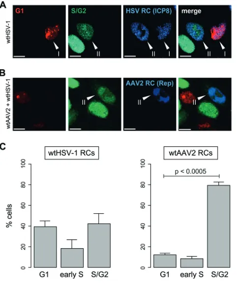
FIG 1Codetection of cell cycle phases and viral replication compartments. (A and B) HeLa Fucci cells were infected with wtHSV-1 (MOI of 3) or coinfected with wtAAV2 (MOI of 4,000) and wtHSV-1 (MOI of 3). After 24 h, the cells were fixed and processed for immunofluorescence analysis and CLSM. (A) HSV-1 RCs were visualized with a primary antibody specific for the HSV-1 major DNA-binding protein ICP8 and an AF-405-labeled secondary antibody (blue). (B) AAV2 RCs were visualized with a primary antibody specific for the AAV2 Rep proteins and an AF-405-labled secondary antibody (blue). Bars⫽10m. Arrowheads in panels A and B indicate cells positive for HSV-1 or AAV2 RCs and either the red fluorescent G1-phase marker Cdt1 (I) or the green fluorescent S/G2-phase marker geminin (II). (C) From the culturesshown in panels A and B, approximately 30 cells containing HSV-1 or AAV2 RCs (blue) were analyzed for their cell cycle phase (G1, early S, and S/G2). The graphs show mean values from six independent
experiments; error bars indicate standard deviations of the means.
Franzoso et al. Journal of Virology
on November 7, 2019 by guest
http://jvi.asm.org/
[image:4.585.85.326.71.362.2]efficient AAV2 replication in these cell cycle phases. The next experiment was
per-formed to investigate whether HSV-1 can replicate in G
1-phase cells despite the
presence of AAV2 genomes or simply because AAV2 genomes are absent from these
cells. For this, HeLa Fucci cells were coinfected with wtAAV2 and recombinant HSV-1
that encodes enhanced cyan fluorescent protein (ECFP) fused with the HSV-1 ICP4
protein (rHSV-1vECFP-ICP4), thus facilitating the observation of HSV-1 RCs due to the
binding of the fluorescent ICP4 fusion protein to the replicating HSV-1 DNA in the cell
nucleus (46). After 24 h, the cells were fixed, and FISH staining was performed by using
an Alexa Fluor 647 (AF-647)-labeled AAV2 DNA probe. The micrographs shown in Fig.
4 demonstrate that HSV-1 RCs are formed in G
1-phase cells despite the presence of
AAV2 genomes.
FIG 2Codetection of cell cycle phases and AAV2 genomes. HeLa Fucci cells were mock infected, infected with wtAAV2 alone (MOI of 4,000), or coinfected with wtAAV2 (MOI of 4,000) and wtHSV-1 (MOI of 3). After 24 h, the cells were fixed and processed for fluorescence in situhybridization (FISH) and CLSM. (A) AAV2 DNA (yellow) was detected with an AF-647-labeled probe as described in Materials and Methods. The red and green signals indicate cells in G1(Cdt1) and S/G2(geminin) phases, respectively. The orange cells observed in some
cultures are in early S phase, where they express both Cdt1 and geminin. Areas in the box were enlarged (right column). Bars⫽10m. (B) From the wtAAV2- and wtHSV-1-coinfected cultures shown in panel A, approximately 30 cells containing AAV2 RCs were analyzed for their cell cycle phase (G1, early S, and S/G2). The graph shows mean values from six independent experiments; error bars indicate standard
deviations of the means.
on November 7, 2019 by guest
http://jvi.asm.org/
[image:5.585.42.480.70.500.2]Cell dependent AAV2 DNA replication is controlled by cell
cycle-dependent AAV2
rep
expression.
In the absence of a helper virus, gene expression
from replication-defective recombinant AAV2 vectors was previously shown to occur
preferentially in the S phase of the cell cycle (47, 48). To address the question of
whether this also applies when a helper virus is present, we coinfected HeLa Fucci cells
with wtHSV-1 and recombinant AAV2 vectors encoding fluorescent reporter proteins
fused in frame with AAV2 Rep (rAAVCFPRep and rAAVYFPRep). The AAV2 p5 promoter
used in these vectors for the transcriptional control of reporter gene expression is
highly regulated during coinfection; in particular, its activity is very low in the absence
of HSV-1. Therefore, we also used a recombinant AAV2 vector that expresses a reporter
gene from the constitutively active human cytomegalovirus (CMV) IE1 enhancer/
promoter (rAAVCFPNeo) to exclude a potential promoter effect responsible for the
observed cell cycle-dependent AAV2 DNA replication/gene expression. Viral gene
expression and the cell cycle phase were determined by fluorescence microscopy or
flow cytometry. Upon coinfection of HeLa Fucci cells with rAAVCFPNeo and wtHSV-1,
the majority of the cyan fluorescent cells was indeed in S/G
2phase, approximately 10%
of the cells were in G
1phase, and 10% were in early S phase (Fig. 5A and B). A similar
distribution was observed upon coinfection of HeLa Fucci cells with rAAVCFPRep and
wtHSV-1: approximately 81% of the AAV2-transduced (ECFP-positive) cells were in S/G
2phase, while only 19% were in G
1phase, as determined by flow cytometry (Table 1). The
inefficient AAV2 gene expression in G
1phase was also observed in other cell lines,
including AT22, HCT116, and normal human fibroblast (NHF) cells (Table 1). For
FIG 3Viral DNA replication in different phases of the cell cycle. HeLa Fucci cells were mock infected, infected with either wtAAV2 (MOI of 8,000) or wtHSV-1 (MOI of 3), or coinfected with wtAAV2 (MOI of 8,000) and wtHSV-1 (MOI of 3). After 24 h, cells positive for Cdt1 (G1phase) (red) or geminin (S/G2phase) (green) were sorted by FACS analysis and subjected to
Southern and Western blot analyses. (A) Southern blot analysis for detection of HSV-1 and AAV2 DNA (equal cell equivalents were loaded into the different lanes) (ss, single stranded; rfm, replication-form monomer; rfd, replication-form dimer). (B to D) Western blot analysis to control for virus infection (HSV-1 ICP0 and AAV2 Rep; actin was used as a loading control) (B) and qPCR to quantify viral DNA (C and D). Graphs in panels C and D show mean values for relative HSV-1 or AAV2 DNA quantities from triplicate experiments; error bars indicate standard deviations of the means.
Franzoso et al. Journal of Virology
on November 7, 2019 by guest
http://jvi.asm.org/
[image:6.585.41.426.67.376.2]example, in AT22 fibroblast cultures coinfected with rAAVYFPRep and wtHSV-1, more
than 80% of AAV2-transduced (enhanced yellow fluorescent protein [EYFP]-positive)
cells were in S or G
2phase, while only approximately 16% of these cells were in G
1phase (Table 1), although the majority (approximately 60%) of the nongated cells in the
culture was in G
1phase (not shown). Therefore, HSV-1 helper functions appear to
support neither AAV2 replication nor AAV2 (
rep
) gene expression in G
1cells,
indepen-dent of the promoter, p5 or CMV. Inefficient AAV2 gene expression in G
1phase and,
consequently, low levels of AAV2 Rep proteins (as indeed shown in Fig. 3B) are likely
responsible for the inefficient AAV2 replication and the inefficient inhibition of HSV-1
replication in this cell cycle phase, as AAV2 Rep proteins are known to be essential for
AAV2 replication and also responsible for the inhibition of HSV-1 replication (43, 49).
Next, we addressed the question of whether, in coinfected cultures, HSV-1
replica-tion is allowed in G
1-phase cells and inhibited in S/G
2-phase cells, because AAV2
rep
is
expressed efficiently only in S/G
2-phase but not in G
1-phase cells. For this, we used
recombinant HSV-1 that encodes the VP16 protein fused with EYFP (rHSV48EYFP) (50).
EYFP-VP16 accumulates on HSV-1 DNA and hence is a marker for HSV-1 RCs (50, 51). To
FIG 4Codetection of AAV2 genomes, HSV-1 RCs, and cell cycle phases. HeLa Fucci cells were coinfected with wtAAV2 (MOI of 4,000) and rHSV-1vECFP-ICP4 (MOI of 3). After 24 h, the cells were fixed and processed for FISH and CLSM. (A) AAV2 DNA (yellow) was detected with an AF-647-labeled probe as described in Materials and Methods. HSV-1 RCs were detected by the binding of the HSV-1 ECFP-ICP4 fusion protein to HSV-1 DNA (blue). The red and green signals indicate cells in G1(Cdt1) and S/G2(geminin) phases, respectively. The area in the
box (top row, merge) is enlarged in the second row and in panel B. Bars⫽20m. (B) Enlargement and sectioning of the HSV-1 RC-positive cell shown in panel A (box in the top row) reveal the subcellular localization of AAV2 genomes (red arrows). For clarity, the red and green channels were omitted.
on November 7, 2019 by guest
http://jvi.asm.org/
[image:7.585.41.490.69.455.2]simultaneously visualize AAV2 Rep, we used rAAVCFPRep, which encodes an ECFP-Rep
fusion protein from the viral p5 promoter and allows not only cell cycle-dependent
replication of the viral genome in the presence of a helper virus but also monitoring
of AAV2 RCs via interactions of the fluorescent Rep fusion protein with replicating
viral DNA (31) (see Movie S1 in the supplemental material). As expected, in cultures
coinfected with rHSV48EYFP and rAAVCFPRep, HSV-1 RCs and AAV2 RCs were not
codetected in individual cells (Fig. 5C, left). However, in cells coinfected with
rHSV48EYFP and rAAVCFPNeo, which encodes ECFP but no AAV2 Rep proteins, yellow
fluorescent HSV-1 RCs were readily detected in both ECFP-positive and ECFP-negative cells
FIG 5Cell cycle-dependent AAV2 gene expression controls the replication of both AAV2 and HSV-1 in coinfected cultures. (A) HeLa Fucci cells were coinfected with rAAVCFPNeo (MOI of 4,000) and wtHSV-1 (MOI of 3). Images were acquired by automated epifluorescence microscopy using ImageXpress Micro XL (Molecular Devices) and fluorescence filters (Semrock) for ECFP (AAV2 gene expression) (blue), EGFP (S/G2phase) (green), and TRITC (G1phase) (red). Bars⫽20m. (B) HeLa Fucci cells were coinfected with
rAAVCFPNeo (MOI of 1,000) and wtHSV-1 (MOI of 3), and 20 h later, the cell cycle phase (G1, early S, and S/G2) of
[image:8.585.43.464.72.345.2]rAAVCFPNeo-transduced cells was determined by the expression of Ctd1 (red) and geminin (green). A total of 40,000 cells (events) per sample were counted by flow cytometry. The graph shows mean values from triplicate experiments; error bars indicate standard deviations of the means. (C) HeLa cells were coinfected with rHSV48EYFP (MOI of 3) and either rAAVCFPRep (MOI of 4,000) or rAAVCFPNeo (MOI of 4,000). After 24 h, cells were examined by using a standard fluorescence microscope. Bars⫽10m. (D) From the cultures shown in panel C, 50 cells containing yellow fluorescent HSV-1 RCs were analyzed for the presence of AAV2 RCs in rHSV48EYFP- and rAAVCFPRep-coinfected cultures or ECFP fluorescence in rHSV48EYFP- and rAAVCFPNeo-coinfected cultures. The graph shows mean values from triplicate experiments; error bars indicate standard deviations of the means.
TABLE 1Cell cycle phases of AAV2-transduced cells
Cell line Infectiond
% of cells in phase
G1 S S/G2 G2
HeLa Fuccia rAAVCFPRep 19 81
AT22b rAAVYFPRep 16 40 44
HCT116c rAAVYFPRep 20 33 47
NHFb rAAVCFPRep 17 35 48
aCell cycle phases were determined by flow cytometry with fluorescent ubiquitination-based cell cycle
indicators (Fucci).
bCell cycle phases were determined by flow cytometry with propidium iodide. cCell cycle phases were determined by flow cytometry with DAPI.
dCoinfected with HSV-1.
Franzoso et al. Journal of Virology
on November 7, 2019 by guest
http://jvi.asm.org/
[image:8.585.43.369.625.693.2](Fig. 5C, right). By using an epifluorescence microscope, cells positive for yellow fluorescent
HSV-1 RCs were examined for ECFP fluorescence (Fig. 5D). In the rHSV48EYFP- and
rAAVCFPRep-coinfected cultures, less than 5% of the cells containing HSV-1 RCs were
ECFP-Rep positive. In contrast, in rHSV48EYFP- and rAAVCFPNeo-coinfected cultures,
ap-proximately 50% of the cells containing HSV-1 RCs were also positive for the AAV2 marker
ECFP. Since rAAVCFPNeo transduction was observed predominantly in the S/G
2phase of
the cell cycle (Fig. 5A and B), these data suggest that AAV2 infection or nonreplicating AAV2
DNA
per se
is not sufficient to inhibit HSV-1 replication in S/G
2phase but that
rep
expression
is required. This is also supported by a previous finding that UV-inactivated wtAAV2
genomes, which do not express any AAV2 genes, did not inhibit HSV-1 replication (43). In
conclusion, cell cycle-dependent AAV2
rep
expression seems to be responsible for the
observed negative correlation between HSV-1 and AAV2 replication efficiencies in different
phases of the cell cycle. Low Rep levels in G
1phase allow HSV-1 but not AAV2 to replicate,
while high Rep levels in S/G
2phase support AAV2 replication and block HSV-1 replication.
Both single-stranded and double-stranded AAV2 vectors predominantly
trans-duce cells in S/G
2phase, independent of the presence or absence of a helper
virus.
We used two different approaches to test whether the single-stranded nature of
AAV2 DNA plays a role in cell cycle-dependent AAV2 gene expression: transfection of
plasmid-cloned AAV2 genomes and infection with self-complementary recombinant
AAV2 (scAAV2) vectors, which can form double-stranded DNA by self-annealing and
therefore do not depend on second-strand synthesis for viral gene expression.
Specif-ically, HeLa Fucci cells were transfected with plasmids containing
rep
-positive
(pAAVCF-PRep) or
rep
-negative (pAAVCFPp5) recombinant AAV2 genomes and 4 h later were
mock infected or infected with wtHSV-1. After 24 h, the cells were examined by CLSM.
The results are shown in Fig. 6 and are summarized as follows: ECFP fluorescent AAV2
RCs were readily detected in both G
1- and in S/G
2-phase cells of cultures transfected
with pAAVCFPRep and infected with the helper virus (Fig. 6A). In the presence of HSV-1,
ECFP fluorescence was also detected both in G
1- and in S/G
2-phase cells of cultures
transfected with pAAVCFPp5, but in these cells, ECFP fluorescence was diffuse, and
AAV2 RCs were not observed because of the absence of
rep
(Fig. 6B). No ECFP
fluorescence was observed in the absence of HSV-1, as both plasmids use the helper
virus-dependent AAV2 p5 promoter for transgene expression (not shown).
To confirm that HSV-1-supported AAV2 DNA replication can indeed take place in G
1phase when the template is a circular double-stranded DNA, HeLa Fucci cells were
transfected with pAAVCFPRep and 4 h later were mock infected or infected with
wtHSV-1. After 24 h, episomal DNA was prepared from G
1- and S/G
2-phase cells
according to methods described previously by Hirt (52) and subjected to qPCR with
primers specific for
rep
. The graph in Fig. 6C shows that the relative AAV2 DNA content
in G
1-phase cells is 80% larger in the presence of the helper virus, presumably because
of DNA replication. We then tested whether HSV-1 DNA replication was also blocked in
G
1-phase cells when
rep
is expressed from a plasmid-cloned recombinant AAV2
ge-nome. The graph in Fig. 6D shows that this is clearly the case. The HSV-1 DNA content
of ECFP (AAV2)- and Cdt1 (G
1-phase) (red)-double-positive HeLa Fucci cells transfected
with pAAVCFPRep prior to HSV-1 infection was reduced by more than 60% compared
to that in cells transfected with
rep
-negative pAAVCFPp5 plasmid DNA prior to infection
with the helper virus. In summary, these data show that AAV2 (
rep
) gene expression,
AAV2 genome replication, and the inhibition of HSV-1 replication can take place in
G
1-phase cells when the AAV2 genome is provided as a circular double-stranded rather
than a linear single-stranded DNA template.
These data suggest that AAV2 single-strand synthesis may be the limiting factor for
efficient AAV2 gene expression/genome replication in G
1phase, and accordingly, one
would expect that scAAV2 vectors would support gene expression in this cell cycle
phase. To investigate this possibility, we infected HeLa Fucci cells with scAAVCFP in
presence or absence of the helper virus and monitored reporter gene expression (ECFP)
and the cell cycle phase from 2 to 20 h after infection (Fig. 7). Indeed, 20 h after
infection, many transgene-positive G
1-phase cells (purple) were observed in
on November 7, 2019 by guest
http://jvi.asm.org/
FIG 6Detection of cell cycle phases, AAV2 gene expression, and virus replication in cells transfected with circular, double-strandedrep-positive orrep-negative recombinant AAV2 genomes. (A and B) HeLa Fucci cells were transfected with pAAVCFPRep (A) or pAAVCFPp5 (B) and infected with wtHSV-1 (MOI of 2) 4 h later. After 24 h, cells were examined by CLSM with filters specific for Cdt1 (G1phase) (red), geminin (S/G2phase)
(green), and ECFP (AAV2) (blue). The framed areas in panels A and B are shown at a higher magnification on the right. Arrowheads in panel A
(Continued on next page)
Franzoso et al. Journal of Virology
on November 7, 2019 by guest
http://jvi.asm.org/
[image:10.585.35.498.77.692.2]infected cultures in the absence of the helper virus (Fig. 7A, left). However, tracking of
individual cells over time revealed that the transgene-positive G
1-phase cells observed
20 h after infection originated from cells that became transgene positive in S/G
2phase
and then went through mitosis (Fig. 7A, right, arrows; see also Movie S2 in the
supplemental material). Occasionally, cells turned transgene positive in G
1phase (not
shown). Coinfection with wtHSV-1 largely prevented the generation of
transgene-positive G
1cells (Fig. 7A), presumably because of HSV-1-mediated G
2arrest (5, 33). As
expected from the above-described data, the vast majority of the cells infected with the
single-stranded AAV2 vector (rAAVCFPNeo) turned transgene positive in S/G
2phase
and did not progress through mitosis, independent of the presence or absence of the
helper virus (Fig. 7A and Movie S3). Also as expected from the above-described data,
transgene-positive G
1-phase cells were also occasionally observed in
rAAVCFPNeo-infected cultures in the absence of the helper virus. These cells often but not always
originated from transgene-positive S/G
2-phase cells that progressed to G
1phase,
sometimes without division (Fig. 7A). To quantify the observations from live
micros-copy, we tracked and counted cells over time from the moment when AAV2
vector-encoded ECFP fluorescence became visible. The graph in Fig. 7B shows that more than
90% of the cells turned ECFP positive in S/G
2phase, independent of the genome
structure, whether single stranded or double stranded (self-complementary), or the
presence or absence of the helper virus. However, in the absence of wtHSV-1, ECFP
fluorescent S/G
2-phase cells progressed through mitosis to G
1phase at a significantly
higher frequency when infected with scAAVCFP than when infected with rAAVCFPNeo
(Fig. 7C).
Overall, we conclude that (i) HSV-1 is not capable of extending its ability to replicate
in all phases of the cell cycle to AAV2, (ii) cell cycle-dependent AAV2 DNA replication
and inhibition of HSV-1 DNA replication are due to cell cycle-dependent AAV2
rep
expression, and (iii) second-strand synthesis is likely not responsible for the cell cycle
preference because both single-stranded and double-stranded linear AAV2 genomes
preferentially transduce cells in S/G
2phase.
DISCUSSION
In this study, we addressed the question of how AAV2 and HSV-1 coordinate their
replication programs. For this, we used a fluorescence microscopy approach, scoring
different cell cycle stages and viral RCs on a single-cell level, and combined the results
with biochemical virus replication measurements in isolated cells at the G
1and S/G
2stages. We find that AAV2 replication occurs predominantly in S/G
2phase. Also, AAV2
gene expression was restricted to S/G
2-phase cells in both the presence and absence
of the helper virus. HSV-1 replication was in turn restricted to G
1-phase cells in
coinfected cultures, while it replicated efficiently in both G
1- and S/G
2-phase cells in the
absence of AAV2 (for an overview, see Tables 2 and 3). On the other hand, HSV-1 RCs
were readily detected in S/G
2-phase cells transduced with a
rep
-negative recombinant
AAV2 vector. Therefore, as Rep proteins are essential for AAV2 replication and have
been found to be at least partially responsible for the AAV2-mediated inhibition of
HSV-1 replication (38, 43, 44), we conclude that cell cycle-dependent AAV2
rep
expres-sion is responsible for the observed restriction of AAV2 replication to S/G
2-phase cells
and of HSV-1 replication to G
1-phase cells.
We have previously shown that the AAV2 Rep68/78 proteins, in particular the
FIG 6Legend (Continued)
indicate cells positive for AAV2 RCs and either the red fluorescent G1-phase marker Cdt1 (I) or the green fluorescent S/G2-phase marker geminin
(II). Bars⫽40m (left) and 10m (right). (C) HeLa Fucci cells were transfected with pAAVCFPRep and 4 h later were mock infected or infected with wtHSV-1 (MOI of 2). After 24 h, extrachromosomal DNA was prepared from Cdt1-positive G1-phase cells (red) and geminin-positive
S/G2-phase cells (green), digested with DpnI, and subjected to qPCR with primers specific for AAV2rep. (D) HeLa Fucci cells were transfected with
pAAVCFPRep or pAAVCFPp5 and infected 4 h later with wtHSV-1 (MOI of 2). Total DNA extracted after 24 h from ECFP (AAV2)- and Cdt1 (G1-phase)
(red)- or ECFP- and geminin (S/G2-phase) (green)-double-positive cells was subjected to qPCR with primers specific for HSV-1 DNA. Graphs in
panels C and D show mean values for relative DNA quantities from triplicate experiments, and samples were prepared from equal numbers of cells; error bars indicate standard deviations of the means.
on November 7, 2019 by guest
http://jvi.asm.org/
FIG 7Live-cell imaging of cell cycle-dependent AAV2 gene expression. (A) HeLa Fucci cells were infected with scAAVCFP (MOI of 3,000) or rAAVCFPNeo (MOI of 3,000) in the absence or presence of wtHSV-1 (MOI of 3), and images were acquired every 30 min from 2 to 20 h after infection by automated time-lapse (37°C with 5% CO2, humidified) epifluorescence microscopy using ImageXpress Micro XL (Molecular Devices) and fluorescence filters (Semrock)
for ECFP (AAV2 gene expression), GFP (G2phase), and TRITC (G1phase). A total of 36 frames was acquired, and the frame numbers are indicated. Overview
images are shown in the left panels. Two different positions highlighted in each of the overview images (boxes 1 and 2) were enlarged and tracked over time (right panels). Arrows point to cells from the moment when ECFP fluorescence becomes visible. (B) Graph showing the percentage of cells that turned ECFP positive in either the G1or S/G2phase. (C) Graph showing the percentage of transgene-positive S/G2-phase cells progressing to G1phase.
Both graphs show mean values from at least 100 ECFP-positive cells tracked in at least 9 different infection-and-acquisition experiments; error bars indicate standard deviations of the means.
Franzoso et al. Journal of Virology
on November 7, 2019 by guest
http://jvi.asm.org/
[image:12.585.40.545.70.608.2]combined DNA-binding and ATPase/helicase activities, can block HSV-1 DNA
replica-tion on the level of DNA synthesis, even in the absence of AAV2 DNA. We also
demonstrated that AAV2 Rep68 can bind to consensus Rep-binding sites on the HSV-1
genome and that the helicase activity can block the replication of any DNA if binding
is facilitated (44). However, the conclusion from the present study, that the inhibition
of HSV-1 replication is limited to S/G
2-phase cells because AAV2
rep
is expressed
efficiently only in this phase, inevitably begs the question of what controls the cell
cycle-dependent expression of
rep
. Recombinant AAV2 vectors have been reported to
transduce cells predominantly in S phase in the absence of a helper virus (47, 48). Our
finding that HSV-1-provided helper functions do not support AAV2 replication and
gene expression in G
1phase, although HSV-1 itself can replicate in G
1and S/G
2phases
and although the HSV-1 protein ICP0 strongly
trans
-activates AAV2
rep
expression,
indicates that a step prior to AAV2 transcription may be blocked. One obvious step is
second-strand synthesis. For example, cellular proteins involved in second-strand
syn-thesis such as polymerases or cofactors may not be available at sufficient
concentra-tions in G
1phase. Likewise, factors that block second-strand synthesis, such as DNA
damage repair proteins, may be abundant or activated in G
1but not in S/G
2phase. We
demonstrate that AAV2 transcription and replication are efficient in S/G
2- as well as in
G
1-phase cells when the template is a circular double-stranded rather than a linear
single-stranded DNA template, indicating that cell cycle-dependent
rep
expression may
indeed be due to cell cycle-dependent second-strand synthesis. However, the circular
versus linear state of the two template DNAs as well as the different means of delivery,
transfection versus infection, may also affect transcription in a cell cycle-dependent
manner, possibilities that were further explored by using scAAV2 vectors. If inefficient
second-strand synthesis in G
1phase was solely responsible for the observed cell
cycle-dependent AAV2 replication and gene expression, one would expect that scAAV2
vectors, which can form double-stranded DNA by self-annealing and therefore do not
depend on second-strand synthesis for gene expression, can transduce both G
1-and
TABLE 2AAV2 and HSV-1 DNA replication in different cell cycle phasesaVirus infection
Efficiency of DNA replication
Fig. and/or
supplemental material G1 S/G2
HSV-1 ⫹⫹⫹⫹ ⫹⫹⫹⫹ Fig. 1⫹3
AAV2 ⫺ ⫺ Fig. 2⫹3
AAV2 (HSV-1 coinfection) ⫹ ⫹⫹⫹⫹ Fig. 1–3, Movie S1 HSV-1 (AAV2 coinfection) ⫹⫹⫹⫹ ⫹ Fig. 3⫹4
a⫹⫹⫹⫹, efficient DNA replication;⫹, inefficient DNA replication;⫺, no DNA replication (AAV2repexpression
from the p5 promoter).
TABLE 3Cell cycle-dependent gene expression from AAV2 vectors and the G2-to-M-to-G1 -phase transitiona
AAV2 vector
Efficiency of gene expression in
phase Efficiency of G2–M–G1
[image:13.585.43.372.82.162.2]transition
Fig., supplemental material, and/or table
G1 S/G2
ssAAV2cmv ⫹ ⫹⫹⫹⫹ ⫹ Fig. 7, Movie S3 ssAAV2cmv (HSV-1
coinfection)
⫹ ⫹⫹⫹⫹ ⫹ Fig. 5⫹7
ssAAV2p5 ⫹ ⫹⫹⫹⫹ ND Table 1 dsAAV2cmv ⫹ ⫹⫹⫹⫹ ⫹⫹⫹⫹ Fig. 7, Movie S2 dsAAV2cmv (HSV-1
coinfection)
⫹ ⫹⫹⫹⫹ ⫹ Fig. 7
assAAV2cmv, single-stranded AAV2 with a cytomegalovirus IE1 enhancer/promoter; dsAAV2cmv,
double-stranded AAV2 with a cytomegalovirus IE1 enhancer/promoter; p5, AAV2 p5 promoter;⫹⫹⫹⫹, efficient gene expression/G2-to-M-to-G1-phase transition;⫹, inefficient replication/G2-to-M-to-G1-phase transition;
ND, not done.
on November 7, 2019 by guest
http://jvi.asm.org/
[image:13.585.42.371.584.703.2]S/G
2-phase cells efficiently. Indeed, in scAAV2 vector-transduced cell cultures, we
observed a considerable proportion of AAV2 transgene-positive G
1-phase cells in
the absence of the helper virus, but interestingly, high-throughput multichannel
time-lapse microscopy of cell cycle progression and viral gene expression showed
that the majority of the scAAV2-transduced G
1-phase cells originated from
scAAV2-transduced S/G
2-phase cells that progressed through mitosis, an event that was
observed at a significantly lower rate upon infection with the single-stranded rAAV2
vector (for an overview, see Table 3). We hypothesize that rAAV2 vectors, similar to
wtAAV2 (53), can induce cell cycle arrest more efficiently than the scAAV2 vectors
because of the single-stranded versus double-stranded nature of the genome. In
the presence of the helper virus, neither single-stranded nor double-stranded AAV2
vector-transduced cells progressed through mitosis, presumably because of
HSV-1-induced G
2arrest (33). This hypothesis is supported by the finding that coinfection
with an ICP0 deletion mutant of HSV-1, which is replication competent but does not induce
cell cycle arrest in G
2phase (33), allowed scAAV2 vector-transduced G
2-phase cells but
not rAAV2 vector-transduced G
2-phase cells to progress through mitosis (data not
shown).
The findings in this study may have important implications for AAV2-mediated gene
therapy. Both single-stranded and double-stranded AAV2 vectors depend on cells in
S/G
2phase for efficient gene expression, which in fact may contribute to their low
transduction efficiency, at least in postmitotic cells. However, when the target cells are
mitotically active, the double-stranded AAV2 vectors may be preferred, as they allow
the cells to go through mitosis, while the single-stranded AAV2 vectors do not. On the
other hand, scAAV2 vectors have been shown to induce a more robust innate immune
response in mouse liver than rAAV2 vectors (54). In any event, it would be interesting
to compare the effects of scAAV2 and rAAV2 vectors on specific factors involved in cell
cycle regulation, including p53, retinoblastoma protein, cyclin-dependent kinases
(CDKs), and CDK inhibitors.
The observation that both single-stranded and double-stranded AAV2 vectors
trans-duce cells predominantly in S/G
2phase argues against second-strand synthesis as the
mechanism responsible for cell cycle-dependent AAV2 gene expression/DNA
replica-tion and inhibireplica-tion of HSV-1 DNA replicareplica-tion. Nevertheless, the preference of AAV2 to
transduce cells in the S/G
2phase of the cell cycle may likely be orchestrated by cellular
factors other than those involved in second-strand synthesis. In fact, many different
cellular proteins are recruited to AAV2 genomes (32, 55–57). Among these, the DNA
damage response (DDR) proteins represent a very prominent group and may
differen-tially affect specific phases in AAV2 genome processing, such as circularization,
repli-cation, and transcription. The DNA damage response is closely linked with the cell cycle
through checkpoint proteins. Moreover, there is increasing evidence that CDKs, which
are the master regulators of cell cycle progression, also have a role in the regulation of
the DNA damage repair (for reviews, see reference 58–60), thereby coordinating DNA
repair and cell cycle progression. For example, it has been shown that CDK1 activity
regulates the choice between different double-strand break (DSB) repair pathways,
homologous recombination (HR) and nonhomologous end joining (NHEJ). In G
1phase,
CDK1 activity is low, and NHEJ is used, while in S/G
2/M phase, CDK1 activity is high, and
HR is employed (61, 62). CDK1 activity stimulates HR by promoting the generation of
the 3
=
single-stranded DNA ends, which are necessary for HR and inhibitory for NHEJ
(61–63). It has also been shown that CDK activity is essential for later steps in HR,
specifically in the recruitment of Rad51 (64). Functional roles in DNA damage repair
pathways have also been demonstrated for other CDKs in complex with cyclins (for a
review, see reference 65).
The licensing factor that allows AAV2 gene expression/DNA replication in S/G
2phase or the inhibitor that blocks it in G
1phase remains to be identified. Nevertheless,
the “partitioning” of the host cell population between the two different viruses is a
novel concept of viral adaptation and a fascinating strategy to minimize competition,
Franzoso et al. Journal of Virology
on November 7, 2019 by guest
http://jvi.asm.org/
allowing both AAV2 and HSV-1 to productively replicate although in distinct sets of
cells.
MATERIALS AND METHODS
Cells.NHF cells were obtained from X. O. Breakefield (Massachusetts General Hospital, Charlestown, MA, USA). HeLa Fucci cells were a gift from B. Kraus (University of Regensburg, Regensburg, Germany); these cells encode cell cycle-specific proteins fused with red fluorescent (Cdt1) (G1-phase) or green
fluorescent (geminin) (S/G2-phase) fusion proteins (45). HCT116 cells were provided by E. Hendrickson
(University of Minnesota, Minneapolis, MN, USA). NHF, HeLa Fucci, HCT116, and HeLa (ATCC) cells were cultured in Dulbecco’s modified Eagle medium (DMEM) supplemented with 10% fetal bovine serum (FBS), 100 U/ml penicillin G, 100g/ml streptomycin, and 0.25g/ml amphotericin B (1% AB). AT22 IJE-T yZ5 cells (kindly provided by Y. Shiloh, Tel Aviv University, Tel Aviv, Israel) were cultured in DMEM supplemented with 10% FBS, 1% AB, and 100g/ml hygromycin B. Vero cells (ECACC) and 293T cells (kindly provided by J. Neidhardt, University of Zurich, Zurich, Switzerland) were grown in DMEM supplemented with 10% FBS and 1% AB. All cells were maintained at 37°C in a 95% air–5% CO2
atmosphere. For all infection experiments, the virus was allowed to adsorb for 30 min at 4°C before the temperature was shifted to 37°C for 1 h. The cells were then washed with phosphate-buffered saline (PBS) and incubated with DMEM containing 2% FBS and 1% AB at 37°C for the indicated times after infection.
Plasmids and viruses.wtHSV-1 strain F was provided by B. Roizman (University of Chicago, Chicago, IL). Recombinant HSV-1 encoding ICP4 fused with ECFP (rHSV-1vECFP-ICP4) (46) was obtained from R. D. Everett (University of Glasgow, Glasgow, UK). Recombinant HSV-1 rHSV48EYFP encoding the VP16 protein fused with EYFP was described previously (50). wtHSV-1 and recombinant HSV-1 were grown and titrated on Vero cells. Plasmid pAAVtCR, which contains a recombinant AAV2 genome that lacks thecap gene and encodes unmodified Rep40/52 under the control of the p19 promoter and Rep68/78 fused at the N terminus with the fluorescent protein mCherry (31) under the control of the p5 promoter, was obtained from A. Salvetti (University of Lyon, Lyon, France). Plasmids containing recombinant AAV2 genomes, pAAVCFPRep and pAAVYFPRep, were constructed by replacing the mCherry coding sequence in the pAAVtCR plasmid with ECFP and EYFP coding sequences, respectively, as follows: pAAVtCR was treated first with BsrGI and then partially with BamHI, and the 5,169-bp fragment was ligated with the 738-bp BsrGI-BamHI fragment containing the ECFP or EYFP coding sequences from pECFP-N2 or pEYFP-N2 (Clontech), respectively. Plasmid pAAVCFPp5, which encodes ECFP from the AAV2 p5 pro-moter, was constructed by replacing the 1,885-bp BsrGI-SwaI fragment of pAAVCFPRep, which contains the AAV2 Rep coding sequence, with the BsrG1-SwaI adaptor oligonucleotide 5=-GTACAAGATCGATATT T-3=. The recombinant AAV2 genome rAAVCFPNeo was constructed by replacing the enhanced green fluorescent protein (EGFP) coding sequence in plasmid pAAVGFP (provided by M. Linden, King’s College London School of Medicine, London, UK) with the ECFP coding sequence, as follows: pECFP-N2 was treated with PstI and NotI, and the 768-bp fragment was ligated with the 6,417-bp PstI-NotI fragment of pAAVGFP. Transgene expression from pAAVCFPNeo is controlled by the human CMV IE1 enhancer/ promoter. Plasmid pscAAVCFP, which encodes the self-complementary recombinant AAV2 genome scAAVCFP, was constructed by replacing the EGFP coding sequence in pscAAVeGFP (kindly provided by J. Neidhardt, University of Zurich, Switzerland) with the ECFP coding sequence. Specifically, a DNA fragment containing the CMV enhancer/promoter-ECFP coding sequence was amplified by PCR from plasmid pCMVeCFP-PolyA (our unpublished data) using forward (5=-AAGATATCACTAGTTAGTTATTAATA GTAATCAATTACG-3=) and reverse (5=-CCGGTACCATGCAGTGAAAAAAATGC-3=) primers (PCR conditions were 30 s at 98°C; 35 cycles of 10 s at 98°C, 45 s at 58°C, and 60 s at 72°C; and 10 min at 72°C), digested with SpeI and NotI, and ligated between the SpeI and NotI sites of pscAAVeGFP. Recombinant AAV2 stocks were produced by transient transfection of 293T cells with pDG (66) and either pAAVCFPRep, pAAVYFPRep, pAAVCFPNeo, or pscAAVCFP. Virus stocks were purified by an iodixanol gradient, and titers were determined as described previously (67). wtAAV2 was grown and titrated as described previously (68).
Antibodies.The following primary antibodies were used: antiactin (catalog number SC-10731; Santa Cruz Biotechnology) (dilution for WB, 1:10,000), anti-ICP8 (catalog number Ab-20193; Abcam) (dilution for WB, 1:1,000; dilution for immunofluorescence [IF] assays, 1:200), anti-AAV2 Rep (catalog number 10R-A111A; Fitzgerald Industries) (dilution for WB, 1:200; dilution for IF assays, 1:50), and anti-ICP0 (catalog number ab6513) (dilution for WB, 1:2,000). The following secondary antibodies were used: rabbit anti-mouse IgG-horseradish peroxidase (HRP) (catalog number A9044; Sigma) (dilution, 1:10,000), goat anti-rabbit IgG-HRP (catalog number A6154; Sigma) (dilution, 1:10,000), and goat anti-mouse IgG(H⫹L)– AF-405 (catalog number A31556; Molecular Probes) (dilution, 1:500).
FACS analysis followed by Western blotting, Southern blotting, or quantitative PCR.For the experiments in Fig. 3, 5⫻106HeLa Fucci cells were seeded onto 10-cm tissue culture plates and 24 h
later were mock infected, infected with wtAAV2 (multiplicity of infection [MOI] of 8,000 genome-containing particles per cell) or wtHSV-1 (MOI of 3), or coinfected with wtAAV2 (MOI of 8,000) and wtHSV-1 (MOI of 3). After 24 h, cells positive for Cdt1 (G1phase) (red) or geminin (S/G2phase) (green)
were sorted by using a FACSAria III sorter (BD Biosciences) and prepared for Western analysis, Southern blotting, or qPCR. For Western blot analysis, cells were trypsinized, washed once with PBS, resuspended in 1⫻loading buffer (4% SDS, 10%-mercaptoethanol, 20% glycerol, 0.005% bromphenol blue, 0.125 M Tris-HCl [pH 6.8]), and boiled for 10 min. Cell lysates were separated on 10% SDS-polyacrylamide gels and transferred onto Protran nitrocellulose membranes (Whatman, Bottmingen, Switzerland). The mem-branes were blocked with PBS-T (PBS containing 0.3% Tween 20) supplemented with 5% nonfat dry milk
on November 7, 2019 by guest
http://jvi.asm.org/
for 1 h at room temperature (RT). Incubation with antibodies was carried out with PBS-T supplemented with 2.5% nonfat dry milk. Primary antibodies were incubated overnight at 4°C, while secondary antibodies were incubated for 1 h at RT. Membranes were washed three times with PBS-T for 10 min after each antibody incubation step. HRP-conjugated secondary antibodies were detected with ECL detection reagent (ECL WB blotting systems; GE Healthcare, Zurich, Switzerland). The membranes were exposed to chemiluminescence detection films (Roche Diagnostics, Rotkreuz, Switzerland). Detection of antiactin served as a loading control for the lysate. For Southern blot analysis, extrachromosomal DNA was extracted according to protocols described previously by Hirt (52). The DNA was separated on 1% agarose gels and transferred onto nylon membranes (Hybond-N⫹, catalog number RPN119B; Amersham). Hybridization with a digoxigenin (DIG)-labeled probe specific for the AAV2 Rep78- or the HSV-1 UL35-coding sequences and subsequent detection by an anti-DIG antibody conjugated with alkaline phosphatase and activation with the chemiluminescence substrate CDP Star were performed according to the manufacturer’s protocols (Roche, Mannheim, Germany). The DIG-labeled probe was produced with the PCR DIG probe synthesis kit (Roche). For qPCR, total DNA was isolated by using the DNeasy blood and tissue kit (Qiagen, Hilden, Germany) according to the manufacturer’s protocol. The viral DNA content was quantified by qPCR using the cellular telomerase reverse transcriptase (TERT) gene as an endoge-nous control (Applied Biosystems, Foster City, CA, USA) and primers and a TaqMan probe specific for the AAV2 Rep78-coding sequence (forward primer 5=-ATTGACGGAACTCAACGAC-3=, reverse primer 5=-CCTC AACCACGTCCTTT-3=, and TaqMan probe 5=-CATGATCCAGACGGCGGGTGA-3=) or primers specific for the HSV-1 UL42 gene (forward primer 5=-CCCTCAAGTTCTTCTTCCTCACG-3=and reverse primer 5=-GGAGTCC TGGCTGTCTGTTG-3=) and SYBR green (Applied Biosystems, Foster City, CA, USA). AAV2 sequences were amplified under the following conditions: 2 min at 50°C; 15 min at 95°C; and 40 cycles of 15 s at 94°C, 30 s at 56°C, and 15 s at 72°C. HSV-1 sequences were amplified under the following conditions: 3 min at 95°C, 39 cycles of 15 s at 95°C and 60 s at 45°C, 10 s at 95°C, 5 s at 65°C, and 5 s at 95°C. For the experiments shown in Fig. 7C and D, 1.5⫻106HeLa Fucci cells in 6-cm tissue culture plates were
transfected with 0.5g of either pAAVCFPRep or pAAVCFPp5 by using Lipofectamine LTX according to the manufacturer’s instructions (Life Technologies) and 4 h later were mock-infected or infected with wtHSV-1 (MOI of 2) as described in the legend. After 24 h, cells positive for the G1-phase marker Cdt1
(red) or double positive for the G1-phase marker and either CFP-Rep or CFP were sorted by using a
FACSAria III sorter (BD Biosciences). Hirt DNA or total DNA was then prepared from the sorted cell populations. To determine the AAV2 DNA content, Hirt DNA was incubated with DpnI prior to qPCRs in order to degrade the transfected input plasmid DNA isolated fromEscherichia coli. To determine the HSV-1 DNA content, total DNA was used for qPCR.
Microscopy.For microscopy, HeLa or HeLa Fucci cells were seeded onto coverslips (12-mm diameter; Glaswarenfabrik Karl Hecht GmbH & Co. KG, Sondheim, Germany) in 24-well tissue culture plates (5⫻104
cells per well) or, specifically for epifluorescence live-cell microscopy, directly onto 96-well tissue culture plates (104cells per well) (see also reference 69). The next day, the cells were infected as indicated in
Results and the figure legends. For the experiments shown in Fig. 6, the cells were transfected with 0.5g of either pAAVCFPRep or pAAVCFPp5 4 h prior to infection. For live-cell microscopy of AAV2 gene expression/replication and cell cycle progression (Fig. 5A and 7A; see also Movies S1 to S3 in the supplemental material), images were acquired every 30 min from 2 to 20 h after infection by automated multisite multichannel live epifluorescence microscopy in a time-lapse mode (37°C with 5% CO2,
humidified) by using ImageXpress Micro XL (Molecular Devices) and fluorescence filters (Semrock) for ECFP (AAV2), GFP (G2phase), and tetramethylrhodamine isocyanate (TRITC) (G1phase) (69, 70); images
were processed by using Fiji software (71). For standard fluorescence microscopy or CSLM, the cells were washed once with cold PBS 24 h after infection and then fixed with 3.7% paraformaldehyde (PFA) in PBS for 10 min at RT. The fixation process was stopped by incubation with 0.1 M glycine for 10 min at RT and two washes with cold PBS. For immunofluorescence analysis (Fig. 1A and B), the cells were permeabilized by treatment with precooled (⫺20°C) acetone for 2 min, followed by 3 washing steps with PBS. The cells were blocked for 30 min with 3% bovine serum albumin (BSA) in PBS. For staining, the cells were incubated with antibodies diluted in PBS-BSA (3%) in a humidified chamber at RT in the dark. The coverslips were placed onto droplets (40l) of a primary antibody solution. After incubation for 1 h, the cells were washed three times with PBS and once with H2O. All coverslips (with and without
anti-body staining) were embedded in Glycergel (DakoCytomation, Carpinteria, CA) containing 1,4-diazabicyclo[2.2.2]octan (DABCO) (26 mg/ml; Fluka, Sigma-Aldrich Chemie GmbH, Munich, Germany), and cells were observed by using a confocal laser scanning microscope (Leica TCS SP2 acousto-optical beam splitter (AOBS); Leica Microsystems, Wetzlar, Germany) (Fig. 1A and B and 6A and B), an automated epifluorescence microscope using ImageXpress Micro XL (Molecular Devices) (Fig. 5A), or a standard fluorescence microscope (Axio Observer inverted microscope, Zeiss AG, Germany) (Fig. 5C). To prevent cross talk between the channels for the different fluorochromes, all channels were recorded separately, and fluorochromes with longer wavelengths were recorded first.
Flow cytometry.HCT116, AT22, NHF, or HeLa Fucci cells were seeded into wells of a 12-well tissue culture plate (105cells per well) and 24 h later were coinfected with wtHSV-1 (MOI of 1.5 for AT22 cells
and MOI of 3 for all other cell lines) and either rAAVCFPRep or rAAVYFPRep (MOI of 1,000 for HeLa Fucci cells and MOI of 4,000 for all other cell lines), as indicated in Table 1. All cells were incubated at 37°C with 5% CO2in DMEM supplemented with 2% FBS and 1% AB. After 20 to 24 h, the cells were trypsinized,
washed once with PBS, centrifuged for 10 min at 1,000⫻g, and resuspended in 1 ml PBS. The cells were fixed by the addition of 2.5 ml ethanol (EtOH) and incubation overnight at⫺20°C. To determine the cell cycle phases in HCT116, NHF, and AT22 cells, DNA was stained for 30 min with DAPI (1g/ml in PBS containing 0.1% Triton X-100) or propidium iodide (PI) (50g/ml in PBS containing 0.1% Triton X-100 and
Franzoso et al. Journal of Virology
on November 7, 2019 by guest
http://jvi.asm.org/
0.1 mg/ml RNase A), as indicated in Table 1. A minimum of 40,000 events were scored for each sample by using a Gallios flow cytometer (Beckman Coulter), and data were analyzed by using Kaluza Flow Analysis software 1.3 (Beckman Coulter).
Fluorescencein situhybridization.FISH was performed essentially as described previously by Lux et al. (68). Briefly, an⬃3.9-kb DNA fragment containing the AAV2 genome without the inverted terminal repeats was amplified by PCR from plasmid pDG (66) using forward (5=-CGGGGTTTTACGAGATTGTG-3=) and reverse (5=-GGCTCTGAATACACGCCATT-3=) primers and the following conditions: 30 s at 95°C; 35 cycles of 10 s at 98°C, 15 s at 58°C, and 75 s at 72°C; and 10 min at 72°C. The PCR product was then digested with DpnI to cut the residual template DNA and purified with a Pure Link PCR purification kit (Qiagen). The DNA fragment was labeled with 5-(3-aminoallyl)dUTP by nick translation, and the incor-porated dUTPs were labeled with amino-reactive Alexa Fluor 647 dye by using the Ares DNA labeling kit (Molecular Probes) according to the manufacturer’s protocols. HeLa Fucci cells were plated onto glass coverslips in 24-well plates at a density of 1.4⫻105cells per well and 24 h later were mock infected,
infected with wtAAV2 (MOI of 4,000), or coinfected with wtAAV2 (MOI of 4,000) and either wtHSV-1 (MOI of 3) or rHSV-1vECFP-ICP4 (MOI of 3). Twenty-four hours after infection, the cells were washed with PBS, fixed for 30 min at RT with 2% PFA (in PBS), and then washed again with PBS. The cells were then quenched for 10 min with 50 mM NH4Cl (in PBS), washed with PBS, permeabilized for 10 min with 0.2%
Triton X-100 (in PBS), blocked for 10 min with 0.2% gelatin (in PBS), and washed again with PBS. Hybridization solution (20l per coverslip) containing 1 ng/l of the labeled DNA probe, 50% form-amide, 7.3% dextran sulfate, 15 ng/l salmon sperm DNA, and 0.74⫻SSC (1⫻SSC is 0.15 M NaCl plus 0.015 M sodium citrate) was denatured for 3 min at 95°C and shock cooled on ice. The coverslips with the fixed, and permeabilized cells facing down were placed onto a drop (20 l) of the denatured hybridization solution and incubated overnight at 37°C in a humidified chamber. (Of note, the cells were not denatured, as the AAV2 genome is a single-stranded DNA.) The next day, the coverslips were washed three times with 2⫻SSC at 37°C, three times with 0.1⫻SSC at 60°C, and twice with PBS at RT. The cells were then embedded in ProLong Anti-Fade mountant (Molecular Probes) and imaged by confocal laser scanning microscopy (Leica SP8; Leica Microsystems, Wetzlar, Germany).
SUPPLEMENTAL MATERIAL
Supplemental material for this article may be found at
https://doi.org/10.1128/JVI
.00357-17
.
SUPPLEMENTAL FILE 1,
MP4 file, 0.3 MB.
SUPPLEMENTAL FILE 2,
MP4 file, 0.2 MB.
SUPPLEMENTAL FILE 3,
MP4 file, 0.2 MB.
SUPPLEMENTAL FILE 4,
PDF file, 0.1 MB.
ACKNOWLEDGMENTS
We thank X. O. Breakefield, B. Kraus, E. Hendrickson, Y. Shiloh, J. Neidhardt, B.
Roizman, R. Everett, A. Salvetti, and M. Linden for providing reagents and E. M. Schraner
for assistance with the preparation of the figures.
This work was supported by grants 31003A_144094/1 and 31003A_166668/1 from
the Swiss National Science Foundation to C.F.
We declare that we have no conflict of interest.
REFERENCES
1. Adeyemi RO, Pintel DJ. 2014. Parvovirus-induced depletion of cyclin B1 prevents mitotic entry of infected cells. PLoS Pathog 10:e1003891.
https://doi.org/10.1371/journal.ppat.1003891.
2. Berk AJ. 2007. Adenoviridae: the viruses and their replication, p 2355–2394.InKnipe DM, Howley PM, Griffin DE, Lamb RA, Martin MA, Roizman B, Straus SE (ed), Fields virology, 5th ed. Lippincott Williams & Wilkins, Philadelphia, PA.
3. Aubert M, Chen Z, Lang R, Dang CH, Fowler C, Sloan DD, Jerome KR. 2008. The antiapoptotic herpes simplex virus glycoprotein J localizes to multiple cellular organelles and induces reactive oxygen species forma-tion. J Virol 82:617– 629.https://doi.org/10.1128/JVI.01341-07. 4. Taylor TJ, Brockman MA, McNamee EE, Knipe DM. 2002. Herpes simplex
virus. Front Biosci 7:d752– d764.https://doi.org/10.2741/A809. 5. Hobbs WE, II, DeLuca NA. 1999. Perturbation of cell cycle progression
and cellular gene expression as a function of herpes simplex virus ICP0. J Virol 73:8245– 8255.
6. Ehmann GL, McLean TI, Bachenheimer SL. 2000. Herpes simplex virus type 1 infection imposes a G(1)/S block in asynchronously growing cells and prevents G(1) entry in quiescent cells. Virology 267:335–349.https:// doi.org/10.1006/viro.1999.0147.
7. Lomonte P, Everett RD. 1999. Herpes simplex virus type 1
immediate-early protein Vmw110 inhibits progression of cells through mitosis and from G(1) into S phase of the cell cycle. J Virol 73:9456 –9467. 8. Song B, Liu JJ, Yeh KC, Knipe DM. 2000. Herpes simplex virus infection
blocks events in the G1phase of the cell cycle. Virology 267:326 –334. https://doi.org/10.1006/viro.1999.0146.
9. Song B, Yeh KC, Liu J, Knipe DM. 2001. Herpes simplex virus gene products required for viral inhibition of expression of G1-phase
func-tions. Virology 290:320 –328.https://doi.org/10.1006/viro.2001.1175. 10. Muzyczka N, Berns KI. 2001. Parvoviridae: the viruses and their
replica-tion, p 2327–2346.InKnipe DM, Howley PM, Griffin DE, Lamb RA, Martin MA, Roizman B, Straus SE (ed), Fields virology, 4th ed, vol 2. Lippincott Williams & Wilkins, Philadelphia, PA.
11. Sonntag F, Schmidt K, Kleinschmidt JA. 2010. A viral assembly factor promotes AAV2 capsid formation in the nucleolus. Proc Natl Acad Sci U S A 107:10220 –10225.https://doi.org/10.1073/pnas.1001673107. 12. Chejanovsky N, Carter BJ. 1989. Mutagenesis of an AUG codon in the
adeno-associated virus rep gene: effects on viral DNA replication. Virol-ogy 173:120 –128.https://doi.org/10.1016/0042-6822(89)90227-4. 13. Im DS, Muzyczka N. 1990. The AAV origin binding protein Rep68 is an
ATP-dependent site-specific endonuclease with DNA helicase activ-ity. Cell 61:447– 457.https://doi.org/10.1016/0092-8674(90)90526-K.
on November 7, 2019 by guest
http://jvi.asm.org/
14. Kotin RM, Menninger JC, Ward DC, Berns KI. 1991. Mapping and direct visualization of a region-specific viral DNA integration site on chromo-some 19q13-qter. Genomics 10:831– 834.https://doi.org/10.1016/0888 -7543(91)90470-Y.
15. Samulski RJ, Zhu X, Xiao X, Brook JD, Housman DE, Epstein N, Hunter LA. 1991. Targeted integration of adeno-associated virus (AAV) into human chromosome 19. EMBO J 10:3941–3950. (Erratum, 11:1228, 1992). 16. King JA. 2001. DNA helicase-mediated packaging of adeno-associated
virus type 2 genomes into preformed capsids. EMBO J 20:3282–3291.
https://doi.org/10.1093/emboj/20.12.3282.
17. McCarty DM, Young SMJ, Samulski RJ. 2004. Integration of adeno-associated virus (AAV) and recombinant AAV vectors. Annu Rev Genet 38:819 – 845.https://doi.org/10.1146/annurev.genet.37.110801.143717. 18. Smith RH, Kotin RM. 1998. The Rep52 gene product of adeno-associated
virus is a DNA helicase with 3=-to-5=polarity. J Virol 72:4874 – 4881. 19. Wu J, Davis MD, Owens RA. 1999. Factors affecting the terminal resolution
site endonuclease, helicase, and ATPase activities of adeno-associated virus type 2 Rep proteins. J Virol 73:8235– 8244.
20. Zhou X, Zolotukhin I, Im DS, Muzyczka N. 1999. Biochemical character-ization of adeno-associated virus rep68 DNA helicase and ATPase activ-ities. J Virol 73:1580 –1590.
21. Yoon-Robarts M, Linden RM. 2003. Identification of active site residues of the adeno-associated virus type 2 Rep endonuclease. J Biol Chem 278: 4912– 4918.https://doi.org/10.1074/jbc.M209750200.
22. Collaco RF, Kalman-Maltese V, Smith AD, Dignam JD, Trempe JP. 2003. A biochemical characterization of the adeno-associated virus Rep40 helicase. J Biol Chem 278:34011–34017. https://doi.org/10 .1074/jbc.M301537200.
23. Hoggan MD, Blacklow NR, Rowe WP. 1966. Studies of small DNA viruses found in various adenovirus preparations: physical, biological, and im-munological characteristics. Proc Natl Acad Sci U S A 55:1467–1474.
https://doi.org/10.1073/pnas.55.6.1467.
24. Buller RM, Janik JE, Sebring ED, Rose JA. 1981. Herpes simplex virus types 1 and 2 completely help adenovirus-associated virus replication. J Virol 40:241–247.
25. Walz C, Deprez A, Dupressoir T, Dürst M, Rabreau M, Schlehofer JR. 1997. Interaction of human papillomavirus type 16 and adeno-associated virus type 2 co-infecting human cervical epithelium. J Gen Virol 78(Part 6):1441–1452.
26. Kotin RM, Linden RM, Berns KI. 1992. Characterization of a preferred site on human chromosome 19q for integration of adeno-associated virus DNA by non-homologous recombination. EMBO J 11:5071–5078. 27. Linden RM, Ward P, Giraud C, Winocour E, Berns KI. 1996. Site-specific
integration by adeno-associated virus. Proc Natl Acad Sci U S A 93: 11288 –11294.https://doi.org/10.1073/pnas.93.21.11288.
28. Linden RM, Winocour E, Berns KI. 1996. The recombination signals for adeno-associated virus site-specific integration. Proc Natl Acad Sci U S A 93:7966 –7972.https://doi.org/10.1073/pnas.93.15.7966.
29. Weindler FW, Heilbronn R. 1991. A subset of herpes simplex virus replication genes provides helper functions for productive adeno-associated virus replication. J Virol 65:2476 –2483.
30. Ward P, Falkenberg M, Elias P, Weitzman M, Linden RM. 2001. Rep-dependent initiation of adeno-associated virus type 2 DNA replication by a herpes simplex virus type 1 replication complex in a reconstituted system. J Virol 75:10250 –10258.https://doi.org/10.1128/JVI.75.21.10250 -10258.2001.
31. Alazard-Dany N, Nicolas A, Ploquin A, Strasser R, Greco A, Epstein AL, Fraefel C, Salvetti A. 2009. Definition of herpes simplex virus type 1 helper activities for adeno-associated virus early replication events. PLoS Pathog 5:e1000340.https://doi.org/10.1371/journal.ppat.1000340. 32. Vogel R, Seyffert M, Strasser R, de Oliveira AP, Dresch C, Glauser DL,
Jolinon N, Salvetti A, Weitzman MD, Ackermann M, Fraefel C. 2011. Adeno-associated virus type 2 modulates the host DNA damage re-sponse induced by herpes simplex virus 1 during coinfection. J Virol 86:143–155.https://doi.org/10.1128/JVI.05694-11.
33. Li H, Baskaran R, Krisky DM, Bein K, Gr