City, University of London Institutional Repository
Citation
:
Chatterjee, S., Phillips, J. P. & Kyriacou, P. A. (2015). Differential pathlength
factor estimation for brain-like tissue from a single-layer Monte Carlo model. 37th Annual
International Conference of the IEEE Engineering in Medicine and Biology Society (EMBC),
2015, pp. 3279-3282. doi: 10.1109/EMBC.2015.7319092
This is the accepted version of the paper.
This version of the publication may differ from the final published
version.
Permanent repository link:
http://openaccess.city.ac.uk/13270/
Link to published version
:
http://dx.doi.org/10.1109/EMBC.2015.7319092
Copyright and reuse:
City Research Online aims to make research
outputs of City, University of London available to a wider audience.
Copyright and Moral Rights remain with the author(s) and/or copyright
holders. URLs from City Research Online may be freely distributed and
linked to.
Abstract—A Monte Carlo simulation-based computational
model has been developed for tracing the pathway of light within a single layer of tissue like bloodless human brain. A reflectance mode source-detector geometry is assumed to illuminate the tissue slab with an irradiation of a near infrared wavelength and to detect the re-emitted light intensity. Light is considered to be attenuated within tissue by scattering and absorption. The model has been used to predict the relationship of mean optical path of photons with variable source-detector geometry and thus, to determine a differential pathlength factor (DPF) of 5.66 for incident light of wavelength 810 nm.
I. INTRODUCTION
Scattered light from biological tissues holds a significant role in the field of non-invasive clinical diagnosis. A wide range of biomedical applications including photoplethysmography (PPG) [1], near-infrared spectroscopy (NIRS) [2], optical blood glucose monitoring [3] and laser Doppler flowmetry (LDF) [4] rely on the analysis of detected light intensity re-emitted or reflected or transmitted from tissue. Despite this there is still a significant lack of understanding of the distribution of photon paths within tissue and the depth photons penetrate into the tissue.
Most human tissues are highly scattering media of varying absorbance at visible and near infrared optical wavelengths. Photons are typically scattered multiple times before absorption. Also, scattering occurs randomly. As a result, the distribution of the paths of photons within tissue is too complex to be modeled analytically. This problem therefore calls for other modeling techniques such as a Monte Carlo simulation. The Monte Carlo simulation is a well-known approach to model photon propagation within tissue [5, 6, 7, 8, 9]. Conventionally, a Monte Carlo model characterizes a scattering and absorbing medium like tissue by its scattering coefficient 𝜇𝑠, absorption co-efficient 𝜇𝑎and anisotropy factor
g [10]. If a collimated monochromatic beam of light is normally incident on the surface of a semi-infinite slab of homogeneous tissue, each individual photon enters the tissue and most likely undergoes a number of scattering events before either being absorbed within the tissue or re-emitted at the surface of the slab. The scatter coefficient (𝜇𝑠) is defined as the mean number of scattering events per unit distance travelled by a photon within the medium. Thus, the free path length between two consecutive scattering event is 1
𝜇𝑠.
Likewise, the free path length between two consecutive
*Research supported by Erasmus Mundus (INTACT) scholarship program. S. Chatterjee, J. P. Phillips and P. A. Kyriacou are with the School of Mathematics, Computer Science and Engineering, City University, London, EC1V 0HB, UK (phone: +44-7438931262; e-mail: [email protected]).
absorption events is 1
𝜇𝑎. Scattering in biological media is
usually considered anisotropic, i.e. the angle of scatter produced at each scattering event is not equally probable for all angles [9,10,11]. Thus together with coefficients of absorption and scattering, a third parameter comes to play which is anisotropy scattering factor g. This term basically defines the average cosine of scattering angle caused by numbers of scattering events [9,10]. The value of g lies between -1 and 1. The usual value of g for tissue lies between 0.85 and 0.99 [13], which is an indicative of the forward scattering in such media. In a medium with 𝑔 = 0, photons would undergo isotropic scattering only [15].
In this paper, a Monte Carlo simulation based model has been developed to trace the path of photons within a highly scattering medium with low absorbance (optically similar to bloodless white brain tissue) for a near infrared wavelength of 810 nm at different source to detector distances. The optical path distribution of photons inside the tissue can be predicted from the measure of the Differential Pathlength Factor (DPF). DPF is a dimensionless quantity, i.e. a scaling factor that relates source-detector separations to the average path length photons travel between source and detector [2,16]. DPF is a very commonly used term in clinical application like NIRS [2], however it can be used for any similar optical detection technique to describe the optical path distribution of photons within a tissue or tissue-like medium. DPF, which is a function of wavelength [16], provides with the information about the mean optical path of photons within tissue in a specific source-detector geometry. The present paper is aimed to show the dependence of the optical path of photons on variable source-detector distances and quantify it in terms of DPF for a particular tissue type at a particular wavelength.
II. METHOD
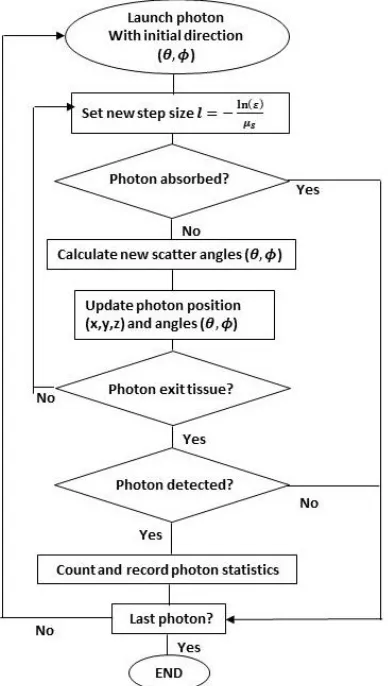
A straightforward Monte Carlo model has been derived. Necessary computational steps are demonstrated in the flowchart in Fig. 1. However, the detailed calculations and explanations of every step can be found elsewhere [12]. When the model is executed, a ‘virtual’ photon is launched onto the ‘tissue surface’ from the emitter. The initial position co-ordinate (x,y,z) and direction (𝜃, 𝜙) of the launched photon is randomly generated but constrained by the limits imposed by the radius and numerical aperture (NA) of the source fiber. When the photon propagates through tissue, it will most likely
Differential pathlength factor estimation for brain-like tissue from a
single-layer Monte Carlo model
to be scattered many times and after a number of scattering events, either be absorbed or exit from the tissue surface. The step-size, i.e. the free path length of the photon between two consecutive scattering events is
𝑙 = −ln(𝜉)
𝜇𝑠 (1)
[image:3.612.62.256.358.701.2]where 𝜉 is a random number (in practice a computer-generated pseudo-random number). It is noteworthy that the Monte Carlo method relies on random sampling of variables from probability distributions [11]. The occurrence of the scattering and absorption events is decided by comparing a random number with the probability of absorption [11,12]. If the photon is not absorbed, scattering takes place. The direction of scattering is calculated depending on the Henyey - Greenstein phase function [12]. The position and direction of the scattered photon are updated [12]. If the scattered photon leaves the tissue medium, a check is made to see if the photon has fulfilled the detection criteria, i.e. the photon must arrive at the tip of detector fiber within its cone of acceptance. If so, several variables are recorded, e.g. the photon’s path, total path length, time of flight, maximum penetration depth etc. The photon might scatter several times within the tissue before it is terminated, i.e. it is detected, absorbed or exits the tissue surface without being detected; and a new photon is launched.
Figure 1. Flow chart for Monte Carlo model for light propagation through a single tissue layer.
In the present piece of work, a homogeneous semi-infinite slab replicated a monolayer tissue section. It was assumed to be illuminated by an optical radiation of near infrared wavelength 810 nm. The tissue- slab was characterized by the optical properties similar to those of tissue like bloodless white human brain matter [13]. The values of the parameters used for this particular wavelength were 𝜇𝑠= 36.36 𝑚𝑚−1,
𝜇𝑎= 0.09 𝑚𝑚−
1
, 𝑔 = 0.876. In a three-dimensional Cartesian geometry, the tissue boundary was defined by the plane 𝑧 = 0. The radiation was supposed to be emitted from a circular source (S) of radius 0.4 mm with its axis along the
z-direction, placed on the tissue surface so that the center of the source was at the origin (0,0,0) of the co-ordinate system. In this ‘reflectance mode’ source-detector architecture, the scattered light intensity from tissue was supposed to be detected by a circular detector (D) of radius 0.8 mm placed on the tissue surface at a distance d from the source so that the co-ordinate of the center of the detector is at (d, 0, 0). The model was executed for different d, e.g. 3, 5, 7 and 11 mm. For each detected photon, its total optical path length, i.e. the sum of its free pathlengths l (Eq. (1)), was recorded. Finally the mean pathlength OP was calculated. The differential pathlength factor (𝐷𝑃𝐹 =𝑀𝑒𝑎𝑛 𝑂𝑃 (𝑚𝑚)𝑑(𝑚𝑚) ) was also calculated for this tissue. The script was written in MATLAB version 8.3.0.532. Program has been executed on a Dell computer with Intel 3.4 GHz dual core processor.
[image:3.612.334.522.381.679.2]III. RESULTS
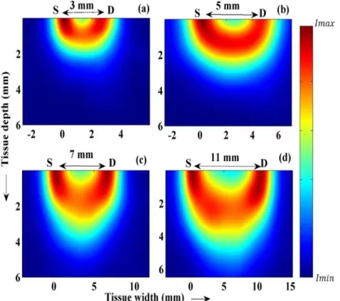
Fig. 2 exhibits the ‘typical’ path of photon(s) within tissue from source S to detector D which are placed on tissue surface (z = 0) with an inter-optode spacing of 7 mm. Fig. 2a shows the path a single photon typically takes within tissue between S and D. Fig. 2b shows how a light beam (photon packet) travels within tissue. It is a smoothed density plot [14] which provides with the information about distribution of the intensity (number density) I of the scattered photons within the medium.
[image:4.612.58.297.226.438.2]Fig. 3 depicts how the mean optical path of photons within tissue varies with variable source-detector distance, i.e. 3, 5, 7 and 11 mm respectively. A clear trend of increasing penetration depth of photons is visible for increasing separation, d. All simulations modelled 107 incident photons.
Figure 3. Difference in optical paths of photon packets for variable source-to-detector distances is shown. Figures (a), (b), (c) and (d) exhibit the S-D separations 3, 5, 7 and 11 mm respectively.
The trend found in Fig. 3 that OP increases with d can be elucidated by Fig. 4, where mean OP is found to exhibit a
Figure 4. Graph of mean optical path length against source-detector separation.
linear relationship with d. The program was run to detect a
107number of photons within the range 0 ≤ d ≤ 7 mm and the
mean optical paths of photons at different d were calculated. Clearly, more number of photons are detected near the source so the curve is very smooth at smaller d. But it gets rougher at higher inter-optode spacing due to detection of comparatively lesser numbers of photons at the same time. The slope of the curve is shown to be 5.66, which in turn is the measure of the DPF. The DPF is found to be constant throughout the range of variable source-detector geometry.
IV. DISCUSSION
The results presented in this paper describe the distribution of optical paths within a certain tissue illuminated by infrared radiation of wavelength 810 nm. It is apparent in Fig. 2 that ‘a typical’ photon travels through a depth roughly equal to half the inter-optode distance before detection at the surface. At smaller and larger depth, the photon density decreases noticeably. As expected, the intensity of photons (Fig. 2b) is maximal near the source and detector. Since the detector diameter is larger than that of the source, the intensity near the source is greater than near the detector. Though a small number of photons take the smallest path between the source and detector (i.e. move approximately laterally), most of the photons take a ‘banana shaped’ path between the source to detector within the tissue.
In this context, another important study is about the relationship of the mean optical path (𝑂𝑃) within tissue to the source-detector separation (𝑑) which illustrated by in Fig. 3. Interestingly, a clear increase in penetration depth is shown for increasing source-detector separation.
Fig. 3 provides with a visual impression of the relationship between mean OP and d, which is explicitly shown in Fig. 4. In this graph, mean OP exhibits a linear relationship with d, suggesting DPF is approximately constant for a particular wavelength and basic geometry, and independent of the source-detector separation.
V. CONCLUSION
The model describes the mean optical path of the photon, shows how the optical path changes with the change of source and detector separations and gives a visual idea about the penetration depth of light within tissue. It estimates the DPF value which basically links quantitatively the path of photons within tissue with source-detector geometry of the probing system.
[image:4.612.46.287.521.699.2]gain better understanding in the field of light-tissue interaction. Such knowledge is invaluable for the design of more effective non-invasive and wearable optical sensors.
REFERENCES
[1] J. Allen et al, ‘Photoplethysmography and its application in clinical physiological measurement’, Physiol. Meas. 28 (2007) 1-39.
[2] Janet S. Soul et al, ‘Near-Infrared Spectroscopy’, Seminars in
Pediatric Neurology 6(2) (1999) 101-110.
[3] Rybynok V. O. et al, ‘A systematic approach for the accurate
non-invasive estimation of blood glucose utilizing a novel light-tissue interaction adaptive modelling scheme’, Journal of Physics: Conference series 85 (2007) 012033.
[4] Marc F. Swiontkowski et al, ‘Laser Doppler Flowmetry-Development and Clinical Application’, Iowa Ortho J. 11 (1991) 119-126.
[5] B. C. Wilson et al, ‘A Monte Carlo model for the absorption and flux distributions of light in tissue’, Med. Phys. 10 (1983) 824-830.
[6] P. Van Der Zee et al, ‘Simulation of the point spread function for
light in tissue by a Monte Carlo method’, Adv. Exp. Med. Biol. 215 (1987) 179-92.
[7] N. S. Zolek et al, ‘Optimization of the Monte Carlo code for
modelling of photon migration in tissue’,Comput. Meth. Prog. Bio. 84 (2006) 50-7.
[8] C. Zhu et al, ‘Review of Monte Carlo modeling of light transport
in tissues’, J. Biomed. Opt. 18(5) (2013) 05902-1-12.
[9] I. Lux et al, ‘Monte Carlo particle transport methods: Neutron and
Photon calculations’, CRC Press, Boca Raton, FL 1991.
[10]R. Splinter et al, ‘An introduction to biomedical optics’, CRC
Press, Boca Raton, FL (2007).
[11]L. Wang et al, ‘MCML- Monte Carlo modeling of light transport
in multi-layered tissues’, Comput. Meth. Prog. Bio. 47 (1995) 131-46.
[12]J. P. Phillips et al, ‘Calculation of Photon path changes due to Scatter in Monte Carlo Simulations’, IEEE EMBC conf. proceeding (2010),
[13]A. N. Yaroslavsky et al, ‘Optical properties of selected native and coagulated human brain tissues in vitro in the visible and near infrared spectral range’, Phys. Med. Biol. 47 (2002), 2059-73.
[14]Paul H. C. et al, ‘Enhancing scatterplots with smoothed densities’, Bioinformatics 20(4) (2004), 623-628.
[15]L. G. Henyey et al, ‘Diffuse Radiation In The Galaxy’, Astrophys. J., 93 (1941) 70-83.